
Hippocampal Dentation in Children and Adolescents: A Cross-Sectional Analysis from Birth to 18 Years Old

 , ,
, ,
Abstract
:1. Introduction
2. Materials and Methods
2.1. Data Acquisition (C-MIND)
2.2. Sample Demographics
2.3. Neuroimaging
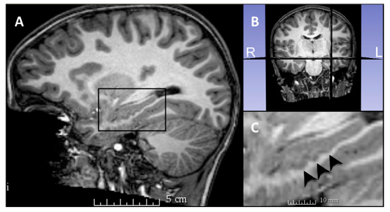
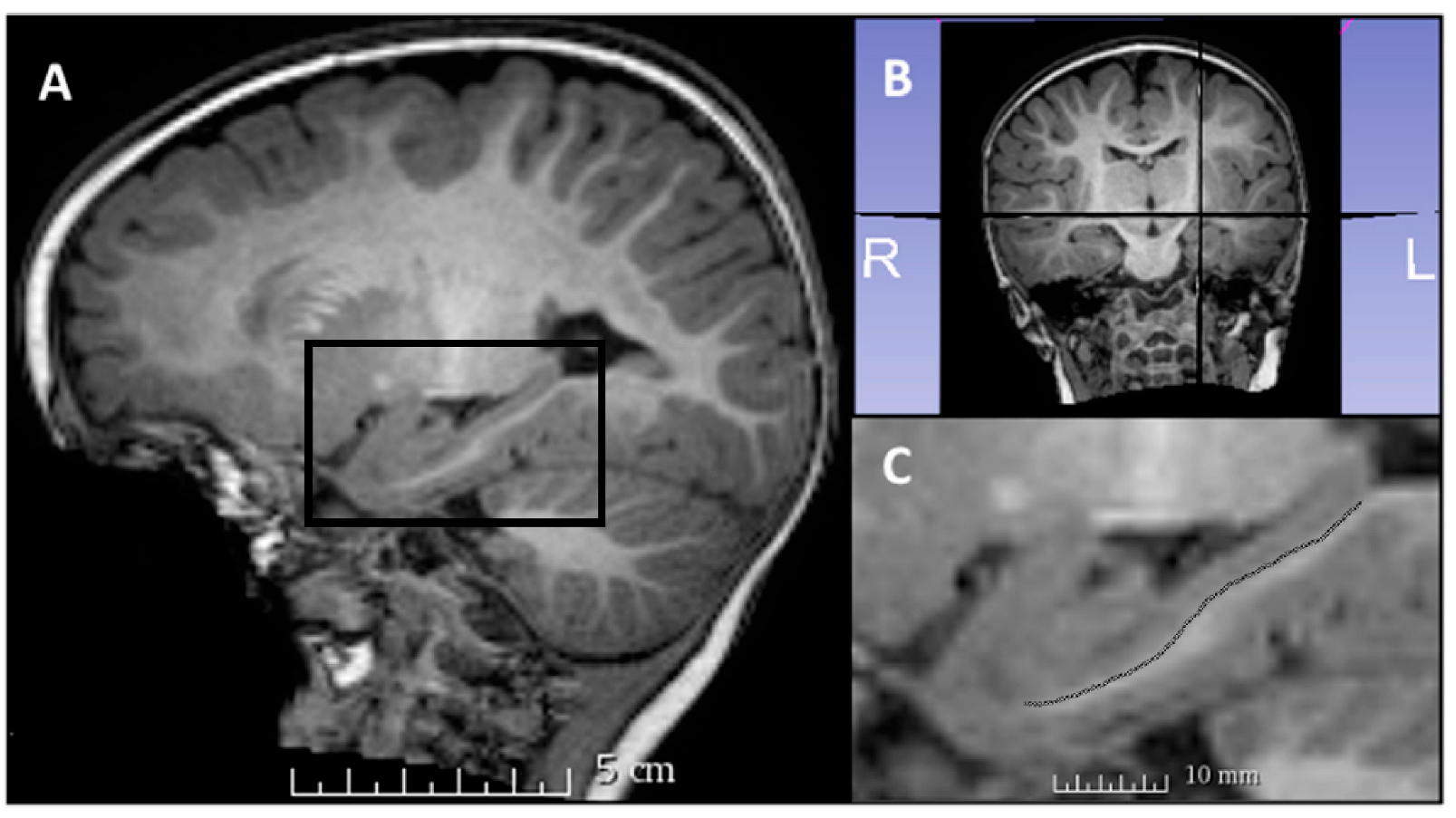
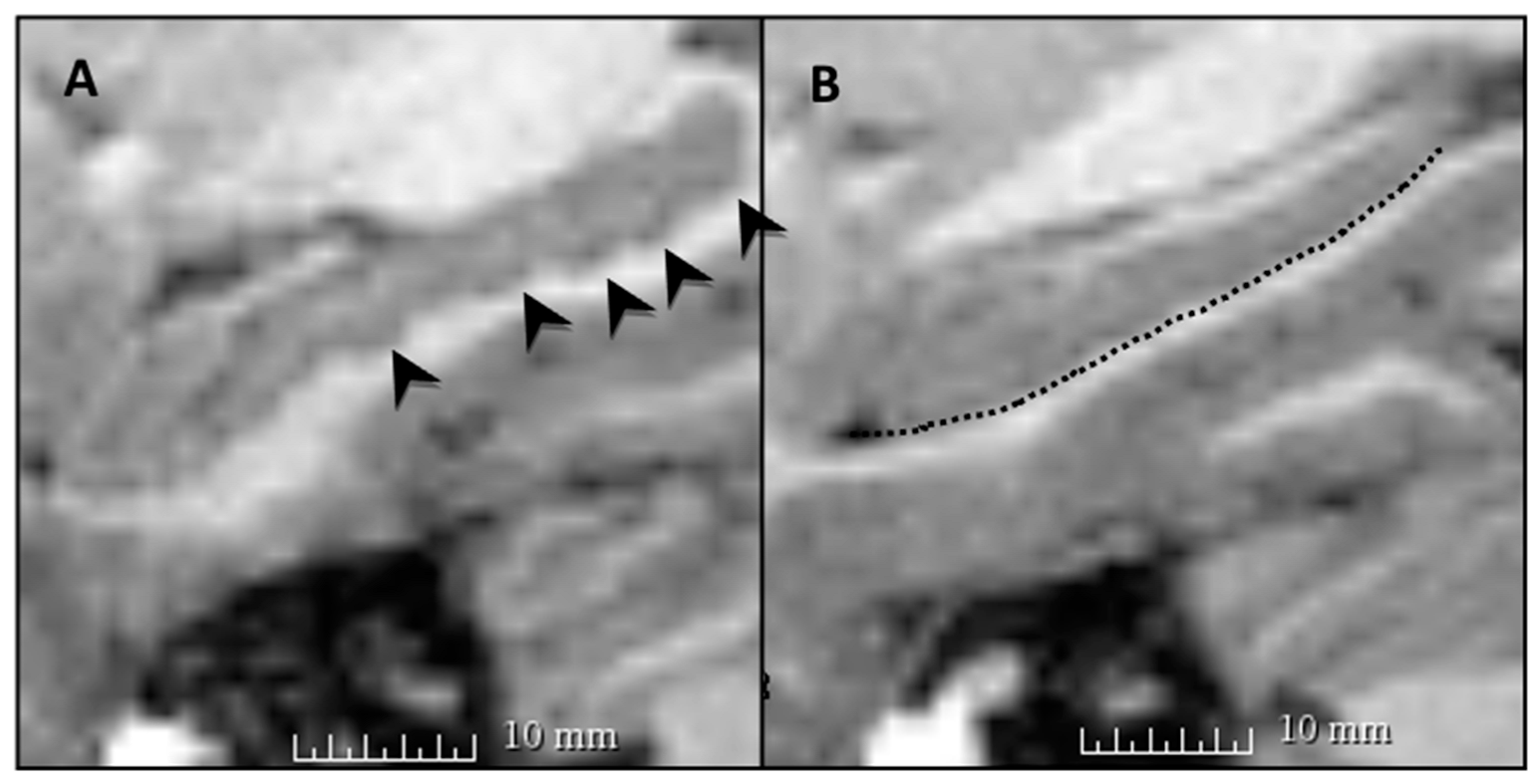
2.4. Dentation Assessment
2.5. Experimental Design and Statistical Analysis
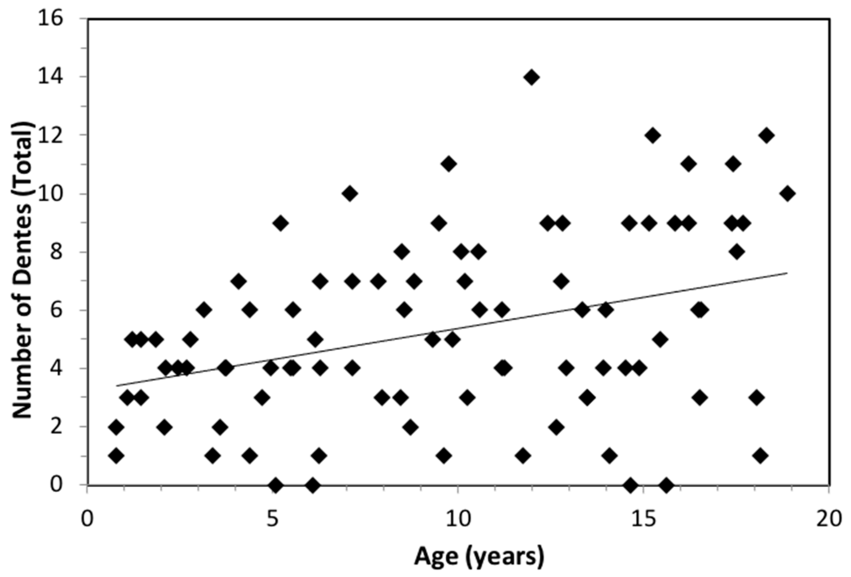
3. Results
4. Discussion
4.1. Hippocampal Dentation and Age
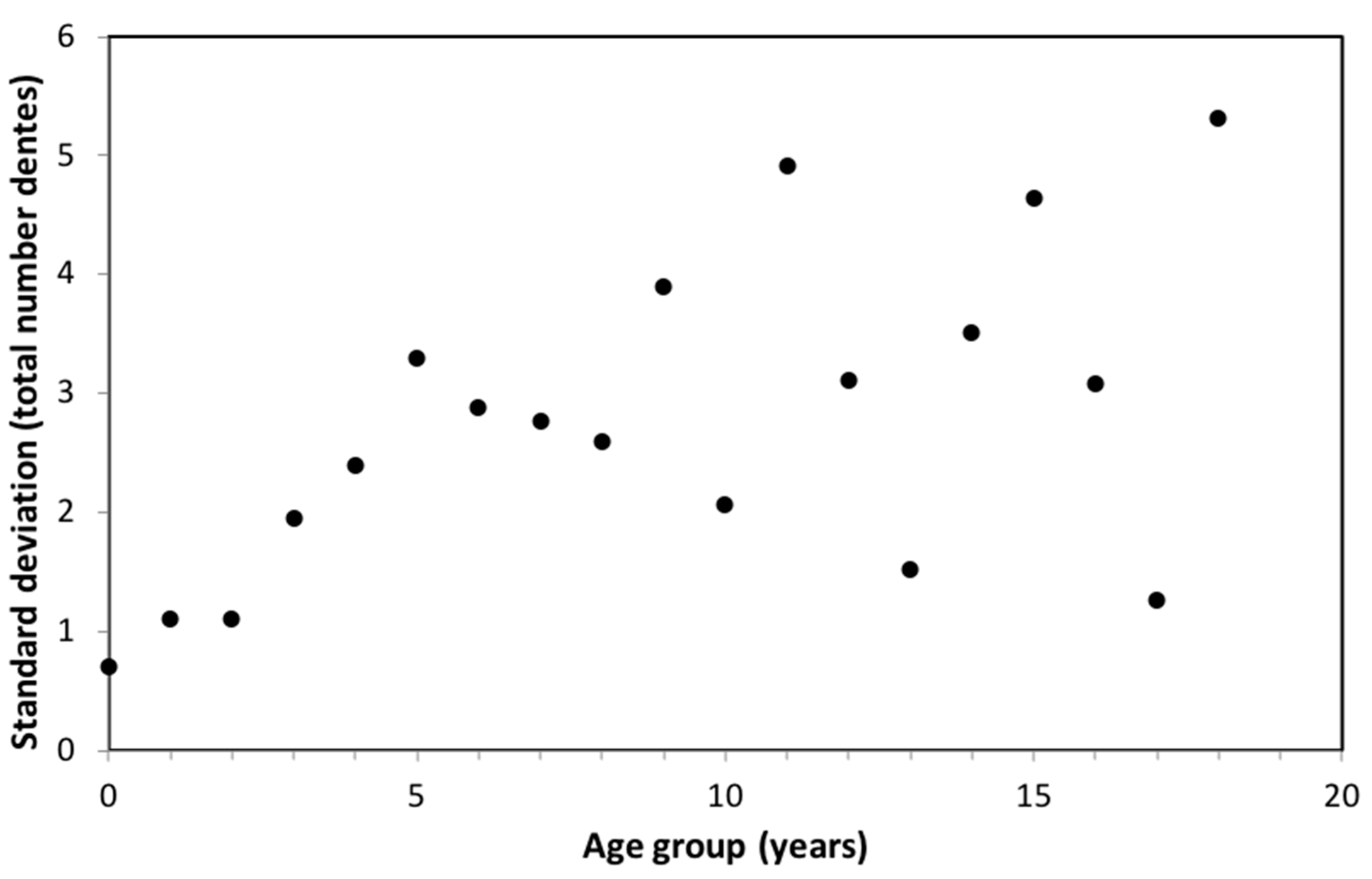
4.2. Variability of Hippocampal Dentation by Age
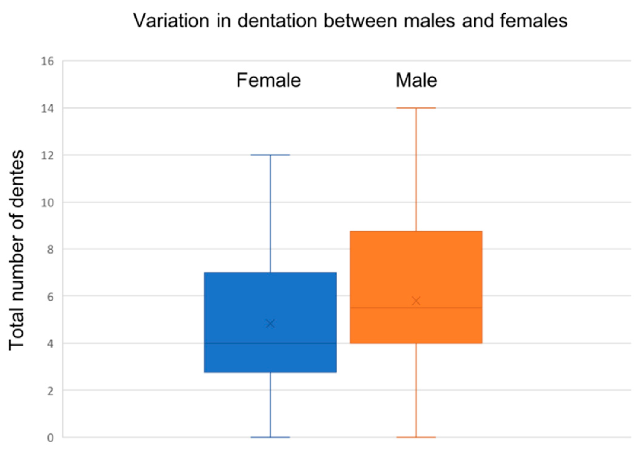
4.3. Hippocampal Dentation and Gender
4.4. Hippocampal Dentation and Demographics
4.5. Reliability of Dentation Assessment
4.6. Limitations
4.7. Future Directions
5. Conclusions
Author Contributions
Funding
Institutional Review Board Statement
Informed Consent Statement
Data Availability Statement
Acknowledgments
Conflicts of Interest
Appendix A

| Mothers | Fathers | |
|---|---|---|
| Less than high school | 4% (n = 4) | 7% (n = 6) |
| High school | 17% (n = 15) | 23% (n = 21) |
| Some college | 19% (n = 17) | 24% (n = 22) |
| College | 30% (n = 27) | 27% (n = 24) |
| Some graduate level | 6% (n = 5) | (n = 0) |
| Graduate level | 21% (n = 19) | 13% (n = 12) |
| Not reported | 3% (n = 3) | 6% (n = 5) |
| Percent of Sample | |
|---|---|
| $0–$5000 | 12% |
| $5000–$10,000 | 1% |
| $10,000–$15,000 | 6% |
| $15,000–$25,000 | 8% |
| $25,000–$35,000 | 8% |
| $35,000–$50,000 | 10% |
| $50,000–$75,000 | 14% |
| $75,000–$100,000 | 21% |
| $100,000–$150,000 | 11% |
| Over $150,000 | 4% |
| Not reported | 4% |
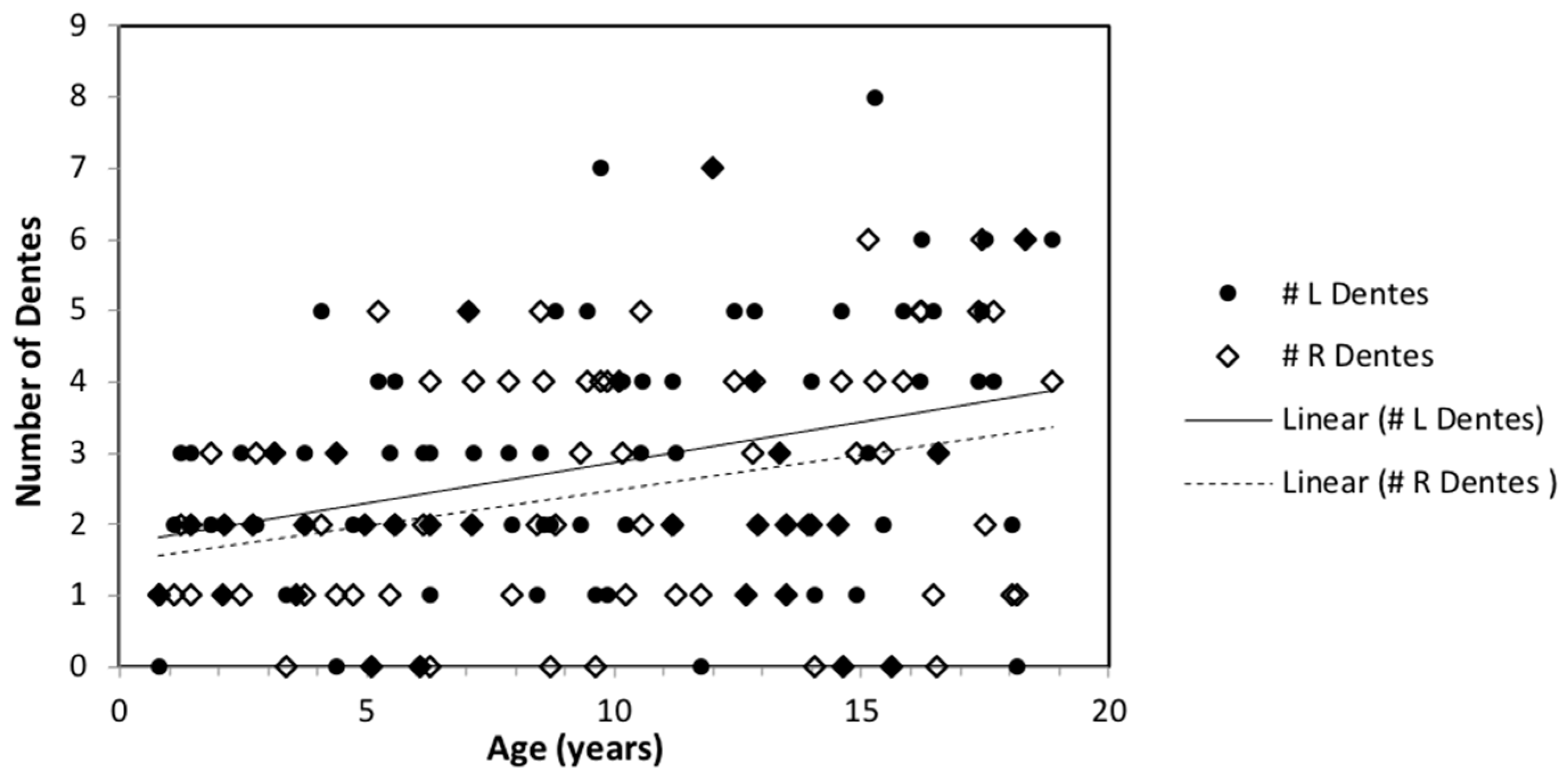
| Number of Dentes | ||||
|---|---|---|---|---|
| Age Group (Years) | Frequency | Left | Right | Total |
| <1 | 2 | 0.50 (0.71) | 1.00 (0.00) | 1.50 (0.71) |
| 1 | 5 | 2.40 (0.55) | 1.80 (0.84) | 4.20 (1.10) |
| 2 | 5 | 2.00 (0.71) | 1.80 (0.84) | 3.80 (1.10) |
| 3 | 5 | 2.00 (1.00) | 1.40 (1.14) | 3.40 (1.95) |
| 4 | 5 | 2.40 (1.82) | 1.80 (0.84) | 4.20 (2.39) |
| 5 | 5 | 2.60 (1.67) | 2.00 (1.87) | 4.60 (3.29) |
| 6 | 5 | 1.80 (1.30) | 1.60 (1.67) | 3.40 (2.88) |
| 7 | 5 | 3.00 (1.22) | 3.20 (1.64) | 6.20 (2.77) |
| 8 | 5 | 2.60 (1.52) | 2.60 (1.95) | 5.20 (2.59) |
| 9 | 5 | 3.20 (2.68) | 3.00 (1.73) | 6.20 (3.90) |
| 10 | 5 | 3.40 (0.89) | 3.00 (1.58) | 6.40 (2.07) |
| 11 | 5 | 3.20 (2.59) | 2.60 (2.51) | 5.80 (4.92) |
| 12 | 5 | 3.40 (1.82) | 2.80 (1.30) | 6.20 (3.11) |
| 13 | 5 | 2.40 (1.14) | 2.00 (0.71) | 4.40 (1.52) |
| 14 | 5 | 1.80 (1.92) | 1.80 (1.79) | 3.60 (3.51) |
| 15 | 5 | 3.60 (3.05) | 3.40 (2.19) | 7.00 (4.64) |
| 16 | 5 | 4.20 (1.30) | 2.80 (2.28) | 7.00 (3.08) |
| 17 | 4 | 4.75 (0.96) | 4.50 (1.73) | 9.25 (1.26) |
| 18 | 4 | 3.50 (3.00) | 3.00 (2.45) | 6.50 (5.32) |
References
- Insausti, R.; Amaral, D.G. Hippocampal formation. Hum. Nerv. Syst. 2004, 2, 871–914. [Google Scholar]
- Ver Hoef, L.; Deshpande, H.; Cure, J.; Selladurai, G.; Beattie, J.; Kennedy, R.E.; Knowlton, R.C.; Szaflarski, J.P. Clear and Consistent Imaging of Hippocampal Internal Architecture with High Resolution Multiple Image Co-registration and Averaging (HR-MICRA). Front. Neurosci. 2021, 15, 546312. [Google Scholar] [CrossRef]
- Benes, F.M.; Turtle, M.; Khan, Y.; Farol, P. Myelination of a key relay zone in the hippocampal formation occurs in the human brain during childhood, adolescence, and adulthood. Arch. Gen. Psychiatry 1994, 51, 477–484. [Google Scholar] [CrossRef]
- Knickmeyer, R.C.; Gouttard, S.; Kang, C.; Evans, D.; Wilber, K.; Smith, J.K.; Hamer, R.M.; Lin, W.; Gerig, G.; Gilmore, J.H. A structural MRI study of human brain development from birth to 2 years. J. Neurosci. 2008, 28, 12176–12182. [Google Scholar] [CrossRef]
- Uematsu, A.; Matsui, M.; Tanaka, C.; Takahashi, T.; Noguchi, K.; Suzuki, M.; Nishijo, H. Developmental trajectories of amygdala and hippocampus from infancy to early adulthood in healthy individuals. PLoS ONE 2012, 7, e46970. [Google Scholar] [CrossRef] [Green Version]
- Ostby, Y.; Tamnes, C.K.; Fjell, A.M.; Westlye, L.T.; Due-Tonnessen, P.; Walhovd, K.B. Heterogeneity in subcortical brain development: A structural magnetic resonance imaging study of brain maturation from 8 to 30 years. J. Neurosci. 2009, 29, 11772–11782. [Google Scholar] [CrossRef]
- Beattie, J.F.; Martin, R.C.; Kana, R.K.; Deshpande, H.; Lee, S.; Curé, J.; Ver Hoef, L. Hippocampal dentation: Structural variation and its association with episodic memory in healthy adults. Neuropsychologia 2017, 101, 65–75. [Google Scholar] [CrossRef]
- Simic, G.; Kostovic, I.; Winblad, B.; Bogdanovic, N. Volume and number of neurons of the human hippocampal formation in normal aging and Alzheimer’s disease. J. Comp. Neurol. 1997, 379, 482–494. [Google Scholar] [CrossRef]
- Ramaniharan, A.K.; Zhang, M.; Martin, R.C.; Parpura, V.; Selladurai, G.; Ver Hoef, L. An objective method to quantify hippocampal dentation and predict the side of seizure onset in temporal lobe epilepsy. In Proceedings of the 2021 IEEE Signal Processing in Medicine and Biology Symposium (SPMB), Philadelphia, PA, USA, 4 December 2021. [Google Scholar]
- Kilpattu Ramaniharan, A.; Zhang, M.W.; Selladurai, G.; Martin, R.; Ver Hoef, L. Loss of hippocampal dentation in hippocampal sclerosis and its relationship to memory dysfunction. Epilepsia 2022. [Google Scholar] [CrossRef]
- Duvernoy, H.M. The Human Hippocampus: Functional Anatomy, Vascularization and Serial Sections with MRI; Springer Science & Business Media: Berlin/Heidelberg, Germany, 2005. [Google Scholar]
- Federau, C.; Gallichan, D. Motion-Correction Enabled Ultra-High Resolution In-Vivo 7T-MRI of the Brain. PLoS ONE 2016, 11, e0154974. [Google Scholar] [CrossRef]
- Isaacson, R. The Limbic System; Springer Science & Business Media: Berlin/Heidelberg, Germany, 2013. [Google Scholar]
- Chang, C.; Huang, C.; Zhou, N.; Li, S.X.; Ver Hoef, L.; Gao, Y. The bumps under the hippocampus. Hum. Brain Mapp. 2018, 39, 472–490. [Google Scholar] [CrossRef] [Green Version]
- Ding, S.L.; Van Hoesen, G.W. Organization and Detailed Parcellation of Human Hippocampal Head and Body Regions Based on a Combined Analysis of Cyto- and Chemoarchitecture. J. Comp. Neurol. 2015, 523, 2233–2253. [Google Scholar] [CrossRef]
- White, T.; Su, S.; Schmidt, M.; Kao, C.-Y.; Sapiro, G. The development of gyrification in childhood and adolescence. Brain Cogn. 2010, 72, 36–45. [Google Scholar] [CrossRef] [Green Version]
- Giedd, J.N.; Snell, J.W.; Lange, N.; Rajapakse, J.C.; Casey, B.J.; Kozuch, P.L.; Vaituzis, A.C.; Vauss, Y.C.; Hamburger, S.D.; Kaysen, D.; et al. Quantitative magnetic resonance imaging of human brain development: Ages 4–18. Cereb. Cortex 1996, 6, 551–560. [Google Scholar] [CrossRef] [Green Version]
- Suzuki, M.; Hagino, H.; Nohara, S.; Zhou, S.Y.; Kawasaki, Y.; Takahashi, T.; Matsui, M.; Seto, H.; Ono, T.; Kurachi, M. Male-specific volume expansion of the human hippocampus during adolescence. Cereb. Cortex 2005, 15, 187–193. [Google Scholar] [CrossRef] [Green Version]
- Byars, A.W.; Holland, S.K.; Strawsburg, R.H.; Bommer, W.; Dunn, R.S.; Schmithorst, V.J.; Plante, E. Practical aspects of conducting large-scale functional magnetic resonance imaging studies in children. J. Child Neurol. 2002, 17, 885–890. [Google Scholar] [CrossRef]
- Vannest, J.; Rajagopal, A.; Cicchino, N.D.; Franks-Henry, J.; Simpson, S.M.; Lee, G.; Altaye, M.; Sroka, C.; Holland, S.K.; the CMIND Authorship Consortium. Factors determining success of awake and asleep magnetic resonance imaging scans in nonsedated children. Neuropediatrics 2014, 45, 370–377. [Google Scholar] [CrossRef]
- Bennett, D.A. How can I deal with missing data in my study? Aust. N. Z. J. Public Health 2001, 25, 464–469. [Google Scholar] [CrossRef]
- Shrout, P.E.; Fleiss, J.L. Intraclass correlations: Uses in assessing rater reliability. Psychol. Bull. 1979, 86, 420–428. [Google Scholar] [CrossRef]
- Cicchetti, D.V.; Sparrow, S.A. Developing criteria for establishing interrater reliability of specific items: Applications to assessment of adaptive behavior. Am. J. Ment. Defic. 1981, 86, 127–137. [Google Scholar]
- Yurgelun-Todd, D.A.; Killgore, W.D.; Cintron, C.B. Cognitive correlates of medial temporal lobe development across adolescence: A magnetic resonance imaging study. Percept. Mot. Ski. 2003, 96, 3–17. [Google Scholar] [CrossRef]
- Isaacs, E.B.; Lucas, A.; Chong, W.K.; Wood, S.J.; Johnson, C.L.; Marshall, C.; Vargha-Khadem, F.; Gadian, D.G. Hippocampal volume and everyday memory in children of very low birth weight. Pediatr. Res. 2000, 47, 713–720. [Google Scholar] [CrossRef]
- Molloy, C.S.; Wilson-Ching, M.; Doyle, L.W.; Anderson, V.A.; Anderson, P.J.; for the Victorian Infant Collaborative Study Group. Visual memory and learning in extremely low-birth-weight/extremely preterm adolescents compared with controls: A geographic study. J. Pediatr. Psychol. 2014, 39, 316–331. [Google Scholar] [CrossRef] [Green Version]
- Aanes, S.; Bjuland, K.J.; Skranes, J.; Lohaugen, G.C. Memory function and hippocampal volumes in preterm born very-low-birth-weight (VLBW) young adults. Neuroimage 2015, 105, 76–83. [Google Scholar] [CrossRef]


| Number of Hippocampal Dentes | |||
|---|---|---|---|
| Left | Right | Total | |
| Birth weight (lb) A | 0.07 (p = 0.54) | −0.10 (p = 0.36) | −0.02 (p = 0.89) |
| Height (cm) B | 0.29 (p = 0.006) ** | 0.27 (p = 0.01) * | 0.31 (p = 0.004) ** |
| Weight (kg) B | 0.21 (p = 0.052) | 0.13 (p = 0.23) | 0.19 (p = 0.08) |
Publisher’s Note: MDPI stays neutral with regard to jurisdictional claims in published maps and institutional affiliations. |
© 2022 by the authors. Licensee MDPI, Basel, Switzerland. This article is an open access article distributed under the terms and conditions of the Creative Commons Attribution (CC BY) license (https://creativecommons.org/licenses/by/4.0/).
Share and Cite
Beattie, J.F.; Martin, R.C.; Cook, E.W., III; Thompson, M.D.; Kana, R.K.; Jacobs, R.Q.; Correya, T.A.; Ramaniharan, A.K.; Ver Hoef, L.W. Hippocampal Dentation in Children and Adolescents: A Cross-Sectional Analysis from Birth to 18 Years Old. Anatomia 2022, 1, 41-53. https://doi.org/10.3390/anatomia1010005
Beattie JF, Martin RC, Cook EW III, Thompson MD, Kana RK, Jacobs RQ, Correya TA, Ramaniharan AK, Ver Hoef LW. Hippocampal Dentation in Children and Adolescents: A Cross-Sectional Analysis from Birth to 18 Years Old. Anatomia. 2022; 1(1):41-53. https://doi.org/10.3390/anatomia1010005
Chicago/Turabian StyleBeattie, Julia F., Roy C. Martin, Edwin W. Cook, III, Matthew D. Thompson, Rajesh K. Kana, Ruth Q. Jacobs, Tanya A. Correya, Anandh K. Ramaniharan, and Lawrence W. Ver Hoef. 2022. "Hippocampal Dentation in Children and Adolescents: A Cross-Sectional Analysis from Birth to 18 Years Old" Anatomia 1, no. 1: 41-53. https://doi.org/10.3390/anatomia1010005