Systematic Review

Non-Multisystem Inflammatory Syndrome in Children—Postacute Sequelae of Paediatric COVID-19: Autoimmune or Autoinflammatory? A Systematic Review of the Reported Cases

by
Antoine Fakhry AbdelMassih
1,2,*,
Maram Hamed Hanafy
3,
Maryam ElAhmady
4,5,
Sylvia Kozman
6,7,
Nourine Diab
8,
Reem Husseiny
3,9,
Ashrakat Deyab
3,
Aalaa Mady
3,
Alia Yasser
3,
Amira R. AbdelHalim
3,
Aya Mohyeldin
3,
Aya Sayed Serour
3,
Ayat AbdelGadir
3,
Eslam Abdelaziz
10,
Farida ElGhamry
3,
Hana Amr
3,
Karim Milad
3,
Lamya Fouda
3,
Mawada Hesham
8,
Mina Adly Riad
3,
Mohamed Aoun
3,
Rana AbdelTawab
3,
Rana Sayed
3,
Salma ElSenbawy
8,
Sara ElAhmady Abdelkhalek
11,12,
Nada Gamal
3 and
Yasmin Omar
3
add Show full author list remove Hide full author list
1
Pediatric Cardiology Unit, Department of Pediatrics, Faculty of Medicine, Cairo University, Cairo 12613, Egypt
2
Cardiac Sciences’ Department, Pediatric Cardiology Division, Sheikh Khalifa Medical City, Abu Dhabi 51900, United Arab Emirates
3
Students, Interns and Residents’ Research Program, Faculty of Medicine, Cairo University, Cairo 12613, Egypt
4
Department of Periodontology, Faculty of Dentistry, Galala University, Suez 43511, Egypt
5
International Association of Dental Students, 1216 Geneva, Switzerland
6
Student and Interns’ Research Program, Faculty of Medicine, Ain Shams University, Cairo 11517, Egypt
7
Department of Health Economics, American University in Cairo, Cairo 11835, Egypt
8
Student and Interns’ Research Program, Faculty of Medicine, New Giza University, New Giza 12256, Egypt
9
Department of Paediatrics, Royal Brompton Hospital, London SW3 6NP, UK
10
Hull University Teaching Hospital, Hull HU3 2JZ, UK
11
Residents’ Research Program, Faculty of Medicine, Zagazig University, Zagazig 44519, Egypt
12
Department of Cardiology, Faculty of Medicine, Helwan University, Helwan 11795, Egypt
*
Author to whom correspondence should be addressed.
Rheumato 2023, 3(2), 132-168; https://doi.org/10.3390/rheumato3020011
Submission received: 7 February 2023 / Revised: 27 April 2023 / Accepted: 16 May 2023 / Published: 30 May 2023
(This article belongs to the Special Issue Post-COVID Rheumatic and Immune-Mediated Diseases)

Abstract

:
Three years after its emergence, coronavirus disease 2019 (COVID-19) continues to be a leading cause of worldwide morbidity and mortality. This systematic review comprises relevant case reports that discuss non-multisystem inflammatory syndrome in children (non-MIS-C) and postacute sequalae of COVID-19 (PASC) in the paediatric population, also known as long COVID syndrome. The study aims to highlight the prevalent time interval between COVID-19 and the development of non-MIS-C post-infectious sequalae (PIS). Databases were searched for studies that met our inclusion and exclusion criteria. The final screening revealed an equal sex distribution where the commonest age intervals were school-age and adolescence, with 38% of the patients being older than six years. Interestingly, hospital admission during the course of COVID-19 was not a predictor of the subsequent PASC; forty-nine patients (44.9%) were hospitalized while sixty patients (55.1%) were not hospitalized. Moreover, the most predominant time interval between COVID-19 and the developing PASC was within 14 days from the start of COVID-19 infection (61%). These findings suggest a crucial link between COVID-19 and immune PIS in the paediatric population, especially those older than six years. Accordingly, follow-up and management are encouraged in case of unusual symptoms and signs following COVID-19 infection, regardless of the COVID-19 infection severity.

1. Introduction

Acute infections are typically defined as self-limiting infections usually lasting less than six months and that usually lead to either complete resolution or death. While many studies cover the typical short-lived course and prognosis of acute infectious diseases, the link between acute infections and chronic disability remains understudied. Consequently, many patients suffering from the long-lasting sequelae of acute infections can easily be wrongly diagnosed or wrongly treated. Furthermore, there is insufficient data relating to postacute infection sequelae (PAIS) since many cases—especially those that are sporadic—remain unrecognized. Postacute sequelae (PAS) are symptoms that occur during the postacute phase of an illness. While the exact definition of the postacute phase is largely debatable and differs from one virus species to another, it is generally known as the phase after the virus becomes no longer detectable by polymerase chain reaction (PCR) [1,2,3,4].
It is not unusual for acute infections to cause fleeting autoimmune symptoms due to disturbances in the innate and adaptive immune signalling pathways. Rarely, however, acute infections can progress to established autoimmune diseases. The main pathogenesis behind this progression is the molecular mimicry of pathogens or the structural similarity between pathogenic proteins and self-proteins, which causes the autoactivation of self-reactive immune cells in some susceptible individuals. Furthermore, viral attacks lead to the release of intracellular components, which, in turn, cause the activation of the innate immune system, the formation of autoantibodies, the stimulation of antigen-presenting cells, and the migration of immune cells to the site of damage [5,6,7,8].
In general, postinfectious sequelae (PIS) can be divided into three broad subtypes based on the time interval between the infection and the sequelae and the duration of the PIS. PIS can be rapidly developing, such as in the case of reactive arthritis, which normally occurs 1–2 weeks after an infection in susceptible individuals. PIS can also take the form of chronic inflammation following nonpersistent viruses, such as in the case of the reovirus, rotavirus A, which causes coeliac disease, an autoimmune disease triggered by gluten. However, the usual pattern is short-lived autoimmunity developing 4 weeks after the infection and does not persist for more than 6 months. Guillain–Barré Syndrome (GBS), idiopathic thrombocytopenic purpura (ITP), and poststreptococcal immune complications are common examples of this pattern [9,10,11].
Ever since the onset of the coronavirus disease 2019 (COVID-19) pandemic, the severe acute respiratory syndrome coronavirus 2 (SARS-CoV-2) virus has quickly become an important focus of research. Due to the atypical cytokine release and immune system dysfunction following a COVID-19 infection, postacute sequelae of COVID-19 (PASC) have appeared in several cases, and such conditions have become known as ‘long COVID’. Previous inflammatory conditions, advanced age, and obesity are all known risk factors. Just like its predecessors, severe acute respiratory syndrome 1 (SARS-CoV-1) and Middle East respiratory syndrome coronavirus (MERS-CoV), the COVID-19 virus can cause symptoms such as fatigue, and dermatological and gastroenterological symptoms. In a recent study, 30% of COVID-19 patients suffered from the persistence of these symptoms, even after the virus became undetectable by PCR [12].
One of the commonest immune sequelae of COVID-19 is a multi-inflammatory syndrome of children, which mimics Kawasaki disease with a more fulminant, yet more reversible course. It usually presents with myocarditis and/or coronary dilatation and responds to treatment with intravenous immunoglobulins. It has gained most of the attention in retrospective, cross-sectional studies as well as in systematic reviews [13].
We choose, in this review, to study immune sequelae developing outside the frame of multisystem inflammatory syndrome in children (MIS-C), hence the term non-MIS-C postacute sequelae of COVID-19. This systematic review aimed to demonstrate all case reports of non-MIS-C PIS of COVID-19 and to determine the prevalent time interval between acute infection and the development of PIS.

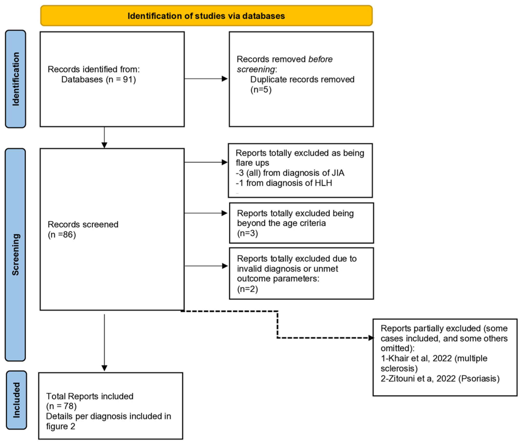
2. Materials and Methods

2.1. Databases Used

A literature search was performed in PubMed, Google Scholar, Google Search, and Scopus.

2.2. Search Terms Inclusion and Exclusion Criteria

2.2.1. Inclusion Criteria

1.
Diagnoses
The following terms and inclusion criteria were included in the search: “COVID-19” OR “SARS-CoV-2” (severe acute respiratory syndrome coronavirus 2) AND “Autoimmune sclerosing cholangitis”, “Diabetes” OR “type 1 diabetes”, “Systemic Lupus Erythematosus” OR “Lupus Erythematosus Disseminatus” OR “Lupus Erythematosus, Systemic”, “Haemolytic–uremic syndrome”, “Juvenile Idiopathic Arthritis”, “Familial Mediterranean Fever”, “Autoimmune thyroid disease” OR “subacute thyroiditis”, “Autoimmune hepatitis”, “ANCA vasculitis” (anti-neutrophil cytoplasmic antibody), “Tumour necrosis factor receptor-associated periodic syndrome”, “ITP”, “HLH” (haemophagocytic lymphohistiocytosis), “Psoriasis”, “Guillain–Barré”, “Multiple Sclerosis” OR “ADEM” (acute disseminated encephalomyelitis) AND “Paediatrics” OR “Children”.
2.
Age: 0–18 Years

2.2.2. Exclusion Criteria

Any case with a multi-inflammatory syndrome of children (MIS-C) or Kawasaki disease was excluded, and any flare-up of a pre-existing autoinflammatory condition was excluded.
Any case not addressing the outcome parameters was excluded.

2.2.3. Outcome Parameters

The main outcome parameters were the age, sex of the included cases, the interval between COVID-19 and the subsequent autoimmune sequelae, and the need for hospitalization during COVID-19 infection that preceded the resultant autoimmune sequelae.

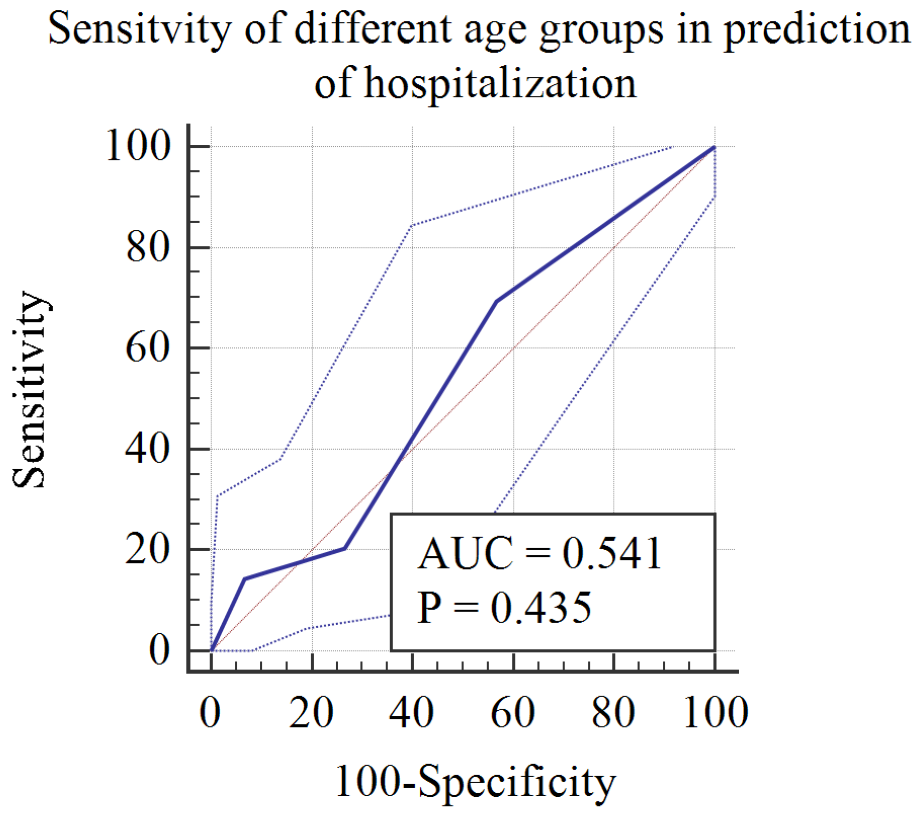
3. Statistical Analyses

For statistical purposes, age was classified into four ranges: 0–2 years (infants), 3–5 years (preschool children), 6–12 years (school-aged children), and 13–18 years (adolescents).
Furthermore, the interval between COVID-19 and subsequent autoimmune sequelae was classified into three intervals: 0–14 days (immediate), 15–28 days (classic), and >28 days (delayed).
Patients were categorized according to the aforementioned age ranges and time intervals as well as the need for hospital admission during COVID-19 and sex. The number and percentage of patients in each category of each outcome parameter were determined, and a comparison between different categories of each outcome parameter was implemented using a chi-square test and illustrated as a pie chart.
Age categories were numbered as follows: Category 1: infancy (<2 y), Category 2: preschool children (3–5 y), Category 3: school children (6–12 y), Category 4: adolescents (13–18 y). A receiver operating characteristic curve analysis was performed where hospitalization was the classification variable, to measure the age categories predicting hospitalization in the context of non-MIS-C postacute sequelae.

4. Results

We gathered a total of 78 reports of autoimmune sequelae following COVID-19, comprising a collective total of 109 patients. (References included in individual results).
Figure 1 is a preferred reporting item for systemic reviews and meta-analysis (PRISMA) flow charts.
Figure 2 is a detailed algorithm for the distribution and overlap of reports and the number of patients per diagnosis.
Both of them, indicate that we included a total of 78 reports, comprising 109 patients.

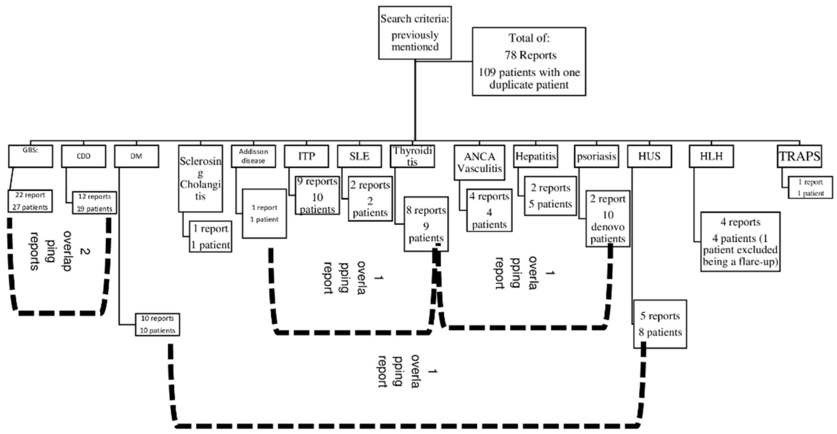
4.1. Overall Results

The sex distribution in retrieved cases was equal between the two genders. Regarding age, the commonest two age intervals involved were school-aged children and adolescents, with each accounting for 38% of the overall cases. Hospital admission during COVID-19 did not seem to be a good predictor of subsequent autoimmune sequelae as there was no statistically significant difference between the number of cases with hospital admission and those who were not admitted, i.e., 49 and 60, respectively. Finally, yet importantly, most of the observed PIS were observed within 14 days of the COVID-19 infection, accounting for 61% of the total cases. (Table 1 and Table 2) illustrate the details of the overall results described in this paragraph.
Figure 3 is a receiver operating characteristic curve (ROC) demonstrating the sensitivity of age categories in predicting hospitalization in the context of non-MIS-C sequelae of COVID-19. It clearly shows that younger age groups, categories 1 and 2 (infants [0–2 years]) and preschool [3–5 years]), were more likely to require hospitalization, with a sensitivity of 69%.

4.2. Individual Results (by Alphabetical Order of the Respective Autoimmune Disorder)

4.2.1. Addison’s Disease (Table 2)

The relationship between COVID-19 and Addison’s disease has been reported in the literature. One case of a 14-year-old female was associated with primary adrenal insufficiency (Addison) as part of autoimmune polyglandular syndrome type 2, which required intensive care unit (ICU) admission, as mentioned by Floras et al.
SARS-CoV-2 releases amino acid sequences similar to ACTH, which should rather cause secondary adrenal insufficiency; however, the presence of anti-21-hydroxylase antibodies was not documented, as in other infectious causes of Addison’s disease [15].

4.2.2. Anti-Neutrophil Cytoplasmic Antibody (Table 3)

Acute ANCA-associated vasculitis is a rare but documented condition following SARS-CoV-2 infection in adults and is even rarer in the paediatric population. Here we present to you four case reports of paediatric ANCA-associated vasculitis following an acute SARS-CoV-2 infection. The male-to-female ratio in the reported cases was 1:1, and the mean age among the four patients was approximately 16 years old. All of the patients (100%) acquired acute ANCA-associated vasculitis as an immediate sequela (within 0–4 weeks after COVID-19 infection), and none had delayed nor persistent sequelae. Two patients developed perinuclear anti-neutrophil cytoplasmic antibody (P-ANCA) vasculitis, as seen in Firenzen et al. and Weston et al., while the other two patients developed cytoplasmic anti-neutrophil cytoplasmic antibody (C-ANCA) vasculitis. Two of the patients had pre-existing asthma, as seen in Firenzen et al. and Bryant et al. The general prognosis for post-COVID ANCA vasculitis in the previous patients was good with mild to moderate COVID-19 courses. However, the patient reported by Weston et al. was admitted to ICU due to a worsening respiratory status. All patients recovered and were discharged after proper treatment.
Molecular mimicry is not the first suggested mechanism for ANCA disorders after COVID-19; T-lymphocyte activation with the subsequent uncontrolled secretion of beta-interferon is regarded as the principal theory underlying ANCA disorders following COVID-19 [16].
Table 3. Anti-neutrophil cytoplasmic antibody vasculitis as a postacute sequela of COVID-19.
Ref.Age (Years)SexInterval between COVID-19 Infection and Autoimmune
Disorder
Course of COVID-19
Infection and
Hospitalization Data
OutcomeNotes
[17]17Male7 daysPresented with fever, drenching night sweats, cough, nasal congestion, haemoptysis, and chest tightness.Recovered
  • Chest X-ray revealed a 5 cm left upper lobe mass and a 3 cm right paratracheal mass.
  • CT showed multiple bilateral cavitary lung lesions, the largest within the left upper lobe measuring 6.5 cm in diameter.
  • Positive c-ANCA and PR3 antibodies.
[18]16Female7 daysMild upper respiratory symptoms with anosmia.Recovered
  • Presented with a worsening persistent cough (at first nonproductive, then productive with green sputum), wheezing, difficulty in breathing, resistant to bronchodilators, and myalgia.
  • Had a history of bronchial asthma.
  • Chest X-ray revealed perihilar and bilateral upper lobe consolidations.
  • CT revealed extensive multifocal pulmonary nodules and regions of consolidation with multiple areas of cavitation and central bronchiectasis with diffuse bronchial wall thickening as well as reactive mediastinal and hilar adenopathy.
  • Positive C-ANCA, PR3, and ANA antibodies.
[19]17Male60 days
  • Pneumonia and respiratory insufficiency requiring a high-flow nasal cannula (HFNC) up to 30 LPM, FiO2 50%.
  • Chest X-ray showed moderate bilateral infiltrates.
Recovered after treatment and resolution of AKI and diffuse alveolar haemorrhage (DAH).
  • Presented with elevated blood pressure, worsening knee and lower back pain as well as generalized body aches, haematuria, and proteinuria, and diagnosed with acute kidney injury (AKI) with a BUN/Cr of 16/1.30.
  • One month later, presented again with a worsening cough, fatigue, exertional dyspnoea, and amber-coloured urine.
  • He developed acute respiratory insufficiency requiring respiratory support with HFNC (40 LPM, FiO2 60%), AKI (BUN/Cr 30/1.52), and was revealed to have significant anaemia (Hb/Hct 5.5/16.8).
  • CT, FFB, and BAL revealed diffuse alveolar haemorrhage.
  • Positive ANA, P-ANCA, and MPO antibodies.
  • History of bronchial asthma.
[20] 12Female14 to 28 daysAsymptomatic
  • Patient was admitted to ICU due to worsening respiratory status.
  • Improved on methylprednisolone, rituximab, and cyclophosphamide.
  • PCR testing during hospitalization was negative.
  • Tested positive for COVID-19 IgG antibodies.
  • Diagnosis of anti-MPO ANCA vasculitis with pulmonary and renal involvement.
Abbreviations: SARS-CoV-2: severe acute respiratory syndrome coronavirus 2; ANCA antibodies: anti-neutrophil cytoplasmic antibody; C-ANCA antibodies: cytoplasmic anti-neutrophil cytoplasmic antibody; P-ANCA antibodies: perinuclear anti-neutrophil cytoplasmic antibody; PR3 antibodies: anti-protease 3 antibodies; ANA antibodies: anti-nuclear antibodies; MPO antibodies: myeloperoxidase antibodies; CT: computed tomography; HFNC: high-flow nasal cannula; LPM: litres per minute; FiO2: fraction of inspired oxygen; AKI: acute kidney injury; BUN/Cr: blood urea nitrogen/creatinine; HB/Hct: haemoglobin/haematocrit; FFB: flexible fibreoptic bronchoscopy; and BAL: bronchoalveolar lavage.

4.2.3. Central Demyelinating Disorders (Table 4)

Concerning the data gathered on post-COVID-19 patients suffering from demyelinating disorders other than Gullain–Barré Syndrome (GBS), we noticed an almost equal ratio of males and females (8 males to 11 females) in the reported cases. The youngest case reported was of a 3-year-old and the oldest was 16 years old. The average age was found to be 11 years.
The course of the preceding COVID infection was mostly mild. Five cases were asymptomatic, and the most reported symptom was fever. The time frame between infection and the neurological presentation ranged from a week to months, with three cases presenting with neurological manifestation during the course of COVID-19 infection.
Of the 19 reported cases, 7 cases were diagnosed as new-onset acute disseminated encephalomyelitis* (ADEM), 1 case was diagnosed as anti-N-methyl-d-aspartate (anti-NMDA)-receptor encephalitis and one case was that of unspecified encephalitis.
Two cases of optic neuritis were reported, as well as two cases of neuromyelitis optica spectrum disorder. Three of the reported cases were of post-COVID multiple sclerosis and one case exhibited an anti-myelin oligodendrocyte glycoprotein (anti-MOG) demyelinating disorder. One case developed longitudinal extensive transverse myelitis (LETM).
A complete recovery was observed in five cases; meanwhile, the rest of the cases suffered from mild remnants, including increased blind spot, persistent gait, residual diffuse weakness, and unilateral papilledema. Furthermore, one patient experienced relapse post-treatment and was placed on rituximab.
It is suggested that direct CNS infection by SARS-CoV-2 through the olfactory pathway weakens the blood–brain barrier via gliosis, the latter mechanism combined with a dysregulated immune response and a cytokine storm can explain the resulting CNS damage seen with COVID-19 [21].
Table 4. Central demyelinating disorders as postacute sequelae of COVID-19.
Ref.Age (Years)SexInterval between Infection and Autoimmune DisorderCourse of COVID-19 Infection and
Hospitalization Data
OutcomeNotes
[22]8MaleOne monthMild respiratory symptoms.Complete recovery.
  • Presented with diplopia, imbalance, and gait ataxia.
  • Diagnosed as anti-MOG antibody demyelinating disorder.
13 Female2 monthsFatigue and loss of sense of smell and taste.Moderate
improvement with residual diffuse weakness.
  • Presented with symptoms prior to COVID infection with flare-ups after infection.
  • Presented with headache, nausea, vomiting, dizziness, numbness, tingling, and walking difficulty.
  • Diagnosed as relapsing neuromyelitis optica spectrum disorder (NMOSD) (tested positive for anti-aquaporin-4 antibodies).
14Female5 to 6 weeksAsymptomatic.Unreported.
  • Presented with right leg weakness and left eye pain.
  • Diagnosed as new-onset MS.
[23]16Female4 monthsUnreportedPatient was placed on Rituximab.
Follow-up
information
unreported.
  • Presented with sudden blindness in the right eye and radicular pain in both lower limbs.
  • Positive family history of autoimmune disease.
  • Diagnosed as new-onset NMOSD.
[24]14Male Positive PCRComplete strength recovery, persistent hyperreflexia in the left lower limb, right eye papilledema, and increased blind spot.
  • Presented with headache, blurry vision, papilledema, right VI cranial nerve palsy, asymmetric mild paraparesis, bilateral ankle clonus, and left Babinski sign.
  • Diagnosis of MS could not be confirmed despite brain and spine MRI showing multifocal demyelinating lesions with signs of activity.
  • Diagnosed as a multifocal demyelinating event.
[25]14Female8 weeksNo respiratory
involvement
No complications
  • Presented with blurred vision.
  • Diagnosed as new-onset MS.
4Male8 weeksNo respiratory
involvement
No complications
  • Presented with fever for 15 days and irritability.
  • Diagnosed as new-onset ADEM.
3Male6 weeksNo respiratory
involvement
No complications
  • Diagnosed as new-onset LETM.
[26]16MaleAt the time of
presentation
AsymptomaticFull recovery
  • Presented with complete bilateral horizontal gaze palsy.
  • MRI showed typical MS findings.
[27]9Male3 daysFever, headache,
and vomiting
Tracheotomized. Discharged after 60 months of hospital stay with incomplete recovery.
  • Presented with status epilepticus.
  • Needed PICU admission.
  • Diagnosed as new-onset ADEM.
9Female5 daysFever, vomiting, and diarrhoeaComplete recovery
  • Presented with afebrile seizures.
  • Needed PICU admission.
  • Diagnosed as new-onset ADEM.
[28]10 MaleDuring disease coursefever, headache, and myalgiaIncomplete
recovery
  • Presented with the inability to walk.
  • Diagnosed as transverse myelitis and ADEM by MRI.
[29]7Female1 weekAsymptomaticIncomplete
recovery with
resolution of
sensory deficits but little improvement in lower limb strength.
  • Presented with acute lower extremity flaccid paralysis and numbness.
  • Diagnosed as new-onset ADEM.
[30]12Female5 daysSkin rash, headache, and feverIncomplete
recovery
  • Presented with acute, progressive, bilateral, and symmetrical motor weakness.
  • Diagnosed as a new-onset ADEM.
[31]6Male10 daysAsymptomaticFull recovery
  • Presented with generalized tonic–clonic seizure with spontaneous resolution.
  • History of Fisher–Evans syndrome.
  • Diagnosed as ADEM.
[32]5Female2 daysMild cough and feverPatient received IVIG, showed
clinical
improvement, and was discharged
after two weeks of hospitalization.
  • Patient presented with painful swelling in the latero-cervical aspect of her neck with a large erythematous patch of the overlying skin.
  • The condition was followed, in the following days, by altered mental status, increased irritability, sleepiness, lack of energy, and lethargy.
  • Diagnosed as encephalitis.
[33]15FemaleDuring the course of the disease Fever, headache,
and vomiting
Needed
hospitalization.Visual acuity fully recovered after treatment.
  • Patient presented with diplopia, bilateral ocular pain, diminished visual acuity, and left VI cranial nerve paresis.
  • MRI revealed optic nerve hyperintensities.
  • Diagnosed as bilateral optic neuritis and left VI cranial nerve paresis.
14FemaleDuring the course of the
disease
Headache, myalgia, and arthralgiaNeeded
hospitalization.
Visual acuity fully recovered after treatment.
  • Presented with headache, left ocular pain, and diminished visual acuity of the left eye.
  • MRI: left optic nerve hyperintensity.
  • Diagnosed as left optic neuritis.
14MaleDuring the course of the
disease
AsymptomaticRankin Score: 0 and
absolute control of epilepsy.
Presence of
psychiatric
symptoms post
discharge.
  • Presented with altered behaviour and mental status, seizures, insomnia, and orolingual dyskinesias.
  • Positive anti-NMDA-R antibodies in CSF.
  • Diagnosed as Anti-NMDA encephalitis.
Abbreviations: Anti-MOG: anti-myelin oligodendrocyte glycoprotein; MS: multiple sclerosis; LETM: longitudinal extensive transverse myelitis; NMSOD: neuromyelitis optica spectrum disorder; ADEM: acute disseminated encephalomyelitis; PICU: paediatric intensive care unit; IVIG: intravenous immune globulin; MRI: magnetic resonance imaging; anti-NMDA-R: anti-N-methyl-d-aspartate (NMDA) receptor encephalitis; and CSF: cerebrospinal fluid.

4.2.4. Guillain–Barré Syndrome (Table 5)

Although the number of adult COVID-19 infections diagnosed with GBS is increasing, the occurrence of cases in the paediatric population remains limited or perhaps underreported.
The research entails that reported paediatric cases of SARS-CoV-2 infection associated with GBS had an average age of 16 years. In general, the age group varied drastically, with the youngest reported case being a 2-month-old male infant 15 days after the course of COVID infection, and the oldest reported patient being a 17-year-old female with a short course of COVID infection 8 days prior to the neurological complications. We assume that the severity of the infection is not directly linked to the Guillain–Barré manifestations since seven of the reported cases were asymptomatic, and the rest of the cases demonstrated variable degrees of severity. Sixteen cases showed a mild course, and eight cases were severe and required paediatric intensive care unit (PICU) admission and mechanical ventilation.
All cases showed immediate post-COVID neurological complications ranging from 0 to 4 weeks after acquiring the infection. To elaborate, the time interval between the disease and the sequelae was around 1 week in 7 cases, 2 weeks in 10 cases, 3 weeks in 2 cases, and 1 month in 11 cases. The shortest interval reported was 2 days.
A full recovery was observed in most cases with the use of IVIG and physiotherapy. However, weakness in neck and limb muscles persisted in 8 of the cases (out of 43), regardless of therapy. One case showed complete recovery after intravenous immunoglobulin (IVIG) except for general hyporeflexia, diminished fine touch sensation in limbs, persistent lower limb weakness, and required home ventilation. Four cases even acquired new deficits and two patients died of respiratory muscle paralysis.
The occurrence of GBS within two weeks rather than 2–4 weeks after the infectious agent mimics the picture seen with the Zika virus. This suggests a proinflammatory state leading to direct nerve damage rather than the presence of autoantibodies [34].
Table 5. Guillain–Barré Syndrome (GBS) as a postacute sequela of COVID-19.
Ref.Age (Years)SexInterval
between COVID-19 Infection and Autoimmune Disorder
Course of COVID-19 Infection and
Hospitalization Data
OutcomeNotes
[35]3Female2 weeksNot specified
  • The patient was treated with 5 cycles of IVIG 0.5 mL/kg/day and then discharged after spending 1 month in the paediatric unit.
  • Presented with progressive and ascending paraesthesia evolving two weeks after respiratory infection.
[36]3Female1 weekMild (Flu-like)
  • The patient improved after the second dose of IVIG.
  • Patient presented with lameness, ataxia, bilateral facial paralysis, ophthalmoplegia, and diplopia.
[37]8Male During the course of COVID-19
infection
Asymptomatic
  • PICU admission, mechanical ventilation for 5 days.
  • The patient improved after IVIG.
  • After 6 weeks, regained bilateral dorsiflexion and plantarflexion, the ability to sit independently, and was working on ambulation.
  • The patient presented with bilateral lower extremity weakness that progressed to paralysis and the inability to walk, which progressed to upper limbs and dyspnoea later.
[38]13Female1 monthFever
  • PICU admission and two weeks of ventilation.
  • Complete neurological recovery and discharge after 6 weeks of hospitalization, IVIG, and plasmapheresis.
  • The patient presented with a high-grade fever, cough, vomiting, and progressive body rash complicated with shock.
  • After 7 days, no response to painful stimuli or spontaneous eye opening, quadriparesis with facial weakness, weak diaphragmatic excursion, and seizures.
[39]11FemaleNot specified Fever
  • PICU admission and mechanical ventilation.
  • After 6 weeks of hospitalization (IVIG and plasmapheresis), the patient was walking independently and had good bowel and urinary control.
  • The patient presented with acute onset of flaccid paralysis and respiratory failure, bowel and bladder incontinence, and lack of sensation.
[40]7MaleNot specifiedAsymptomatic
  • PICU admission and mechanical ventilation.
  • Extubated after 3 days.
  • Clinical improvement.
  • The patient presented with bilateral, symmetrical, lower limb weakness and paresthesia for 8 days, with no antecedent viral illness.
  • He had areflexia, poor gag reflex, and a single breath count of 8, requiring mechanical ventilation.
[41]15 MaleDuring the course of COVID-19
infection
Mild
(no respiratory symptoms)
  • Patient received IVIG; however, improvement was mild and he received physiotherapy.
  • Patient presented with frontal headaches with retro-orbital pain accompanied by fever, evolving to weakness and pain in the lower limbs, which ascended to the upper limbs.
[42]14Female 3 weeksUpper respiratory tract infection 3 weeks
Earlier.
  • Complete recovery after IVIG except for general hyporeflexia and decreased light touch sensation in the distal limbs.
  • The patient presented with bilateral progressive limb weakness and quadriparesthesia, headaches, dizziness, and absent deep tendon reflexes.
  • His father had the same symptoms.
[43]11Male3 weeksVomiting,
diarrhoea,
abdominal pain, and headache for 3 weeks.
  • PICU admission and intubation.
  • Discharged after IVIG administration with normal conscious level, normal muscle tone, cranial nerve palsy, normal muscle tone, grade 4 muscle power, and normal gag and cough reflexes.
  • The patient was lethargic, tachypnoeic, fatigued, no fever, bilateral sixth nerve palsy, and double vision of lateral gaze.
  • The diagnosis of Miller–Fischer syndrome (MFS) with posterior reversible encephalopathy syndrome in association with COVID-19 infection was made.
[33] 9MaleNot specifiedAsymptomatic
  • The patient recovered the ability to walk and run independently.
  • The patient presented with pain in the lower limbs, ascending weakness, hypotonia, and diminished muscle strength in lower limbs.
14MaleNot specifiedFever and
rhinorrhoea
  • The patient recovered the ability to walk and run independently.
  • Paresthesia in feet, ascending weakness, hypotonia, and diminished tendon reflexes in the lower limbs.
12FemaleNot specifiedNot specified
  • The patient recovered the ability to walk and run independently.
  • Dysphonia, hypotonia, ascending weakness, diminished tendon reflexes in upper limbs, and absent reflexes in lower limbs.
[44] 16FemaleDuring the course of COVID-19
infection
Mild
  • The patient received acyclovir, IVIG, and methylprednisolone.
  • After 15 days of hospitalization, the patient was discharged after clinical improvement.
  • Patient presented with diarrhoea and 5 days later with paresthesia and progressive difficulty in walking.
15Male15 daysMild
(no respiratory
involvement)
  • Patient received IVIG after which there was significant clinical improvement.
  • The patient presented with pain, paresthesia, weakness in the lower limbs followed by the upper limbs’ involvement, and without respiratory impairment.
5FemaleDuring the course of the diseaseMild (No respiratory involvement)
  • Symptoms decreased after CSF withdrawal and acetazolamide.
  • The patient presented with intense headache, fever, vomiting, and horizontal diplopia.
  • High CSF pressure (70 cm H2O) and normal analysis.
0.2Male15 daysDry cough, fever, and diarrhoea.
15 days later,
symptoms
developed into dyspnoea and hypoxemia requiring mechanical
ventilation.
  • The patient was treated with intravenous phenobarbital, with an improvement in the epileptic events, and was discharged after 25 days of hospitalization without any apparent neurologic deficits.
  • Three days after being mechanically ventilated, the patient presented with a deviation of the eyes and automatic masticatory movements.
[45]Adolescent
(age is not specified)
Male2 weeksMild; fever
  • Not specified.
  • Patient presented with progressive proximal weakness of the bilateral lower limbs without bladder or bowel involvement.
[46]4Female2 weeksMild; fever
  • Muscle weakness involved respiratory muscles requiring mechanical ventilation.
  • Patient received IVIG, showed clinical improvement, and was discharged on day 10 of hospitalization.
  • Patient presented with a two-day history of pain in the neck, neck floppiness, change in voice, drooling, and bilateral arm weakness.
[47]9FemaleNot specifiedNot specified
  • The patient received IVIG with mild improvement and was discharged after 15 days of hospitalization with a strict neurological follow-up and physiotherapy.
  • Patient presented with progressive weakness and gait instability over the last month.
[48]16FemaleDuring the course of COVID-19
infection
Asymptomatic
  • The patient received IVIG 1 gm/kg/day and showed significant improvement.
  • She was discharged after 5 days with a steppage gait but was able to walk independently.
  • Presented with a seven-day history of progressive symmetric ascending quadriparesis and paresthesia.
  • The patient developed urinary incontinence and constipation on the 9th day of illness.
[49]9MaleDuring the course of COVID-19
infection
Asymptomatic
  • The patient received analgesics-only for his back pain and did not receive IVIG.
  • The patient showed gradual but promising improvement after a few months of conservative treatment.
  • Presented with unbalanced gait, back pain, and lower limb weakness.
[50]6Male1 weekTwo days of fever followed by severe respiratory muscle weakness requiring mechanical ventilation.
  • The patient received a plasma exchange sessions with 5% albumin, methylprednisolone, and IVIG.
  • He was discharged on day 60 of hospitalization with absent reflexes, weakness in upper and lower limbs, and on home ventilation.
  • The patient presented with symmetrical ascending paralysis.
[51]12Male1 weekMild and treated symptomatically at home.
  • The patient was admitted to PICU; however, he was extubated accidentally on day five in PICU and died despite resuscitation attempts.
  • The patient presented with acute progressive symmetric ascending quadriparesis with bilateral facial paresis.
[52]11Male20 daysAn acute upper
respiratory tract
infection with
low-grade fever treated at home with acetaminophen and azithromycin.
Chest CT showed patchy subsegmental faint opacifications with atelectasis in the lingula.
  • Patient received IVIG and clinically improved.
  • The patient presented with acute onset of unsteady gait and the inability to walk or climb stairs, associated with a tingling sensation felt in both the legs and feet of 1-day duration.
[53]6Female1 monthAsymptomatic
  • Patient received IVIG over the course of two days and was discharged after clinical improvement.
  • The patient presented with acute progressive weakness of the lower limbs with no history of recent infection.
[54]17Female8 daysFever, nausea,
severe vomiting, and diarrhoea.
  • Clinical improvement after IVIG administration.
  • The patient presented with severe low back pain that had anteriorized to the groin area, followed by weakness of extremities with loss of ambulation.
  • Acute flaccid tetraparesis is worse in the lower limbs with areflexia in the patella and Achilles tendon, and hyporeflexia in the upper limbs.
[24]8Male During the course of COVID-19Asymptomatic
  • Discharged after 18 days.
  • Moderate improvement in facial diparesis, ophthalmoparesis, and strength; walking with aids.
  • On admission the patient presented with ophthalmoparesis, facial diparesis, acute progressive ascending flaccid tetraparesis, areflexia, and headache.
  • EMG/NCS: moderate acute motor axonal neuropathy (AMAN) with incipient signs of reinnervation.
  • Guillain–Barré syndrome AMAN variant with multiple cranial nerve impairment.
Abbreviations: GBS: Guillain–Barré syndrome; PICU: paediatric intensive care unit; IVIG: intravenous immune globulin; and EMG/NCS: electromyography/nerve conduction studies.

4.2.5. Hepatitis (Table 6)

Hepatic involvement has been widely described as part of the acute setting of SARS-CoV-2 infection, manifesting as a mild increase in liver enzymes without hepatic dysfunction, which eventually subsides as the clinical course of COVID-19 improves. Severe COVID-19 infection in the paediatric population can result in MIS-C and multiorgan failure including hepatic failure. With that being said, here we present five case reports of isolated hepatitis with or without hepatic failure as the main presentation of COVID-19 infection in children. The female-to-male ratio was found to be 3:2 with 150% of females being more susceptible to acquiring the reported complications. The mean age among patients was approximately 6 years old. All of the patients (100%) developed an immediate (within 0–4 months from the start of COVID-19 infection) post-COVID-19 sequelae and none suffered from delayed or persistent sequelae.
The course of COVID-19 infection was mild in three patients and moderate to severe in two infant patients, as seen in Antala et al., at the ages of 6 months and 4 months. Three of the five patients acquired complications, such as acute liver failure with resistant coagulopathy, which is seen in Osborn et al. and the two infants in Antala et al. The 4-month-old infant in Antala et al. also acquired acute kidney injury as well as seizures. Two patients developed hepatic encephalopathy, as seen in Osborn et al. and the 16-year-old male patient in Antala et al. It should be noted that four out of the five patients were admitted to the PICU with an average length of stay of approximately 5 days.
Ultimately, all patients received all of the needed treatment and were discharged accordingly.
Molecular mimicry with the activation of autoreactive T cells and the secretion of proinflammatory mediators has been proposed as a potential mechanism for the occurrence of hepatitis following COVID-19 [55].
Table 6. Hepatitis as a postacute sequela of COVID-19.
Ref.Age (years)SexInterval between COVID-19
Infection and
Autoimmune Disorder
Course of COVID-19
Infection and
Hospitalization Data
OutcomeNotes
[56]3Female21 days
  • The patient had a mild fever and cough.
  • She received no medications, did not require hospitalization, and symptoms resolved in 5 days.
Recovered and discharged after 18 days of hospitalization on azathioprine as steroid-sparing maintenance therapy.
  • Presented to the ER with jaundice, fatigue, and oliguria.
  • She showed worsening coagulopathy (INR 2.7), cholestasis (conjugated bilirubin of 3.8 mg/dL), and hyperammonemia to 317 µmol/L along with altered mental status consistent with hepatic encephalopathy grade I–II.
  • A liver biopsy showed acute submassive hepatic necrosis, lobular collapse, and an intense mixed inflammatory infiltrate consisting primarily of CD3+ T lymphocytes.
  • Elevated anti-liver–kidney–microsomal antibody (anti-LKM) titer of 1:1280, suggestive of type II autoimmune hepatitis.
[57]0.5FemaleDuring the course of COVID-19
infection
  • Presented to ER with new-onset irritability, poor feeding, recurrent emesis, and progressive lethargy over a span of 24 h.
  • GCS of 8, unresponsive and shallow breathing.
  • Hospitalized for 13 days, and admitted to PICU for 5 days where she became more responsive.
  • Recovered and was discharged eventually.
  • Presented to ER with shallow breathing, GCS of 8, hypothermia, epistaxis, and decreased pupillary light response.
  • Coagulopathy was resistant to vitamin K suggesting acute liver failure.
0.33MaleDuring the course of COVID-19
infection
Presented to ER with feeding difficulties, vomiting, hypotonia, diaphoresis, and progressive lethargy over 12 h.
  • Hospitalized for 15 days, and admitted to PICU for 10 days where he was intubated, resuscitated with isotonic saline and dextrose, and started on an epinephrine infusion.
  • Recovered and was discharged eventually.
  • The patient was febrile (38.78 C), tachycardic, tachypneic, hypotensive, and unresponsive.
  • Worsening coagulopathy was resistant to vitamin K suggesting acute liver failure.
  • Associated kidney injury (Creatinine = 0.7, normal level for age= 0.1–0.4) and seizure activity on video EEG.
16Female3 daysPresented with cough, congestion, and fever.
  • Hospitalized for 2 days, and admitted to the ICU for 3 days where she received 80 mg IV methylprednisolone for the empiric treatment of COVID-19.
  • Recovered and was discharged eventually.
  • Presented to the ER with emesis and abdominal pain.
  • Stage 1 encephalopathy, which was resolved by the time of discharge.
11Male2 daysAfebrile without other symptoms.
  • Admitted to ICU for 1 day where he received intravenous fluids for dehydration.
  • Recovered and was discharged eventually.
Presented with non-bloody, non-bilious emesis and abdominal pain.
Abbreviations: Yrs: years; ER: emergency room; INR: international normalized ratio; GCS: Glasgow coma scale; PICU: paediatric intensive care unit; EEG: electroencephalography; ICU: intensive care unit; IV: intravenous; and CD3: cluster of differentiation 3.

4.2.6. Haemophagocytic Lymphohistiocytosis (Table 7)

Four cases of de novo HLH were reported following COVID-19 infections. It was found that the age of the patients varied from neonates to school age in both diseases, with a predominance of preschool age (mean age = 3). HLH appeared equally in both males and females (1:1). Three cases presented with symptoms of HLH several weeks after COVID, but one had symptoms during the course of COVID. The severity of the preceding COVID infection ranged from unremarkable to severe, with two of the cases having required ICU admission during their COVID infection.
It is worth noting that one case presented with concomitant post-COVID viral encephalitis with cerebral atrophy, and another case was diagnosed as Chédiak–Higashi syndrome.
A triggered immune response could be the mechanism of HLH development in COVID-19 patients. Severe acute respiratory syndrome coronavirus 2 (SARS-CoV-2) could activate the NLR family pyrin domain-containing 3 (NLRP3) inflammasome, a potent activator of macrophages, with a significant release of interleukin 1 beta (IL-1b) subsequently leading to the release of interleukin-6 (IL-6) [58].
Table 7. Haemophagocytic lymphohistiocytosis as a postacute sequela of COVID-19.
Ref.Age
(years)
SexInterval between
Infection and
Autoimmune
Disorder
Course of COVID-19
Infection and
Hospitalization Data
OutcomeNotes
[59]5 yearsFemale4.5 weeks Presented with fever and papular rash for three days.8 months in remission.Condition caused:
  • Steroid-induced myopathy, hyperglycaemia, thrush, and Cushingoid features.
  • Haematopoietic cell transplant causing engraftment syndrome.
  • Respiratory failure.
  • Secondary adrenal insufficiency.
  • Veno-occlusive disease.
  • Transplant-associated thrombotic microangiopathy.
  • The patient needed ICU admission.
[60]7 yearsMale2 weeksMild attack.Recovery after 3 days of steroid therapy.
[61]2 yearsMale2 weeksThe disease course showed feeding intolerance, fever (39.6 °C), diarrhoea, and vomiting for two days.Monitored in PICU at the time of publishing.
  • Associated with post-COVID viral encephalitis with cerebral atrophy.
[62]6 weeksfemaleDuring the courseFever of up to 40 °C and poor feeding.Recovery.
  • The patient was diagnosed with Chédiak–Higashi syndrome and required haematopoietic stem cell transplantation.
Abbreviations: Yrs: years; HUS: haemolytic–uremic syndrome; HLH: haemophagocytic lymphohistiocytosis; and TRAP: TNF receptor-associated periodic syndrome.

4.2.7. Haemolytic–Uremic Syndrome (Table 8)

Eight cases were documented with HUS following COVID-19 in the paediatric age groups. The mean age of the patients was 7 years. All HUS cases were males, with only one case report of COVID related to HUS in a female. Only two cases required ICU admission during the course of the preceding COVID.
It is worth noting that all HUS cases were atypical HUS, except for one case of concomitant COVID-19 and Shiga-toxin-associated HUS. In all cases, treatment was given with zero mortality.
Direct endothelial damage by SARS-CoV-2 may be the trigger for the activation of complementary and subsequent HUS in COVID-19 cases [63].
Table 8. Haemolytic–uremic syndrome as a postacute sequela of COVID-19.
Ref.AgeSexInterval
between
Infection and HUS
Course of COVID-19
Infection and Hospitalization Data
OutcomeNotes
[64]4 monthsMale 4 weeksFever and mild respiratory symptoms.
  • Admitted to PICU on day 8 of hospitalization.
  • Received Eculizumab, antibiotic (piperacillin-Tazobactam), and peritoneal dialysis.
  • Received lisinopril, amlodipine, and clonidine due to severe hypertension.
  • Five months after discharge, the patient still suffered from hypertension and persistent proteinuria.
  • The patient had a history of prematurity (26 weeks of gestation) and known neurological abnormalities since birth (microcephaly, joint contractures, and axial hypotonia).
4.5 monthsMale During the course of COVID-19 infection Presented with pyrexia, diarrhoea, and reduced drinking.
  • Treated empirically with eculizumab, triple therapy for hypertension, and peritoneal dialysis for acute kidney injury and anuria.
  • Five months after discharge, the patient still had hypertension and mild persistent proteinuria.
[65]16 monthsMale During the course of COVID-19 infection Fever, emesis, and respiratory distress.
  • The patient was discharged after being treated with Eculizumab and advised to receive it every 3 weeks for aHUS.
  • The patient presented with fever, emesis, and respiratory distress.
  • Diagnosed as DKA on top of type 1 diabetes mellitus and atypical haemolytic–Uremic syndrome.
  • Admitted to PICU for DKA management.
  • The patient had a history of prematurity at 34 weeks of gestation, intrauterine growth restriction, severe failure to thrive, microcephaly, pachygyria, agenesis of the corpus callosum, postnatal embolic stroke with residual cranial nerve IV palsy, retinopathy of prematurity, and multiple dysmorphisms without a unifying genetic disorder.
[66]3 years Male During the course of COVID-19 infection The patient presented with fever, coryza, cough, decreased urine output lasting for 3 days, and a history of non-bloody diarrhoea 1 week prior to admission.
  • Peritoneal dialysis was performed due to anuria and acute kidney injury.
  • The patient was discharged on day 22 of hospitalization, after proper treatment, with mild anaemia, normal platelet count, persistent proteinuria, and haematuria.
[67]
3 patients were excluded as they are flare-ups of pre-existing conditions
10 yearsFemale10Fever without respiratory manifestations.
  • Persistent CKD
4 yearsMale21
  • Fully recovered with no residual CKD.
[68]6 yearsMaleDuring the course of the disease
  • Both patients developed CKD
10 yearsMaleDuring the course of the diseaseBloody diarrhoea,
oliguria, and
thrombocytopenia.
Abbreviations: Yrs: years; HUS: haemolytic–uremic syndrome; DKA: diabetic ketoacidosis; and CKD: chronic kidney disease.

4.2.8. Immune Thrombocytopenic Purpura (Table 9)

The literature investigated 10 paediatric case reports discussing post-COVID-19 ITP. It was found that ITP in children can be triggered by various viruses, including human immunodeficiency virus (HIV), hepatitis B, hepatitis C, cytomegalovirus (CMV), varicella zoster virus (VZV), and, recently, SARS-CoV-2. Despite ITP being more common in males, the female-to-male ratio among the cases collected from the literature is 3:2. The mean age was 8 years. Results found that only three patients developed ITP during the course of COVID, while the remaining seven developed symptoms an average of 3.7 weeks after being infected with COVID-19. Seven out of ten cases had a mild course of COVID-19 infection prior to ITP, while only one case required ICU admission for 14 days after the infection progressed to acute respiratory distress syndrome (ARDS).
All patients recovered successfully after receiving the proper steroid and IVIG treatment.
Several mechanisms have been postulated to induce thrombocytopenia in the context of COVID-19, but not all of them are mediated by autoantibodies. Viral infection and inflammation result in lung damage. Damaged lung tissues and pulmonary endothelial cells may activate platelets in the lungs, resulting in aggregation and the formation of microthrombi, which increases platelet consumption. Most patients with COVID-19 who have thrombocytopenia have elevated D-dimer levels and impaired coagulation time, which further proves the above hypothesis that there is low intravascular coagulation [69].
Table 9. Idiopathic thrombocytopenic purpura (ITP) as postacute sequela of COVID-19.
Ref.Age (Yrs)SexInterval between
Infection and
Autoimmune
Disorder
Course of
COVID-19
Infection and
Hospitalization Data
OutcomeNotes
[70]0.75MaleDuring the course of COVID-19 infectionYesRecovery after megadose methylprednisolone.
[71]8FemaleDuring the course of COVID-19 infection YesRecovery after IV methylprednisolone, platelet concentrate, and two doses of IVIG.
  • Presented to the emergency department with generalized petechiae, ecchymoses, and fever.
[72]5.5Female22 daysYesHospitalized for 4 weeks.
Recovery after prednisolone (tapering dose) and eltrombopag.
  • Presented with reddish-purple spots on the neck, shoulder, arms, and legs.
  • History of concomitant ALL.
[73]11Male4 weeksYesRecovery after 2 doses of IVIG.
  • Presented with diffuse petechiae and ecchymoses.
[74]15Male5 weeksNo Recovered after IVIG.
  • Presented with epistaxis, petechiae, and bruises for 7 days.
3Female3 weeksNo Recovered after IVIG.
  • Presented with a low-grade fever for 24 h, epistaxis, and melaena.
[75]
(One patient excluded only AIHA)
16Male3–4 weeksNoRecovery after corticosteroid therapy.
  • Presented with rash and mouth sores.
[76]1.5Female5 weeksNoRecovery after a single dose of IVIG.
  • Presented with fever and ecchymoses over limbs for two weeks.
[77]10Female3 weeksNoClinical improvement after acetaminophen, diphenhydramine, and IVIG.
  • Presented with generalized petechiae and bruises.
[78]12FemaleDuring the course of COVID-19 infectionYesRecovery after IVIG and corticosteroids. ARDS improved with tocilizumab and remdesivir.
  • Presented with 5 days of fever, non-productive cough, 2 days of non-bloody emesis, worsening shortness of breath, and haematuria.
Abbreviations: Yrs: years; ITP: immune thrombocytopenic purpura; COVID: coronavirus disease; COVID-19: coronavirus disease 2019; IV: intravenous; IVIG: intravenous immunoglobulin; ALL: acute lymphocytic leukaemia. AIHA: acute haemolytic anaemia; ARDS: acute respiratory distress syndrome; and ICU: intensive care unit.

4.2.9. Psoriasis (Table 10)

Most of the published cases in the literature reported exacerbations of pre-existing psoriasis following an attack of COVID. However, two papers reported de novo cases. The first reported nine cases of de novo appearance, consisting of six males and three females. The mean age was 10 years.
Eight of the nine cases had a mild course of the preceding COVID infection, and only one patient needed hospital admission. The patients developed various variants of psoriasis, with guttate psoriasis being the most common. Six of the patients had a previous family history of psoriasis. The second paper reported a 13-year-old male with a previously mild course of COVID-19 infection that developed psoriasis vulgaris, which responded fully to topical steroids.
Psoriasis is a sustained inflammation led by a T-cell-driven autoimmune response with elevated levels of interleukin (IL)-23, IL-17, and tumour necrosis factor-alpha (TNF-a). Psoriasis has also been associated with higher levels of angiotensin-converting enzyme type 2 (ACE2) than the general population. COVID-19 spike protein has been noted to have a high affinity for ACE2 receptors. This could be a possible causal mechanism of reactivity in the association between psoriasis and COVID-19 infection and vaccination [79].
Table 10. Psoriasis as a postacute sequela of COVID-19.
Ref.AgeSexInterval between
Infection and
Autoimmune Disorder
Course of
COVID-19
Infection and
Hospitalization Data
OutcomeNotes
[80]2.5 yrsMaleMean interval of 28 daysOnly one of the nine patients needed
hospital admission.
Unreported.
  • Six patients had a positive family history of psoriasis.
  • Different types of psoriasis:
    Three patients had guttate psoriasis.
    Two patients had palmoplantar.
    Two patients had a plaque.
    One had scalp psoriasis.
15 yrsMale
9 yrsMale
9 yrsFemale
7 yrsMale
16 yrsFemale
8 yrsMale
10 yrsMale
16 yrsFemale
[81]13 yrsMale8 weeks NoFull recovery after receiving topical steroids.
  • Presented with groin rash refractory to different antifungal treatments.
  • Diagnosed as psoriasis vulgaris.
Abbreviations: Yrs: years.

4.2.10. Sclerosing Cholangitis (Table 11)

Only one case of post-COVID development of autoimmune sclerosing cholangitis (AISC) had been reported in the paediatric age groups at the time of data collection. It manifested as a delayed post-COVID autoimmune sequelae 2 months after the setting of a SARS-CoV-2 infection in a 14-year-old male patient. The presence of advanced fibrosis observed in the patient’s liver biopsy suggests that the autoimmune process may have started before COVID-19, and the infection itself accelerated the progression of the disease. However, the lack of other reported cases makes this theory hard to prove.
The patient had a mild course of the preceding COVID-19 infection. All symptoms of AISC subsided after receiving a two-month course of prednisone as well as azathioprine.
Cholangiopathy following COVID-19 might not only be the result of autoinflammation or autoimmune; a possible confounder is a hypoxic injury. Hypoxia leads to biliary necrosis, and previous reports of influenza cases have demonstrated sclerosing cholangitis as a result of severe hypoxia [82].
Table 11. Autoimmune sclerosing cholangitis as a postacute sequela of COVID-19.
Ref.AgeSexInterval between Infection and Autoimmune DisorderCourse of COVID-19
Infection and Hospitalization Data
OutcomeNotes
[83]14 yrsMale8 weeks Yes
  • Treatment was given for 2 months, it included UDCA and prednisone at first, then, prednisone was tapered and azathioprine introduced.
  • The patient was asymptomatic (with a significant decrease in ALT, GGT) following treatment.
  • Patient admitted for investigation of raised ALT and AST 2 months after setting of COVID-19 infection.
  • The needle liver biopsy showed a morphological picture of autoimmune liver disease with advanced fibrosis (Batts and Ludwig score 3) corresponding to the AIH/PSC overlap syndrome.
Abbreviations: ALT: alanine aminotransferase; AST: aspartate aminotransferase; GGT: gamma-glutamyl transferase; UDCA: ursodeoxycholic acid; and AIH/PSC: autoimmune hepatitis/primary sclerosing cholangitis.

4.2.11. Systemic Lupus Erythematosus (Table 12)

As for systemic lupus erythematosus (SLE) triggered by COVID-19, two cases were reported at the time of this paper. Both cases were female patients. The first case was a 13-year-old patient who was hospitalized after developing severe pneumonia during the course of COVID-19 infection. The interval between COVID-19 infection and the development of SLE was 2 months. The patient required plasma exchange to show improvement.
The second patient was an 18-year-old female who had a simultaneous onset of SLE with COVID-19 infection. She was hospitalized and needed mechanical ventilation. She also developed severe attacks of deep venous thrombosis (DVT) with positive antiphospholipid antibodies and lupus anticoagulant, and, unfortunately, went into cardiac arrest after developing cardiac tamponade and could not be resuscitated.
Genome-wide association studies show that there is a genetic component shared between SLE and COVID-19. The locus with the most evidence of shared association is TYK2, a gene critical to the type I interferon pathway, where the local genetic correlation is negative. Another shared locus is CLEC1A [84].
Table 12. Systemic lupus erythematosus as a postacute sequela of COVID-19.
Ref.AgeSexInterval between
Infection and
Autoimmune
Disorder
Course of COVID-19 Infection and
Hospitalization Data
OutcomeNotes
[85]13 yrsFemale2 monthsYesImprovement only after
receiving six sessions of plasma exchange.
  • Presented with diffuse alveolar haemorrhage and proteinuria.
[86]18 yrsFemaleDuring COVIDYesDeath.
  • Presented with cardiac tamponade, renal affection, and hypercoagulable state with DVT.
Abbreviations: yrs: years; and DVT: deep venous thrombosis.

4.2.12. Thyroiditis (Table 13)

Much like other endocrine post-COVID-19 sequelae, paediatric thyroid complications are not uncommon. Some would even hypothesize that COVID-19 is an endocrine disorder given the number of sites it affects besides the respiratory system. Many papers have attributed this to the fact that the COVID-19 virus utilizes an entry receptor, the ACE-2 receptor. The ACE-2 receptor is expressed in many endocrine tissues, one of which is the thyroid follicular cell, rendering it more susceptible to the relentless virus.
Table 13 contains the relevant papers found against our search criteria, describing the most prominent paediatric thyroid complications among the post-intensive care syndrome (PICS), age, sex, course of the COVID-19 infection, and whether or not it was a de novo complication. Accordingly, the female-to-male ratio was found to be 5:4, with the female sex being the most predominant and the mean age being approximately 14 years.
While four patients acquired an immediate post-COVID-19 sequelae ranging between 2–4 weeks after COVID-19 infection and five other patients acquired delayed post-COVID19 sequelae ranging between 4 weeks up to 6 months, no patients were reported with persistent paediatric post-COVID19 thyroid complications lasting for more than 6 months.
Seven out of nine of the patients were previously healthy, while two out of the nine had pre-existing hyperthyroid states at the time of COVID-19 infection. Seven out of nine patients had a rather mild self-limiting course of COVID-19, while two required ICU admission, as seen in Victoria et al. However, all patients recovered and were discharged eventually after adequate treatment.
Three of the nine patients were found to have acquired autoimmune hypothyroidism, one case of which was associated with primary adrenal insufficiency as part of autoimmune polyglandular syndrome type 2 in Flokas et al. (mentioned in Addison’s Table). Two patients developed a thyrotoxic storm on top of a pre-existing state of hyperthyroidism, while two others developed de novo Grave’s disease, one of which was also associated with a thyrotoxic storm, as seen in Qureshi et al. and Rocket et al.
Unlike thyroid complications in adults, subacute thyroiditis was much less commonly reported, with one case in Brancatella et al. Lastly, a post-COVID-19 thyroid abscess was reported by Maithani et al., despite the absence of any relevant congenital anomalies.
Table 13. Thyroiditis as a postacute sequela of COVID-19.
Ref.AgeSexInterval between COVID-19
Infection and Disorder
Course of COVID-19 Infection and
Hospitalization Data
OutcomeNotes
[81]13 Male56 daysMild course with low-grade fever, congestion, cough, and body aches that resolved in a few days.
  • The patient was started on methimazole 10 mg once daily and propranolol 10 mg twice a day for symptomatic relief.
  • Clinical and biochemical improvement after treatment.
  • The patient was diagnosed with psoriasis vulgaris and was treated with topical steroids resulting in a complete resolution.
  • Presented with dizziness, easy fatigability, difficulty in sleeping, a presyncopal episode, heat intolerance, and weight loss (8 lbs during 2 months) despite an increased appetite.
  • Physical examination showed tachycardia at 102 beats per minute with mild exophthalmos and palpable thyroid. An erythematous lesion was found in the intertriginous left groin.
  • Elevated free T4 at 2.5 ng/dL (normal range: 0.7–1.5 NG/DL), undetectable TSH at <0.01 uIU/mL (normal range 0.50–4.80 uIU/mL).
  • Elevated anti-thyroid peroxidase (TPO) antibodies at 946.2 IU/mL (normal <9.0 IU/ML) and elevated thyroid-stimulating immunoglobulins (TSIs) at 28.2 IU/L (normal <0.10 IU/L).
  • With the presence of antibodies for both Graves’ and Hashimoto’s (with TSI predominance), the patient was diagnosed with hyperthyroidism from autoimmune thyroid disease.
[87]16Male56 daysPresented with a diminished sense of smell, cough, chills, nausea, and fatigue.Patients improved after methimazole and propranolol.
  • Presented to the emergency room with shortness of breath, chest pain, and worsening anxiety 19 days after onset of symptoms.
  • Enlarged thyroid and tremors in both hands.
  • Laboratory analysis revealed a thyroid-stimulating hormone (TSH) level of <0.005 mcunit/mL (normal = 0.27–4.20) and a free thyroxine level of >7.77 ng/dl (normal = 0.93–1.70), which was consistent with hyperthyroidism.
  • Thyroid ultrasound demonstrated diffuse enlargement with heterogeneous echogenicity of bilateral thyroid lobes and isthmus with multiple small hypoechoic nodules.
  • A diagnosis of thyrotoxicosis secondary to Graves’ disease was made.
[88]16Female3 days
  • She was afebrile, tachycardic, and hypertensive.
  • Chest X-ray showed cardiomegaly with pulmonary oedema.
  • The patient received methimazole 20 mg a day in addition to oral heart failure drugs such as Lasix, Lisinopril, and Aldactone.
  • The patient improved clinically but presented 3 months later with an echocardiogram showing severely decreased left ventricular systolic function (ejection fraction 25%), severe dilation with the left ventricular end-diastolic volume of 167 mL/m2, and mildly depressed right ventricular systolic function (ejection fraction 47%).
  • Presented to the emergency room with difficulty in breathing, tachycardia, hypertension, exophthalmos, thyromegaly, and a gallop on physical exam.
  • Her echocardiogram showed severely decreased left ventricular systolic function (ejection fraction 14%), moderate-to-severe left ventricular dilation, and mildly decreased right ventricular systolic function without any evidence of coronary artery dilation or pericardial effusion.
  • The thyroid-stimulating hormone level was extremely low (0.01 mICU/mL) with markedly elevated triiodothyronine (1070 pg/dL) and free thyroxine levels (3760 ng/dL).
  • Diagnosed with decompensated heart failure and thyroid storm on top of Graves’ disease.
[89]16MaleDuring the course of the
disease
  • Diaphoresis and shortness of breath.
  • The patient was admitted to a paediatric ICU and started on methimazole 20 mg every 8 h, potassium iodide 250 mg every 8 h, propranolol 40 mg every 8 h, and hydrocortisone 50 mg every 8 h.
  • The patient recovered and was discharged on day 9 of hospitalization.
  • Patient with recently diagnosed hyperthyroidism presented with URI symptoms, tremors, palpitations, weight loss, fever, and hypertension.
  • TSH < 0.02 mIU/L and FT4 at 6.86 ng/dL on day 0.
  • Diagnosed with thyroid storm on top of hyperthyroidism.
[14]14Female21 days
  • Congestion and fatigue for three weeks.
  • Patient recovered.
  • Presented in shock following one week of fever, lethargy, diarrhoea, vomiting, and worsening anaemia (Hb 6.9 g/dL).
  • The patient was resuscitated with normal saline first, then, a blood transfusion, epinephrine drip, and admitted to ICU.
  • The family reported she had a history of fatigue, constipation, dry skin, oligomenorrhea, cold intolerance, and a family history of autoimmune hypothyroidism in her grandmothers.
  • Diagnosed with autoimmune thyroiditis and primary adrenal insufficiency as part of APS2.
[90]14 Female and male twins 56 days
  • Mild course with anosmia, mild fever and myalgia.
  • Both of them started treatment of levothyroxine sodium 25 μg every day.
  • They both recovered biochemically and clinically after the last follow-up.
  • Presented with easy fatigability, decreased appetite, and hair loss.
  • The thyroid function test results showed a hypothyroid state with thyroid-stimulating hormone (TSH) levels of 16 mIU/mL (up to 4.7 mIU/mL) and a free T4 of 0.5 ng/dL (0.7–1.8 ng/dL).
  • The thyroid antibody profile was positive for anti-thyroglobulin antibodies (Tg) of 252 IU/mL (less than 50 IU/mL) and anti-thyroid peroxidase antibodies (TPO) of 71.2 IU/mL (less than 50 IU/mL).
  • A thyroid ultrasound scan showed the presence of heterogeneous and diffusely hypoechoic tissue.
  • Both were diagnosed with autoimmune thyroiditis and primary hypothyroidism.
[91]18Female14 days
  • Mild; rhinorrhea and cough.
  • Recovered after 2 weeks of treatment with Prednisone.
  • The patient presented with a sudden fever (37.5 °C), fatigue, palpitations, and anterior neck pain radiating to the jaw.
  • Thyroid ultrasound showed multiple, diffuse hypoechoic areas.
  • At laboratory exams, free thyroxine (FT4) and free triiodothyronine (FT3) were both mildly elevated, thyrotropin (TSH) was undetectable, and thyroglobulin (Tg) was detectable at low levels with positive Tg Ab. TPO Ab and antibodies to the TSH receptor were negative.
  • A diagnosis of subacute thyroiditis was made.
[92]3Female 42 days
  • Mild; managed by home isolation.
  • The patient underwent incision and drainage under general anaesthesia and 6 cc of thick pus was drained from the thyroid gland and sent for microbiological analysis.
  • Secondary suturing was carried out after 5 days.
  • The patient completely recovered.
  • Presented with sudden onset painful swelling in the neck region accompanied by intermittent high-grade fever for five days.
  • The swelling was tender and firm with central fluctuations and fingers could be insinuated below the swelling.
  • Thyroid function tests revealed a euthyroid status.
  • Fine needle aspiration cytology findings were suggestive of acute suppurative thyroiditis.
  • A diagnosis of post-COVID-19 thyroid abscess was established based on clinical evaluations and investigations.
Abbreviations: Hb: haemoglobin; APS2: autoimmune polyglandular syndrome type 2; TSH: thyroid-stimulating hormone; TPO: thyroid peroxidase; TSI: thyroid-stimulating immunoglobulins; Tg: thyroglobulin; FT3: free triiodothyronine; FT4: free thyroxine; URI: upper respiratory infection; ICU: intensive care unit; and APS 2: autoimmune polyglandular syndrome type 2.

4.2.13. Tumour Necrosis Factor Receptor-Associated Periodic Syndrome (Table 14)

Regarding TRAPS, only a single case report of post-COVID TRAPS was found in a 6-year-old female of delayed onset, 4 months after a COVID infection of unspecified severity.
The patient suffered from three attacks of macrophage activation syndrome (MAS) as a presentation of TRAPS for which she was hospitalized and treated.
She recovered after being admitted to the PICU for 22 days and received methylprednisolone and anakinra.
Table 14. Tumour necrosis factor receptor-associated periodic syndrome as a postacute sequela of COVID-19.
Ref.Age
(Yrs)
SexInterval between
Infection and
Autoimmune Disorder
Course of COVID-19
Infection and Hospitalization Data
OutcomeNotes
[93]6Female4 monthsAsymptomatic
  • Admitted to PICU and moved to the in-patient ward 22 days later.
  • Patient improved on methylprednisolone and anakinra.
  • Presented with fever and pancytopenia.
  • Diagnosed with MAS for which pulse dexamethasone and anakinra were administered.
  • Two more MAS attacks followed.
  • The patient was diagnosed with TRAPS by genetic studies.
Abbreviations: Yrs: years; TRAPS: tumour necrosis factor receptor-associated periodic syndrome; and MAS: macrophage activation syndrome.

4.2.14. Type 1 Diabetes Mellitus (Table 15)

Similarly, SARS-CoV-2 has proven to manifest itself through catalyzing diabetic ketoacidosis (DKA) and unmasking autoimmune type 1 diabetes mellitus, particularly in children. The mechanism for that is hypothesized to be similar to that of thyroid-related sequelae: through ACE-2 receptors found in the endocrine part of the pancreas. Furthermore, a recent study by Govender et al. reported that COVID-19 can precipitate insulin resistance in some patients causing chronic metabolic disorders that would not have existed otherwise. All in all, the exact relationship between SARS-CoV-2 and type 1 diabetes mellitus remains uncertain and requires further research.
According to the 10 case reports collected, we have concluded a male predominance with a male: female ratio of 7:3, where females are 43% less likely to acquire post-COVID-19 type 1 diabetes mellitus. The mean age for said complication is approximately 9 years old. Moreover, 100% of the patients developed de novo type 1 diabetes mellitus with none having a pre-existing disease.
In nine patients out of ten, post-COVID-19 type 1 diabetes mellitus was immediate (manifesting anytime between the start of COVID-19 infection and 4 weeks after) with only one case being delayed, as reported by Naguib et al. (manifesting 1–6 months after the start of COVID-19 infection), and none reporting persistent (lasting more than 6 months) post-COVID19 complications. Only one patient exhibited mild COVID-19 symptoms, as seen in Lanca et al., while the rest of the patients exhibited high severity.
Eight patients were admitted to the PICU with a median length of stay of approximately 3 days. However, seven of them were eventually discharged after clinical improvement.
Out of the 10 patients, one death was reported in Brothers et al. due to multisystem failure, metabolic acidosis, and fungal urosepsis from Candida glabrata resistance to azoles, despite DKA resolution.
Table 15. Type 1 diabetes mellitus as a postacute sequela of COVID-19.
Ref.Age (Years)SexInterval
between
Infection and Autoimmune Disorder
Course of COVID-19 Infection and
Hospitalization Data
OutcomeNotes
[94]10Male0Presented with respiratory distress and drowsiness.
  • Hospitalized for 10 days (7 days in ICU and 3 days in ward)
  • Discharged after recovery.
  • 10-day history of polyuria and polydipsia and a 3-day history of vomiting.
[95]13 MaleDuring the course of COVIDAfebrile, tachycardic, tachypneic.
  • After 17 days of hospitalization, a negative PCR for SARS-CoV-2, he was discharged, on a multiple daily injection insulin regimen.
  • 2-month history of progressive non-intentional weight loss with polyphagia and polyuria. Later, he developed abdominal pain, sporadic non-bilious vomits, diarrhoea and progressive weakness with nasal congestion, anosmia, and odynophagia. He was afebrile, tachycardic, and tachypnoeic.
8MaleDuring the course of COVID-19 infectionAfebrile and mildly dehydrated.
  • Discharged after stabilization.
  • Presented with polyphagia, polyuria with nocturia, and a non-quantified weight loss in the last 2 weeks.
[96]12Female4 daysPresented with rhinorrhea progressing to dry cough, post-tussive non-bilious emesis, shortness of breath, mottled skin, and altered mental status.
  • Death due to fungal sepsis on top of DM type 1 (C Galbrata resistance to Azoles).
  • After 24 h in ICU, DKA resolved but progressive multiorgan failure, anion gap metabolic acidosis, and fungal urosepsis developed.
[97]7MaleDuring the course of COVID-19 infectionAsymptomatic
  • Admitted to PICU for 4 days until stabilized. He was discharged later with a recommendation to quarantine.
  • Presented with progressive anorexia and a 10-pound weight loss over 3 weeks.
  • 3-day history of polydipsia, abdominal pain, nausea, and headache.
[98]3MaleDuring the course of COVID-19 infection
  • Hospitalized for 10 days.
  • Chest CT showed bilateral ground-glass opacities.
  • Admitted to ICU for 2 days until clinical improvement.
  • Respiratory distress required noninvasive ventilation.
  • Discharged after stabilization with quarantine recommendation.
  • Presented with acute dyspnoea accompanied by asthenia, vomiting, and respiratory distress was evident with a swollen throat and conjunctival hyperaemia.
  • A 2-week history of polyuria, polydipsia, and 2 kg weight loss over the previous month.
[99]16MaleDuring the course of COVID-19 infection.Mild dyspnoea and productive cough.
  • 2-day ICU admission for hydration and insulin treatment.
  • The patient was discharged with a basal-bolus insulin regimen after clinical improvement.
  • Presented with a seven-day history of fatigue, weakness, nausea, polyuria, polydipsia, abdominal pain, and a 2 kg weight loss over the previous 2 weeks.
[100]0.7MaleDuring the course of COVID-19 infectionTachycardia, tachypnoea, and fever.
  • PICU admission for 1 day until stabilization.
  • Discharged after clinical improvement.
  • Presented with a two-day history of fever, vomiting, 10% dehydration, and rapid breathing.
[101]8Female8 weeksCough, rhinorrhoea, anorexia, and weight loss.
  • PICU admission for 5 days due to neurological and respiratory deterioration.
  • Discharged from the hospital on day 10 after resuscitation, infliximab, and IVIG.
  • Presented with four days of polyuria, nocturia, polydipsia, anorexia, fever, diarrhoea, vomiting, lethargy, rash, and conjunctivitis.
[102]15FemaleDuring the course of COVID-19 infectionFever, abdominal pain, and vomiting.
  • ICU admission for 5 days; 14-day hospitalization.
  • Discharged after stabilization.
  • Presented with acute onset of abdominal pain and vomiting.
[65]16 monthsMale During the course of COVID-19 infectionFever, emesis, and respiratory distress.
  • The patient was discharged after being treated with Eculizumab and advised to receive it every 3 weeks for aHUS.
  • The patient presented with fever, emesis, and respiratory distress.
  • Diagnosed as DKA on top of diabetes mellitus type 1 and atypical haemolytic–uremic syndrome.
  • Admitted to PICU for DKA management.
  • The patient had a history of prematurity at 34 weeks’ gestation, intrauterine growth restriction, severe failure to thrive, microcephaly, pachygyria, agenesis of the corpus callosum, postnatal embolic stroke with residual cranial nerve IV palsy, retinopathy of prematurity, and multiple dysmorphisms without a unifying genetic disorder.
Abbreviations: SARS-CoV-2: severe acute respiratory syndrome coronavirus 2; ICU: intensive care unit; PCR: polymerase chain reaction; DM: diabetes mellitus; PICU: paediatric intensive care unit; DKA: diabetic ketoacidosis; and aHUS: atypical haemolytic–uremic syndrome.

5. Discussion

Throughout this systematic review of all non-MIS-C postinfectious immune sequelae of COVID-19, the two key findings uncovered were the rapid development of those immune sequelae in less than 14 days from the onset of COVID-19 and the high prevalence of these complications in children older than 6 years old.
It also seems that no age is spared—postacute sequelae have affected the whole spectrum from infancy to adolescence.
The interval between infectious disease and its postacute sequelae is important as it might be suggestive of the underlying mechanism.
Different pathogeneses underlie different types of similar postinfectious disorders, and mechanisms can be predictable from the time interval until the development of such sequelae. For instance, reactive arthritis is known to develop immediately following the related infection, and despite the incomplete understanding of the pathogenesis of reactive arthritis, it is hypothesized that T lymphocytes are induced by bacterial fragments such as lipopolysaccharide and nucleic acids when invasive bacteria reach the systemic circulation. These activated cytotoxic T cells then attack the synovium. It is still unclear whether reactive arthritis involves the production of autoantibodies or not, but the rapid development of this postinfectious complication within days of the initial infection suggests a T-cell-mediated autoinflammatory process rather than a classic autoimmune disorder [103,104].
Another extreme example of postinfectious sequelae is the celiac disease process, which is rarely observed after a rotavirus infection. It differs from reactive arthritis in being a delayed postacute sequelae, which necessitates weeks, up to months, to develop after infection. Surprisingly, it shares similarities to reactive arthritis, a T-cell-mediated mechanism with hypercytokinemia. This occurs because rotaviruses disrupt intestinal immune homeostasis, eventually facilitating T-cell-mediated immunity against dietary antigens. Type I interferon (IFN) and interferon regulatory factor 1 signalling play a central role by blocking regulatory T-cell conversion and promoting helper T-cell immunity.
Rheumatic fever is another classic example of postinfectious sequelae. Rheumatic fever develops within a 2–4 weeks interval after the initial infection. Autoantibodies to myosin, tropomyosin, and collagens have been identified [105].
According to our study, in the case of post-COVID-19 infections, 61% of the PIS occurred within 14 days of the infections, with many occurring during the course of the disease. This rapid onset of PIS to SARS-CoV-2 suggests a rather similar autoinflammatory process to the postinfectious diseases previously mentioned, notably reactive arthritis, with dysregulated immunity leading to widespread activation of T cells and hypercytokinemia.
In COVID-19, next-generation sequencing has revealed activated CD8+, T-helper type 1, Th17, natural killer (NK), and natural killer T (NKT) cells together with other innate immune cells that secrete additional cytokines to target virus-infected cells, and their overstimulation, together with effector innate immune cells, may lead to tissue damage [106].
Moreover, CD8+ T cells expressing high levels of PD-1, CTLA-4, TIGIT, granzyme B, and perforin were increased in the severe group compared with the mild group. This data suggests that SARS-CoV-2 infection may lead to functional impairment in CD4+ T cells and uphold excessive activation of CD8+ T cells [106].
Another interesting finding in our study was the high prevalence of postinfectious sequelae in children and adolescents older than six years. This is thought to be due to a distinct group of lymphocytes known as regulatory T cells (Tregs), which are key inflammatory response regulators and play a pivotal role in immune tolerance and homeostasis. Treg-mediated robust immunosuppression provides self-tolerance and protection against autoimmune diseases. However, once this system fails to operate or poorly operates, it leads to an extreme situation where the immune system reacts against self-antigens and destroys host organs and, consequently, causes autoimmune and autoinflammatory diseases. There is established evidence that Tregs decline with age. An interesting study focused on T-cell differentiation from infancy to childhood, and it was noted that the proinflammatory Th17 cell increases after the age of three years of age. Moreover, the functionality of CD4 cells increased with age; secretion of IL-17 and TNF was positively correlated with increasing age after the age of three years. These findings might explain why postacute sequelae were more prominent after the age of six years [107,108].

6. Limitations

The biggest challenge and limitation we faced in the construction of this systematic review was the diversity of outcome parameters in the collected reports. The available reports did not always include the same information about the discussed patients. This led to a significant limitation in the retrieved outcome parameters.
Any risk of bias has been illustrated in Figure 4 [109].
A PRISMA chart has been designed using the updated guidelines for reporting for systematic reviews [110].

7. Conclusions and Clinical Implications

This is the largest systematic review to date of all non-MIS-C post-infectious immune sequelae (PIS) of COVID-19. The results suggest that PIS commonly occur immediately (within 14 days) after infection with COVID-19, which prompts the conclusion of an autoinflammatory process rather than a classic autoimmune pathology. On that account, more evidence is needed to focus on the underlying mechanisms, as this can contribute to enhancing the management of patients by giving a variety of immune modulators immediately after COVID-19 infection. In addition, equal care should be given to hospitalized and non-hospitalized patients after infection because the severity of COVID-19 did not prove to be a predictor of the occurrence of post-infectious immune sequelae. Close attention should be given to patients above 6 years of age as our data suggest a high predilection for complications in this age group.

Author Contributions

Conceptualization, A.F.A., N.G., M.H.H. and Y.O.; methodology, N.D., R.H., A.D., A.M. (Aalaa Mady), A.Y., A.R.A., A.M. (Aya Mohyeldin), A.S.S., A.A., E.A., F.E., H.A., K.M., L.F., M.H., M.A.R., M.A., R.A., R.S. and S.E.; software, A.F.A., S.K. and N.D.; formal analysis, A.F.A., M.H.H., M.E., S.K., N.D., R.H., A.D., A.M. (Aalaa Mady), A.Y., A.R.A., A.M. (Aya Mohyeldin), A.S.S., A.A., E.A., F.E., H.A., K.M., L.F., M.H., M.A.R., M.A., R.A., R.S., S.E., S.E.A., N.G. and Y.O.; investigation, A.F.A., M.H.H., M.E., S.K., N.D., R.H., A.D., A.M. (Aalaa Mady), A.Y., A.R.A., A.M. (Aya Mohyeldin), A.S.S., A.M. (Aya Mohyeldin), E.A., F.E., H.A., K.M., L.F., M.H., M.A.R., M.A., R.A., R.S., S.E., S.E.A., N.G. and Y.O.; resources, A.F.A., M.H.H., M.E., S.K., N.D., R.H., A.D., A.M. (Aalaa Mady), A.Y., A.R.A., A.M. (Aya Mohyeldin), A.S.S., A.A., E.A., F.E., H.A., K.M., L.F., M.H., M.A.R., M.A., R.A., R.S., S.E., S.E.A., N.G. and Y.O.; data curation, A.F.A., M.H.H., M.E., S.K., N.D., R.H., A.D., A.M. (Aalaa Mady), A.Y., A.R.A., A.M. (Aya Mohyeldin), A.S.S., A.A., E.A., F.E., H.A., K.M., L.F., M.H., M.A.R., M.A., R.A., R.S., S.E., S.E.A., N.G. and Y.O.; writing—original draft preparation, A.F.A., M.H.H., M.E., S.K., N.D., R.H., A.D., A.M. (Aalaa Mady), A.Y., A.R.A., A.M. (Aya Mohyeldin), A.S.S., A.A., E.A., F.E., H.A., K.M., L.F., M.H., M.A.R., M.A., R.A., R.S., S.E., S.E.A., N.G. and Y.O.; writing—review and editing, A.F.A., M.H.H., M.E., S.K., N.D., R.H., A.D., A.M. (Aalaa Mady), A.Y., A.R.A., A.M. (Aya Mohyeldin), A.S.S., A.A., E.A., F.E., H.A., K.M., L.F., M.H., M.A.R., M.A., R.A., R.S., S.E., S.E.A., N.G. and Y.O.; visualization, N.D., R.H., A.D., A.M. (Aalaa Mady), A.Y., A.R.A., A.M. (Aya Mohyeldin), A.S.S., A.A., E.A., F.E., H.A., K.M., L.F., M.H., M.A.R., M.A., R.A., R.S. and S.E.; supervision, A.F.A., N.G., M.H.H. and Y.O.; project administration, A.F.A., N.G., M.H.H. and Y.O.; funding acquisition, (none). All authors have read and agreed to the published version of the manuscript.

Funding

This research received no external funding.

Institutional Review Board Statement

Not applicable as this study is a systematic review of reported cases.

Informed Consent Statement

Not applicable as this study is a systematic review of reported cases.

Data Availability Statement

Data is made available upon request to the corresponding author.

Acknowledgments

As a first author, I wanted to thank the students, interns and residents who are co-authoring this work with me. I wanted also to thank everyone who is struggling to be him or herself, to succeed without support; one day, you will realize the worth of this struggle.

Conflicts of Interest

The authors declare no conflict of interest.

List of Abbreviations

AbbreviationDefinition
A-ANCAacute anti-neutrophil cytoplasmic antibody
ACE-2angiotensin-converting enzyme 2
ACTHadrenocorticotropic hormone
ADEMacute disseminated encephalomyelitis
AHAacute haemolytic anaemia
AIHautoimmune hepatitis
AIHAautoimmune haemolytic anaemia
AKIacute kidney injury
ALLacute lymphocytic leukaemia
ALTalanine aminotransferase
ANA antibodiesanti-nuclear antibodies
ANCAanti-neutrophil cytoplasmic antibody
Anti-MOGanti-myelin oligodendrocyte glycoprotein
Anti-NMDA-Ranti-N-methyl-d-aspartate (NMDA)-receptor encephalitis
APS2autoimmune polyglandular syndrome type 2
ARDSacute respiratory distress syndrome
ASTaspartate aminotransferase
BALbronchoalveolar lavage
BUNblood urea nitrogen
C-ANCAcytoplasmic anti-neutrophil cytoplasmic antibody
CDceliac disease
CD4+cluster of differentiation 4 (a co-receptor for t-helper receptor)
CD8+cluster of differentiation 8
CDDcentral demyelinating disorders
CMVcytomegalovirus
COVIDcoronavirus disease
COVID 19:coronavirus disease of 2019
Crcreatinine
CSFcerebrospinal fluid
CTcomputed tomography
CTLA-4cytotoxic T-lymphocyte-associated antigen 4
DAMPsdamage-associated molecular patterns
DKAdiabetic ketoacidosis
DVTdeep venous thrombosis
EEGelectroencephalography
ERemergency room
FFBflexible fibreoptic bronchoscopy
FiO2fraction of inspired oxygen
FT3free triiodothyronine
FT4free thyroxine
GBSGuillain–Barré syndrome
GCSGlasgow Coma Scale
GGTbamma-glutamyl transferase
Hbhaemoglobin
Hcthaematocrit
HFNChigh-flow nasal cannula
HIVhuman immunodeficiency virus
HLHhaemophagocytic lymphocytic histiocytosis
HUShaemolytic–uremic syndrome
ICUintensive care unit
IFNinterferon
INRinternational normalized ratio
ITPidiopathic thrombocytopenic purpura
ITPimmune thrombocytopenic purpura
IVintravenous
IVIGintravenous immune globulin
LETMlongitudinal extensive transverse myelitis
MASmacrophage activation syndrome
MERS-CoV Middle East respiratory syndrome coronavirus
MIS-Cmulti-inflammatory syndrome of children
MPO antibodiesmyeloperoxidase antibodies
MRImagnetic resonance imaging
MSmultiple sclerosis
NKnatural killer cell
NKTnatural killer T cell
NMSODneuromyelitis optica spectrum disorder
PAISpostacute infection sequelae
P-ANCAperinuclear anti-neutrophil cytoplasmic antibody
PASpostacute sequelae
PASCpostacute sequelae of COVID-19
PCRpolymerase chain reaction
PD-1programmed cell death protein 1
PICSpost-intensive care syndrome
PICUpaediatric intensive care unit
PISpost-infectious sequelae
PR3 antibodies anti-protease 3 antibodies
PSCprimary sclerosing cholangitis
SARS-CoV-2severe acute respiratory syndrome coronavirus 2
SLEsystemic lupus erythematosus
T1DMtype 1 diabetes mellitus
Tgthyroglobulin
Th1T-helper type 1
Th17T-helper type 17
TIGITT-cell immunoreceptor with Ig and ITIM domains
TPOthyroid peroxidase
TRAPStumour necrosis factor receptor-associated periodic syndrome
Tregsregulatory T cells
TSHthyroid-stimulating hormone
TSIthyroid-stimulating immunoglobulins
UDCAursodeoxycholic acid
URI upper respiratory infection
VZVvaricella zoster virus
Yrsyears

References

  1. Bozzola, E.; Spina, G.; Valeriani, M.; Papetti, L.; Ursitti, F.; Agostiniani, R.; Mascolo, C.; Ruggiero, M.; Di Camillo, C.; Quondamcarlo, A.; et al. Management of pediatric post-infectious neurological syndromes. Ital. J. Pediatr. 2021, 47, 17. [Google Scholar] [CrossRef]
  2. Kundu, S.; Rogal, S.; Alam, A.; Levinthal, D.J. Rapid improvement in post-infectious gastroparesis symptoms with mirtazapine. World J. Gastroenterol. 2014, 20, 6671–6674. [Google Scholar] [CrossRef] [PubMed]
  3. Tiwari, N.; Kapoor, P.; Dhole, T.N. Antibody and inflammatory response-mediated severity of pandemic 2009 (pH1N1) influenza virus. J. Med. Virol. 2014, 86, 1034–1040. [Google Scholar] [CrossRef] [PubMed]
  4. Li, Y.-N.; Liu, L.; Qiao, H.-M.; Cheng, H.; Cheng, H.-J. Post-infectious bronchiolitis obliterans in children: A review of 42 cases. BMC Pediatr. 2014, 14, 238. [Google Scholar] [CrossRef] [PubMed]
  5. Plesca, D.A.; Luminos, M.; Spatariu, L.; Stefanescu, M.; Cinteza, E.; Balgradean, M. Postinfectious arthritis in pediatric practice. Maedica 2013, 8, 164–169. [Google Scholar]
  6. Lilleberg, H.S.; Eide, I.A.; Geitung, J.T.; Svensson, M.H.S. Akutt glomerulonefritt utløst av parvovirus B19. Tidsskr. Den Nor. Legeforen. 2018. Available online: https://tidsskriftet.no/2018/10/kort-kasuistikk/akutt-glomerulonefritt-utlost-av-parvovirus-b19 (accessed on 30 October 2018). [CrossRef]
  7. Mancera-Páez, O.; Román, G.C.; Pardo-Turriago, R.; Rodríguez, Y.; Anaya, J.-M. Concurrent Guillain-Barré syndrome, transverse myelitis and encephalitis post-Zika: A case report and review of the pathogenic role of multiple arboviral immunity. J. Neurol. Sci. 2018, 395, 47–53. [Google Scholar] [CrossRef]
  8. Blitz, J.; Riddle, M.S.; Porter, C.K. The Risk of Chronic Gastrointestinal Disorders Following Acute Infection with Intestinal Parasites. Front. Microbiol. 2018, 9, 17. [Google Scholar] [CrossRef]
  9. Jubber, A.; Moorthy, A. Reactive arthritis: A clinical review. J. R. Coll. Physicians Edinb. 2021, 51, 288–297. [Google Scholar] [CrossRef]
  10. Hermann, E. T cells in reactive arthritis. APMIS 1993, 101, 177–186. [Google Scholar] [CrossRef]
  11. Rostami, K.; Rostami-Nejad, M.; Al Dulaimi, D. Post gastroenteritis gluten intolerance. Gastroenterol. Hepatol. Bed Bench 2015, 8, 66–70. [Google Scholar]
  12. Joli, J.; Buck, P.; Zipfel, S.; Stengel, A. Post-COVID-19 fatigue: A systematic review. Front. Psychiatry 2022, 13, 947973. [Google Scholar] [CrossRef] [PubMed]
  13. Messiah, S.E.; Xie, L.; Mathew, M.S.; Shaikh, S.; Veeraswamy, A.; Rabi, A.; Francis, J.; Lozano, A.; Ronquillo, C.; Sanchez, V.; et al. Comparison of Long-Term Complications of COVID-19 Illness among a Diverse Sample of Children by MIS-C Status. Int. J. Environ. Res. Public Health 2022, 19, 13382. [Google Scholar] [CrossRef] [PubMed]
  14. Flokas, M.E.; Bustamante, V.H.; Kanakatti Shankar, R. New-Onset Primary Adrenal Insufficiency and Autoimmune Hypothyroidism in a Pediatric Patient Presenting with MIS-C. Horm. Res. Paediatr. 2022, 95, 397–401. [Google Scholar] [CrossRef] [PubMed]
  15. Sánchez, J.; Cohen, M.; Zapater, J.L.; Eisenberg, Y. Primary Adrenal Insufficiency After COVID-19 Infection. AACE Clin. Case Rep. 2022, 8, 51–53. [Google Scholar] [CrossRef]
  16. Christodoulou, M.; Iatridi, F.; Chalkidis, G.; Lioulios, G.; Nikolaidou, C.; Badis, K.; Fylaktou, A.; Papagianni, A.; Stangou, M. ANCA-Associated Vasculitis May Result as a Complication to Both SARS-CoV-2 Infection and Vaccination. Life 2022, 12, 1072. [Google Scholar] [CrossRef]
  17. Reiff, D.D.; Meyer, C.G.; Marlin, B.; Mannion, M.L. New onset ANCA-associated vasculitis in an adolescent during an acute COVID-19 infection: A case report. BMC Pediatr. 2021, 21, 333. [Google Scholar] [CrossRef]
  18. Bryant, M.C.; Spencer, L.T.; Yalcindag, A. A case of ANCA-associated vasculitis in a 16-year-old female following SARS-CoV-2 infection and a systematic review of the literature. Pediatr. Rheumatol. 2022, 20, 65. [Google Scholar] [CrossRef]
  19. Fireizen, Y.; Shahriary, C.; Imperial, M.E.; Randhawa, I.; Nianiaris, N.; Ovunc, B. Pediatric P-ANCA vasculitis following COVID-19. Pediatr. Pulmonol. 2021, 56, 3422–3424. [Google Scholar] [CrossRef]
  20. Powell, W.T.; Campbell, J.A.; Ross, F.; Peña Jiménez, P.; Rudzinski, E.R.; Dickerson, J.A. Acute ANCA Vasculitis and Asymptomatic COVID-19. Pediatrics 2021, 147, e2020033092. [Google Scholar] [CrossRef]
  21. MacDougall, M.; El-Hajj Sleiman, J.; Beauchemin, P.; Rangachari, M. SARS-CoV-2 and Multiple Sclerosis: Potential for Disease Exacerbation. Front. Immunol. 2022, 13, 871276. [Google Scholar] [CrossRef] [PubMed]
  22. Khair, A.M.; Nikam, R.; Husain, S.; Ortiz, M.; Kaur, G. Para and Post-COVID-19 CNS Acute Demyelinating Disorders in Children: A Case Series on Expanding the Spectrum of Clinical and Radiological Characteristics. Cureus 2022, 14, e23405. [Google Scholar] [CrossRef] [PubMed]
  23. Das, D.; Bhattacharjee, H.; Rehman, O.; Deori, N.; Magdalene, D.; Bharali, G.; Mishra, S.; Godani, K. Neuromyelitis optica spectrum disorder post-COVID-19 infection: A rare case report from Northeast India. Indian J. Ophthalmol. 2022, 70, 1833. [Google Scholar] [CrossRef]
  24. Sandoval, F.; Julio, K.; Méndez, G.; Valderas, C.; Echeverría, A.C.; Perinetti, M.J.; Suarez, N.M.; Barraza, G.; Piñera, C.; Alarcón, M.; et al. Neurologic Features Associated With SARS-CoV-2 Infection in Children: A Case Series Report. J. Child Neurol. 2021, 36, 853–866. [Google Scholar] [CrossRef]
  25. Saini, L.; Krishna, D.; Tiwari, S.; Goyal, J.P.; Kumar, P.; Khera, D.; Choudhary, B.; Didel, S.; Gadepalli, R.; Singh, K. Post-COVID-19 Immune-Mediated Neurological Complications in Children: An Ambispective Study. Pediatr. Neurol. 2022, 136, 20–27. [Google Scholar] [CrossRef] [PubMed]
  26. Carta, A.; Bellucci, C.; Tagliavini, V.; Turco, E.C.; Farci, R.; Cerasti, D.; Bozzetti, F.; Mora, P. Atypical presentation of juvenile multiple sclerosis in a patient with COVID-19. Eur. J. Ophthalmol. 2022. [Google Scholar] [CrossRef]
  27. Akçay, N.; Bektaş, G.; Menentoğlu, M.E.; Oğur, M.; Sofuoğlu, A.İ.; Palabiyik, F.B.; Şevketoğlu, E. COVID-19–associated Acute Disseminated Encephalomyelitis–like Disease in 2 Children. Pediatr. Infect. Dis. J. 2021, 40, e445–e450. [Google Scholar] [CrossRef]
  28. Poyrazoğlu, H.G.; Kırık, S.; Sarı, M.Y.; Esen, İ.; Toraman, Z.A.; Eroğlu, Y. Acute demyelinating encephalomyelitis and transverse myelitis in a child with COVID-19. Turk. J. Pediatr. 2022, 64, 133. [Google Scholar] [CrossRef]
  29. Cay-Martínez, K.C.; Shen, M.Y.; Silver, W.G.; Vargas, W.S. Postinfectious Encephalomyelitis Associated With Myelin Oligodendrocyte Glycoprotein Antibody in a Pediatric Patient with COVID-19. Pediatr. Neurol. 2021, 124, 40–41. [Google Scholar] [CrossRef]
  30. de Miranda Henriques-Souza, A.M.; de Melo, A.C.M.G.; de Aguiar Coelho Silva Madeiro, B.; Freitas, L.F.; Sampaio Rocha-Filho, P.A.; Gonçalves, F.G. Acute disseminated encephalomyelitis in a COVID-19 pediatric patient. Neuroradiology 2021, 63, 141–145. [Google Scholar] [CrossRef]
  31. Manzo, M.L.; Galati, C.; Gallo, C.; Santangelo, G.; Marino, A.; Guccione, F.; Pitino, R.; Raieli, V. ADEM post-SARS-CoV-2 infection in a pediatric patient with Fisher-Evans syndrome. Neurol. Sci. 2021, 42, 4293–4296. [Google Scholar] [CrossRef] [PubMed]
  32. Urso, L.; Distefano, M.G.; Cambula, G.; Colomba, A.I.; Nuzzo, D.; Picone, P.; Giacomazza, D.; Sicurella, L. The case of encephalitis in a COVID-19 pediatric patient. Neurol. Sci. 2022, 43, 105–112. [Google Scholar] [CrossRef] [PubMed]
  33. Sánchez-Morales, A.E.; Urrutia-Osorio, M.; Camacho-Mendoza, E.; Rosales-Pedraza, G.; Dávila-Maldonado, L.; González-Duarte, A.; Herrera-Mora, P.; Ruiz-García, M. Neurological manifestations temporally associated with SARS-CoV-2 infection in pediatric patients in Mexico. Child’s Nerv. Syst. 2021, 37, 2305–2312. [Google Scholar] [CrossRef] [PubMed]
  34. Ivan, A.P.; Odajiu, I.; Popescu, B.O.; Davidescu, E.I. COVID-19 Associated Guillain–Barré Syndrome: A Report of Nine New Cases and a Review of the Literature. Medicina 2022, 58, 977. [Google Scholar] [CrossRef]
  35. El Mezzeoui, S.; Aftiss, F.z.; Aabdi, M.; Bkiyar, H.; Housni, B. Guillan barre syndrome in post COVID-19 infection in children. Ann. Med. Surg. 2021, 67, 102524. [Google Scholar] [CrossRef]
  36. Costa, H.S.C.; de Castro, P.N.P.; Ventura, N.; Leite, L.C.; Rego, C.T.O.; Santos, R.Q.D.; Machado, D.C.; Zaeyen, E.J.B. COVID-19-related Guillain-Barré Syndrome variant with multiple cranial neuropathies in a child. EuroRad 2022. [Google Scholar] [CrossRef]
  37. Curtis, M.; Bhumbra, S.; Felker, M.V.; Jordan, B.L.; Kim, J.; Weber, M.; Friedman, M.L. Guillain-Barré syndrome in a child with COVID-19 infection. Pediatrics 2021, 147, e2020015115. [Google Scholar] [CrossRef]
  38. Mehra, B.; Aggarwal, V.; Kumar, P.; Kundal, M.; Gupta, D.; Kumar, A.; Dugaya, S.K. COVID-19 associated severe multisystem inflammatory syndrome in children with encephalopathy and neuropathy in an adolescent girl with the successful outcome: An unusual presentation. Indian J. Crit. Care Med. 2020, 24, 1276–1278. [Google Scholar] [CrossRef] [PubMed]
  39. Khera, D.; Didel, S.; Panda, S.; Tiwari, S.; Singh, K. Concurrent Longitudinally Extensive Transverse Myelitis and Guillain-Barré Syndrome in a Child Secondary to COVID-19 Infection. Pediatr. Infect. Dis. J. 2021, 40, e236–e239. [Google Scholar] [CrossRef]
  40. Das, K.Y.; Midhun Raj, K.T.; Samprathi, M.; Sridhar, M.; Adiga, R.; Vemgal, P. Guillain–Barré Syndrome Associated with SARS-CoV-2 Infection. Indian J. Pediatr. 2021, 88, 479. [Google Scholar] [CrossRef]
  41. Frank, C.H.M.; Almeida, T.V.R.; Marques, E.A.; de Sousa Monteiro, Q.; Feitoza, P.V.S.; Borba, M.G.S.; Vasconcelos, H.L.; de Souza Bastos, M.; Lacerda, M.V.G. Guillain–Barré Syndrome Associated with SARS-CoV-2 Infection in a Pediatric Patient. J. Trop. Pediatr. 2021, 67, fmaa044. [Google Scholar] [CrossRef] [PubMed]
  42. Paybast, S.; Gorji, R.; Mavandadi, S. Guillain-Barré Syndrome as a Neurological Complication of Novel COVID-19 Infection. Neurologist 2020, 25, 101–103. [Google Scholar] [CrossRef] [PubMed]
  43. Al Haboob, A.A. Miller Fischer and posterior reversible encephalopathy syndromes post COVID-19 infection. Neurosciences 2021, 26, 295–299. [Google Scholar] [CrossRef] [PubMed]
  44. Krueger, M.B.; Montenegro, R.C.; de Araújo Coimbra, P.P.; de Queiroz Lemos, L.; Fiorenza, R.M.; da Silva Fernandes, C.J.; Pessoa, M.S.L.; Rodrigues, C.L.; da Cruz, C.G.; de Araújo Verdiano, V.; et al. A wide spectrum of neurological manifestations in pediatrics patients with the COVID-19 infection: A case series. J. Neurovirol. 2021, 27, 782–786. [Google Scholar] [CrossRef]
  45. Krishnakumar, A.; Kewalramani, D.; Mahalingam, H.; Manokaran, R.K. Guillain–Barré Syndrome with Preserved Reflexes in a Child after COVID-19 Infection. Indian J. Pediatr. 2021, 88, 831–832. [Google Scholar] [CrossRef]
  46. Michael, S.N.; Madaan, P.; Shekhar, M. An Unusual Descending Presentation of Pediatric Guillain-Barre Syndrome Following COVID-19: Expanding the Spectrum. Pediatr. Neurol. 2021, 124, 13–14. [Google Scholar] [CrossRef]
  47. Mussinatto, I.; Benevenuta, C.; Caci, A.; Calvo, M.; Impastato, M.; Barra, M.; Genovese, E.; Timeus, F. Possible association between Guillain-Barré syndrome and SARS-CoV-2 infection in children: A case report and literature review. Exp. Ther. Med. 2022, 24, 462. [Google Scholar] [CrossRef]
  48. Terencio, B.B.; Patiño, R.F.; Jamora, R.D.G. Guillain-Barré Syndrome in a Pediatric Patient with COVID-19: A Case Report and Review of Literature. Acta Med. Philipp. 2021, 56, 53–60. [Google Scholar] [CrossRef]
  49. Kanou, S.; Wardeh, L.; Govindarajan, S.; Macnay, K. Guillain-Barre syndrome (GBS) associated with COVID-19 infection that resolved without treatment in a child. BMJ Case Rep. 2022, 15, e245455. [Google Scholar] [CrossRef]
  50. Akçay, N.; Menentoğlu, M.E.; Bektaş, G.; Şevketoğlu, E. Axonal Guillain-Barre syndrome associated with SARS-CoV-2 infection in a child. J. Med. Virol. 2021, 93, 5599–5602. [Google Scholar] [CrossRef]
  51. Manji, H.K.; George, U.; Mkopi, N.P.; Manji, K.P. Guillain-Barré syndrome associated with COVID-19 infection. Pan Afr. Med. J. 2020, 35, 118. [Google Scholar] [PubMed]
  52. Khalifa, M.; Zakaria, F.; Ragab, Y.; Saad, A.; Bamaga, A.; Emad, Y.; Rasker, J.J. Guillain-Barré syndrome associated with severe acute respiratory syndrome coronavirus 2 detection and coronavirus disease 2019 in a child. J. Pediatr. Infect. Dis. Soc. 2020, 9, 510–513. [Google Scholar] [CrossRef] [PubMed]
  53. Qamar, T.; Kumar, S.; Gupta, S.; Awasthi, S. Guillain–Barré Syndrome with Normal Nerve Conduction Study Associated with COVID-19 Infection in a Child. Indian J. Pediatr. 2022, 89, 631. [Google Scholar] [CrossRef] [PubMed]
  54. Araújo, N.M.; Ferreira, L.C.; Dantas, D.P.; Silva, D.S.; dos Santos, C.A.; Cipolotti, R.; Martins-Filho, P.R. First Report of SARS-CoV-2 Detection in Cerebrospinal Fluid in a Child with Guillain-Barré Syndrome. Pediatr. Infect. Dis. J. 2021, 40, e274–e276. [Google Scholar] [CrossRef] [PubMed]
  55. Pinazo-Bandera, J.M.; Hernández-Albújar, A.; García-Salguero, A.I.; Arranz-Salas, I.; Andrade, R.J.; Robles-Díaz, M. Acute hepatitis with autoimmune features after COVID-19 vaccine: Coincidence or vaccine-induced phenomenon? Gastroenterol. Rep. 2022, 10, goac014. [Google Scholar] [CrossRef] [PubMed]
  56. Osborn, J.; Szabo, S.; Peters, A.L. Pediatric Acute Liver Failure Due to Type 2 Autoimmune Hepatitis Associated with SARS-CoV-2 Infection: A Case Report. JPGN Rep. 2022, 3, e204. [Google Scholar] [CrossRef]
  57. Antala, S.; Diamond, T.; Kociolek, L.K.; Shah, A.A.; Chapin, C.A. Severe Hepatitis in Pediatric Coronavirus Disease 2019. J. Pediatr. Gastroenterol. Nutr. 2022, 74, 631–635. [Google Scholar] [CrossRef]
  58. Retamozo, S.; Brito-Zerón, P.; Sisó-Almirall, A.; Flores-Chávez, A.; Soto-Cárdenas, M.-J.; Ramos-Casals, M. Haemophagocytic syndrome and COVID-19. Clin. Rheumatol. 2021, 40, 1233–1244. [Google Scholar] [CrossRef]
  59. Greenmyer, J.R.; Wyatt, K.D.; Milanovich, S.; Kohorst, M.A.; Ferdjallah, A. COVID-19-associated secondary hemophagocytic lymphohistiocytosis requiring hematopoietic cell transplant. eJHaem 2022, 3, 1025–1028. [Google Scholar] [CrossRef]
  60. Rjoop, A.; Barukba, M.; Rusan, O.A. A rare case of post COVID-19 hemophagocytic lymphohistiocytosis in a pediatric patient. Am. J. Clin. Pathol. 2021, 156, S93. [Google Scholar] [CrossRef]
  61. Kalita, P.; Laishram, D.; Dey, B.; Mishra, J.; Barman, B.; Barman, H. Secondary Hemophagocytic Lymphohistiocytosis in Post-COVID-19 Patients: A Report of Two Cases. Cureus 2021, 13, e17328. [Google Scholar] [CrossRef] [PubMed]
  62. Lange, M.; Linden, T.; Müller, H.L.; Flasskuehler, M.A.; Koester, H.; Lehmberg, K.; Ledig, S.; Ehl, S.; Heep, A.; Beske, F. Primary haemophagocytic lymphohistiocytosis (Chédiak-Higashi Syndrome) triggered by acute SARS-CoV-2 infection in a six-week-old infant. Br. J. Haematol. 2021, 195, 198–200. [Google Scholar] [CrossRef] [PubMed]
  63. Kurian, C.J.; French, Z.; Kukulich, P.; Lankiewicz, M.; Ghimire, S.; Maarouf, O.H.; Rizk, S.; Rhoades, R. Case series: Coronavirus disease 2019 infection as a precipitant of atypical hemolytic uremic syndrome: Two case reports. J. Med. Case Rep. 2021, 15, 587. [Google Scholar] [CrossRef] [PubMed]
  64. Van Quekelberghe, C.; Latta, K.; Kunzmann, S.; Grohmann, M.; Hansen, M. Atypical hemolytic uremic syndrome induced by SARS-CoV2 infection in infants with EXOSC3 mutation. Pediatr. Nephrol. 2022, 37, 2781–2784. [Google Scholar] [CrossRef]
  65. Alizadeh, F.; O’Halloran, A.; Alghamdi, A.; Chen, C.; Trissal, M.; Traum, A.; DeCourcey, D. Toddler With New Onset Diabetes and Atypical Hemolytic-Uremic Syndrome in the Setting of COVID-19. Pediatrics 2021, 147, e2020016774. [Google Scholar] [CrossRef] [PubMed]
  66. Azukaitis, K.; Stankute-Kolosova, A.; Burokiene, V.; Saulyte Trakymiene, S.; Jankauskiene, A. Possible microangiopathic overlap between COVID-19 and Shiga toxin-associated hemolytic uremic syndrome. Pediatr. Blood Cancer 2022, 69, e29798. [Google Scholar] [CrossRef]
  67. Khandelwal, P.; Krishnasamy, S.; Govindarajan, S.; Kumar, M.; Marik, B.; Sinha, A.; Hari, P.; Bagga, A. Anti-factor H antibody associated hemolytic uremic syndrome following SARS-CoV-2 infection. Pediatr. Nephrol. 2022, 37, 2151–2156. [Google Scholar] [CrossRef]
  68. Richardson, G.M.; Su, S.W.; Iragorri, S. Case report: Diarrhea-associated hemolytic uremic syndrome in the Era of COVID-19. Front. Pediatr. 2022, 10, 979850. [Google Scholar] [CrossRef]
  69. Xu, P.; Zhou, Q.; Xu, J. Mechanism of thrombocytopenia in COVID-19 patients. Ann. Hematol. 2020, 99, 1205–1208. [Google Scholar] [CrossRef]
  70. Ringoringo, H.P.; Hartoyo, E. Megadose Methylprednisolone for Immune Thrombocytopenia in an Infant Positive for SARS-CoV-2: A Case Report. Am. J. Case Rep. 2021, 22, e931517. [Google Scholar] [CrossRef]
  71. Marinescu, A.R.; Lazureanu, V.E.; Musta, V.F.; Nicolescu, N.D.; Mocanu, A.; Cut, T.G.; Muresan, C.O.; Tudoran, C.; Licker, M.; Laza, R. Severe Thrombocytopenic Purpura Associated with COVID-19 in a Pediatric Patient. Infect. Drug Resist. 2022, 15, 3405–3415. [Google Scholar] [CrossRef] [PubMed]
  72. Dongre, A.; Jameel, P.Z.; Deshmukh, M.; Bhandarkar, S. Immune thrombocytopenic purpura secondary to SARS-CoV-2 infection in a child with acute lymphoblastic leukaemia: A case report and review of literature. BMJ Case Rep. 2021, 14, e245869. [Google Scholar] [CrossRef] [PubMed]
  73. Ceglie, G.; De Ioris, M.A.; Mercadante, S.; Olivini, N.; Del Bufalo, F.; Marchesani, S.; Cocca, F.; Monteferrario, E.; Boccieri, E.; Pianese, J.; et al. Immune thrombocytopenia in a child with COVID-19: Is it the calm after the (cytokine) storm? Pediatr. Blood Cancer 2022, 69, e29326. [Google Scholar] [CrossRef] [PubMed]
  74. Behlivani, E.; Tragiannidis, A.; Hatzipantelis, E.; Panagopoulou, P. Immune thrombocytopenia secondary to COVID-19 infection: Report of two cases. Pediatr. Blood Cancer 2021, 68, e29175. [Google Scholar] [CrossRef]
  75. Rosenzweig, J.D.; McThenia, S.S.; Kaicker, S. SARS-CoV-2 infection in two pediatric patients with immune cytopenias: A single institution experience during the pandemic. Pediatr. Blood Cancer 2020, 67, e28503. [Google Scholar] [CrossRef]
  76. Vadakkekara, J.; Mathew, R.; Khera, S. COVID-19–Associated Immune Thrombocytopenia in a Toddler. Indian J. Pediatr. 2022, 89, 623. [Google Scholar] [CrossRef]
  77. Tsao, H.S.; Chason, H.M.; Fearon, D.M. Immune Thrombocytopenia (ITP) in a Pediatric Patient Positive for SARS-CoV-2. Pediatrics 2020, 146, e20201419. [Google Scholar] [CrossRef]
  78. Patel, P.A.; Chandrakasan, S.; Mickells, G.E.; Yildirim, I.; Kao, C.M.; Bennett, C.M. Severe Pediatric COVID-19 Presenting with Respiratory Failure and Severe Thrombocytopenia. Pediatrics 2020, 146, e20201437. [Google Scholar] [CrossRef]
  79. Shah, H.; Busquets, A.C. Psoriasis Flares in Patients With COVID-19 Infection or Vaccination: A Case Series. Cureus 2022, 14, e25987. [Google Scholar] [CrossRef]
  80. Zitouni, J.; Bursztejn, A.-C.; Fortina, A.B.; Beauchet, A.; Di Lernia, V.; Lesiak, A.; Thomas, J.; Topkarci, Z.; Murashkin, N.; Brzezinski, P.; et al. Children with psoriasis and COVID-19: Factors associated with an unfavourable COVID-19 course, and the impact of infection on disease progression (Chi-PsoCov registry). J. Eur. Acad. Dermatol. Venereol. 2022, 36, 2076–2086. [Google Scholar] [CrossRef]
  81. Qureshi, N.K.; Bansal, S.K. Autoimmune Thyroid Disease and Psoriasis Vulgaris after COVID-19 in a Male Teenager. Case Rep. Pediatr. 2021, 2021, 7584729. [Google Scholar] [CrossRef] [PubMed]
  82. Klindt, C.; Jensen, B.; Brandenburger, T.; Feldt, T.; Killer, A.; Schimmöller, L.; Antoch, G.; Senff, T.; Hauka, S.; Timm, J.; et al. Secondary sclerosing cholangitis as a complication of severe COVID-19: A case report and review of the literature. Clin. Case Rep. 2021, 9, e04068. [Google Scholar] [CrossRef] [PubMed]
  83. Zdanowicz, K.; Bobrus-Chociej, A.; Kopiczko, A.; Uścinowicz, M.; Tomczuk-Ostapczuk, M.; Janica, J.; Łotowska, J.M.; Białokoz-Kalinowska, I.; Lebensztejn, D.M. Autoimmune sclerosing cholangitis might be triggered by SARS-CoV-2 infection in a child—A case report. Cent. Eur. J. Immunol. 2022, 47, 183–187. [Google Scholar] [CrossRef] [PubMed]
  84. Wang, Y.; Guga, S.; Wu, K.; Khaw, Z.; Tzoumkas, K.; Tombleson, P.; Comeau, M.E.; Langefeld, C.D.; Cunninghame Graham, D.S.; Morris, D.L.; et al. COVID-19 and systemic lupus erythematosus genetics: A balance between autoimmune disease risk and protection against infection. PLoS Genet. 2022, 18, e1010253. [Google Scholar] [CrossRef]
  85. Asseri, A.A.; Al-Murayeh, R.; Abudiah, A.M.; Elgebally, E.I.; Aljaser, A.M. A case report of pediatric systemic lupus erythematosus with diffuse alveolar hemorrhage following COVID-19 infection: Causation, association, or chance? Medicine 2022, 101, e30071. [Google Scholar] [CrossRef]
  86. Cardoso, E.M.; Hundal, J.; Feterman, D.; Magaldi, J. Concomitant new diagnosis of systemic lupus erythematosus and COVID-19 with possible antiphospholipid syndrome. Just a coincidence? A case report and review of intertwining pathophysiology. Clin. Rheumatol. 2020, 39, 2811–2815. [Google Scholar] [CrossRef]
  87. Rockett, J.; Nelson, C.; Pierce, R.; Morlan, A. Van A case report of Graves’ disease following SARS-CoV-2 infection. Int. J. Contemp. Pediatr. 2021, 8, 1260. [Google Scholar] [CrossRef]
  88. Das, B.B.; Shakti, D.; Akam-Venkata, J.; Obi, O.; Weiland, M.D.; Moskowitz, W. SARS-CoV-2 infection induced thyroid storm and heart failure in an adolescent girl. Cardiol. Young 2022, 32, 988–992. [Google Scholar] [CrossRef]
  89. Nicksic, V.B.; Logel, S.; Srinivasan, S.; Mann, E.; Rehm, J.L. Thyroid Storm with Concurrent COVID-19 Infection in a Pediatric Patient. J. Endocr. Soc. 2021, 5, A962–A963. [Google Scholar] [CrossRef]
  90. Kumar, V.S.; Dhananjaya, S.R.; Sathish, H.S.; Gowda, S. Auto-immune thyroiditis in SARS-CoV-2 exposed twins. Eur. Rev. Med. Pharmacol. Sci. 2022, 26, 4881–4883. [Google Scholar] [CrossRef]
  91. Brancatella, A.; Ricci, D.; Viola, N.; Sgrò, D.; Santini, F.; Latrofa, F. Subacute Thyroiditis After SARS-CoV-2 Infection. J. Clin. Endocrinol. Metab. 2020, 105, 2367–2370. [Google Scholar] [CrossRef] [PubMed]
  92. Maithani, T.; Gupta, M.; Dogra, R.; Hernot, S. Pediatric thyroid abscess: An unusual late complication of COVID-19 infection. Int. J. Otorhinolaryngol. Head Neck Surg. 2022, 8, 406. [Google Scholar] [CrossRef]
  93. Çağlayan, Ş.; Ulu, K.; Çakan, M.; Sözeri, B. A rare onset in tumour necrosis factor receptor–associated periodic syndrome: Recurrent macrophage activation syndrome triggered by COVID-19 infection. Rheumatology 2022, 61, e366–e367. [Google Scholar] [CrossRef] [PubMed]
  94. Ordooei, M.; Behniafard, N.; Soheilipour, F.; Akbarian, E. New onset of diabetes in a child infected with COVID-19: A case report. J. Diabetes Metab. Disord. 2021, 20, 2129–2132. [Google Scholar] [CrossRef] [PubMed]
  95. Lança, A.; Rodrigues, C.; Diamantino, C.; Fitas, A.L. COVID-19 in two children with new-onset diabetes: Case reports. BMJ Case Rep. 2022, 15, e247309. [Google Scholar] [CrossRef] [PubMed]
  96. Brothers, E.M.; Lidsky, K.; Simmons, J.; Nakagawa, T. A Child With COVID-19, Type 1 Diabetes, and Candida glabrata : A Case Report and Literature Review. Clin. Pediatr. 2021, 60, 554–558. [Google Scholar] [CrossRef]
  97. Nielsen-Saines, K.; Li, E.; Olivera, A.M.; Martin-Blais, R.; Bulut, Y. Case Report: Insulin-Dependent Diabetes Mellitus and Diabetic Keto-Acidosis in a Child with COVID-19. Front. Pediatr. 2021, 9, 628810. [Google Scholar] [CrossRef]
  98. Benyakhlef, S.; Abdellaoui, W.; Tahri, A.; Rouf, S.; Latrech, H. Diabetic Ketoacidosis at Onset of Pediatric Type-1 Diabetes Triggered by COVID-19: An Original Case Report. Cureus 2021, 13, e13958. [Google Scholar] [CrossRef]
  99. Rabizadeh, S.; Hajmiri, M.; Rajab, A.; Emadi Kouchak, H.; Nakhjavani, M. Severe diabetic ketoacidosis and coronavirus disease 2019 (COVID-19) infection in a teenage patient with newly diagnosed diabetes. J. Pediatr. Endocrinol. Metab. 2020, 33, 1241–1243. [Google Scholar] [CrossRef]
  100. Soliman, A.; Al-Amri, M.; Ellithy, K.; Alaaraj, N.; Hamed, N.; De Sanctis, V. Newly-onset type 1 diabetes mellitus precipitated by COVID-19 in an 8-month-old infant. Acta Biomed. 2020, 91, e2020046. [Google Scholar] [CrossRef]
  101. Naguib, M.N.; Raymond, J.K.; Vidmar, A.P. New onset diabetes with diabetic ketoacidosis in a child with multisystem inflammatory syndrome due to COVID-19. J. Pediatr. Endocrinol. Metab. 2021, 34, 147–150. [Google Scholar] [CrossRef] [PubMed]
  102. Daniel, S.; Gadhiya, B.; Parikh, A.; Joshi, P. COVID-19 in a Child With Diabetic Ketoacidosis: An Instigator, a Deviator or a Spectator. Indian Pediatr. 2020, 57, 969–970. [Google Scholar] [CrossRef] [PubMed]
  103. Márquez-Hernández, J.D. Parasitic arthritis. In Infections and the Rheumatic Diseases; Springer: Berlin, Germany, 2019; pp. 301–314. [Google Scholar]
  104. El-Shebiny, E.M.; Zahran, E.S.; Shoeib, S.A.; Habib, E.S. Bridging autoinflammatory and autoimmune diseases. Egypt. J. Intern. Med. 2021, 33, 11. [Google Scholar] [CrossRef]
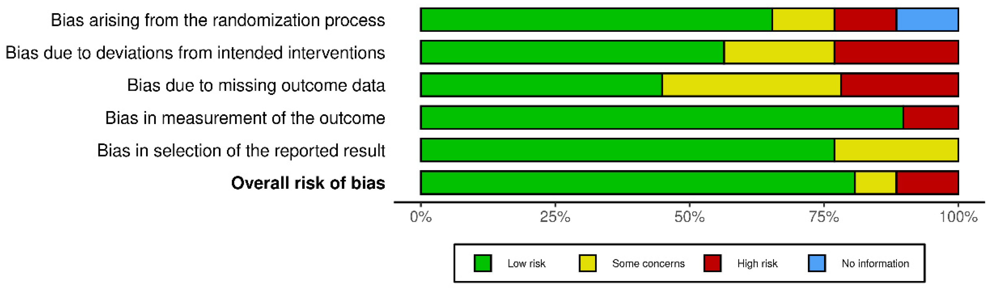
  105. Sterne, J.A.C.; Savović, J.; Page, M.J.; Elbers, R.G.; Blencowe, N.S.; Boutron, I.; Cates, C.J.; Cheng, H.-Y.; Corbett, M.S.; Eldridge, S.M.; et al. RoB 2: A revised tool for assessing risk of bias in randomised trials. BMJ 2019, 366, l4898. [Google Scholar] [CrossRef] [PubMed]
  106. AbdelMassih, A.F.; Fouda, R.; Kamel, A.; Mishriky, F.; Ismail, H.-A.; El Qadi, L.; Malak, L.; Mohamed, M.; Arsanyous, M.; Hazem, M.; et al. Single cell sequencing unraveling genetic basis of severe COVID19 in obesity. Obes. Med. 2020, 20, 100303. [Google Scholar] [CrossRef] [PubMed]
  107. Eggenhuizen, P.J.; Ng, B.H.; Ooi, J.D. Treg Enhancing Therapies to Treat Autoimmune Diseases. Int. J. Mol. Sci. 2020, 21, 7015. [Google Scholar] [CrossRef]
  108. Knolle, J.; Pierau, M.; Hebel, K.; Lampe, K.; Jorch, G.; Kropf, S.; Arens, C.; Brunner-Weinzierl, M.C. Children From the Age of Three Show a Developmental Switch in T-Cell Differentiation. Front. Immunol. 2020, 11, 1640. [Google Scholar] [CrossRef]
  109. Batiha, G.E.S.; Al-kuraishy, H.M.; Al-Gareeb, A.I.; Welson, N.N. Pathophysiology of Post-COVID syndromes: A new perspective. Virol. J. 2022, 19, 158. [Google Scholar] [CrossRef]
  110. Page, M.J.; McKenzie, J.E.; Bossuyt, P.M.; Boutron, I.; Hoffmann, T.C.; Mulrow, C.D.; Shamseer, L.; Tetzlaff, J.M.; Akl, E.A.; Brennan, S.E.; et al. The PRISMA 2020 statement: An updated guideline for reporting systematic reviews. BMJ 2021, 372, n71. [Google Scholar] [CrossRef]
Figure 1. PRISMA 2020 flow diagram for our systematic review to show the study selection process.
Rheumato 03 00011 g001
Figure 2. A detailed number of case series and reports per diagnosis. Abbreviations: CDD: central demyelinating disorder, GBS: Guillain–Barré syndrome, DM: Type 1 diabetes mellitus, HLH: haemophagocytic lymphohistiocytosis, HUS: haemolytic–uremic syndrome, SLE: systemic lupus erythematosus, TRAPS: tumour necrosis factor receptor-associated periodic syndrome.
Rheumato 03 00011 g002
Figure 3. ROC curve for the diagnostic accuracy of age groups in predicting hospitalization during postacute sequelae of COVID-19.
Rheumato 03 00011 g003
Figure 4. Risk-of-bias assessment.
Rheumato 03 00011 g004
Table 1. Age, sex distribution, hospital admission, and interval to immune sequelae of patients with postacute COVID-19 sequelae.
p-Value
Age distribution, N (%)Infants (0–2 y) 11 (10)<0.001
Preschool children (3–5 y) 15 (14)
School children (6–12 y) 42 (38)
Adolescents
(13–18 y) 41 (38)
Sex distribution, N (%)Female 54 (50)0.92
Male 55 (50)
Hospital admission, N (%)Yes 49 (45)0.29
No 60 (55)
Interval between COVID-19 and autoimmune sequelae, N (%)Immediate (0–14 days) 67 (61)<0.001
Classic (15–28 days) 15 (14)
Delayed (>28 days) 27 (25)
Abbreviations: N: number.
Table 2. Addison’s disease as a postacute sequela of COVID-19.
ReferenceAgeSexInterval
between COVID-19 Infection and Disorder
Course of
COVID-19
Infection and
Hospitalization Data
OutcomeNotes
[14]14Female21 days Congestion and
fatigue for three weeks.
  • The patient was restarted on hydrocortisone 50 mg/m2/day and weaned down to a maintenance physiologic dose.
  • Aldosterone was <1 ng/dL and plasma renin was 0.43 ng/mL/h (normal range 0.25–5.82), and she started on fludrocortisone 0.05 mg daily.
  • Due to persistent haemodynamic instability and catecholamine dependence, despite improvement in her inflammatory markers, random cortisol was drawn and was <1 μg/dL.
  • Hydrocortisone stress dose at 50 mg/m2/day was initiated, and this led to improvement in her clinical condition and was able to be weaned off vasopressor support.
  • The baseline ACTH level was elevated to >1250 pg/mL (normal range 9–57 pg/mL), and both baseline and stimulated cortisol was <1 μg/dL, confirming a diagnosis of primary adrenal insufficiency.
  • 21-hydroxylase anti-adrenal antibodies were positive.
  • A diagnosis of primary adrenal insufficiency and autoimmune hypothyroidism in addition to MIS-C was made.
Abbreviations: ACTH: adrenocorticotropic hormone; and MIS-C: multisystem inflammatory syndrome in children.
Disclaimer/Publisher’s Note: The statements, opinions and data contained in all publications are solely those of the individual author(s) and contributor(s) and not of MDPI and/or the editor(s). MDPI and/or the editor(s) disclaim responsibility for any injury to people or property resulting from any ideas, methods, instructions or products referred to in the content.

