
The Phenomenon of the Cross-Resistance of Breast Cancer to Target and Hormonal Drugs: The Role of Epigenetic Reconstruction †

 ,
,  ,
,  ,
, 
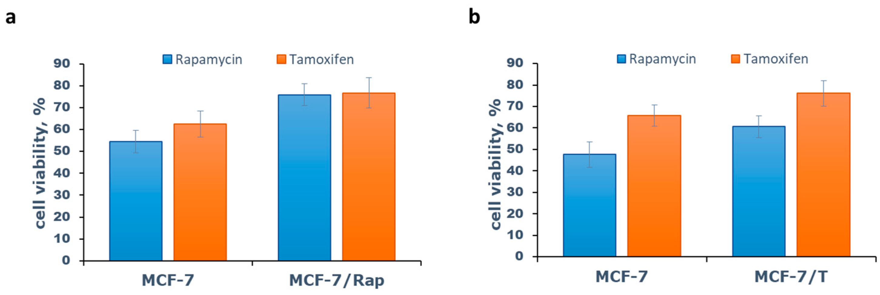
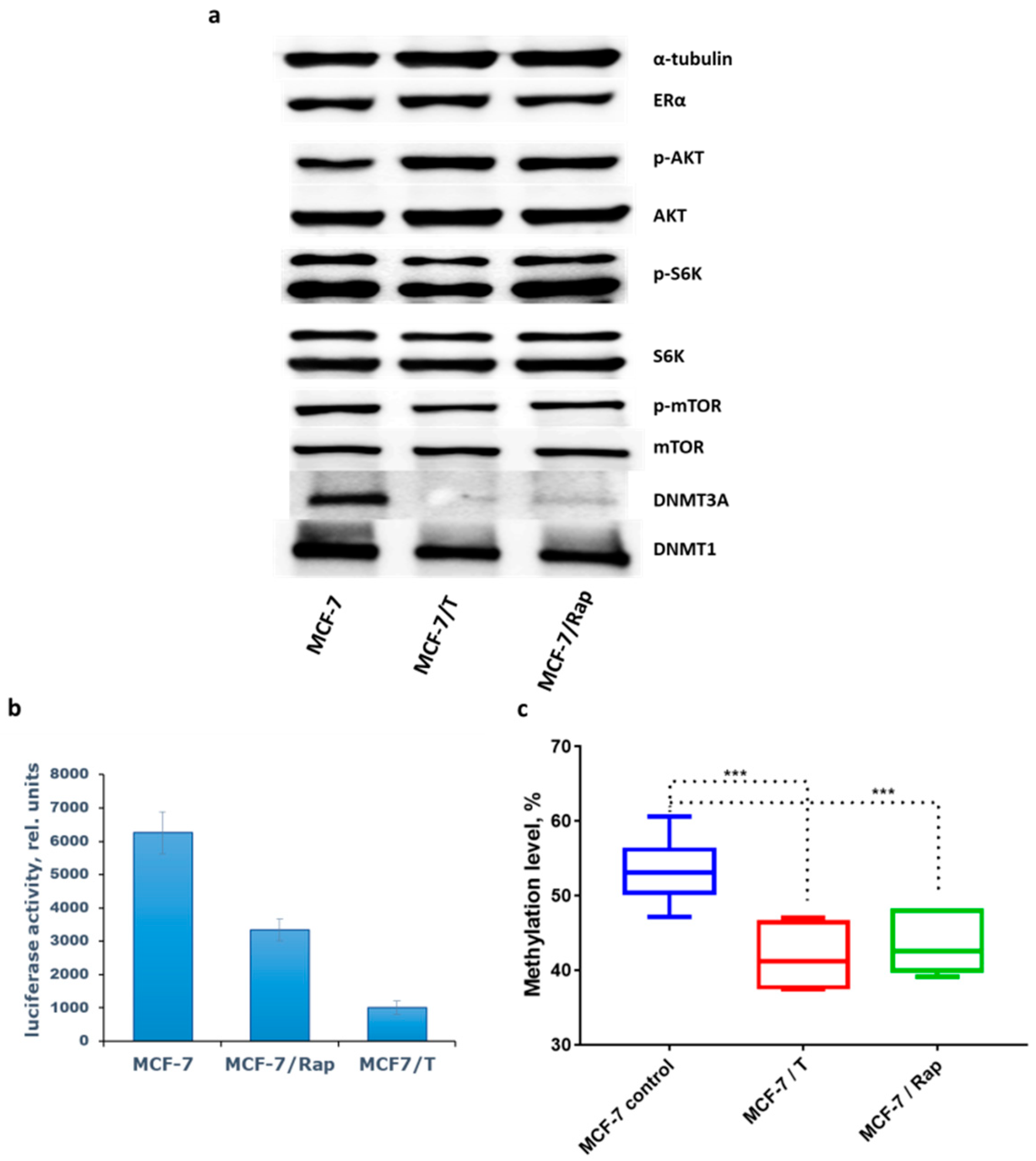
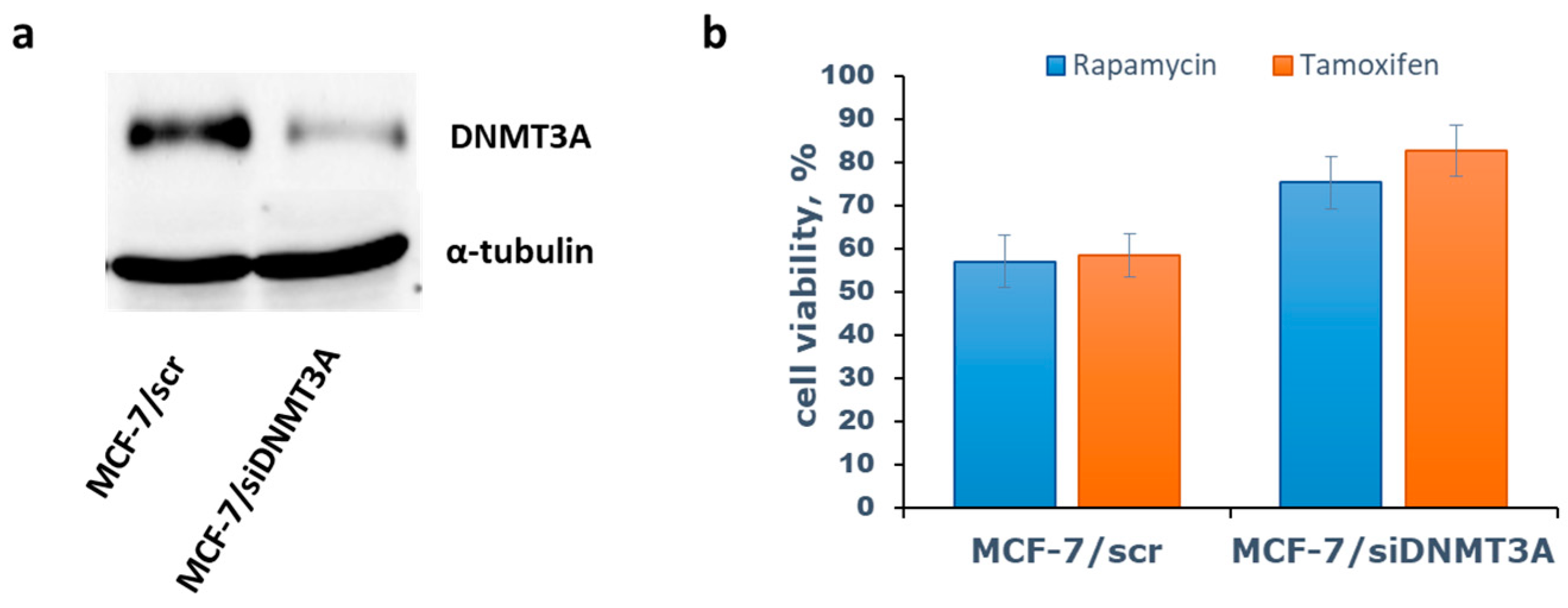
Abstract
:1. Introduction
2. Methods
2.1. Cell Cultures and Evaluation of Antiproliferative Activity
2.2. Transient Transfection and Measurement of Reporter Gene Activity
2.3. Transfection of Small Interfering RNA
2.4. Immunoblotting
2.5. Bisulfite Pyrosequencing for LINE-1 Methylation Analysis
2.6. Statistical Analysis
3. Results and Discussion
4. Conclusions
Author Contributions
Funding
Institutional Review Board Statement
Informed Consent Statement
Data Availability Statement
Conflicts of Interest
References
- Clarke, R.; Tyson, J.J.; Dixon, J.M. Endocrine resistance in breast cancer—An overview and update. Mol. Cell. Endocrinol. 2015, 418, 220–234. [Google Scholar] [CrossRef]
- Araki, K.; Miyoshi, Y. Mechanism of resistance to endocrine therapy in breast cancer: The important role of PI3K/Akt/mTOR in estrogen receptor-positive, HER2-negative breast cancer. Breast Cancer 2017, 25, 392–401. [Google Scholar] [CrossRef]
- Citi, V.; Del Re, M.; Martelli, A.; Calderone, V.; Breschi, M.C.; Danesi, R. Phosphorylation of AKT and ERK1/2 and mutations of PIK3CA and PTEN are predictive of breast cancer cell sensitivity to everolimus in vitro. Cancer Chemother. Pharm. 2018, 81, 745–754. [Google Scholar] [CrossRef] [PubMed]
- Xie, W.; Sun, H.; Li, X.; Lin, F.; Wang, Z.; Wang, X. Ovarian cancer: Epigenetics, drug resistance, and progression. Cancer Cell Int. 2021, 21, 434. [Google Scholar] [CrossRef] [PubMed]
- Hazra, A.; Bose, P.; Sunita, P.; Pattanayak, S.P. Molecular epigenetic dynamics in breast carcinogenesis. Arch. Pharm. Res. 2021, 44, 741–763. [Google Scholar] [CrossRef] [PubMed]
- Shchegolev, Y.; Sorokin, D.; Scherbakov, A.; Shunaev, A.; Andreeva, O.; Mikhaevich, E.; Gudkova, M.; Bure, I.; Berstein, L.; Nemtsova, M.; et al. Upregulation of Akt/Raptor signaling is associated with rapamycin resistance of breast cancer cells. Chem. Biol. Interact. 2020, 330, 109243. [Google Scholar] [CrossRef] [PubMed]
- Iselt, M.; Holtei, W.; Hilgard, P. The tetrazolium dye assay for rapid in vitro assessment of cytotoxicity. Arzneim. Forsch. 1989, 39, 747–749. [Google Scholar]
- Ilovaisky, A.I.; Scherbakov, A.M.; Merkulova, V.M.; Chernoburova, E.I.; Shchetinina, M.A.; Andreeva, O.E.; Salnikova, D.I.; Zavarzin, I.V.; Terent’ev, A.O. Secosteroid–quinoline hybrids as new anticancer agents. J. Steroid Biochem. Mol. Biol. 2023, 228, 106245. [Google Scholar] [CrossRef] [PubMed]
- Scherbakov, A.M.; Lobanova, Y.S.; Shatskaya, V.A.; Onopchenko, O.V.; Gershtein, E.S.; Krasil’nikov, M.A. Activation of mitogenic pathways and sensitization to estrogen-induced apoptosis: Two independent characteristics of tamoxifen-resistant breast cancer cells? Breast Cancer Res. Treat. 2006, 100, 1–11. [Google Scholar] [CrossRef]
- Mruk, D.D.; Cheng, C.Y. Enhanced chemiluminescence (ECL) for routine immunoblotting: An inexpensive alternative to commercially available kits. Spermatogenesis 2011, 1, 121–122. [Google Scholar] [CrossRef] [PubMed]
- Lander, E.S.; Linton, L.M.; Birren, B.; Nusbaum, C.; Zody, M.C.; Baldwin, J.; Devon, K.; Dewar, K.; Doyle, M.; FitzHugh, W.; et al. Initial sequencing and analysis of the human genome. Nature 2001, 409, 860–921. [Google Scholar] [PubMed]
- Yang, A.S.; Estécio, M.R.; Doshi, K.; Kondo, Y.; Tajara, E.H.; Issa, J.P. A simple method for estimating global DNA methylation using bisulfite PCR of repetitive DNA elements. Nucleic Acids Res. 2004, 32, e38. [Google Scholar] [CrossRef] [PubMed]
- Sadakierska-Chudy, A. MicroRNAs: Diverse Mechanisms of Action and Their Potential Applications as Cancer Epi-Therapeutics. Biomolecules 2020, 10, 1285. [Google Scholar] [CrossRef] [PubMed]
- Stone, A.; Valdés-Mora, F.; Gee, J.M.W.; Farrow, L.; McClelland, R.A.; Fiegl, H.; Dutkowski, C.; McCloy, R.A.; Sutherland, R.L.; Musgrove, E.A.; et al. Tamoxifen-induced epigenetic silencing of oestrogen-regulated genes in anti-hormone resistant breast cancer. PLoS ONE 2012, 7, e40466. [Google Scholar] [CrossRef] [PubMed]


Disclaimer/Publisher’s Note: The statements, opinions and data contained in all publications are solely those of the individual author(s) and contributor(s) and not of MDPI and/or the editor(s). MDPI and/or the editor(s) disclaim responsibility for any injury to people or property resulting from any ideas, methods, instructions or products referred to in the content. |
© 2023 by the authors. Licensee MDPI, Basel, Switzerland. This article is an open access article distributed under the terms and conditions of the Creative Commons Attribution (CC BY) license (https://creativecommons.org/licenses/by/4.0/).
Share and Cite
Andreeva, O.E.; Shchegolev, Y.Y.; Scherbakov, A.M.; Sorokin, D.V.; Vinokurova, S.V.; Katargin, A.N.; Salnikova, D.I.; Krasil’nikov, M.A. The Phenomenon of the Cross-Resistance of Breast Cancer to Target and Hormonal Drugs: The Role of Epigenetic Reconstruction. Med. Sci. Forum 2023, 20, 5. https://doi.org/10.3390/IECC2023-14220
Andreeva OE, Shchegolev YY, Scherbakov AM, Sorokin DV, Vinokurova SV, Katargin AN, Salnikova DI, Krasil’nikov MA. The Phenomenon of the Cross-Resistance of Breast Cancer to Target and Hormonal Drugs: The Role of Epigenetic Reconstruction. Medical Sciences Forum. 2023; 20(1):5. https://doi.org/10.3390/IECC2023-14220
Chicago/Turabian StyleAndreeva, Olga E., Yuri Y. Shchegolev, Alexander M. Scherbakov, Danila V. Sorokin, Svetlana V. Vinokurova, Alexey N. Katargin, Diana I. Salnikova, and Mikhail A. Krasil’nikov. 2023. "The Phenomenon of the Cross-Resistance of Breast Cancer to Target and Hormonal Drugs: The Role of Epigenetic Reconstruction" Medical Sciences Forum 20, no. 1: 5. https://doi.org/10.3390/IECC2023-14220