Proceeding Paper

Targeting AKT Kinase in Hydroxytamoxifen-Resistant Breast Cancer Cells †

by
Alexander M. Scherbakov
*,‡,
Fedor B. Bogdanov
,
Alexandra L. Mikhaylova
,
Olga E. Andreeva
and
Diana I. Salnikova
N.N. Blokhin National Medical Research Center of Oncology, The Ministry of Health of the Russian Federation, Moscow 115522, Russia
*
Author to whom correspondence should be addressed.
Presented at the 3rd International Electronic Conference on Cancers: New Targets for Cancer Therapies, 16–30 March 2023; Available online: https://iecc2023.sciforum.net/.
These authors contributed equally to this work.
Med. Sci. Forum 2023, 20(1), 4; https://doi.org/10.3390/IECC2023-14224
Published: 16 March 2023

Abstract

:
More than 650,000 people die each year from breast cancer, making it a particularly significant disease worldwide. The development of about 70% of breast tumors depends on steroid hormones, namely, estrogens. Estrogens trigger signaling pathways that support tumor growth and progression. Hydroxytamoxifen (HT) halting estrogen-induced tumor growth is among the most effective drugs in current anticancer therapy. The purpose of this work was to investigate approaches to overcome breast cancer cell resistance to hydroxytamoxifen. Cells with resistance to the antiestrogen hydroxytamoxifen were obtained by long-term incubation of parental MCF7 cells with this drug. Estrogen receptor α (ERα) expression and progesterone receptor (PR) expression were analyzed by immunoblotting. The resistant MCF7/HT cells were found not to lose ERα expression. These cells were found to have slightly reduced ERα activity when compared to parental MCF7 cells. The expression of PR, one of the ERα targets, was downregulated in hydroxytamoxifen-resistant cells. AKT kinase belongs to the PI3K/AKT/mTOR signaling pathway, and its activity is associated with resistance. Three types of AKT inhibitors were evaluated, including AKT inhibitor IV (6-(2-benzothiazolyl)-1-ethyl-2-[2-(methylphenylamino)ethenyl]-3-phenyl-1H-benzimidazolium, monoiodide), 10-DEBC (2-chloro-N,N-diethyl-10H-phenoxazine-10-butanamine, monohydrochloride), and luminespib (HSP90 inhibitor, 5-[2,4-dihydroxy-5-(1-methylethyl)phenyl]-N-ethyl-4-[4-(4-morpholinylmethyl)phenyl]-3-isoxazolecarboxamide). All three compounds showed high antiproliferative activity against hydroxytamoxifen-resistant cells. The IC50 value of 10-DEBC was 4.2 µM, and this was when the AKT inhibitor IV was more active with IC50 value of 390 nM. The HSP90 inhibitor luminespib, which reduces AKT expression, showed the highest activity against parental and hydroxytamoxifen-resistant breast cancer cells, with IC50 values of 14 and 18 nM, respectively. Thus, the hydroxytamoxifen-resistant cells were found to partially retain hormone signaling and to be sensitive to selective AKT inhibitors. The best effects were discovered for HSP90-AKT blocker luminespib, with an IC50 value of about 20 nM.

1. Introduction

The incidence of breast cancer occupies a leading position among other oncological pathologies in patients around the world. The most common (more than 70% of cases) is the luminal subtype A of breast cancer, which is positive for estrogen (ERα+) or progesterone (PR+) receptors, and it is negative for epidermal growth factor receptor type 2 (HER2-). Estrogens, by activating nuclear receptors, promote the survival and proliferation of cancer tissue through gene transcription and activation of cell molecular pathways [1].
Treatment of patients with ERα-positive breast cancer is aimed at either lowering estrogen levels or blocking ERα signaling [2]. Three classes of endocrine agents are currently used in clinical practice: selective estrogen receptor modulators (SERMs), which block ERα activity, inhibitors of estrogen synthesis (aromatase inhibitors), and selective estrogen receptor regulators (SERDs), which cause destabilization and degradation of ERα [3,4].
Tamoxifen, as well as its active metabolite, 4-hydroxytamoxifen (HT), are first-line drugs in the treatment of breast cancer, acting as selective receptor modulators and thereby inhibiting tumor tissue proliferation [5]. Despite the proven efficacy of endocrine therapy in primary cancer and its reducing the mortality rate by up to 40%, over time, patients can develop resistance, which can lead to cancer recurrence and metastasis [6]. In addition, although less frequently, patients may develop innate resistance to SERMs or SERDs, including tamoxifen and fulvestrant [7].
Tamoxifen resistance is multifactorial and involves the modulation of multiple physiological processes and signaling pathways. Mechanisms of endocrine resistance include changes in signaling through disruption of biochemical cascades and modulation of ERα expression, including ESR1 mutations [8], changes in regulatory proteins and transcription factors, nuclear receptors and epigenetic modulators, changes in the tumor microenvironment, and tamoxifen metabolic activity [9].
Since about 650,000 people worldwide die from breast cancer every year, that is why understanding the patterns and factors in the development of resistance to endocrine therapy drugs is extremely important [10]. It is also essential to find novel strategies to inhibit the growth of resistant cells. Therefore, the purpose of this work was to investigate approaches to overcome breast cancer cell resistance to hydroxytamoxifen.

2. Methods

2.1. Cells and Reagents

The MCF7 breast cancer cells were obtained from ATCC. To develop resistant clones, MCF7 cells were treated with hydroxytamoxifen for a long period (12 months), as described in our recent work [11]. Subsequent experiments were performed three weeks after withdrawal of the drug. The resistance index, defined as IC50 of HT against MCF7/HT divided by its IC50 against parental MCF7 cells, was 2 [11]. The hydroxytamoxifen-resistant line was named MCF7/HT. Hydroxytamoxifen (4-[(1Z)-1-[4-[2-(dimethylamino)ethoxy]phenyl]-2-phenyl-1-buten-1-yl]-phenol), 17β-estradiol, and AKT inhibitors were purchased from Cayman Chemical (Denver, CO, USA). Cell culture reagents were provided by PanEco (Moscow, Russia).

2.2. Evaluation of Antiproliferative Activity

The cells were seeded into plates, and the compounds were added 24 h later. Cell viability was assessed by the MTT assay after 72 h, as described previously in the work [12].

2.3. Immunoblotting

Immunoblotting with modifications was held, as described earlier [13]. ERα and PR expression was evaluated using Cell Signaling Technology (CST) antibodies. Antibodies to α-tubulin (CST) were applied to normalize and control the loading of samples into a gel. The detection was performed using secondary antibodies to rabbit Ig conjugated with horseradish peroxidase (Jackson ImmunoResearch: Ely, UK) and an ImageQuant LAS 4000 imager (GE Healthcare, Chicago, IL, USA), as described in Mruk and Cheng’s protocol [14]. Densitometry for immunoblotting data was performed using ImageJ software 1.53t (Wayne Rasband: Bethesda, MD, USA). The protocol for densitometry was provided by The University of Queensland, with recommendations from the work [15].

2.4. Reporter Assay

The transcriptional activity of the estrogen receptor α was assessed by reporter analysis, as described in work [12]. The cells were transfected using Lipofectamine 2000 and the following plasmids: ERE-LUC, carrying the luciferase gene under the control of estrogen responsive elements [16], and a plasmid for the expression of β-galactosidase to control the efficiency of transfection.
To reduce the effects of serum steroids on cells, the experiments were carried out in a steroid-free medium (HyClone: Marlborough, MA, USA). Luciferase activity was measured on a Tecan Infinite M200 Pro microplate reader (Männedorf, Switzerland), according to the Promega protocol. The relative activity of luciferase was considered as the ratio of the activity of luciferase to β-galactosidase.

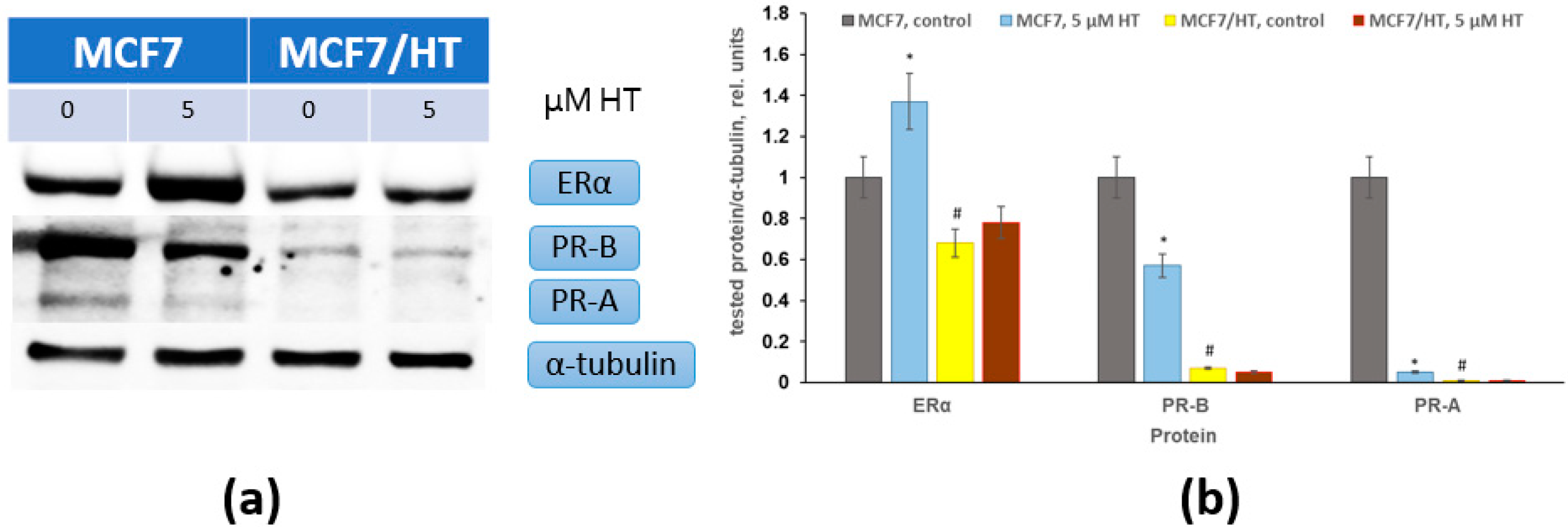
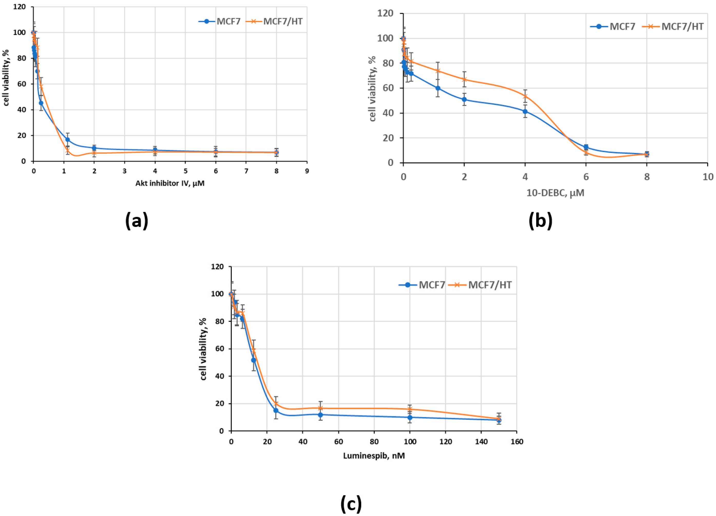
3. Results and Discussion

In this study, MCF7/HT cells obtained by long-term cultivation of MCF7 breast cancer cells with antiestrogen hydroxytamoxifen were studied [11]. Hydroxytamoxifen is one of the major active metabolites of antiestrogen tamoxifen [17]. ERα is one of major drivers of cell growth and survival of hormone-dependent breast cancer cells. To assess ERα activity, MCF7 and MCF7/HT cells were transfected with the ERE-LUC plasmids, containing the luciferase reporter gene under the ERα-dependent promoters. Luciferase activity can be used to estimate the activity of ERα; 17β-estradiol (10 nM), a natural ERα ligand, was used as an inducer of luciferase activity. As can be seen in Table 1, 17β-estradiol-induced luciferase activity was 1.5 times higher in MCF7 cells than that in MCF7/HT cells. Thus, the reporter assay, based on 17β-estradiol-induced luciferase activity, indicates the loss of sensitivity of MCF7/HT cells to hormonal stimuli.
Immunoblotting analysis was performed to assess the expression of estrogen and progesterone receptors. It was found that ERα expression was substantially decreased in hormone-resistant cells. Progesterone receptor expression is actively regulated by estrogens, and high PR expression indicates ERα activity in cells. As shown in Figure 1, PR expression was quite high in parental MCF7 cells, whereas, in MCF7/HT, it was significantly reduced.
AKT kinase belongs to the PI3K/AKT/mTOR signaling pathway, and its increased activity is associated with resistance [18]. Previously, we showed that the expression of AKT phosphorylated at Ser473 is increased in cells subjected to long-term treatment with HT (MCF7/HT) [11]. The involvement of AKT in the development of resistance to tamoxifen has been confirmed in a number of works performed by other researchers [19,20,21]. This makes it possible to use AKT inhibitors to suppress the growth of the established resistant cells. Three types of AKT inhibitors were evaluated, including AKT inhibitor IV (6-(2-benzothiazolyl)-1-ethyl-2-[2-(methylphenylamino)ethenyl]-3-phenyl-1H-benzimidazolium, monoiodide), 10-DEBC (2-chloro-N,N-diethyl-10H-phenoxazine-10-butanamine, monohydrochloride), and luminespib (HSP90 inhibitor, 5-[2,4-dihydroxy-5-(1-methylethyl)phenyl]-N-ethyl-4-[4-(4-morpholinylmethyl)phenyl]-3-isoxazolecarboxamide). All three compounds showed high antiproliferative activity against hydroxytamoxifen-resistant cells, as can be seen in Figure 2. The IC50 value of 10-DEBC was 4.2 µM, when Akt inhibitor IV was more active with IC50 value of 390 nM. The HSP90 inhibitor luminespib showed the highest activity against parental and hydroxytamoxifen-resistant breast cancer cells, with an IC50 value of 14 and 18 nM, respectively.
The efficacy of antiestrogen treatments, particularly tamoxifen and hydroxytamoxifen, in ERα(+) breast cancer, can be measured by the expression levels of certain biomarkers, the most common of which are insulin-like growth factor 1 receptor (IGF-1R) and epidermal growth factor receptor (EGFR) [22]. Tamoxifen-resistant breast cancer cells show reduced expression of IGF-1R and increased expression of EGFR. In addition, β1-integrin [23] is overexpressed in resistant cells, which is involved in focal adhesion between cells and has a significant effect on cancer cell proliferation and metastasis through activation of focal adhesion kinase (FAK), mitogen-activated protein kinase (MAPK), and phosphoinositide-3-kinase (PI3K).
Protein kinases can be considered as key targets for targeted breast cancer therapies, since their activation plays an important role in cell survival, metabolism, and proliferation. The protein kinase AKT, an important participant in the PI3K/AKT/mTOR signaling pathway [24], contributes to the development of drug resistance and tumor progression [19]. Inhibition of AKT as a central component of the PI3K/AKT signaling pathway has been an attractive therapeutic approach in breast cancer since the 1990s. There are two classes of AKT inhibitors: ATP-competitive inhibitors that bind to the active site of the protein, as well as allosteric inhibitors that bind to the PH domain and prevent AKT phosphorylation. However, despite their attractiveness, AKT inhibitors are quite toxic compounds and have limited clinical efficacy as a monotherapy [25].
Overexpression of certain protein molecules can contribute to the development of drug resistance, and their assessment is an important diagnostic feature in the formulation of a therapeutic regimen for breast cancer. Thus, a significant increase in HOXA5 expression in breast cancer cells leads to activation of the PI3K/AKT signaling cascade and a decrease in p53 and p21, which, in turn, leads to the development of resistance to tamoxifen therapy due to impaired apoptosis [19]. In addition, overexpression of the lysosomal serine protease PRCP contributes to the development of 4-hydroxytamoxifen resistance in ERα(+) breast cancer cells by activating AKT and increasing signaling along the IGF-1R signaling pathway [26].
The PI3K/AKT signaling pathway is involved in the regulation of the M1 Forkhead box (FOXM1) protein’s expression, which is an important transcription factor that regulates proliferation, invasion, and metastasis. AKT stimulates FOXM1 activity by inhibiting FOXO3a, which has the opposite effect. In ERα-positive breast cancer, FOXM1 expression increases, which is associated with increased cell invasiveness and resistance to endocrine treatments [27]. In addition, the activated PI3K/AKT signaling pathway in tamoxifen-resistant breast cancer cells contributes to a significantly higher expression of BARD1 and BRCA1, which determine drug resistance to cytotoxic drugs [28].
Here, we characterized hydroxytamoxifen-resistant cells, which were found to partially retain hormone signaling. MCF7/HT cells were sensitive to selective AKT inhibitors. The best effects were discovered for HSP90-AKT blocker luminespib, which had an IC50 value of about 20 nM.
The combination of AKT inhibitors and agents acting on other molecular targets may increase the effectiveness of cancer therapy and be considered as a potential opportunity to overcome resistance in ERα(+) breast cancers.

Author Contributions

Conceptualization, A.M.S.; methodology, A.M.S. and O.E.A.; formal analysis, A.M.S., O.E.A. and D.I.S.; investigation, A.M.S., O.E.A., D.I.S., A.L.M. and F.B.B.; writing—original draft preparation, A.M.S. and F.B.B.; writing—review and editing, A.M.S., O.E.A., A.L.M. and D.I.S.; visualization, A.M.S.; supervision, A.M.S. All authors have read and agreed to the published version of the manuscript.

Funding

This research was funded by the Russian Science Foundation, grant number 19-15-00245 (https://rscf.ru/project/22-15-35008/, accessed on 4 January 2023).

Institutional Review Board Statement

Not applicable.

Informed Consent Statement

Not applicable.

Data Availability Statement

Data are available from the authors.

Acknowledgments

The authors thank Mikhail A. Krasil’nikov for the helpful suggestions regarding the manuscript. The plasmids used in the work were kindly provided by F. Gannon and G. Reid. The study was carried out using the unique scientific facility Transgenebank.

Conflicts of Interest

The authors declare no conflict of interest. The funders had no role in the design of the study, in the collection, analyses, or interpretation of data, in the writing of the manuscript, or in the decision to publish the results.

References

  1. Hua, H.; Zhang, H.; Kong, Q.; Jiang, Y. Mechanisms for estrogen receptor expression in human cancer. Exp. Hematol. Oncol. 2018, 7, 24. [Google Scholar] [CrossRef]
  2. Heel, R.C.; Brogden, R.N.; Speight, T.M.; Avery, G.S. Tamoxifen: A review of its pharmacological properties and therapeutic use in the treatment of breast cancer. Drugs 1978, 16, 1–24. [Google Scholar] [CrossRef]
  3. Duan, L.; Calhoun, S.J.; Perez, R.E.; Macias, V.; Mir, F.; Gattuso, P.; Maki, C.G. Prolylcarboxypeptidase promotes IGF1R/HER3 signaling and is a potential target to improve endocrine therapy response in estrogen receptor positive breast cancer. Cancer Biol. Ther. 2022, 23, 1–10. [Google Scholar] [CrossRef]
  4. Scherbakov, A.M.; Krasil’nikov, M.A.; Kushlinskii, N.E. Molecular mechanisms of hormone resistance of breast cancer. Bull. Exp. Biol. Med. 2013, 155, 384–395. [Google Scholar] [CrossRef] [PubMed]
  5. Jordan, V.C. 50th anniversary of the first clinical trial with ICI 46,474 (tamoxifen): Then what happened? Endocr. -Relat. Cancer 2021, 28, R11–R30. [Google Scholar] [CrossRef] [PubMed]
  6. Wang, J.; Han, Y.; Wang, J.; Li, Q.; Xu, B. Endocrine Therapy-Based Strategies for Metastatic Breast Cancer with Different Endocrine Sensitivity Statuses: A Systematic Review and Network Meta-Analysis. Cancers 2022, 14, 6100. [Google Scholar] [CrossRef] [PubMed]
  7. Patel, H.K.; Bihani, T. Selective estrogen receptor modulators (SERMs) and selective estrogen receptor degraders (SERDs) in cancer treatment. Pharmacol. Ther. 2018, 186, 1–24. [Google Scholar] [CrossRef]
  8. García-Becerra, R.; Santos, N.; Díaz, L.; Camacho, J. Mechanisms of resistance to endocrine therapy in breast cancer: Focus on signaling pathways, miRNAs and genetically based resistance. Int. J. Mol. Sci. 2012, 14, 108–145. [Google Scholar] [CrossRef] [PubMed]
  9. Hanker, A.B.; Sudhan, D.R.; Arteaga, C.L. Overcoming Endocrine Resistance in Breast Cancer. Cancer Cell 2020, 37, 496–513. [Google Scholar] [CrossRef]
  10. Wilkinson, L.; Gathani, T. Understanding breast cancer as a global health concern. Br. J. Radiol. 2022, 95, 20211033. [Google Scholar] [CrossRef]
  11. Scherbakov, A.M.; Basharina, A.A.; Sorokin, D.V.; Mikhaevich, E.I.; Mizaeva, I.E.; Mikhaylova, A.L.; Bogush, T.A.; Krasil’nikov, M.A. Targeting hormone-resistant breast cancer cells with docetaxel: A look inside the resistance. Cancer Drug Resist. 2023, 6, 103–115. [Google Scholar] [CrossRef] [PubMed]
  12. Ilovaisky, A.I.; Scherbakov, A.M.; Merkulova, V.M.; Chernoburova, E.I.; Shchetinina, M.A.; Andreeva, O.E.; Salnikova, D.I.; Zavarzin, I.V.; Terent’ev, A.O. Secosteroid-quinoline hybrids as new anticancer agents. J. Steroid Biochem. Mol. Biol. 2023, 228, 106245. [Google Scholar] [CrossRef] [PubMed]
  13. Scherbakov, A.M.; Lobanova, Y.S.; Shatskaya, V.A.; Onopchenko, O.V.; Gershtein, E.S.; Krasil’nikov, M.A. Activation of mitogenic pathways and sensitization to estrogen-induced apoptosis: Two independent characteristics of tamoxifen-resistant breast cancer cells? Breast Cancer Res. Treat. 2006, 100, 1–11. [Google Scholar] [CrossRef] [PubMed]
  14. Mruk, D.D.; Cheng, C.Y. Enhanced chemiluminescence (ECL) for routine immunoblotting: An inexpensive alternative to commercially available kits. Spermatogenesis 2011, 1, 121–122. [Google Scholar] [CrossRef]
  15. Taylor, S.C.; Berkelman, T.; Yadav, G.; Hammond, M. A defined methodology for reliable quantification of Western blot data. Mol. Biotechnol. 2013, 55, 217–226. [Google Scholar] [CrossRef]
  16. Reid, G.; Hübner, M.R.; Métivier, R.; Brand, H.; Denger, S.; Manu, D.; Beaudouin, J.; Ellenberg, J.; Gannon, F. Cyclic, proteasome-mediated turnover of unliganded and liganded ERalpha on responsive promoters is an integral feature of estrogen signaling. Mol. Cell 2003, 11, 695–707. [Google Scholar] [CrossRef]
  17. Sarkaria, J.N.; Miller, E.M.; Parker, C.J.; Jordan, V.C.; Mulcahy, R.T. 4-Hydroxytamoxifen, an active metabolite of tamoxifen, does not alter the radiation sensitivity of MCF-7 breast carcinoma cells irradiated in vitro. Breast Cancer Res. Treat. 1994, 30, 159–165. [Google Scholar] [CrossRef]
  18. Tokunaga, E.; Kimura, Y.; Mashino, K.; Oki, E.; Kataoka, A.; Ohno, S.; Morita, M.; Kakeji, Y.; Baba, H.; Maehara, Y. Activation of PI3K/Akt signaling and hormone resistance in breast cancer. Breast Cancer 2006, 13, 137–144. [Google Scholar] [CrossRef]
  19. Kim, C.Y.; Kim, Y.C.; Oh, J.H.; Kim, M.H. HOXA5 confers tamoxifen resistance via the PI3K/AKT signaling pathway in ER-positive breast cancer. J. Cancer 2021, 12, 4626–4637. [Google Scholar] [CrossRef]
  20. Hamadneh, L.; Abuarqoub, R.; Alhusban, A.; Bahader, M. Upregulation of PI3K/AKT/PTEN pathway is correlated with glucose and glutamine metabolic dysfunction during tamoxifen resistance development in MCF-7 cells. Sci. Rep. 2020, 10, 21933. [Google Scholar] [CrossRef]
  21. Yoo, Y.A.; Kim, Y.H.; Kim, J.S.; Seo, J.H. The functional implications of Akt activity and TGF-beta signaling in tamoxifen-resistant breast cancer. Biochim. Biophys. Acta 2008, 1783, 438–447. [Google Scholar] [CrossRef]
  22. Bitton, A.; Zheng, Y.; Houston, J.P.; Houston, K.D. Investigating differences between tamoxifen resistant and sensitive breast cancer cells with flow cytometry. Cytometry Part A J. Int. Soc. Anal. Cytol. 2021, 99, 164–169. [Google Scholar] [CrossRef] [PubMed]
  23. Hu, S.; Yang, Q.; Chen, Z.; Fu, W. Role of β1-integrin in promoting cell motility and tamoxifen resistance of human breast cancer MCF-7 cells. Asia-Pac. J. Clin. Oncol. 2022; Early View. [Google Scholar] [CrossRef]
  24. Hua, H.; Zhang, H.; Chen, J.; Wang, J.; Liu, J.; Jiang, Y. Targeting Akt in cancer for precision therapy. J. Hematol. Oncol. 2021, 14, 128. [Google Scholar] [CrossRef] [PubMed]
  25. Miricescu, D.; Totan, A.; Stanescu, S., II; Badoiu, S.C.; Stefani, C.; Greabu, M. PI3K/AKT/mTOR Signaling Pathway in Breast Cancer: From Molecular Landscape to Clinical Aspects. Int. J. Mol. Sci. 2020, 22, 173. [Google Scholar] [CrossRef] [PubMed]
  26. Saatci, O.; Huynh-Dam, K.T.; Sahin, O. Endocrine resistance in breast cancer: From molecular mechanisms to therapeutic strategies. J. Mol. Med. 2021, 99, 1691–1710. [Google Scholar] [CrossRef]
  27. Pham, T.H.; Page, Y.L.; Percevault, F.; Ferrière, F.; Flouriot, G.; Pakdel, F. Apigenin, a Partial Antagonist of the Estrogen Receptor (ER), Inhibits ER-Positive Breast Cancer Cell Proliferation through Akt/FOXM1 Signaling. Int. J. Mol. Sci. 2021, 22, 470. [Google Scholar] [CrossRef]
  28. Zhu, Y.; Liu, Y.; Zhang, C.; Chu, J.; Wu, Y.; Li, Y.; Liu, J.; Li, Q.; Li, S.; Shi, Q.; et al. Tamoxifen-resistant breast cancer cells are resistant to DNA-damaging chemotherapy because of upregulated BARD1 and BRCA1. Nat. Commun. 2018, 9, 1595. [Google Scholar] [CrossRef]
Figure 1. Immunoblotting analysis of ERα and PR in parental (MCF7) and hydroxytamoxifen-resistant (MCF7/HT) breast cancer cells; (a) immunoblots; (b) densitometry of immunoblots; HT—hydroxytamoxifen; PR-B, PR-A—progesterone receptor B and A; ERα—estrogen receptor alpha. Antibodies to α-tubulin were used to normalize and control the loading of samples into a gel. * p < 0.05 versus corresponding control cells; # p < 0.05 versus MCF7 cells.
Msf 20 00004 g001
Figure 2. Antiproliferative activity of AKT inhibitors against parental (MCF7) and hydroxytamoxifen-resistant (MCF7/HT) breast cancer cells (72 h treatments with compounds): (a) AKT inhibitor IV; (b) 10-DEBC; (c) luminespib).
Msf 20 00004 g002
Table 1. Characteristics of parental and hydroxytamoxifen-resistant breast cancer cells (* p < 0.05 versus MCF7 cells).
Cells17β-Estradiol-Induced Luciferase Activity, Rel. Units
MCF72985 ± 301
MCF7/HT2008 ± 191 *
Resistance index1.5
Disclaimer/Publisher’s Note: The statements, opinions and data contained in all publications are solely those of the individual author(s) and contributor(s) and not of MDPI and/or the editor(s). MDPI and/or the editor(s) disclaim responsibility for any injury to people or property resulting from any ideas, methods, instructions or products referred to in the content.

