Review

Overview of Pharmacological Therapies for Diffuse Tenosynovial Giant Cell Tumor

1
Department of Oncology, Lausanne University Hospital (CHUV), 1011 Lausanne, Switzerland
2
Department of Orthopedics, Lausanne University Hospital (CHUV), 1011 Lausanne, Switzerland
3
Department of Diagnostic Radiology and Interventional Radiology, Lausanne University Hospital (CHUV), 1011 Lausanne, Switzerland
4
Faculty of Biology and Medicine, University of Lausanne, 1011 Lausanne, Switzerland
*
Author to whom correspondence should be addressed.
Future Pharmacol. 2023, 3(4), 926-937; https://doi.org/10.3390/futurepharmacol3040056
Submission received: 11 September 2023 / Revised: 8 November 2023 / Accepted: 21 November 2023 / Published: 1 December 2023

Abstract

:
Tenosynovial giant cell tumor (TGCT) is a rare and locally aggressive benign tumor arising from the synovium of joints, bursae, and tendon sheaths. It is classified into localized (L-TGCT) and diffuse (D-TGCT) forms based on the extent of involvement. Surgical resection is the primary treatment, though achieving a definitive cure remains challenging due to the high recurrence rates, especially in D-TGCT. Systemic therapies targeting the CSF1-CSF1R axis have emerged as promising treatment options. CSF1R tyrosine kinase inhibitors (TKIs) such as imatinib, nilotinib, pexidartinib, and vimseltinib, alongside anti-CSF1R antibodies like emactuzumab, cabiralizumab, and lacnotuzumab, have shown encouraging results in managing TGCT, particularly when surgery is not feasible or poses significant morbidity. Other potential therapies, including local treatments and anti-inflammatory drugs, are being explored for TGCT management. This review provides an overview of systemic treatment options for D-TGCT, highlighting emerging therapeutic modalities and their potential implications. Effective management is crucial due to TGCT’s significant morbidity despite its non-life-threatening nature, necessitating novel approaches to improve patient prognosis and quality of life.

1. Introduction

Tenosynovial giant cell tumor (TGCT), also known as pigmented villonodular synovitis (PVNS), is a rare, locally aggressive, benign neoplasm originating in the synovium of joints, bursae and tendon sheaths, exhibiting synovial differentiation. The World Health Organization (WHO) 2013 classifies TGCT into localized (L-TGCT) and diffuse (D-TGCT) forms. L-TGCT encompasses the previous entities giant cell tumor of the tendon sheath (GCTTS) and nodular tenosynovitis, affecting a part of the synovium and commonly localized in the digits as a well-circumscribed nodule. D-TGCT is a locally aggressive tumor that affects the entire synovium of large joints, most commonly the knee, often extending into extra-articular structures [1,2,3].
Annual incidence falls between 1.8 per million for diffuse and 50 per million for localized forms and the affected people’s ages range mainly between 20 and 50 years old. Prevalence in 2012 was 44.3% for L-TGCT and 11.5% for L-TGCT according to a Danish cohort study [1]. TGCTs typically manifest as monoarticular conditions, and their clinical presentation is non-specific and varies depending on the localization of occurrence. Extra-articular and tendon sheath forms usually present as slow-growing painless nodules, while intra-articular and diffuse forms present with symptoms including pain, stiffness and edema of the affected joint, hemorrhagic joint effusions, and erosions of the cartilage potentially leading to secondary osteoarthritis [4,5].
Magnetic resonance imaging (MRI) is the gold-standard imaging modality to establish diagnosis as well as for preoperative evaluation of TGCT. L-TGCT appears as well-demarcated lesions of the tendon, whereas D-TGCT typically presents as a soft tissue intra-articular mass with irregular margins, often associated with joint effusion and eventually extra-articular extension. D-TGCT appears more multinodular on MRI than L-TGCT. Bone and cartilage involvement is more common in D-TGCT while peripheral hypointensity is more common in L-TGCT [4,6,7,8].
Biopsy is not mandatory for the diagnosis of TGCT when clinical and MRI findings are typical but should be performed in unusual presentations. Histologically, TGCT is characterized by an hypervascular and hyperplastic synovium containing multinucleated osteoclast-like giant cells, macrophages, and hemosiderin [4,9,10].
When feasible, surgical resection is the mainstay of treatment for treatment-naïve TGCT, although surgery can be debilitating and achieving a definitive cure is difficult [11]. The risk of recurrence for D-TGCT after surgery is high, with a 10-year estimated recurrence rate of around 19.1% in one Danish cohort study [1] and up to 44% according to Mastboom, M.J.L., et al. (2019) [3], whereas the risk of recurrence for L-TGCT after surgery is comparatively lower, approximately 10% [1]. Adjuvant or perioperative radiotherapy is generally not recommended due to potential toxicity such as joint stiffness, necrosis, or malignant transformation to sarcoma [12].
Despite being non-life-threatening and rarely metastatic, disease progression of D-TGCT can cause significant morbidity, such as long-term pain and joint dysfunction, and in some cases, amputation may be considered, thus negatively affecting quality of life (QoL) [13,14]. Systemic therapy targeting the CSF1/CSF1R axis is an option in patients with disease relapse or progression where further surgery would be associated with significant morbidity as well as in patients with advanced primary disease. Current evidence for treatment of TGCT is mostly based on retrospective data and few prospective trials exist. Of note, CS1R inhibitors are considered a standard treatment for D-TGCT but are not approved in all countries, but they can be obtained off-label through participation in clinical trials.
In our review, we provide a comprehensive overview of pharmacotherapy for D-TGCT, showcasing novel treatment approaches aimed at improving patient outcome and quality of life. In the text, we describe the pathogenesis of this disease and summarize systemic treatment options as well as pharmacological treatments administered locally. We analyze mechanisms of action, efficacy, and toxicities of different treatments, with the scope of defining the optimal clinical management for this disease that provokes significant morbidity in young patients. Many data presented in this article are derived from early-phase trials and more phase III trials are necessary.

2. Materials and Methods

We conducted a literature research in PubMed, Google Scholar, and Science Direct using the terms “Tenosynovial Giant Cell Tumor”, Pathogenesis of TGCT, Treatment of TGCT, and Imaging of TGCT, as well as searched for specific research using the treatments listed in this article for keywords. Articles included in the reference list were written in English and were published between 1998 and February 2023. Articles including original research were favored rather than review articles. Furthermore, we performed research in the ClinicalTrials.gov database for ongoing trials.

3. Pathogenesis

TGCT pathogenesis has been long debated. TGCT has been considered of inflammatory origin, as supported by the polyclonal proliferation of tumor cells described by Vogrincic et al. (1997) [15]. Cytogenetic studies have since demonstrated the presence of clonal structural chromosomal aberrations, frequently involving the 1p11-13 region, supporting a neoplastic process [16]. In many cases of TGCT, overexpression of colony stimulating factor 1 (CSF1) is driven by a t(1;2) translocation, fusing the CSF1 gene on chromosome 1p11 with the collagen type VI a3 (COL6A3) promoter on chromosome 2q37 [17,18]. Other fusion partners have also been reported to result in CSF1 overexpression [19]. CSF1 promotes the differentiation of myeloid progenitors into monocytes, macrophages, dendritic cells, and bone-resorbing osteoclasts, and controls cell proliferation [20].
West et al. (2006) [21] showed that in most cases of TGCT harboring the t(1;2) translocation, the translocation is present only in a restricted amount of cells. Thus, only few cells within the tumor express CSF1 and are neoplastic, while most of the tumor cells express CSF1 receptor (CSF1R). CSF1 attracts CSF1R-expressing inflammatory cells such as macrophages, resulting in the formation of osteoclast-like multinucleated giant cells [17]. Consequently, TGCT tumors contain only a small number of neoplastic cells and a large number of reactive cells [21].

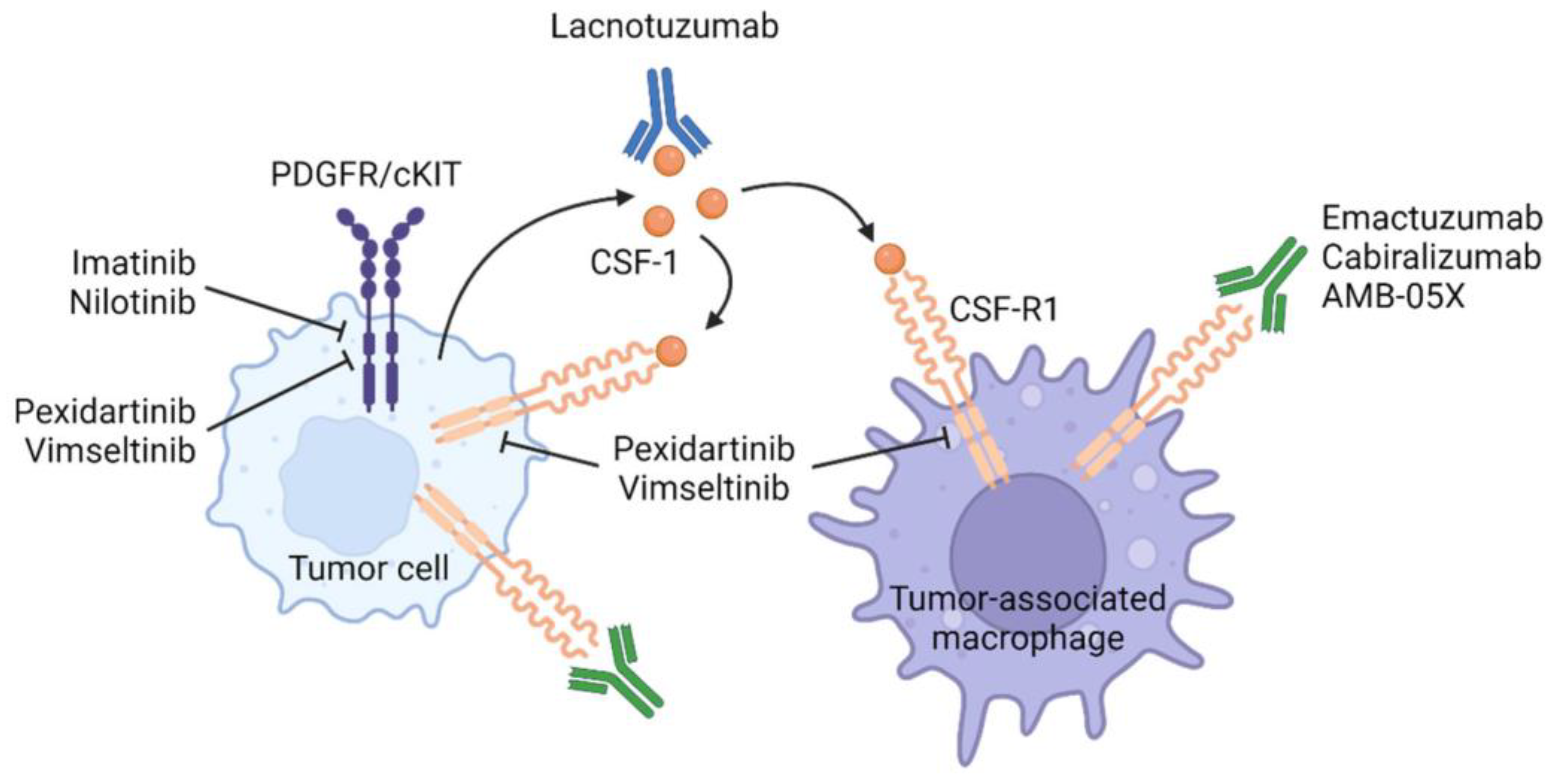
4. Systemic Therapies Targeting the CSF1-CSF1R Axis (Figure 1)

Due to its role in TGCT pathogenesis, targeting the CSF1-CSF1R axis is key in TGCT treatment. CSF1R tyrosine kinase inhibitors (TKIs) and antibodies have shown efficacy in tumor control in D-TGCT.
Figure 1. Mechanism of action of CSF1/CSFR1 inhibitors.
Futurepharmacol 03 00056 g001
A retrospective single-center study of 39 patients with advanced and/or inoperable D-TGCT treated with CSF1R TKI or antibodies showed a progression-free rate at 30 months of 56% for 1st-line treatment, 25% for 2nd-line, and 20% for 3rd-line. The median times to progression (TTPs) were 12 months and 9 months for 2nd- and 3rd-line treatments, respectively. Although the median duration of treatment was short, tumor control could be achieved beyond treatment interruption, and 50% of patients required multiple lines of treatment [22].

4.1. CSF1R TKIs

4.1.1. Imatinib (Table 1)

Imatinib is a multi-kinase inhibitor that targets the chronic myeloid leukemia-specific tyrosine kinase, breakpoint cluster region-Abelson proto-oncogene (Bcr-Abl) complex, Abl, c-Kit proto-oncogene (KIT), platelet-derived growth factor receptor (PDGFR), and the abl-related gene (ARG). Activity against CSF1R has also been described and its efficacy in TGCT has been demonstrated in case reports and retrospective case series (Table 1).
Two multicenter retrospective studies evaluated imatinib safety and efficacy based on anonymous data from patients treated for advanced TGCT. Objective response rate (ORR) was 19% in the first trial and 31% in the latter [23,24]. A retrospective study including 25 patients treated for locally advanced or recurrent TGCT of the knee evaluated imaging response to imatinib. The trial compared metabolic response based on PET-CT measured by maximum standardized uptake value (SUV-max) to radiological response by MRI measured by tumor volume score (TVS). Consistently with the above-mentioned reports, the ORR was 32% when assessed as per TVS. Interestingly, there was no correlation between MRI and PET-CT response in 15/25 patients [25].
Case reports have also described an efficacy of imatinib as a second-line treatment in patients who have progressed on nilotinib, despite a similar mechanism of action [26,27].
Regarding toxicity, most patients treated with imatinib experienced grade 1–2 adverse effects (AEs), mostly fatigue and edema. A drop-off rate ranging from 22 to 66% was reported with up to 12% of patients presenting grade 3–4 toxicities [23,24,25].
Table 1. Studies assessing imatinib efficacy in TGCT.
PhaseNORR (%)PFS Symptomatic Improvement (%)AEs
Cassier et al. (2012) [23]R2719
1 CR
4 PR
20.9 months73Fatigue, edema, rash
Grade ≥ 3.3%
Verspoor et al. (2019) [24]R6231
2 CR
1 year 71%
5 years 48%
(metastatic patients excluded)
78Fatigue, edema, rash
Grade ≥ 3.11%
Mastboom et al. (2020) [25]R2532N/AN/ADiarrhea, nausea
Grade ≥ 3.12%
TGCT, tenosynovial giant cell tumor; N, number; ORR; objective response rate; PFS, progression-free survival; AEs, adverse events; R, retrospective; CR, complete response; PR, partial response; N/A, not applicable.

4.1.2. Nilotinib (Table 2)

Nilotinib is a TKI targeting ABL, KIT, PDGFR, and CSF1R. In a phase II open-label single-arm trial, 56 patients with advanced inoperable D-TGCT were treated with nilotinib until progression, unacceptable toxicity, or one year of treatment. The primary endpoint was the progression-free rate at 12 weeks, which was estimated to be 92.6%. ORR after one year of treatment was 6% [28]. Long-term analysis of the study showed that, after 8.5 years, half of the patients had experienced progression with a need for additional treatment, while disease control persisted in several patients after nilotinib cessation [29]. Stable disease after nilotinib cessation was also reported in case reports [30].
Toxicity with nilotinib is generally manageable, with grade 3 AEs present in 11% of patients (headache, dizziness, liver toxicity, diarrhea, and toxidermia) and no grade 4 or 5 AEs [28,29,31,32].
Preliminary results of an ongoing phase II study suggest promising efficacy of nilotinib in patients with TGCT (NCT01207492), with a 6-month PFS rate of 82%.
Table 2. Studies assessing nilotinib efficacy in TGCT.
PhaseNORR (%)PFSAEs
Gelderblom et al. (2018) [28]II56612 weeks 92.6%
4 years 57%
Headache, nausea, increased ALT
Grade ≥ 3.11%
One treatment-related SAE (skin toxicity)
Spierenburg et al. (2022) [29]II48N/A77 monthsNo long-term AEs
NCT01207492 [32]II1706 months 82%Fatigue, nausea, constipation
SAEs 12%
TGCT, tenosynovial giant cell tumor; N, number; ORR; objective response rate; PFS, progression-free survival; AEs, adverse events; N/A, not applicable; SAEs, serious adverse events.

4.1.3. Pexidartinib (Table 3)

Pexidartinib is a selective CSF1R TKI and is the first medication to be approved for the treatment of TGCT by the US Food and Drug Administration (FDA).
Pexidartinib was first evaluated in a two-part phase I trial including patients with inoperable TGCT and other advanced solid tumors. The dose escalation part resulted in dose selection of 1000 mg/day. In the extension part, six cohorts of solid tumors were included, of which one cohort contained 39 patients with inoperable TGCT. The patients in the TGCT cohort had a response rate of 62% as well as improvement of pain and QoL, with a median duration of treatment of 511 days. Toxicity was acceptable. Of note, patients with tumors other than TGCT did not benefit from significant tumor regression or pain improvement and had lower median duration of treatment. The majority of tissue biopsies in TGCT patients showed abnormal CSF1 transcripts [33]. In the phase II expansion part, 23 patients with inoperable TGCT were enrolled. Half of patients experienced tumor reduction as measured by response evaluation criteria in solid tumors (RECIST) v1.1 and TVS [34].
Efficacy and safety of pexidartinib was further evaluated versus placebo in the two-part randomized phase III trial ENLIVEN as first-line systemic therapy in TGCT, mainly in unresectable cases. Patients assigned to pexidartinib showed an ORR of 39%, as well as improved QoL [14]. In the second part, patients on the placebo were allowed to cross over. In this cohort of 30 patients, the response rate was of 53% at the data cutoff. An analysis of patient-reported outcome data in ENLIVEN showed an improvement in stiffness and physical function, sustained after 50 weeks of treatment [35]. Median duration of response (DoR) was not reached [14]. The rate of serious adverse effects (SAEs) in the ENLIVEN trial was 13% in the pexidartinib versus 2% in the placebo group, with hair discoloration and fatigue being the most common [18] and with mixed or cholestatic hepatoxicity being the most serious [14]. Hepatotoxicity has also been observed in patients treated with pexidartinib for non-TGCT indications and a boxed warning exists about potentially fatal liver toxicity [36]. This is attributed to pexidartinib’s effect on Kupffer cells. Two types of liver toxicity provoked by pexidartinib exist: a most common, usually reversible, low-grade, predictable, and dose-dependent transaminase elevation; and a rarer, unpredictable, idiopathic, mixed or cholestatic hepatotoxicity, which can be more severe and irreversible. Toxic liver effects are more frequent and more serious in women [37,38].
A pooled analysis of three pexidartinib-treated TGCT cohorts (phase I extension study, ENLIVEN patients randomized to pexidartinib, and ENLIVEN crossover patients), showed an ORR of 60% [39].
An exposure–response analysis of efficacy and safety of pexidartinib in patients with TGCT showed a maximum efficacy at dose of 800 mg per day, while higher doses increased the risk of toxicity without increasing efficacy [40]. Based on the benefit/risk balance for patients with inoperable TGCT, the FDA granted its approval of pexidartinib for this indication in June 2019. On the other hand, the European Medicines Agency’s Committee for Medicinal Products for Human Use refused approval of pexidartinib based on judging the risk of hepatotoxicity unacceptable for a non-metastatic disease [38,39].
An ongoing Japanese phase II, multicenter, two-part, open-label, single-arm study (NCT04703322) is recruiting and will assess the safety, tolerability, pharmacokinetics, and efficacy of pexidartinib in enrolled subjects with symptomatic TGCT that cannot be surgically treated.
Table 3. Studies assessing pexidartinib efficacy in TGCT.
PhaseNORR RECIST 1.1 (%)ORR
TVS (%)
mDoR (months)AEs
Tap et al. (2015) [34]II
(expansion)
2352788 (at data cutoff)Fatigue, hair color changes, nausea, dysgeusia, periorbital edema, ALT elevation
Tap et al. (2022) [33]I
(extension)
39657151Fatigue, hair color changes, nausea, periorbital edema,
dysgeusia and pruritus
Grade ≥ 3: hypophosphatemia, ALT
increase, AST increase
Tap et al. (2019) [14]III
ENLIVEN
120 a
30 b
39 a
53 b
56 a
67 b
Not reached at data cutoffAny grade 98% vs. 93%
Grade ≥ 3.44% vs. 2% (increases in liver enzymes)
SAEs 13% vs. 2%
Gelderblom et al. (2021) [39]Pooled analysis39 c
61 d
30 e
606546.8 (TVS)Hair color changes, fatigue, and nausea
SAEs 18% (hepatic)
TGCT, tenosynovial giant cell tumor; N, number; ORR; objective response rate; RECIST, response evaluation criteria in solid tumor; TVS, tumor volume score; DoR, duration of response; AEs, adverse events; ALT, alanine aminotransferase; AST, aspartate aminotransferase; SAEs, serious adverse events. a part one; b part two (crossover); c phase 1 extension study; d ENLIVEN; e ENLIVEN crossover.

5. Vimseltinib (Table 4)

Vimseltinib (DCC-3014) is an oral, highly selective switch control kinase inhibitor of CSF1R. Preliminary results of phase I (dose escalation) of a phase I/II study (NCT03069469) were presented at the ESMO 2021 Congress and showed an ORR of 33–50% [41]. Updated results from the phase II expansion part, for patients treated with the recommended phase II dose (30 mg twice weekly), showed an ORR of 49% in patients not previously treated with anti-CSF1/CSF1R therapy except imatinib and/or nilotinib (cohort 2A), and 44% in patients pretreated with anti-CSF1/CSF1R therapy (cohort 2B). Pain improvement was observed in >50% of patients [42].
Toxicity was generally manageable and comparable between the two study phases. AEs were usually low-grade, but grade 3–4 AEs occurred in more than 5% of patients and consisted of asymptomatic increases in blood creatine phosphokinase, as well as increases in transaminases lipase, amylase, and hypertension. Two patients presented treatment-related grade 3 SAEs metabolic encephalopathy and vaginal hemorrhage in phase I [41,42].
An ongoing phase III, randomized, placebo-controlled, double-blinded study (MOTION Trial, NCT05059262) is assessing the efficacy and safety of vimseltinib in patients with inoperable TGCT. Eligible patients for this trial should not have received previous CSF1R treatment with the exception of imatinib and nilotinib. In the first part of this trial, participants will receive either vimseltinib at 30 mg twice a week (optimal dose according to phase II trial) or the placebo for 24 weeks. The primary outcome measure is ORR at 25 weeks, assessed using RECIST v1.1, while the secondary outcome measures are ORR per tumor volume score, range of motion, and patient-reported outcomes. In the second part of the trial, participants assigned to the placebo in part 1 can opt to cross over to open-label vimseltinib in a long-term treatment phase [43].
Table 4. Studies assessing vimseltinib efficacy in TGCT.
PhaseNORR (%)AEs
Gelderblom et al. (2021) [41]I/II3233–50Mostly grade ≤ 2
2 SAEs
Blay et al. (2022) [42]II46 a
11 b
49 a
44 b
Mostly grade ≤ 2
Grade ≥ 3 > 5% (elevated CK)
TGCT, tenosynovial giant cell tumor; N, number; ORR; objective response rate; AEs, adverse events; CK, creatine phosphokinase; a no prior anti-CSF1/CSF1R therapy; b prior anti-CSF1/CSF1R therapy.

5.1. Anti-CSFR1 Antibodies (Table 5)

5.1.1. Emactuzumab

Emactuzumab (RG7155) is a humanized monoclonal antibody (mAb) that blocks the activation of the CSF1R expressed on the surface of macrophages, monocytes, and other cells. Emactuzumab specifically inhibits tumor-associated macrophages (M2 polarized) that promote tumor growth while sparing granulocyte-macrophage colony-stimulating factor-dependent tumor-killing M1 macrophages [44].
Emactuzumab was evaluated in a phase I trial to assess the safety, tolerability, and optimal dosage in patients treated for D-TGCT. Emactuzumab was administered at 900 mg, 1350 mg, or 2000 mg every two weeks in the dose-escalation phase and at the optimal biological dose (1000 mg) in a dose-expansion phase [45]. A median number of four cycles were administered and the ORRs were 70% after one year and 64% after two years. Moreover, biopsy samples taken from 36 patients showed a decrease in CSF1R-positive and CD68/CD163-positive macrophages. An improvement in symptoms and QoL was observed. The most frequent AEs were facial edema asthenia, and pruritus [46,47]. Five patients experienced an SAE (periorbital edema, lupus, erythema, and dermohypodermitis), of which periorbital edema, lupus erythematosus, and dermohypodermitis were evaluated as grade 3 [45].
An upcoming phase III, randomized, double-blind, placebo-controlled study (TANGENT, NCT05417789) will further assess the efficacy and safety of emactuzumab for the treatment of patients with inoperable TGCT.

5.1.2. Cabiralizumab

Cabiralizumab is a mAb that impedes the interaction of the CSF1 and IL-34 ligands with CSF1R. Preliminary results of a phase I/II study (NCT02471716) assessing the safety, tolerability, and pharmakokinetics of cabiralizumab administered intravenously every two weeks for six months at different doses in patients with inoperable D-TGCT have been presented. Data from the first 22 patients showed an improvement in functional status in objective responders, as indicated by the Ogilvie–Harris score. The most common ≥ grade 2 AEs were CK elevation, fatigue, skin disorders, and facial edema, and four drug-related SAEs were reported. Preliminary results of the dose escalation part showed a PR in 5 out of 11 patients in the 4 mg/kg cohort. Updated data are awaited [48].

5.1.3. Lacnotuzumab (MCS110)

Lacnotuzumab is humanized anti-CSF1 mAb. Preliminary data of a single-dose, randomized, placebo-controlled phase II study (NCT01643850) evaluating the safety, tolerability, and efficacy of MCS110 in patients with inoperable D-TGCT showed an ORR of 100% in four patients as assessed by the reduction in TVS, using MRI, with a mean TVS decrease from the baseline of 40%, versus only 2% in the placebo control patient. QoL was also improved. Updated results are awaited (Cheng E, Kudlidjian A, Block J. eds. MCS110, an Anti-CSF-1 Antibody, for the Treatment of Pigmented Villonodular Synovitis (PVNS). In: ISOLS/MSTS 2015 Annual Meeting, Abstract. Orlando: ISOLS/MSTS). Lacnotuzumab was well-tolerated in this trial with few low-grade AEs and no drug-related AEs. In contrast, ≥grade 3 AEs (most commonly asthenia, hyponatremia, and aminotransferase elevation) and drug-related AEs (aminotransferase elevation, creatine kinase elevation, and periorbital edema) were described in a phase Ib/II study (NCT02807844) associating lacnotuzumab with spartalizumab in advanced solid tumors other than TGCT [49].
Table 5. Studies assessing the efficacy of anti-CSF1/CSF1R mAbs in TGCT.
PhaseDrugNORR (%)AEs
Cassier et al. (2015)
(2020) [45]
IEmactuzumab12 a
51 b
71Pruritus, asthenia, edema
Five SAEs (periorbital edema, lupus erythematosus, erythema, and dermohypodermitis)
Sankhala et al. (2017) [48]I/IICabiralizumab6645 (5/11)CK elevation, rash, fatigue, edema
Four SAEs (hypertension, fever, CRP elevation, and myocarditis)
Cheng et al. (2015)IILacnotuzumab (MCS110)36100% (4/4)No drug-related adverse events
TGCT, tenosynovial giant cell tumor; N, number; ORR; objective response rate; AEs, adverse events; CK, creatine phosphokinase; a dose escalation cohort; b dose expansion cohort.

6. Local Treatments

AMB-05X (Table 6)

AMB-05X is a mAb targeting CSF1R. A multicenter phase II trial studying safety, tolerability, efficacy, and pharmacokinetics of intra-articular injections of AMB-05X in patients with TGCT of the knee is ongoing (NCT04731675). Exclusion criteria are prior exposure to CSF1, CSF1R, or oral TKIs. Patients receive six injections of AMB-05X once every two weeks for 12 weeks. Interim data after three doses show a clinical benefit, with 5/5 patients presenting tumor reduction as per RECIST v1.1 and improvements in patient-reported function. Pharmacokinetic data were presented at the ESMO 2022 congress and confirmed high local concentrations and activity at the tumor site, which was maintained post treatment, as well as low systemic exposure and fewer AEs in the TGCT patients treated [50]. Based on these results, AMB-05X was granted fast-track designation by the FDA in 2022. An ongoing phase II trial (NCT04938180) is investigating intravenous AMB-05X in patients with TGCT.
AMB-051-07 (NCT05349643) is a future phase II open-label, adaptive, dose-ranging study with long-term extension, which will include 48 patients treated with intra-articular injection doses over 24 weeks (Part 1). Part 2 will include six treatment and/or observational cycles of 12 weeks and then 12 weeks of follow-up.
Table 6. Ongoing AMB-05X trials.
PhaseAdministrationNORR (%)AEs
NCT04731675 [50]IIIntra-articular injections11100 (5/5)Few low-grade AEs
NCT04938180IIIntravenous
injections
4N/AN/A
NCT05349643IIIntra-articular injections48N/AN/A
N, number; ORR; objective response rate; AEs, adverse events.

7. Potential Therapies

7.1. TNFa Blockade

High expression levels of tumor necrosis factor (TNF)-α, a pro-inflammatory cytokine, have been described in TGCT. Promising results of intra-articular anti-TNFα injections have been observed in seven patients with knee TGCT (infliximab in one patient and etanercept in six). Symptomatic improvement was noted as well as a decrease in synovial stromal fibrosis and vasculogenesis [51,52,53,54].

7.2. Immunotherapy

Immune checkpoint inhibitors have revolutionized the management of many cancer types and represent a promising strategy in tumor treatment. To our knowledge, no clinical trials are investigating checkpoint inhibitors in TGCT and no cases reports have been published in this sense. Two case series investigated the expression of programmed cell death ligand 1 (PD-L1) in TGCT cases, with a cut-off value for immunopositivity at 5% when conducting immunohistochemistry. The expression was found, respectively, in 52.5% (21 of the total 40, with a statistically significant relationship between PD-L1 expression and larger tumor size) and 70% (14 of the total 20) of all patients.
Based on these results, treatment with anti-PD-L1 agents may be a valuable therapeutic option and further investigation is warranted [55].

7.3. Intra-Articular Application of Bevacizumab

Bevacizumab, an antivascular endothelial growth factor (VEGF) antibody, has been administered through intra-articular injection as an adjuvant treatment post arthroscopic synovectomy in a patient with dTGCT of the knee. Injections were initiated three weeks after surgery and bevacizumab was administered monthly for 12 months. The patient presented a complete response and no adverse events at follow-up 2 months after the last injection [56].

7.4. Zaltoprofen

The REALIZE trial, a phase II randomized, placebo-controlled, double-blind, multicenter trial, suggests that zaltoprofen 480 mg/day is safe for patients with L-TGCT or D-TGCT. In this study, the treatment was administered for 48 weeks. The primary outcome was the progression-free rate (PFR) at week 48 after treatment administration. The number of patients was small, at forty-one patients, who were divided between zaltoprofen (n = 21; Z) and the placebo (n = 20; P), including both primary and recurrent disease. The primary outcome of PFR for group Z was not higher than that for the group P at week 48 (84.0% vs. 90.0%; p = 0.619). Zaltoprofen appeared to improve the limb function. Given the limitation of the study, further analysis with long-term administration of zaltoprofen needs to be investigated [57].

8. Conclusions

D-TGCT is a rare disease mainly affecting young adults. Although not life-threatening, D-TGCT is responsible for significant morbidity. Surgical excision is the standard of care when feasible. Unfortunately, limited effective options regarding systemic therapy are available. Targeting the CSF1/CSF1R axis is an option in patients with disease relapse or progression and/or inoperable disease. Currently, pexidartinib is the only FDA-approved systemic treatment for D-TGCT, although not approved by the EMA due to its risk of hepatotoxicity, which is considered unacceptably high for a non-fatal disease. Inhibiting this pathway with several other agents is a potentially effective and less toxic treatment option that needs prospective studies. It should be highlighted that clinical trials assessing the efficacy of emactuzumab and vimseltinib are pending. Although D-TGCT is a sarcoma with a well-described molecular alteration, the molecular pathogenesis and the downstream targets of the CSF1/CSF1R axis are still unknown. New molecules targeting them could be the answer to the therapeutic challenge that currently exists, as well as exploring targeting other pathways such as immune checkpoint inhibitors. Our review describes the lack of approved effective treatment options for D-TGCT and summarizes the data of the current off-label regimen that is used. Furthermore, we highlight the lack of optimal primary endpoints such as quality of life, impact on mobility, and PFS, which would be more appropriate for this disease. To date, the prospective Tenosynovial Giant Cell Tumor Observational Platform Project (TOPP) registry, aiming to generate high-quality outcome-focused data to better understand the outcomes for these patients, is ongoing. Still, translational research is needed to fully understand the molecular pathways involved in the oncogenesis of this disease and identify potential targets.

Author Contributions

Conceptualization, A.S., L.W. and A.D.; methodology, A.S., L.W. and A.D.; writing—original draft preparation, A.S., L.W. and A.D.; writing—review and editing, A.S., T.N.-N., L.W., A.-M.D., G.D.T., S.C., P.O. and A.D. All authors have read and agreed to the published version of the manuscript.

Funding

This research received no external funding.

Conflicts of Interest

The authors declare no conflict of interest.

References

  1. Ehrenstein, V.; Andersen, S.L.; Qazi, I.; Sankar, N.; Pedersen, A.B.; Sikorski, R.; Acquavella, J.F. Tenosynovial Giant Cell Tumor: Incidence, Prevalence, Patient Characteristics, and Recurrence. A Registry-based Cohort Study in Denmark. J. Rheumatol. 2017, 44, 1476–1483. [Google Scholar] [CrossRef]
  2. Gounder, M.M.; Thomas, D.M.; Tap, W.D. Locally Aggressive Connective Tissue Tumors. J. Clin. Oncol. 2018, 36, 202–209. [Google Scholar] [CrossRef] [PubMed]
  3. Mastboom, M.J.L.; Palmerini, E.; Verspoor, F.G.M.; Rueten-Budde, A.J.; Stacchiotti, S.; Staals, E.L. Surgical outcomes of patients with diffuse-type tenosynovial giant-cell tumours: An inter-national, retrospective, cohort study. Lancet Oncol. 2019, 20, 877–886. [Google Scholar] [CrossRef] [PubMed]
  4. Gouin, F.; Noailles, T. Localized and diffuse forms of tenosynovial giant cell tumor (formerly giant cell tumor of the tendon sheath and pigmented villonodular synovitis). Orthop. Traumatol. Surg. Res. 2017, 103, S91–S97. [Google Scholar] [CrossRef]
  5. Vaynrub, A.; Healey, J.H.; Tap, W.; Vaynrub, M. Pexidartinib in the Management of Advanced Tenosynovial Giant Cell Tumor: Focus on Patient Selection and Special Considerations. OncoTargets Ther. 2022, 15, 53–66. [Google Scholar] [CrossRef] [PubMed]
  6. Dürr, H.R.; Stäbler, A.; Maier, M.; Refior, H.J. Pigmented villonodular synovitis. Review of 20 cases. J. Rheumatol. 2001, 28, 1620–1630. [Google Scholar]
  7. Jeong, H.S.; Lee, S.K.; Kim, J.-Y.; Yoo, C.; Joo, M.W.; Kim, J.-H. Tenosynovial giant cell tumors of digits: MRI differentiation between localized types and diffuse types with pathology correlation. Skelet. Radiol. 2023, 52, 593–603. [Google Scholar] [CrossRef]
  8. Kim, J.-H.; Lee, S.K.; Kim, J.-Y. MRI Prediction Model for Tenosynovial Giant Cell Tumor with Risk of Diffuse-type. Acad. Radiol. 2023, 30, 2616–2624. [Google Scholar] [CrossRef]
  9. Darling, J.M.; Goldring, S.R.; Harada, Y.; Handel, M.L.; Glowacki, J.; Gravallese, E.M. Multinucleated cells in pigmented villonodular synovitis and giant cell tumor of tendon sheath express features of osteoclasts. Am. J. Pathol. 1997, 150, 1383–1393. [Google Scholar]
  10. Robert, M.; Farese, H.; Miossec, P. Update on Tenosynovial Giant Cell Tumor, an Inflammatory Arthritis with Neoplastic Features. Front. Immunol. 2022, 13, 820046. [Google Scholar] [CrossRef]
  11. Hamlin, B.R.; Duffy, G.P.; Trousdale, R.T.; Morrey, B.F. Total Knee Arthroplasty in Patients Who Have Pigmented Villonodular Synovitis. Minerva Anestesiol. 1998, 80, 76–82. [Google Scholar] [CrossRef] [PubMed]
  12. Stephan, S.R.; Shallop, B.; Lackman, R.; Kim, T.W.B.; Mulcahey, M.K. Pigmented Villonodular Synovitis: A Comprehensive Review and Proposed Treatment Algorithm. JBJS Rev. 2016, 4, e3. [Google Scholar] [CrossRef]
  13. Palmerini, E.; Staals, E.L. Treatment updates on tenosynovial giant cell tumor. Curr. Opin. Oncol. 2022, 34, 322–327. [Google Scholar] [CrossRef]
  14. Tap, W.D.; Gelderblom, H.; Palmerini, E.; Desai, J.; Bauer, S.; Blay, J.-Y.; Alcindor, T.; Ganjoo, K.; Martín-Broto, J.; Ryan, C.W.; et al. Pexidartinib versus placebo for advanced tenosynovial giant cell tumour (ENLIVEN): A random-ised phase 3 trial. Lancet 2019, 394, 478–487. [Google Scholar] [CrossRef] [PubMed]
  15. Vogrincic, G.S.; O’Connell, J.X.; Gilks, C. Giant cell tumor of tendon sheath is a polyclonal cellular proliferation. Hum. Pathol. 1997, 28, 815–819. [Google Scholar] [CrossRef] [PubMed]
  16. Nilsson, M.; Höglund, M.; Panagopoulos, I.; Sciot, R.; Dal Cin, P.; Debiec-Rychter, M.; Mertens, F.; Mandahl, N. Molecular cytogenetic mapping of recurrent chromosomal breakpoints in tenosynovial giant cell tumors. Virchows Arch. 2002, 441, 475–480. [Google Scholar] [CrossRef]
  17. Brahmi, M.; Vinceneux, A.; Cassier, P.A. Current Systemic Treatment Options for Tenosynovial Giant Cell Tumor/Pigmented Villonodular Synovitis: Targeting the CSF1/CSF1R Axis. Curr. Treat. Options Oncol. 2016, 17, 10. [Google Scholar] [CrossRef]
  18. Tap, W. ENLIVEN study: Pexidartinib for tenosynovial giant cell tumor (TGCT). Futur. Oncol. 2020, 16, 1875–1878. [Google Scholar] [CrossRef]
  19. Panagopoulos, I.; Brandal, P.; Gorunova, L.; Bjerkehagen, B.; Heim, S. Novel CSF1-S100A10 fusion gene and CSF1 transcript identified by RNA sequencing in te-nosynovial giant cell tumors. Int. J. Oncol. 2014, 44, 1425–1432. [Google Scholar] [CrossRef]
  20. Hume, D.A.; MacDonald, K.P.A. Therapeutic applications of macrophage colony-stimulating factor-1 (CSF-1) and antagonists of CSF-1 receptor (CSF-1R) signaling. Blood 2012, 119, 1810–1820. [Google Scholar] [CrossRef]
  21. West, R.B.; Rubin, B.P.; Miller, M.A.; Subramanian, S.; Kaygusuz, G.; Montgomery, K.; Zhu, S.; Marinelli, R.J.; De Luca, A.; Downs-Kelly, E.; et al. A landscape effect in tenosynovial giant-cell tumor from activation of CSF1 expression by a trans-location in a minority of tumor cells. Proc. Natl. Acad. Sci. USA 2006, 103, 690–695. [Google Scholar] [CrossRef] [PubMed]
  22. Brahmi, M.; Cassier, P.; Dufresne, A.; Chabaud, S.; Karanian, M.; Meurgey, A.; Bouhamama, A.; Gouin, F.; Vaz, G.; Garret, J.; et al. Long term term follow-up of tyrosine kinase inhibitors treatments in inoperable or relapsing diffuse type tenosynovial giant cell tumors (dTGCT). PLoS ONE 2020, 15, e0233046. [Google Scholar] [CrossRef] [PubMed]
  23. Cassier, P.A.; Gelderblom, H.; Stacchiotti, S.; Thomas, D.; Maki, R.G.; Kroep, J.R.; van der Graaf, W.T.; Italiano, A.; Seddon, B.; Dômont, J.; et al. Efficacy of imatinib mesylate for the treatment of locally advanced and/or metastatic tenosyno-vial giant cell tumor/pigmented villonodular synovitis. Cancer 2012, 118, 1649–1655. [Google Scholar] [CrossRef] [PubMed]
  24. Verspoor, F.G.M.; Mastboom, M.J.L.; Hannink, G.; Maki, R.G.; Wagner, A.; Bompas, E.; Desai, J.; Italiano, A.; Seddon, B.M.; van der Graaf, W.T.A.; et al. Long-term efficacy of imatinib mesylate in patients with advanced Tenosynovial Giant Cell Tumor. Sci. Rep. 2019, 9, 14551. [Google Scholar] [CrossRef] [PubMed]
  25. Mastboom, M.; Lips, W.; van Langevelde, K.; Mifsud, M.; Ng, C.; McCarthy, C.; Athanasou, N.; Gibbons, C.; van de Sande, M. The effect of Imatinib Mesylate in diffuse-type Tenosynovial Giant Cell Tumours on MR imaging and PET-CT. Surg. Oncol. 2020, 35, 261–267. [Google Scholar] [CrossRef]
  26. Blay, J.-Y.; El Sayadi, H.; Thiesse, P.; Garret, J.; Ray-Coquard, I. Complete response to imatinib in relapsing pigmented villonodular synovitis/tenosynovial giant cell tumor (PVNS/TGCT). Ann. Oncol. 2008, 19, 821–822. [Google Scholar] [CrossRef] [PubMed]
  27. Stacchiotti, S.; Crippa, F.; Messina, A.; Pilotti, S.; Gronchi, A.; Blay, J.Y.; Casali, P.G. Response to imatinib in villonodular pigmented synovitis (PVNS) resistant to nilotinib. Clin. Sarcoma Res. 2013, 3, 8. [Google Scholar] [CrossRef]
  28. Gelderblom, H.; Cropet, C.; Chevreau, C.; Boyle, R.; Tattersall, M.; Stacchiotti, S.; Italiano, A.; Piperno-Neumann, S.; Le Cesne, A.; Ferraresi, A.; et al. Nilotinib in locally advanced pigmented villonodular synovitis: A multicentre, open-label, single-arm, phase 2 trial. Lancet Oncol. 2018, 19, 639–648. [Google Scholar] [CrossRef] [PubMed]
  29. Spierenburg, G.; Grimison, P.; Chevreau, C.; Stacchiotti, S.; Piperno-Neumann, S.; Le Cesne, A.; Ferraresi, V.; Italiano, A.; Duffaud, F.; Penel, N.; et al. Long-term follow-up of nilotinib in patients with advanced tenosynovial giant cell tumours: Long-term follow-up of nilotinib in TGCT. Eur. J. Cancer 2022, 173, 219–228. [Google Scholar] [CrossRef]
  30. Verspoor, F.; Mastboom, M.; Weijs, W.; Koetsveld, A.; Schreuder, H.; Flucke, U. Treatments of tenosynovial giant cell tumours of the temperomandibular joint: A report of three cases and a review of literature. Int. J. Oral. Maxillofac. Surg. 2018, 47, 1288–1294. [Google Scholar] [CrossRef]
  31. Gouin, F. Nilotinib in locally advanced pigmented villonodular synovitis: Challenges of a new targeted therapy. Lancet Oncol. 2018, 19, 584–586. [Google Scholar] [CrossRef] [PubMed]
  32. Staals, E.L.; Ferrari, S.; Donati, D.M.; Palmerini, E. Diffuse-type tenosynovial giant cell tumour: Current treatment concepts and future perspectives. Eur. J. Cancer 2016, 63, 34–40. [Google Scholar] [CrossRef]
  33. Tap, W.D.; Singh, A.S.; Anthony, S.P.; Sterba, M.; Zhang, C.; Healey, J.H.; Chmielowski, B.; Cohn, A.L.; Shapiro, G.I.; Keedy, V.L.; et al. Results from Phase I Extension Study Assessing Pexidartinib Treatment in Six Cohorts with Solid Tumors including TGCT, and Abnormal CSF1 Transcripts in TGCT. Clin. Cancer Res. 2022, 28, 298–307. [Google Scholar] [CrossRef] [PubMed]
  34. Tap, W.D.; Wainberg, Z.A.; Anthony, S.P.; Ibrahim, P.N.; Zhang, C.; Healey, J.H.; Chmielowski, B.; Staddon, A.P.; Cohn, A.L.; Shapiro, G.I.; et al. Structure-Guided Blockade of CSF1R Kinase in Tenosynovial Giant-Cell Tumor. N. Engl. J. Med. 2015, 373, 428–437. [Google Scholar] [CrossRef] [PubMed]
  35. Van De Sande, M.; Tap, W.D.; Gelhorn, H.L.; Ye, X.; Speck, R.M.; Palmerini, E.; Stacchiotti, S.; Desai, J.; Wagner, A.J.; Alcindor, T.; et al. Pexidartinib improves physical functioning and stiffness in patients with tenosynovial giant cell tumor: Results from the ENLIVEN randomized clinical trial. Acta Orthop. 2021, 92, 493–499. [Google Scholar] [CrossRef] [PubMed]
  36. Palmerini, E.; Longhi, A.; Donati, D.M.; Staals, E.L. Pexidartinib for the treatment of adult patients with symptomatic tenosynovial giant cell tumor: Safety and efficacy. Expert. Rev. Anticancer. Ther. 2020, 20, 441–445. [Google Scholar] [CrossRef] [PubMed]
  37. Lewis, J.H.; Gelderblom, H.; Sande, M.; Stacchiotti, S.; Healey, J.H.; Tap, W.D.; Wagner, A.J.; Pousa, A.L.; Druta, M.; Lin, C.-C.; et al. Pexidartinib Long-Term Hepatic Safety Profile in Patients with Tenosynovial Giant Cell Tumors. Oncologist 2021, 26, e863–e873. [Google Scholar] [CrossRef] [PubMed]
  38. Monestime, S.; Lazaridis, D. Pexidartinib (TURALIO™): The First FDA-Indicated Systemic Treatment for Teno-synovial Giant Cell Tumor. Drugs R D 2020, 20, 189–195. [Google Scholar] [CrossRef] [PubMed]
  39. Gelderblom, H.; Wagner, A.J.; Tap, W.D.; Palmerini, E.; Wainberg, Z.A.; Desai, J.; Healey, J.H.; Sande, M.A.J.; Bernthal, N.M.; Staals, E.L.; et al. Long-term outcomes of pexidartinib in tenosynovial giant cell tumors. Cancer 2021, 127, 884–893. [Google Scholar] [CrossRef]
  40. Yin, O.; Zahir, H.; French, J.; Polhamus, D.; Wang, X.; van de Sande, M.; Tap, W.D.; Gelderblom, H.; Wagner, A.J.; Healey, J.H.; et al. Exposure–response analysis of efficacy and safety for pexidartinib in patients with tenosynovial giant cell tumor. CPT Pharmacometrics Syst. Pharmacol. 2021, 10, 1422–1432. [Google Scholar] [CrossRef]
  41. Gelderblom, H.; Razak, A.A.; Sánchez-Gastaldo, A.; Rutkowski, P.; Wilky, B.; Wagner, A.; van de Sande, M.; Michenzie, M.; Vallee, M.; Sharma, M.; et al. 1821P Safety and preliminary efficacy of vimseltinib in tenosynovial giant cell tumor (TGCT). Ann. Oncol. 2021, 32, S1233–S1234. [Google Scholar] [CrossRef]
  42. Blay, J.-Y.; Gelderblom, H.; Rutkowski, P.; Wagner, A.; van de Sande, M.; Gonzalez, A.F.; Stacchiotti, S.; Le Cesne, A.; Alcindor, T.; Serrano, C.; et al. 1509P Efficacy and safety of vimseltinib in tenosynovial giant cell tumour (TGCT): Phase II expansion. Ann. Oncol. 2022, 33, S1236–S1237. [Google Scholar] [CrossRef]
  43. Tap, W.D.; Wagner, A.J.; Sharma, M.G.; Vallee, M.; Michenzie, M.F.; Sherman, M.L.; Ruiz-Soto, R.; Stacchiotti, S.; van de Sande, M.A.; Gelderblom, H. MOTION: A randomized, phase 3, placebo-controlled, double-blind study of vimseltinib (DCC-3014) for the treatment of tenosynovial giant cell tumor. J. Clin. Oncol. 2022, 40, TPS11590. [Google Scholar] [CrossRef]
  44. Smart, K.; Bröske, A.-M.; Rüttinger, D.; Mueller, C.; Phipps, A.; Walz, A.-C.; Ries, C.; Baehner, M.; Cannarile, M.; Meneses-Lorente, G.; et al. PK/PD Mediated Dose Optimization of Emactuzumab, a CSF1R Inhibitor, in Patients with Advanced Solid Tumors and Diffuse-Type Tenosynovial Giant Cell Tumor. Clin. Pharmacol. Ther. 2020, 108, 616–624. [Google Scholar] [CrossRef] [PubMed]
  45. Cassier, P.A.; Italiano, A.; Gomez-Roca, C.A.; Le Tourneau, C.; Toulmonde, M.; Cannarile, M.A.; Ries, C.; Brillouet, A.; Müller, C.; Jegg, A.-M.; et al. CSF1R inhibition with emactuzumab in locally advanced diffuse-type tenosynovial giant cell tumours of the soft tissue: A dose-escalation and dose-expansion phase 1 study. Lancet Oncol. 2015, 16, 949–956. [Google Scholar] [CrossRef]
  46. Bissinger, S.; Hage, C.; Wagner, V.; Maser, I.-P.; Brand, V.; Schmittnaegel, M.; Jegg, A.-M.; Cannarile, M.; Watson, C.; Klaman, I.; et al. Macrophage depletion induces edema through release of matrix-degrading proteases and prote-oglycan deposition. Sci. Transl. Med. 2021, 13, 598. [Google Scholar] [CrossRef]
  47. Cassier, P.A.; Italiano, A.; Gomez-Roca, C.; Le Tourneau, C.; Toulmonde, M.; D’Angelo, S.P.; Weber, K.; Loirat, D.; Jacob, W.; Jegg, A.-M.; et al. Long-term clinical activity, safety and patient-reported quality of life for emactuzumab-treated patients with diffuse-type tenosynovial giant-cell tumour. Eur. J. Cancer 2020, 141, 162–170. [Google Scholar] [CrossRef]
  48. Sankhala, K.K.; Blay, J.-Y.; Ganjoo, K.N.; Italiano, A.; Hassan, A.B.; Kim, T.M.; Ravi, V.; Cassier, P.A.; Rutkowski, P.; Sankar, N.; et al. A phase I/II dose escalation and expansion study of cabiralizumab (cabira; FPA-008), an an-ti-CSF1R antibody, in tenosynovial giant cell tumor (TGCT, diffuse pigmented villonodular synovitis D-PVNS). J. Clin. Oncol. 2017, 35, 11078. [Google Scholar] [CrossRef]
  49. Calvo, A.; Joensuu, H.; Sebastian, M.; Naing, A.; Bang, Y.-J.; Martin, M.; Roda, D.; Hodi, F.S.; Veloso, A.; Mataraza, J.; et al. Phase Ib/II study of lacnotuzumab (MCS110) combined with spartalizumab (PDR001) in patients (pts) with advanced tumors. J. Clin. Oncol. 2018, 36, 3014. [Google Scholar] [CrossRef]
  50. Gelderblom, H.; Huang, M.; van de Sande, M.; Scharschmidt, T.; Kostogryz, O.; Alani, L.; Huang, T.; Hsu, L.; Johnson, K. 1486MO The synovial and systemic pharmacokinetics and pharmacodynamics of in-tra-articular administration of the CSF1 receptor antibody AMB-05X in a phase II proof-of-concept trial in tenosynovial gi-ant cell tumor. Ann. Oncol. 2022, 33, S1226. [Google Scholar] [CrossRef]
  51. Fiocco, U.; Sfriso, P.; Lunardi, F.; Pagnin, E.; Oliviero, F.; Scagliori, E.; Cozzi, L.; Vezzù, M.; Molena, B.; Scanu, A.; et al. Molecular pathways involved in synovial cell inflammation and tumoral proliferation in diffuse pigmented villonodular synovitis. Autoimmun. Rev. 2010, 9, 780–784. [Google Scholar] [CrossRef]
  52. Fiocco, U.; Sfriso, P.; Oliviero, F.; Sovran, F.; Scagliori, E.; Pagnin, E.; Vezzu, M.; Cozzi, L.; Botsios, C.; Nardacchione, R.; et al. Intra-articular treatment with the TNF-alpha antagonist, etanercept, in severe diffuse pigmented villonodular synovitis of the knee. Reumatismo 2006, 58, 268–274. [Google Scholar] [PubMed]
  53. Kroot, E.-J.A.; Kraan, M.C.; Smeets, T.J.M.; Maas, M.; Tak, P.P.; Wouters, J.M.G.W. Tumour necrosis factor blockade in treatment resistant pigmented villonodular synovitis. Ann. Rheum. Dis. 2005, 64, 497–499. [Google Scholar] [CrossRef] [PubMed]
  54. Spierenburg, G.; van der Heijden, L.; van Langevelde, K.; Szuhai, K.; Bovée, J.V.G.M.; van de Sande, M.A.J.; Gelderblom, H. Tenosynovial giant cell tumors (TGCT): Molecular biology, drug targets and non-surgical pharmacological approaches. Expert. Opin. Ther. Targets 2022, 26, 333–345. [Google Scholar] [CrossRef]
  55. Zheng, B.; Yu, L.; Hu, J.; Xu, H.; Wang, J.; Shi, Y.; Luo, X.; Yan, W. Expression of PD-L1 in mononuclear cells, multinucleated cells, and foam cells in tenosynovial giant cell tumors. Int. J. Clin. Exp. Pathol. 2019, 12, 876–884. [Google Scholar] [PubMed]
  56. Nissen, M.J.; Boucher, A.; Brulhart, L.; Menetrey, J.; Gabay, C. Efficacy of intra-articular bevacizumab for relapsing diffuse-type giant cell tumour. Ann. Rheum. Dis. 2014, 73, 947–948. [Google Scholar] [CrossRef] [PubMed]
  57. Takeuchi, A.; Endo, M.; Kawai, A.; Nishida, Y.; Terauchi, R.; Matsumine, A.; Aiba, H.; Nakamura, T.; Tandai, S.; Ozaki, T.; et al. Randomized placebo-controlled double-blind phase II study of zaltoprofen for patients with diffuse-type and unresectable localized tenosynovial giant cell tumors: The REALIZE study. Front. Oncol. 2022, 12, 900010. [Google Scholar] [CrossRef]
Disclaimer/Publisher’s Note: The statements, opinions and data contained in all publications are solely those of the individual author(s) and contributor(s) and not of MDPI and/or the editor(s). MDPI and/or the editor(s) disclaim responsibility for any injury to people or property resulting from any ideas, methods, instructions or products referred to in the content.

