Article

Influence of Polyether Backbone PEO–PPO on the Drug Release Behavior of Polyurea Xerogels

Laboratório Sol-Gel, Universidade de Franca, Av. Dr. Armando Salles Oliveira 201, Franca 14404-600, SP, Brazil
*
Author to whom correspondence should be addressed.
Future Pharmacol. 2023, 3(2), 426-439; https://doi.org/10.3390/futurepharmacol3020026
Submission received: 10 February 2023 / Revised: 15 April 2023 / Accepted: 18 April 2023 / Published: 21 April 2023

Abstract

:
To evaluate possible structural changes and thermal stability of the polyurea unloaded and loaded with diclofenac sodium, polyurea networks based on polyetheramine containing polypropylene oxide (PPO) or polyethylene oxide (PEO) and hexamethylene diisocyanate trimer-HDI were synthesized. The formation of the network was controlled by sol-gel reactions, and the obtained materials were then characterized by different techniques (FTIR, XRD, TGA). Moreover, the amount of diclofenac released could be modulated as a function of time, studying the water absorption or swelling capacity, the cytotoxicity of the material and the amount of drug released. A choice was therefore made on the hydrophilicity of PEO- or PPO-based polyetheramine (with similar molecular weight), and the release profile was hereafter correlated with the water absorption by the PEO/PPO polyurea matrix. Links could finally be established between the release of diclofenac and the polyurea matrices properties, such as the nature of polymer (PEO/PPO) and the hydrophilicity (water uptake). Our objective here is to identify challenges and opportunities for the development of innovative functional biomaterials for health applications.

1. Introduction

Controlled drug delivery involves the strategic administration of dosing bioactive molecules over a certain period [1,2,3]. Thanks to technological advances, the new design of membrane-based devices on a macroscopic scale allows regulation of the rate of release over long periods of time. Sophisticated systems, called nano-transporters, are also available to form controlled drug release systems [3,4,5].
The release of bioactive molecules in polymeric membranes is controlled by mechanisms such as osmosis, diffusion, chemical degradation, swelling and dissolution. Diffusion plays a fundamental and primary role in many controlled drug release systems [6].
Degradable polymeric membranes release drugs by erosion of the polymeric matrix surface, breakage of the polymeric bonds, or diffusion of physically entrapped drugs. Typically, drug release results from a simultaneous combination of all these three mechanisms [7,8].
Diffusion refers to the intricate process of transferring individual molecules of a substance from one part of a system to another, driven by a force arising from a concentration gradient [9]. The diffusion of the active molecules through a carrier can trigger a swelling process upon contact with water, owing to their hydrophilicity. This affinity of the matrix’s structural groups with water molecules causes a polymeric structure to swell (expansion of free volume), forming new chemical or physical bonds within its bulk. This process could lead to control of the release/diffusion of bioactive molecules in an aqueous medium [10].
Erosion of the polymeric networks is characterized by a superficial degradation of the matrix, e.g., by hydrolysis and is dependent on the aqueous environment [11].
Polyetheramines, traded by the name Jeffamine®, are modified polymers containing monoamines, diamines or triamines attached to a polyether backbone typically based on ethylene oxide (EO), propylene oxide (PO) or a mix of such compounds. This family of polyetheramines is renowned for its enhanced flexibility and toughness, and its low viscosity properties make it easy to handle [12,13,14]. With a wide range of molecular weights, amine group functionalities, types of repeating unit types and distribution of amine groups, the polyetheramines provide great opportunities for the design of new compounds or blends with enhanced mechanical properties and stability [15]. In this respect, polyurea matrices based on polyetheramine-isocyanates present interesting properties (physical and chemical) for their use as drug delivery systems.
Polyureas have been obtained from the reaction between a polyisocyanate and a polyamine to form urea groups that can interact via hydrogen bonds [16,17,18]. Presently, polyurea-derivated materials have gained widespread acceptance in biomedical applications (implants or medical devices) due to their favorable biocompatibility and physical properties [19,20,21]. Our research group has been exploring the synthesis and potential applications of polyureas based on polyetheramine-isocyanate as multifunctional materials, particularly for controlled drug delivery [21,22,23,24]. So far, we have successfully synthetized a PEO-based polyurea with a Molecular Weight (Mw) of 1900 g mol−1. It is to note that, in addition to the polymeric nature, the Mw of a polymer can directly influence its properties, such as wettability, crystallinity, mechanical properties, etc. The PEO polymer with molecular weight 1900 g mol−1 presents, for instance, a certain degree of crystallinity, while the same polymer with a molecular weight of 500 g mol−1 is completely amorphous. Moreover, the arrangement of the polymer chains, directly related to the molecular weight, impacts the glass transition temperature of these PEO/PPO based materials. These differences clearly cause changes in the physicochemical properties of synthesized polyureas, leading to varying degrees of water uptake, distinct drug release profiles and different mechanisms of absorption of water. These materials can easily be prepared by sol-gel process at room temperature, which increases the possibility to scale up the production [24]. Lowinger et al. [25] highlighted the significance of polyurethanes as vehicles for sustained drug releases. This class of polymers has been used in a wide range of applications, such as stent coating for adjustable drug release, patches for transdermal drug delivery, matrices for tissue scaffolds, and as microspheres and nanospheres [26,27,28,29]. Polyureas typically exhibit superior mechanical properties compared to polyurethanes due to the presence of urea linkages, which facilitate increased hydrogen bonding [30]. Furthermore, polyureas do not require a catalyst to quicken the chemical reaction, owing to the high nucleophilicity of amines. The reactivity of isocyanates, when combined with amines to form polyureas, is faster than that of alcohols and polyurethanes. Polyurea-derived materials exhibit significantly higher thermal and melting properties, along with improved resistance to high pH, than polyurethanes, hybrids and other polymers [31,32]. The most beneficial qualities of polyureas are their exceptional resistance to abrasion and chemicals, which could make them a viable option for transdermal drug delivery systems and stents. In this work, by choosing the hydrophilicity of the polyetheramine based on PEO or PPO (with similar Mw ~500 mg L−1), the amount of diclofenac released could be modulated slowly or rapidly according to the time demand. The release profile was correlated with the water uptake by PEO/PPO polyurea matrix. The final materials were characterized by different techniques (FTIR, XRD, TGA) to evaluate any structural changes and the thermal stability of polyurea unloaded and loaded with diclofenac sodium drug. Additionally, the swelling behavior and cytotoxicity XTT of the material were studied and correlated with release assays.

2. Materials and Methods

2.1. Materials

O,O′-bis-(2-aminopropyl)polypropylene glycol-block-polyethylene glycol-block-polypropylene (Jeffamine ED600-PEO block with average molecular weight of 500 g mol−1), O,O′-bis(2-aminopropyl)-poly(propylene oxide) (Jeffamine D400-PPO with average molecular weight of 400 g mol−1) and the hexamethylene diisocyanate trimer-HDI (Desmodur N3300 with a molecular weight of ~504 g mol−1) were donated by Huntsman Performance Products (Brazil) and Bayer Corporation, respectively. Diclofenac sodium salt DCF (C14H10Cl2NNaO2) and acetone (ACS reagent, ≥99.5%) were purchased from Sigma-Aldrich. All reagents were used without further purification.

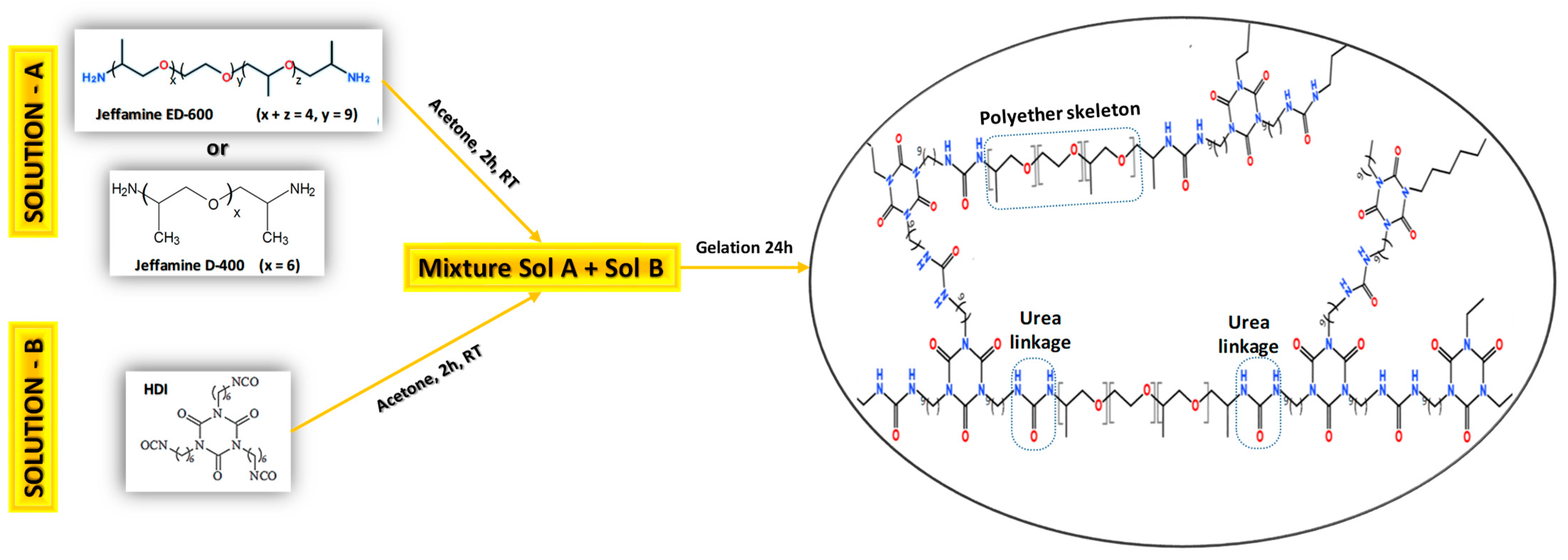
2.2. Synthesis of Polyureas

The polyurea xerogel was obtained by the sol-gel process, as reported in the literature [18,24,33]. The monomers Jeffamine (ED600 or D400) and trimer-HDI were separately dissolved in acetone with the molar ratio of jeffamine: HDI at 1:1. The solutions were sealed and stirred for 2 h at room temperature (RT) to form a homogenous solution. After, the HDI solution was added dropwise to the jeffamine solution until complete dissolution (see details Scheme 1). The resulting solution was poured into Teflon® mold. During the gelation reaction, to obtain polyurea networks, the acetone solvent is ambient dried (evaporated) before being used for characterization, cytotoxicity and release assays.

2.3. Synthesis of Polyureas Containing Drug

Polyurea PEO/PPO samples containing DCF were prepared via one-step sol-gel process. The desired drug amount (1 and 10 wt% with respect to the mass of jeffamine) was dissolved in acetone and then mixed with PEO or PPO to obtain a homogeneous solution. The HDI solution was dissolved in a separated beaker and added, as cited above, to form DCF-loaded polyurea samples. The resulting mixture was poured into Teflon® mold and gelled using the same conditions (temperature and time) as for the pure polyurea. The final materials were named PEO500, PPO400 (polyurea based on Jeffamine ED600 and D400, respectively) and PEO-DCFx or PPO-DCFx (loaded polyurea containing diclofenac DCF and x for the wt% of drug loaded). Since the acetone was evaporated at ambient conditions to form the final polyurea membranes, the mass of loaded DCF used was completely incorporated into PEO or PPO matrixes.

2.4. Characterization

X-ray diffraction XRD patterns were recorded on a Rigaku MiniFlex II Desktop X-ray Diffractometer using Cu Kα (λ = 1.54 Å) radiation at a scan speed of 0.04°·s−1. Fourier Transform Infrared Spectroscopy FTIR spectra were recorded at 3100–700 cm−1 with ATR mode using a Perkin Elmer Frontier spectrometer, with a resolution of 2 cm−1 and an average of 32 scans. Thermogravimetric analysis TGA was carried out applying SDTQ600 TA Instruments. The polyurea samples were cut into small pieces (~10 mg). Measurements were carried out under a nitrogen atmosphere and typical heating rate of 10 °C/min to 25 °C to 700 °C.
The swelling behavior (water uptake) of the PEO- and PPO-based polyurea membranes was studied gravimetrically. A fraction of the samples (2 cm × 2 cm) was immersed in distilled water. The swollen samples were removed from the water at a preset time (5, 10, 20, 30, 40, 50, 60, 90, 120, 150, 180, 240 and 300 min), then lightly squeezed with filter papers to remove any excess water on the surface before being weighed in an electronic analytical balance. The swelling ratio (Sr) was determined using the equation:
S w = W s W d W d × 100
where Ws and Wd are the mass of swollen and dry polyurea samples, respectively.
The in vitro temporal release of DCF was assayed in distilled water (100 mL, pH 6.5) using ~0.3 g of loaded PEO or PPO containing different amounts of DCF drug (1 and 10% wt). The release trials were carried out at a controlled temperature of 37 °C under constant stirring (100 rpm). Spectroscopic analysis of the in vitro release was conducted on the UV-Vis band using a Cary60 dual-beam spectrophotometer (Agilent Technologies Brasil Ltda, Barueri, São Paulo, Brazil) connected to an immersion probe (with a 2 mm optical path length). The quantitative determination of the cumulative DCF release was based on a calibration curve compiled from the maximum absorbance values at λmax= 275 nm of drug solutions at different concentrations. The in vitro release and swelling tests were all performed with three independent samples, and the results presented are mean data. Regarding the release mechanism, two well-known approaches were used for the analysis of drug release data (see details in results and discussion Section 3.5).
The cell viability assay XTT was conducted using the cell line GM07492-A obtained from Coriell Cell Repositories (Camden, NJ, USA). Cultivated in DMEM + HAM F10 (1:1, v/v) (Sigma, St Louis, MO, USA), it was supplemented with 10% fetal calf serum (Sigma, St Louis, MO, USA) and 1% of penicillin/streptomycin stabilized solution (Sigma, St Louis, MO, USA,). To determine the cell viability, 1.5 × 10⁵ cells/well were seeded on a 24-well plate. After 24 h, the cell culture was exposed to a 0.04 cm2 of PEO 500 and PPO 400 membranes and incubated for an additional 24 h. The plates were washed twice in phosphate buffer saline (PBS 1X) and incubated for 4 h at 37 °C with DMEM without phenol red and supplemented with the reagents of the Cell Proliferation Kit (Roche, Mannhein, Germany) as recommended by the manufacturer [34]. Afterwards, the culture media solution was placed on a 96-well plate. Total absorbance was measured at 492 and 690 nm (reference energy) using a microplate reader (ASYS, Eugendorf, Salzburg, Austria). With the results of total absorbance being considered directly proportional to the number of viable cells as a percentage of the negative control (100% of cell viability), a statistical analysis of experimental data was run through the software GraphPad Prism 5.0. The comparative analysis of the experimental groups, based on the Analysis of Variance (ANOVA), was followed by the Tukey test when significant differences among treatments were found. The significance was set at p < 0.05, and the results were reported as means and standard deviations (SD) of three independent experimental samples assayed in triplicate.

3. Results and Discussion

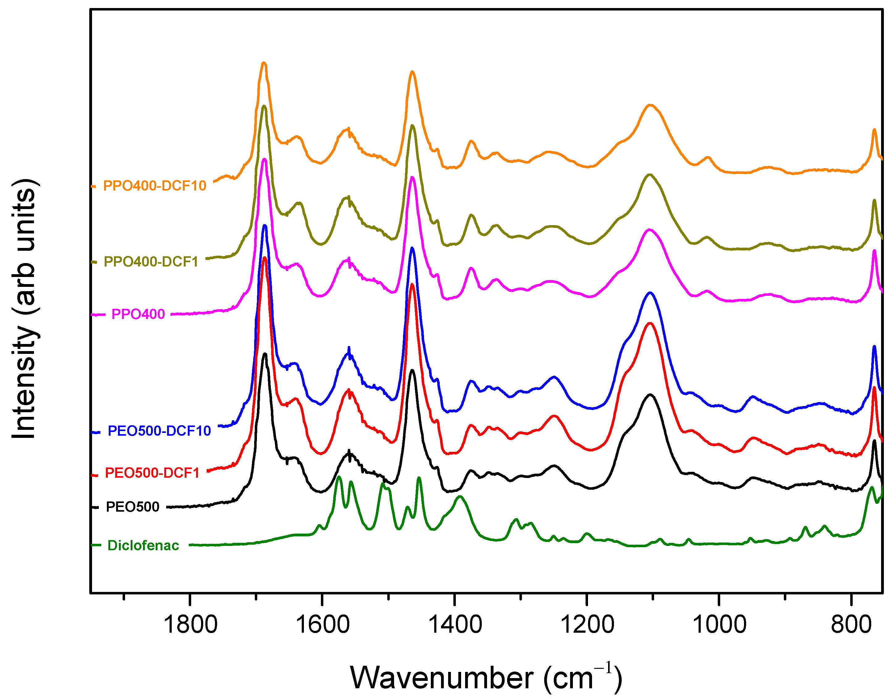
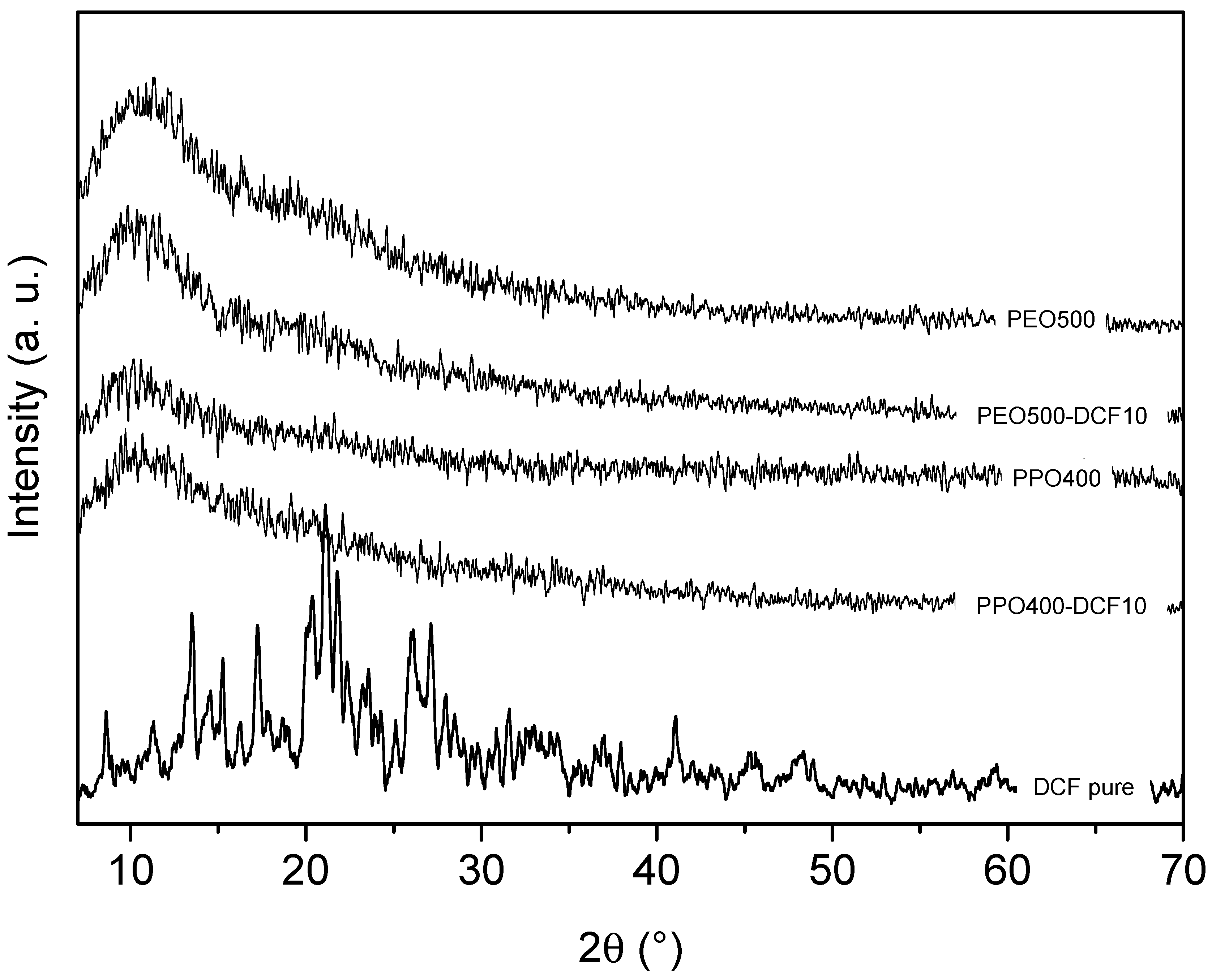
3.1. Structural Characterization
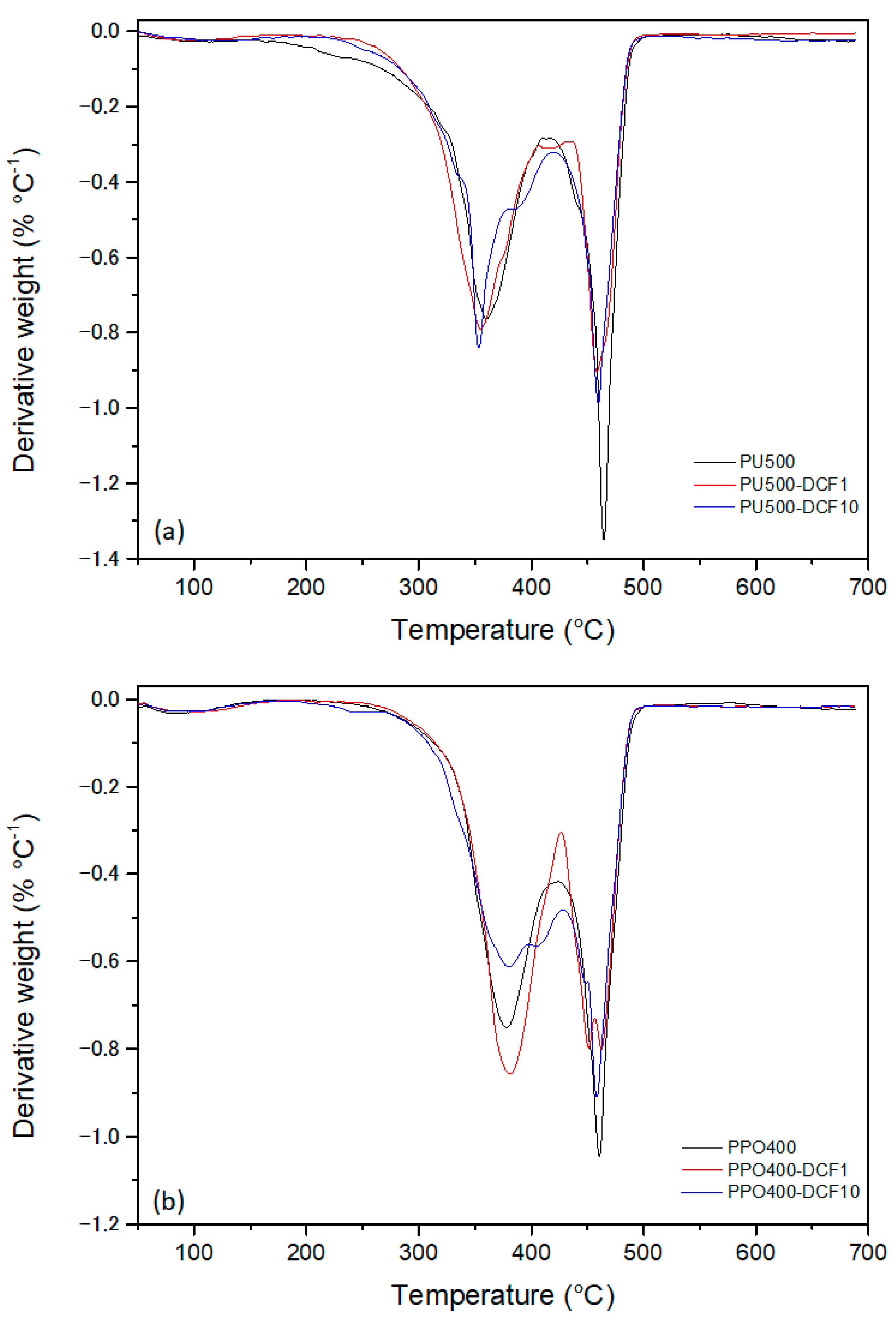
The synthesis of polyurea is a straightforward method for encapsulating an active pharmaceutical ingredient (API). To prepare the loaded samples, DCF drug was dissolved in acetone and then blended with PEO to create a homogeneous solution (see details in experimental section). It is noteworthy to mention that after gelation, samples began to shrink (acetone extraction). This was due to the drying process at room temperature, and this led to the formation of a homogeneous and transparent polyurea xerogel containing the amount of drug initially dissolved. Thus, 100% encapsulation efficiency is recorded for the drug concentration used in this work (1 and 10% wt), with no precipitation of diclofenac after the gelation process. The structural elucidation of unloaded and loaded polyureas was carried out by FTIR spectroscopy. The FTIR spectra plots of PEO500 and PPO400, without and with DCF drug (1 and 10 wt%), are depicted in Figure 1. A superposition of characteristic bands of unloaded PEO/PPO matrices and the DCF drug was revealed in the region of the FT-IR spectrum studied, and the analysis of potential peak displacements was discussed in relation to the characteristic vibrations of polyurea. The absorption bands at 765, 949 and 1104 cm−1 are attributed to the ethylene rocking vibrations (CH2) and stretching vibrations of the polyether backbone (C–O–C) present in PEO and PPO structure [35]. Three bands centered at approximately 1560, 1641 and 1686 cm−1, characteristic of CO–N–H amide II, C=O amide I and free C=O group, respectively, can be noticed. The presence of these bands ascribed to amide II and amide I evidence the successful obtention of polyurea matrices based on PEO or PPO (Figure 1, black line and pink line, respectively) with the formation of urea groups. By comparing the spectrum of unloaded polyureas (PEO500 and PPO400) with the spectrum of DCF loaded PEO500-DCF and PPO400-DCF containing 1 and 10% wt of the drug, it can clearly be seen that the polyether characteristic absorption bands do not show changes in peak position (without displacement) after incorporation of the DCF. These results indicate good solvation of DCF molecules through the polyurea matrices. The amount of drug release by PEO/PPO polyureas evidences the DCF encapsulation and excellent solvation of the drug by these matrices, as reported in the release studies.
The X-ray diffraction results of PEO500 and PPO400 xerogels, both unloaded and loaded with 10% wt content of DCF drug, are presented in Figure 2. Interestingly, the diffractograms of all samples, whether loaded or not, exhibit a sole broad Gaussian-shaped peak centered at 2θ = 11°, which can be attributed to the amorphous lattice. However, when crystalline DCF (10% wt) was blended into the polyurea PEO/PPO, its crystalline diffraction peaks disappeared, indicating that the DCF was highly soluble in the matrices. These results highlight the remarkable solubility of DCF in PEO/PPO matrices, which could have significant implications for drug delivery applications.

3.2. TGA Analysis

The thermal degradation of polyureas and the impact of DCF on thermal stability were studied by TGA. Figure 3 displays the DTGA curves for both unloaded and loaded PEO/PPO polyurea samples. The thermal degradation process of the polyurea samples (PEO and PPO) was observed to have two main stages. The first stage of degradation involved the urea bond decomposition, resulting in a dissociation into isocyanate and alcohol. The second stage is characteristic of polyol and isocyanate degradation into amine compounds (primary/secondary amines), terminal olefinic group and CO2 [36,37]. For PEO500 polyurea xerogel (unloaded and loaded), the first stage of polymer degradation started at approximately 165 °C and ended at around 418 °C. According to Figure 3a, increasing the amount of drug loaded into PEO500 leads to an increase in thermal stability during the first stage of degradation. This may be attributed to the possibility of hydrogen bonding between hydrogen atoms from urea groups and oxygen atoms from the diclofenac [24]. In the case of PPO polyurea xerogel (unloaded and loaded), the first stage of polymer degradation started at approximately 200 °C and ended at around 425 °C (Figure 3b). These findings indicate that the PPO400 polyurea matrix is thermally more stable than PEO500. Table 1 summarizes the parameters of mass loss (%) at the first and second stages and char residue (%) obtained from TGA curves of unloaded and loaded PEO500 and PPO400 polyurea samples.

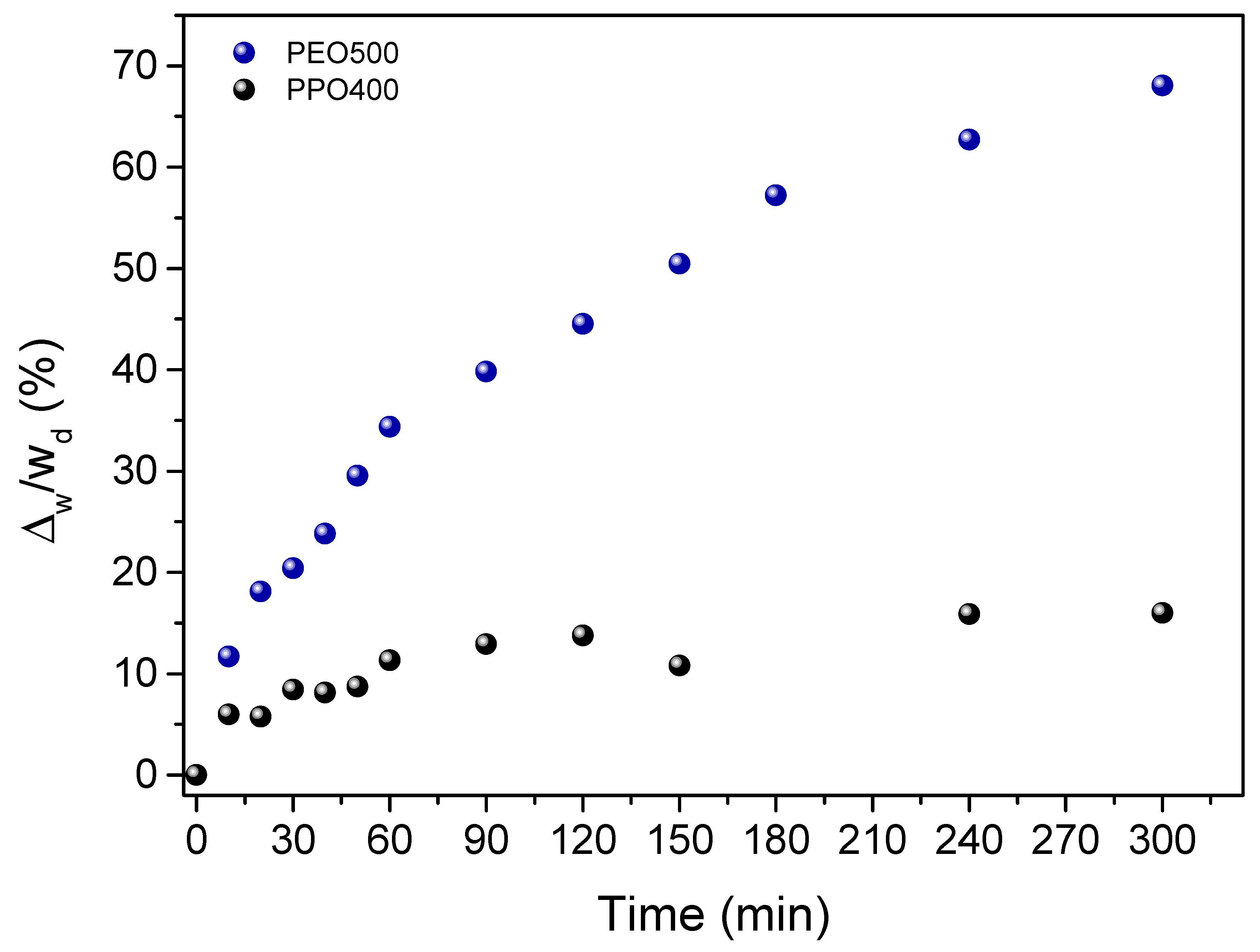
3.3. Swelling Behavior of Polyurea PEO/PPO Matrixes

The swelling behaviors of polyurea xerogels at room temperature were examined by gravimetric method, as displayed in Figure 4. The results indicate that for PEO500 xerogel, the rapid increase of the swelling ratio during the 6 h of assay leading to 80% can be attributed to the hydration of the matrix due to the hydrophilic nature of PEO chains. On the other hand, PPO400 xerogel showed a slower water uptake during the swelling assay, at around 10%. This behavior is due to the hydrophobic nature of PPO chains. Considering the differences between PEO500 and PPO400 swelling ratios, these results open perspectives of controlling the water uptake capacity by selecting the appropriate polyether nature and suggest fine-tuning these properties by mixing PEO/PPO amounts during the polyurea preparation.

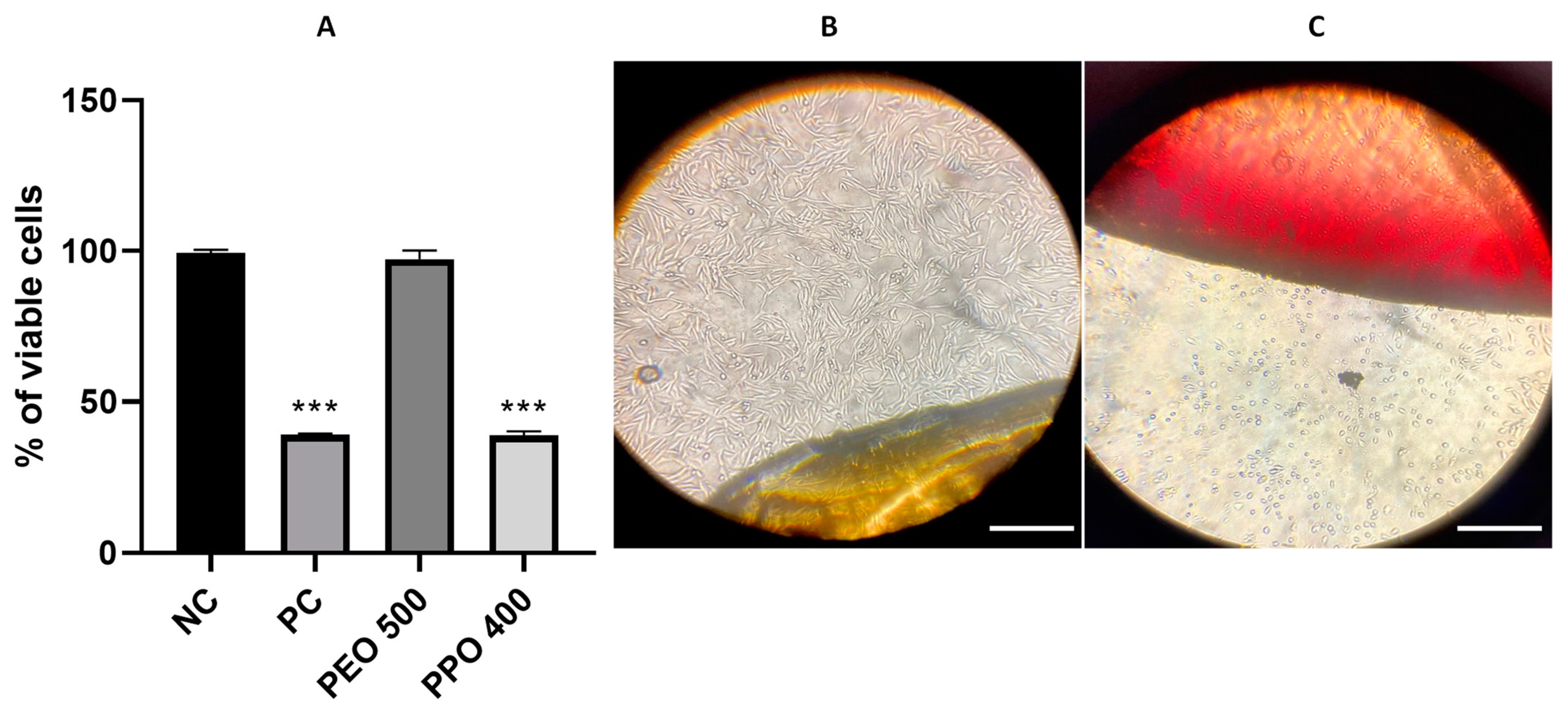
3.4. Cell Viability Assays

The cytotoxicity evaluation of PEO/PPO polyurea membranes involved the GM07492-A cell line exposure to both PEO500 and PPO400 membranes through direct contact with the culture medium (Figure 5). The XTT studies (Figure 5A) revealed that PEO500 demonstrates high cell viability (about 98%). While using PPO400, the percentage decreased to approximately 40%. Under the experimental conditions, after 24 h in contact with the culture medium, the PEO500 sample (Figure 5B) did not demonstrate changes in adherence and morphology of the cells, indicating healthy and living cells with a fibrous characteristic. In contrast, exposure to PPO400 (Figure 5C) induced a loss of cellular adherence, alteration in morphology (like spherical, in this case, dead cells), and consequently, the inability of cell growth and survival. These cytotoxicity results suggest that the PEO500 matrix is biocompatible (without cytotoxicity when exposed to cell lines) compared to PPO400, which showed lower cell viability when exposed to the culture medium. These results are in agreement with the XTT assays (Figure 5A).

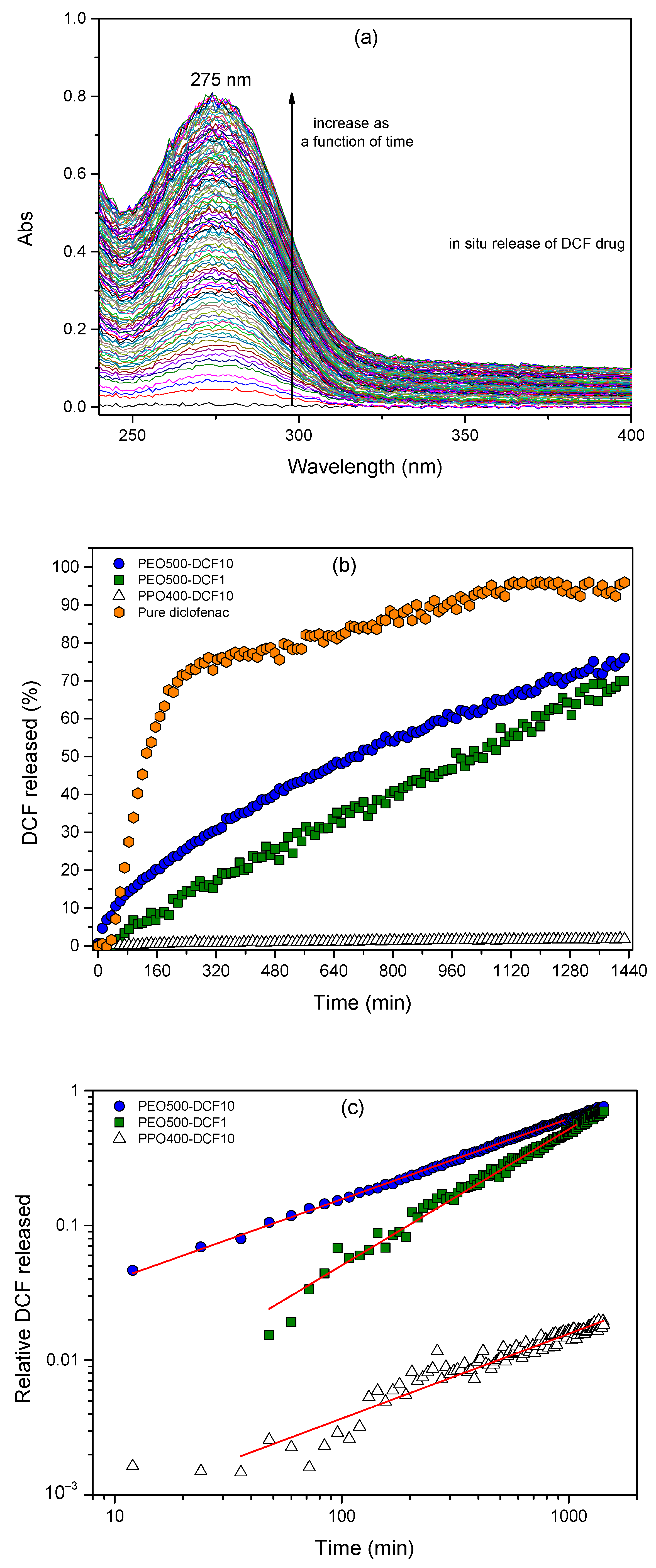
3.5. In Situ Diclofenac Release Assays

Diclofenac drug release studies: The cumulative in situ release of pure DCF and DCF from PEO500/PPO400 polyurea samples are displayed in Figure 6. The DCF drug release monitoring was done with UV-vis spectrometry at 275 nm (see Figure 6a). In vitro studies showed that 80% of the DCF (pure from patch) was released over a period of 6 h (Figure 6b). The PEO500-DCF profiles showed a well-controlled sustained manner, with a release amount of 67% and 69% by PEO500 polyurea containing 1 and 10% wt of drug, respectively (see Figure 6b, blue circles and green squares). On the other hand, the DCF molecules are barely released from polyurea PPO400-DCF10 (containing 10% wt of drug), reaching about 1.8% after 24 h of assay (see Figure 6b, black triangles). For the PPO400-DCF1 (containing 1% wt of drug), no release of the drug was observed. These different release behaviors of DCF by PEO500 and PPO400 polyurea samples were attributed mainly to the hydrophilicity of the matrixes, i.e., the capacity to swell (water uptake) through the polyurea network. Comparing the two matrices, it is worth noting that (i) PEO500 xerogel showed a faster and higher water uptake after 5 h (see Figure 4, blue circles), characteristic of a swollen network due to the hydrophilic nature of polyether chains, and (ii) PPO400 xerogel displayed a slow and lower water uptake (see Figure 4, black circles) that can be correlated to the hydrophobic nature of the polyether. The findings suggest that utilizing polyurea systems designed for controlled drug delivery molecules is promising for achieving a sustained-release profile by using polyurea based on PEO nature. Nevertheless, the possibility to add a small amount of PPO into the PEO system during the polyurea synthesis encourages further development of polymeric blends as a potential candidate for fine-tuning the amount of drug released and could enhance the cellular viability of the PPO characterized above (Figure 5B) for lower cell viability.
The drug release kinetics of DCF drug in polyurea samples were investigated using the Korsmeyer–Peppas model [38]. This model is based on Fick’s law of diffusion, and the equation is given by Equation (2):
M t M = k t n
where Mt/M is the fractional solute release, t is the release time, k is a kinetic constant characteristic of the drug/polymer system, and n is an exponent which characterizes the mechanism of release, where n values of 0.50 and 1 are characterized by Fickian diffusion and Case II transport mechanism, respectively. Values of n between 0.55 and 0.90 can indicate the superposition of both phenomena (anomalous transport).
Figure 6c shows the DCF release curves plotted as log (Mt/M) as a function of log t from loaded PEO500 and PPO400 polyurea samples. The DCF amount released in the final assays (t = 1440 min) and the parameters obtained by the Korsmeyer–Peppas model are displayed in Table 2. The coefficient of determination (R2) for loaded PEO500 samples was ~0.99; and for PPO400 containing 10% wt of drug, it was ~0.95. The experimental n values obtained for PEO500 containing 1 and 10% wt of DCF were n = 1 and 0.59, respectively. These n values indicate a Case II transport mechanism (for the PEO500-DCF1 sample) and an anomalous transport (for the PEO500-DCF10 sample) attributed to the diffusion/zero-order rate of drug controlled by the penetration of the water-swollen, respectively. The PPO400-DCF10 sample showed an n value of 0.62, characteristic of anomalous transport. Higuchi model [39] was used to compare the release mechanism results. In this case, when linear regression of Mt/M versus t1/2 is applied, the curve does not fit well, presenting the R2 values < 0.98 for loaded PEO500 containing 1 and 10% wt of the drug, respectively. Thus, we conclude that the Korsmeyer–Peppas model nicely describes all data sets analyzed.
Recently, Chen et al. [40] developed an ocular drug delivery system based on miltefosine loaded in a contact lens hydrogel. The authors demonstrate the importance of a slow and steady release of the antiparasitic miltefosine for the treatment of Acanthamoeba keratitis. In this sense, polyurea based PEO–PPO materials could be promising candidates for this scenario since they are (i) easy to obtain by one-pot process, (ii) low in cost, (iii) high in transparency, and have (iv) desired shape and thickness according to needs [24]. Moreover, with a judicious PEO–PPO mixture during the synthesis followed by fabricate process, it is possible to produce microneedle patch-based polyurea devices which could be used, for example, as a sustained release of insulin. Cao et al. used silk fibroin for the purpose that the swelling degree of the needle controls the release of insulin [41].

4. Conclusions

The influence of the polymeric nature on the physicochemical properties of polyureas using PEO and PPO of similar molecular weight (Mw ~500 g mol−1), exhibiting sustained-release profiles, has been herein evidenced. These xerogels were selected as the carrier for sodium diclofenac DCF at different drug loads. Polyureas based on PEO500 and PPO400 have high thermal stability (onset of degradation above 200 °C), as shown in TGA studies. Therefore, the degree of swelling can be controlled by the nature of the matrix (PEO or PPO), which plays an important role in the profile release of DCF drug. PEO500 matrix was very compatible with the GM07492-A cell, and the fibrous morphology of the cells did not visibly alter after contact with the membrane. After contact with PPO400, on the contrary, the cells changed their morphology to spherical, resulting in low cellular viability. The polyurea xerogels can then be considered a favorable carrier for the sustained release of drug molecules. In our study, commercially available reagents (PEO/PPO and HDI) can be used to produce polyurea membranes with potential applications as transdermal drug delivery systems. This approach is very advantageous owing to the low cost and simplicity of manufacture of the sol-gel synthesis. Thus, basing our conclusion on the properties of polyurea PPO400, this formulation could also be exploited as a matrix for high-performance advanced composite materials, such as membranes for gas separation and as a coating.

Author Contributions

J.G.V. and H.E.A. contributed equally to this work for all the steps (conceptualization, methodology, investigation, writing/editing and discussion of the results, the drug release assays and data treatment). B.A.F. contributed to data treatment and discussed the structural results and drug release mechanism. J.M.P., N.N.S. and R.A.d.S. realized the biological assays and results discussion. E.F.M. (supervisor of J.G.V.) contributed to all steps, suggesting experiments, discussing the results and writing the manuscript. The manuscript was written based on contributions from all authors. All authors have read and agreed to the published version of the manuscript.

Funding

Fundação de Amparo à Pesquisa do Estado de São Paulo FAPESP (n° 2021/06552-1 and 2022/06507-9), Coordenação de Aperfeicoamento de Pessoal de Nível Superior CAPES (Finance Code 001) and Conselho Nacional de Desenvolvimento Científico e Tecnológico CNPq (307696/2021-9).

Institutional Review Board Statement

Not applicable.

Informed Consent Statement

Not applicable.

Data Availability Statement

Not applicable.

Acknowledgments

The authors would like to thank Huntsman Performance Products and Bayer Group for donating the Jeffamine® and triisocyanate crosslinker reagents, respectively. We are grateful to Brazilian funding agencies FAPESP, CAPES and CNPq.

Conflicts of Interest

The authors declare no conflict of interest.

References

  1. Kaul, L.; Grundmann, C.E.; Köll-weber, M.; Löffler, H.; Weiz, A.; Zannettino, A.C.W.; Richter, K.; Süss, R. A Thermosensitive, Chitosan-Based Hydrogel as Delivery System for Antibacterial Liposomes to Surgical Site Infections. Pharmaceutics 2022, 14, 2841. [Google Scholar] [CrossRef] [PubMed]
  2. Jiang, J.; Cheng, W.; Tang, Q.; Pan, X.; Li, J.; Zhao, L.; Xi, Z.; Yuan, W. Multiblock Poly(Ether-b-Amide) Copolymers Comprised of PA1212 and PPO-PEO-PPO with Specific Moisture-Responsive and Antistatic Properties. Chin. J. Chem. Eng. 2022, 53, 421–430. [Google Scholar] [CrossRef]
  3. Sanopoulou, M.; Papadokostaki, K.G. Controlled Drug Release Systems: Mechanisms and Kinetics. Biomed. Membr. (Bio) Artif. Organs 2017, 2, 1–33. [Google Scholar] [CrossRef]
  4. Macha, I.J.; Ben-Nissan, B.; Vilchevskaya, E.N.; Morozova, A.S.; Abali, B.E.; Müller, W.H.; Rickert, W. Drug Delivery from Polymer-Based Nanopharmaceuticals-an Experimental Study Complemented by Simulations of Selected Diffusion Processes. Front. Bioeng. Biotechnol. 2019, 7, 37. [Google Scholar] [CrossRef]
  5. Rajabi, M.; Srinivasan, M.; Mousa, S.A. Nanobiomaterials in Drug Delivery; William Andrew Publishing: Norwich, NY, USA, 2016; Volume 9, pp. 1–37. ISBN 9780323428897. [Google Scholar] [CrossRef]
  6. Bruschi, M.L. Strategies to Modify the Drug Release from Pharmaceutical Systems/Edited by Marcos Luciano Bruschi; Woodhead Publishing Series in Biomedicine; Elsevier: Cambridge, UK, 2015; p. 85. ISBN 0-08-100112-6. [Google Scholar]
  7. Kamaly, N.; Yameen, B.; Wu, J.; Farokhzad, O.C. Degradable Controlled-Release Polymers and Polymeric Nanoparticles: Mechanisms of Controlling Drug Release. Chem. Rev. 2016, 116, 2602–2663. [Google Scholar] [CrossRef]
  8. Malekjani, N.; Jafari, S.M. Modeling the Release of Food Bioactive Ingredients from Carriers/Nanocarriers by the Empirical, Semiempirical, and Mechanistic Models. Compr. Rev. Food Sci. Food Saf. 2021, 20, 3–47. [Google Scholar] [CrossRef]
  9. Anghel, N.; Melinte, V.; Spiridon, I.; Pertea, M. Antioxidant, Antimicrobial, and Kinetic Studies of Β-Cyclodextrin Crosslinked with Lignin for Drug Delivery. Pharmaceutics 2022, 14, 2260. [Google Scholar] [CrossRef]
  10. Uboldi, M.; Pasini, C.; Pandini, S.; Baldi, F.; Briatico-vangosa, F.; Inverardi, N.; Maroni, A.; Moutaharrik, S.; Melocchi, A.; Gazzaniga, A.; et al. Expandable Drug Delivery Systems Based on Shape Memory Polymers: Impact of Film Coating on Mechanical Properties and Release and Recovery Performance. Pharmaceutics 2022, 14, 2814. [Google Scholar] [CrossRef]
  11. Ferrero, C.; Casas, M.; Caraballo, I. Redox-Responsive Polymersomes as Smart Doxorubicin Delivery Systems. Pharmaceutics 2022, 14, 1724. [Google Scholar] [CrossRef]
  12. Huang, Y.; Yan, H.; Cai, M.; Song, S.; He, C.; Fan, X.; Zhu, M. Polyurea as a Reinforcing Filler for the Anti-Corrosion and Wear-Resistant Application of Epoxy Resin. Prog. Org. Coat. 2022, 171, 107049. [Google Scholar] [CrossRef]
  13. Tang, S.; Shi, Z.; Cao, Y.; He, W. Facile Aqueous-Phase Synthesis of Multi-Responsive Nanogels Based on Polyetheramines and Bisepoxide. J. Mater. Chem. B 2013, 1, 1628–1634. [Google Scholar] [CrossRef] [PubMed]
  14. Qin, C.; Lv, Y.; Xu, C.; Li, J.; Yin, L.; He, W.; Lv, Y.; Zhao, Y.; Xu, C.; Jin, Z.; et al. Compositional Tuning of Epoxide-Polyetheramine “Click” Reaction toward Cytocompatible, Cationic Hydrogel Particles with Antimicrobial and DNA Binding Activities. Int. J. Pharm. 2020, 484, 15–40. [Google Scholar] [CrossRef]
  15. Bar, H.; Bianco-Peled, H. Modification of Shellac Coating Using Jeffamine® for Enhanced Mechanical Properties and Stability. Prog. Org. Coat. 2020, 141, 105559. [Google Scholar] [CrossRef]
  16. Huang, H.; Wei, H.; Huang, L.; Fan, T.; Li, X.; Zhang, Z.; Shi, T. Spontaneous Alternating Copolymerization of Aziridines with Tosyl Isocyanate toward Polyureas. Eur. Polym. J. 2023, 182, 111731. [Google Scholar] [CrossRef]
  17. Ren, G.; Zhou, C.; Fan, X.; Zheng, M.; Wang, S. Investigating the Rheological and Tribological Properties of Polyurea Grease via Regulating Ureido Amount. Tribol. Int. 2022, 173, 107643. [Google Scholar] [CrossRef]
  18. Sánchez-Ferrer, A.; Rogez, D.; Martinoty, P. Synthesis and Characterization of New Polyurea Elastomers by Sol/Gel Chemistry. Macromol. Chem. Phys. 2010, 211, 1712–1721. [Google Scholar] [CrossRef]
  19. Zhang, C.; Lu, L.; Ouyang, R.; Zhou, C. Polyurethane/Liquid Crystal Microfibers with PDNA Polyplex Loadings for the Optimal Release and Promotion of HUVEC Proliferation. Pharmaceutics 2022, 14, 2489. [Google Scholar] [CrossRef]
  20. Du, J.; Ibaseta, N.; Guichardon, P. Characterization of Polyurea Microcapsules Synthesized with an Isocyanate of Low Toxicity and Eco-Friendly Esters via Microfluidics: Shape, Shell Thickness, Morphology and Encapsulation Efficiency. Chem. Eng. Res. Des. 2022, 182, 256–272. [Google Scholar] [CrossRef]
  21. Resende, M.A.; Pedroza, G.A.; Macêdo, L.H.G.M.C.; Oliveira, R.; Amela-Cortes, M.; Molard, Y.; Molina, E.F. Design of Polyurea Networks Containing Anticancer and Anti-inflammatory Drugs for Dual Drug Delivery Purposes. J. Appl. Polym. Sci. 2022, 139, 51970. [Google Scholar] [CrossRef]
  22. Caravieri, B.B.; de Jesus, N.A.M.; de Oliveira, L.K.; Araujo, M.D.; Andrade, G.P.; Molina, E.F. Ureasil Organic–Inorganic Hybrid as a Potential Carrier for Combined Delivery of Anti-Inflammatory and Anticancer Drugs. ACS Appl. Bio Mater. 2019, 2, 1875–1883. [Google Scholar] [CrossRef]
  23. Bonattini, V.H.; Paula, L.A.L.; Jesus, N.A.; Tavares, D.C.; Nicolella, H.D.; Magalhães, L.G.; Molina, E.F. One-step Formation of Polyurea Gel as a Multifunctional Approach for Biological and Environmental Applications. Polym. Int. 2020, 69, 476–484. [Google Scholar] [CrossRef]
  24. Pedroza, G.A.; Macêdo, L.H.G.M.C.; de Oliveira, R.; Silveira, N.N.; Orenha, R.P.; Parreira, R.L.T.; dos Santos, R.A.; Molard, Y.; Amela-Cortes, M.; Molina, E.F. Cost-Efficient Polyurea Carrier for Precise Control of an Anti-Inflammatory Drug Loading and Release. J. Drug Deliv. Sci. Technol. 2022, 76, 103744. [Google Scholar] [CrossRef]
  25. Lowinger, M.B.; Barrett, S.E.; Zhang, F.; Williams, R.O. Sustained Release Drug Delivery Applications of Polyurethanes. Pharmaceutics 2018, 10, 55. [Google Scholar] [CrossRef] [PubMed]
  26. Guo, Q.; Knight, P.T.; Mather, P.T. Tailored Drug Release from Biodegradable Stent Coatings Based on Hybrid Polyurethanes. J. Control. Release 2009, 137, 224–233. [Google Scholar] [CrossRef] [PubMed]
  27. Chen, X.; Liu, W.; Zhao, Y.; Jiang, L.; Xu, H.; Yang, X. Preparation and Characterization of PEG-Modified Polyurethane Pressure-Sensitive Adhesives for Transdermal Drug Delivery. Drug Dev. Ind. Pharm. 2009, 35, 704–711. [Google Scholar] [CrossRef]
  28. Li, B.; Yoshii, T.; Hafeman, A.E.; Nyman, J.S.; Wenke, J.C.; Guelcher, S.A. The Effects of RhBMP-2 Released from Biodegradable Polyurethane/Microsphere Composite Scaffolds on New Bone Formation in Rat Femora. Biomaterials 2009, 30, 6768–6779. [Google Scholar] [CrossRef]
  29. Martinelli, A.; D’Ilario, L.; Francolini, I.; Piozzi, A. Water State Effect on Drug Release from an Antibiotic Loaded Polyurethane Matrix Containing Albumin Nanoparticles. Int. J. Pharm. 2011, 407, 197–206. [Google Scholar] [CrossRef]
  30. Castagna, A.M.; Pangon, A.; Choi, T.; Dillon, G.P.; Runt, J. The Role of Soft Segment Molecular Weight on Microphase Separation and Dynamics of Bulk Polymerized Polyureas. Macromolecules 2012, 45, 8438–8444. [Google Scholar] [CrossRef]
  31. Choi, T.; Fragiadakis, D.; Roland, C.M.; Runt, J. Microstructure and Segmental Dynamics of Polyurea under Uniaxial Deformation. Macromolecules 2012, 45, 3581–3589. [Google Scholar] [CrossRef]
  32. Zheng, T.; Li, T.; Shi, J.; Wu, T.; Zhuang, Z.; Xu, J.; Guo, B. Molecular Insight into the Toughness of Polyureas: A Hybrid All-Atom/Coarse-Grained Molecular Dynamics Study. Macromolecules 2022, 55, 3020–3029. [Google Scholar] [CrossRef]
  33. Sánchez-Ferrer, A.; Soprunyuk, V.; Engelhardt, M.; Stehle, R.; Gilg, H.A.; Schranz, W.; Richter, K. Polyurea Networks from Moisture-Cure, Reaction-Setting, Aliphatic Polyisocyanates with Tunable Mechanical and Thermal Properties. ACS Appl. Polym. Mater. 2021, 3, 4070–4078. [Google Scholar] [CrossRef]
  34. Scudiero, D.A.; Shoemaker, R.H.; Paull, K.D.; Monks, A.; Tierney, S.; Nofziger, T.H.; Currens, M.J.; Seniff, D.; Boyd, M.R. Evaluation of a Soluble Tetrazolium/Formazan Assay for Cell Growth and Drug Sensitivity in Culture Using Human and Other Tumor Cell Lines. Cancer Res. 1988, 48, 4827–4833. [Google Scholar] [PubMed]
  35. Chaker, J.A.; Santilli, C.V.; Pulcinelli, S.H.; Dahmouche, K.; Briois, V.; Judeinstein, P. Multi-Scale Structural Description of Siloxane-PPO Hybrid Ionic Conductors Doped by Sodium Salts. J. Mater. Chem. 2007, 17, 744–757. [Google Scholar] [CrossRef]
  36. Xu, D.; Yu, K.; Qian, K. Thermal Degradation Study of Rigid Polyurethane Foams Containing Tris(1-Chloro-2-Propyl)Phosphate and Modified Aramid Fiber. Polym. Test. 2018, 67, 159–168. [Google Scholar] [CrossRef]
  37. Levchik, S.V.; Weil, E.D. Thermal Decomposition, Combustion and Fire-Retardancy of Polyurethanes—A Review of the Recent Literature. Polym. Int. 2004, 53, 1585–1610. [Google Scholar] [CrossRef]
  38. Korsmeyer, R.W.; Gurny, R.; Doelker, E.; Buri, P.; Peppas, N.A. Mechanisms of Solute Release from Porous Hydrophilic Polymers. Int. J. Pharm. 1983, 15, 25–35. [Google Scholar] [CrossRef]
  39. Higuchi, T. Rate of release of medicaments from ointment bases containing drugs in suspension. J. Pharm. Sci. 1961, 50, 874–875. [Google Scholar] [CrossRef]
  40. Chen, L.; Kuang, L.; Ross, A.E.; Farhat, W.; Boychev, N.; Sharfi, S.; Kanu, L.N.; Liu, L.; Kohane, D.S.; Ciolino, J.B. Topical Sustained Delivery of Miltefosine Via Drug-Eluting Contact Lenses to Treat Acanthamoeba Keratitis. Pharmaceutics 2022, 14, 2750. [Google Scholar] [CrossRef]
  41. Cao, J.; Liu, Y.; Qi, Z.; Tao, X.; Kundu, S.C.; Lu, S. Sustained Release of Insulin from Silk Microneedles. J. Drug Deliv. Sci. Technol. 2022, 74, 103611. [Google Scholar] [CrossRef]
Scheme 1. Sol-gel route for the preparation of polyurea based on Jeffamine ED-600 (PEO500) and Jeffamine D-400 (PPO400) and schematic network showing urea linkage and polyether backbone.
Futurepharmacol 03 00026 sch001
Figure 1. FTIR spectra of the DCF drug, unloaded and loaded polyurea PEO500 and PPO400 containing 1 and 10% wt of DCF.
Futurepharmacol 03 00026 g001
Figure 2. XRD patterns of DCF drug, unloaded and loaded PEO500 and PPO400 containing 10% wt of drug.
Futurepharmacol 03 00026 g002
Figure 3. DTGA curves of the polyurea samples (a) unloaded and loaded PEO500 and (b) unloaded and loaded PPO400.
Futurepharmacol 03 00026 g003
Figure 4. Evolution of swelling degree as a function of the immersion time in water for polyurea PEO500 and PPO400.
Futurepharmacol 03 00026 g004
Figure 5. (A) Mean values of cell viability obtained by XTT assay in GM07492-A cell line after 24 h of treatment with PEO 500 and PPO 400. NC: negative control; PC: positive control (DMSO 10%). *** p < 0.0001 compared to negative control; and photograph of (B) PEO500 and (C) PPO400 when exposed to the cell line GM07492-A. The white scale bars represent 50 μm (20× magnification).
Futurepharmacol 03 00026 g005
Figure 6. (a,b) Evolution of the cumulative DCF release in water at 37 °C from PEO500 and PPO400 and (c) relative DCF release (see Equation (1)), log (Mt/M) plotted as a function of log t, from PEO500 and PPO400 (red line means fitting to data generated from Equation (2)).
Futurepharmacol 03 00026 g006
Table 1. Different weight loss percentages at the degradation stages (first and second stage), char residues (%) for unloaded and loaded PEO500 and PPO400 polyurea samples.
Polyurea Sample
Mass LossPEO500PEO500-DCF1PEO500-DCF10PPO400PPO400-DCF1PPO400-DCF10
at first stage (%)545563616659
at second stage (%)363526302530
Char residue (%)445334
Table 2. Amount of DCF released by polyurea PEO/PPO xerogel, the Korsmeyer–Peppas parameters, and release mechanism of the drug from polyurea systems.
Amount
Released (%)
Korsmeyer–Peppas ParametersRelease Mechanism
Sample nR2
PEO500-DCF167.01.000.98Fickian diffusion
PEO500-DCF1069.00.590.99anomalous transport
PPO400-DCF101.830.620.95anomalous transport
Disclaimer/Publisher’s Note: The statements, opinions and data contained in all publications are solely those of the individual author(s) and contributor(s) and not of MDPI and/or the editor(s). MDPI and/or the editor(s) disclaim responsibility for any injury to people or property resulting from any ideas, methods, instructions or products referred to in the content.

