Article

Hemodynamic Responses to a Handgrip Exercise Session, with and without Blood Flow Restriction, in Healthy Volunteers

by
Spyridoula Filippou
*,
Paris Iakovidis
,
Dimitrios Lytras
,
Konstantinos Kasimis
,
Freideriki Solomonidou
and
Christos Kopsidas
Department of Physiotherapy, Faculty of Health Sciences, International Hellenic University, Alexander Campus, P.O. Box 141, Sindos, 57 400 Thessaloniki, Greece
*
Author to whom correspondence should be addressed.
Physiologia 2023, 3(2), 259-271; https://doi.org/10.3390/physiologia3020018
Submission received: 15 March 2023 / Revised: 10 April 2023 / Accepted: 18 April 2023 / Published: 26 April 2023
(This article belongs to the Special Issue Exercise Physiology and Biochemistry)

Abstract

:
Exercising at submaximal intensity with a hand dynamometer causes mild hemodynamic adaptations that can improve cardiovascular function. However, hemodynamic responses and fatigue have not been adequately studied in an isometric exercise protocol combined with blood flow restriction (BFR). Our study aimed to examine and compare acute hemodynamic responses and muscle fatigue after an isometric exercise session using a handgrip dynamometer, with and without BFR. Twelve volunteers performed the exercise protocol, with and without BFR, at random, with the BFR pressure set at 140 mmHg. Arterial blood pressure (BP), heart rate (HR), oxygen saturation (SpO2), and muscle fatigue were measured before, during, and 15 min after the completion of the exercise session. Without BFR, we noticed a slight, albeit statistically insignificant, HR increase. The variations found in systolic and diastolic pressure were small and statistically insignificant. Furthermore, blood oxygen saturation (SpO2) did not change significantly. Significantly higher levels of fatigue were found in exercise with BFR, compared to without BFR, at the end of each set of isometric contractions. In conclusion, a handgrip exercise session with mild BFR does not alter the acute hemodynamic responses to exercise in healthy volunteers. However, it results in higher muscle fatigue compared to that experienced after exercise without BFR.

1. Introduction

Exercise helps maintain good health in a healthy population while also serving as the cornerstone of the rehabilitation of people with health problems or injuries [1]. A type of exercise that has begun to gain ground in recent years is blood flow restriction training (BFRT), which is exercise with partial restriction of arterial circulation and complete blocking of the venous return of the limbs [2,3]. Restriction of blood circulation is achieved through devices with custom cuffs that are applied to the limbs of the trainee [4,5].
Restriction of blood flow causes hypoxia in the contracted muscles, which accelerates the induction of muscle spasm in conditions of submaximal-intensity exercise and causes adjustments akin to high-intensity training [5]. Some fields of application for BFRT include the coaching and rehabilitation of sports injuries incurred by amateur and professional athletes [6], therapeutic exercise in the context of the rehabilitation of special population groups, such as older adults [7,8], and patients with chronic diseases in which high-intensity exercise may be a contraindication [9,10] . Research has shown that muscle hypertrophy is achieved with resistance training at a load rate greater than 65% of one-repetition maximum (1RM) [11,12]. Training under BFRT, the athlete can achieve muscular hypertrophy and increase muscle strength at submaximal intensities, as it has been found that performing low-intensity exercises with BFRT at 20–40% 1RM causes similar adjustments [4,13]. Similar benefits of BFRT have been reported in aerobic exercise protocols [14,15,16], while several studies have shown that blood flow restriction significantly contributes to muscle atrophy prevention, even when applied without simultaneous exercise [17,18].
The mechanism behind these adjustments is based on processes occurring due to the hypoxic environment created in the muscles by reduced blood flow. Local hypoxia decreases proteolysis and increases anabolic processes by increasing anaerobic metabolism and lactic acid production [19]. In addition, an increase in type II fast-twitch muscle fibers, a decrease in inhibitory enzymes such as myostatin, and an increase in anabolic hormones and enzymes have been observed [20].
According to previous research data, isometric exercise heavily burdens the myocardium and is aggravating for older adult patients or patients with coronary heart disease [21] due to the Valsalva effect, which stimulates the parasympathetic system, leading to increased heart rate (HR) and blood pressure (BP) [22,23]. Recent research, however, supports the view that isometric exercise is beneficial for the cardiovascular system [24,25] and lowers arterial BP, especially in hypertensive individuals [26]. Furthermore, it seems that BFRT and its role in vasodilatory regulation and influence on endothelial functions is very important and is of particular research interest. Low intensity BFRT is more efficient in the stimulation of angiogenesis compared to the same training performed without blood flow restriction (BFR) [27]. Horiuchi et. al. (2012) [28] mention that the effect of reactive hyperemic blood flow caused by BFR increased shear stress and led to vasodilation and stimulation of endothelial-factor production, which could improve endothelial functions and significantly enhance angiogenesis. A potential mechanism is the exercise-induced elevations in key angiogenic stimuli, such as the vascular endothelial growth factor (VEGF), which is elevated in low-load BFRT compared to low-load exercise without BFR [29,30]. The VEGF, in combination with other hypoxic stimuli, may stimulate the mobilization and recruitment of endothelial progenitor cells (EPCs) from the bone marrow, which can then stimulate vascular repair in areas of endothelial damage/dysfunction [31]. The main stimuli for inducing skeletal muscle capillarization are hypoxia, sheer stress, and increases in the concentrations of growth factors such as the VEGF [32].
Several researchers have pointed out the positive cardiovascular system effects of isometric handgrip exercise at 30% or 50% of the maximum isometric contraction [33]. Specifically, it causes a modest hemodynamic effect [34], an increase in sympathetic modulation, and a decrease in parasympathetic modulation after exercise [33]. However, the combination of this type of exercise with simultaneous BFR does not appear to have been sufficiently studied. Isometric exercise with a hand dynamometer, in combination with restricting blood flow, could increase the adjustments and benefits to the exercised muscles. On the other hand, due to the hypoxic environment created by BFRT, the cardiovascular system may be negatively affected regarding the hemodynamic responses of the body. The effect of isometric contraction with simultaneous BFR has been studied in knee extensors [35]. To date, however, there is not enough evidence regarding the effect of isometric handgrip exercise with BFR [36]. Dizziness, numbness, pain, discomfort, increased muscle spasm, and even fainting are side effects experienced by BFRT practitioners [4]. For this reason, we chose to conduct this pilot study in apparently healthy adults to assess any adverse effects on the cardiovascular system.
The aim of this study was to investigate and compare the acute hemodynamic responses and muscle fatigue after an isometric exercise session, with and without BFR, in apparently healthy volunteers.

2. Materials and Methods

2.1. Study Design

Our randomized crossover study was conducted under the supervision of the Department of Physiotherapy of the International Hellenic University. Ethical approval was granted by the Ethics Committee of the International Hellenic University (No. EC-02/2023). Written informed consent was obtained from all subjects in our study.
For our research, four separate visits were held at three-day intervals. The purpose of the first visit was to inform and recruit the participants, as well as to familiarize them with the measurement procedures and the exercise protocol. All 12 volunteers who attended were deemed eligible to participate and signed a consent form for their voluntary participation in the research.
The second visit was a scheduled appointment of the participant with the members of the research team and involved the collection of baseline measurements. Participants initially completed an anonymous record sheet requesting their demographics (gender, age) and demographic characteristics (BMI) (Table 1). Consequently, they remained seated for 15 min to perform the measurements at rest. The same member of the research team then performed the baseline measurements, taking the average resting values for HR, BP, and blood oxygen saturation (SpO2).
In the next two visits, participants performed the handgrip isometric exercise protocols (combined and not combined with BFR) at random. Simple randomization was selected to start the procedures.

2.2. Participants

The sample was made up of 12 apparently healthy volunteers (n = 6 women) with a mean age of 23.3 ± 1.9 years. Inclusion criteria were participants to be apparently healthy, have a body mass index (BMI) less than 30 (the World Health Organization reports 30 kg/m² as the BMI limit before the first stage of obesity [37]), and provide written consent for their participation in the research. Exclusion criteria were taking medications that affect hemodynamic responses, recent upper limb musculoskeletal injuries, a history of cardiovascular disease, and resting systolic and diastolic BP values ≤ 120 mmHg and ≤ 80 mmHg, respectively (upper limits according to the American Heart Association [38]). Participants were instructed not to smoke for at least four hours before the measurements, not to consume coffee and alcohol for at least 24 h, and not to exercise vigorously during the same period.

2.3. Measures

2.3.1. Blood Pressure with Digital Sphygmomanometer

A digital sphygmomanometer () Omron 705 IT monitor (Omron Healthcare Europe BV, Hoofddorp, The Netherlands) was used to measure BP before (at rest), during (immediately after each set: first, second, third, and fourth break), and 15 min after the exercise. All measurements were made from the left upper limb (i.e., opposite the one to which BFR was applied), with the forearm placed on the examination table. The sphygmomanometer cuff was placed on the arm throughout the exercise program to save time. After the completion of each set, the examiner pressed the START indicator, the cuff inflated, and the systolic and diastolic pressure values were recorded. For the measurement of BP at each time point, three consecutive measurements were performed, and the average of the three measurements was recorded. This type of sphygmomanometer has a high validity and reliability index (ICC = 0.92) [39] and is a commonly accepted tool for measuring BP [40].

2.3.2. Heart Rate with Oscilloscope

A portable HR monitor (Polar® RS800CX, Polar Electro, Helsinki, Finland) was used to measure HR. The measurement was performed before (at rest), during the sets (first, second, third, and fourth), and 15 min after the exercise. The heart rate sensor was located around the chest of the subject, while the wrist unit was located on the wrist of the right hand. For the recording of HR during the intermediate measurements throughout the exercise program (first, second, third, and fourth set), the highest recorded value of the wrist unit from the beginning of each set until immediately after its completion was considered. This tool displays high ratings of validity and reliability (ICC = 0.95–0.99) [41] and is a commonly accepted tool for assessing HR.

2.3.3. Blood Oxygen Saturation with Pulse Oximeter

A pulse oximeter (Onyx II 9550, Nonin Medical, Plymouth, MN, USA) was used to measure SpO2. The measurement was performed before (at rest), during the exercise (first, second, third, and fourth set), and 15 min after the exercise. The oximeter was placed at the index finger of the right hand. During the intermediate measurements, SpO2 was measured immediately after the completion of each set in the same hand in which BFR was applied. This tool is widely accepted by the scientific community for measuring SpO2 [42].

2.3.4. Rating of Perceived Exertion with the Borg Scale

The Borg Rating of Perceived Exertion (RPE) scale was used to assess exercise fatigue [43,44]. The Borg scale scores the exercise intensity through the fatigue that the trainee feels while performing the exercise. The rating of the scale ranges from 6 (no exertion) to 20 (maximal exertion). Each participant was given appropriate clarifications by the members of the research team and then was asked to rate the intensity of their effort based on the scale, considering the feelings of physical stress and fatigue and focusing on the feeling of generalized fatigue (not local muscle fatigue). The trainees were asked to complete the scale immediately after each set. The validity of the scale has been confirmed by its correlation with HR [45]. Moreover, the Centers for Disease Control and Prevention (CDC) has determined exercise intensity values in specific population groups based on this scale [46].

2.4. Exercise Protocol

The exercise protocol for the first two visits included four two-minute periods of isometric exercise at 30% of the maximum voluntary contraction (MVC), with a two-minute break between repetitions. All participants performed the same exercise protocol under both conditions (i.e., exercise with and without BFR) with the right hand. A 57 × 9 cm Riester Komprimeter Pneumatic Tourniquet upper arm cuff was used to restrict the blood flow with an applied tightening pressure of 140 mmHg. Pressure was applied for two minutes during the exercise, whereas during the break, the cuff was deflated. The isometric handgrip protocol was based on a protocol implemented by da Silva et al. [33].
In the next two visits, all participants performed a random isometric exercise at 30% of the MVC with reduced blood flow (by tightening the arm to 140 mmHg), or performed the exercise without restricted blood flow.

2.5. Statistical Analysis

Data were analyzed using SPSS Statistics for Windows, Version 25.0 (SPSS Inc., Chicago, IL, USA). Normal distribution was checked using the Shapiro–Wilk test, as well as with the appropriate graphs (Q-Q plots and P-P plots). The normally distributed variables were presented with mean value and standard deviation. A two-way analysis of variance (ANOVA) with repeated measures was applied. The ANOVA was applied to examine the interaction effect of “group” and “time of measurement”. Each different exercise condition (exercise with BFR and exercise without BFR) was considered as a separate group, while for “time” each dependent variable was measured at six different time points: baseline, after each set (first, second, third, fourth set), and after the completion of the exercise (15 min later). The “time” factor for the Borg scale was checked at only four levels (at each break between sets). The overall comparisons between groups were made using the “group” × “time” interaction effects. If the interaction was statistically significant, the simple main effects were reported using Tukey’s post hoc test (HSD). Demographic characteristics are presented as mean ± standard mean error (S.E.M) and outcome measures as mean ± standard deviation. The significance level was set at p < 0.05, with bilateral control.

3. Results

No missed visits or drop-outs occurred during the entire research period. Moreover, no side effects were reported by any of the participants, both during and after the exercise program. The demographic characteristics of the participants are presented in Table 1. The outcome measures mean score (SD) for each time point of measurement in the two participant groups are presented in Table 2.

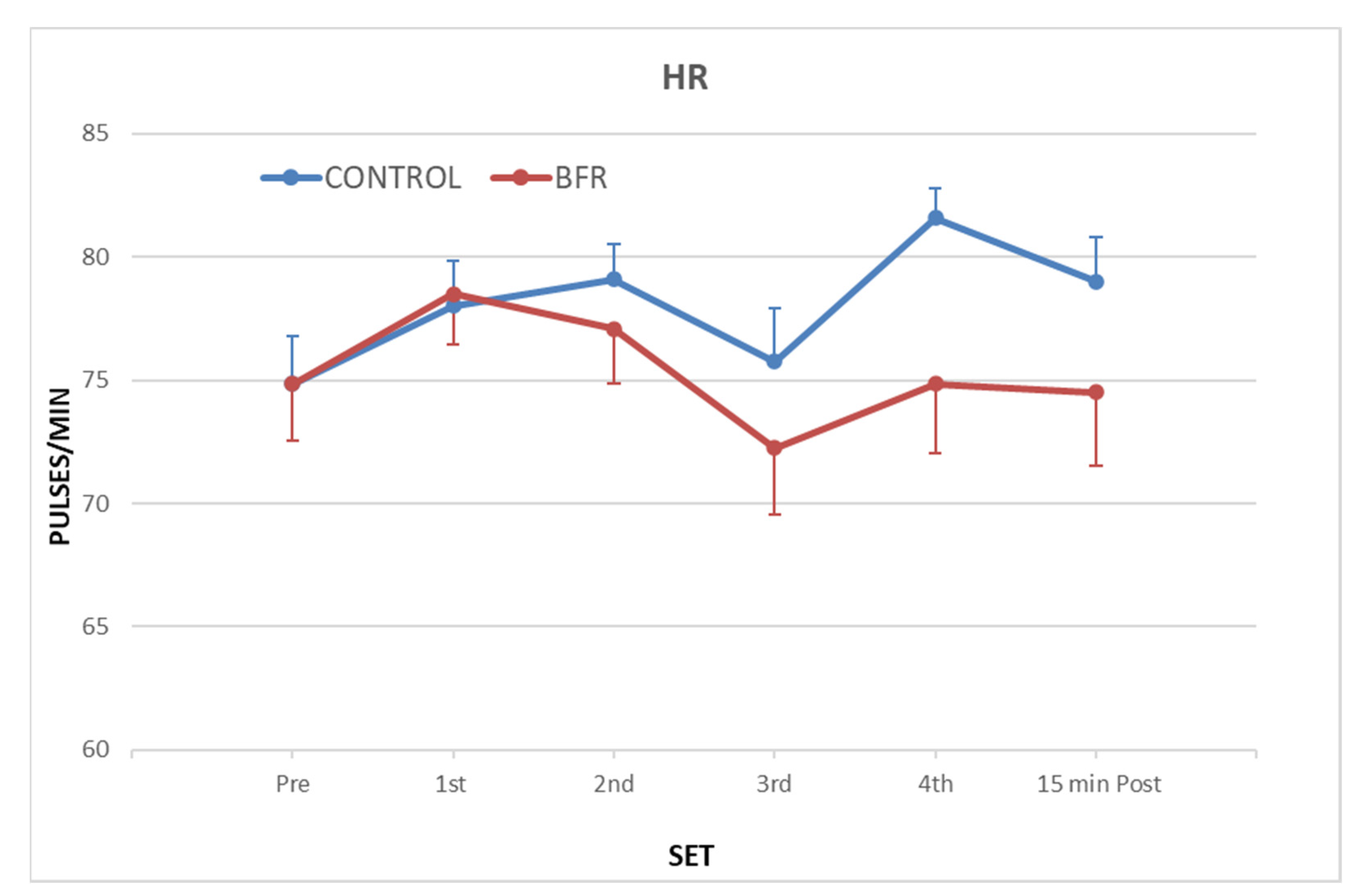
3.1. Heart Rate (HR)

The two-way ANOVA analysis displayed a non-significant “group” × “time” interaction effect (p > 0.05) for HR, while a main effect on the “time” factor was observed (p < 0.05) (Figure 1). The results showed that the HR did not change significantly in the two exercise conditions. However, during exercise without BFR, HR slightly increased compared to exercise with restriction. The minimal increase was statistically insignificant, and HR returned close to resting values after the end of 15 min.

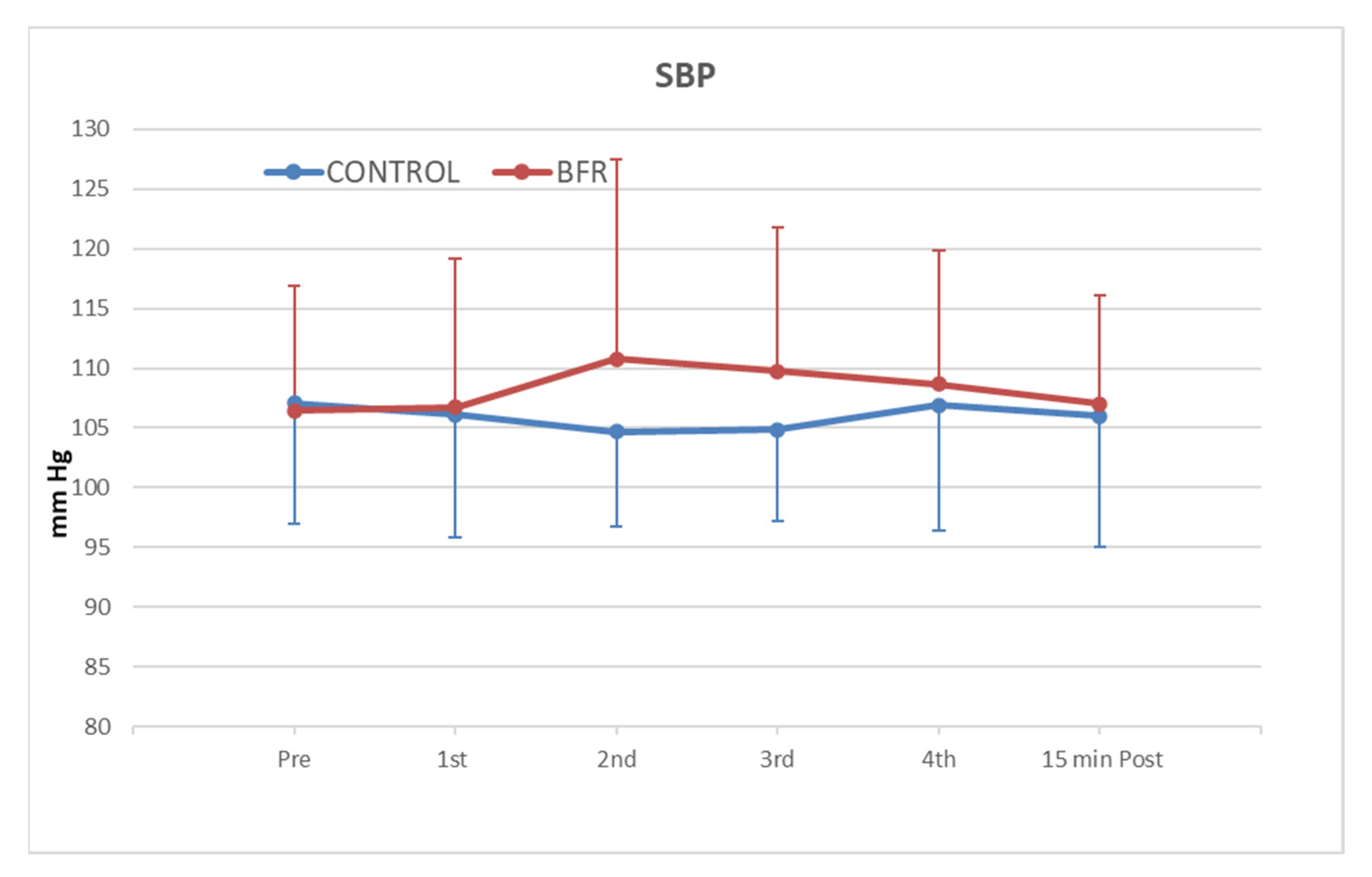
3.2. Systolic Blood Pressure (SBP)

The two-way ANOVA analysis did not display a significant “group” × “time” interaction effect (p > 0.05) for the systolic blood pressure (SBP) value, whereas a main effect on the “time” factor was observed (p < 0.001) (Figure 2). The results showed that the SBP did not change significantly in comparison to resting values, and that there were no statistically significant differences between the groups at any measurement time. The SBP value slightly increased when exercising with BFR compared to without BFR in all intermediate measurements (first to fourth set). However, the difference was statistically insignificant and after 15 minutes, SBP returned close to resting values.

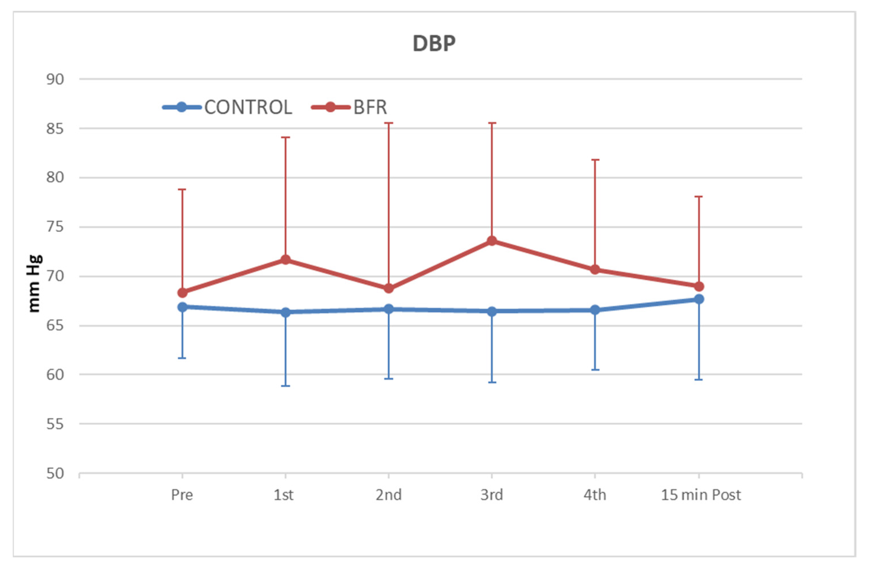
3.3. Diastolic Blood Pressure (DBP)

The two-way ANOVA analysis did not display a significant “group” × “time” interaction effect (p > 0.05) for the diastolic blood pressure (DBP) value, whereas a main effect on the “time” factor was observed (p < 0.05) (Figure 3). The results showed that the DBP did not change significantly in comparison to resting values, and that there were no statistically significant differences between the groups at any time of measurement. The DBP exhibited slightly higher values in exercise with BFR compared to exercise without BFR in all intermediate measurements (first to fourth set). However, the difference was statistically insignificant, and after 15 min, DBP returned close to resting values.

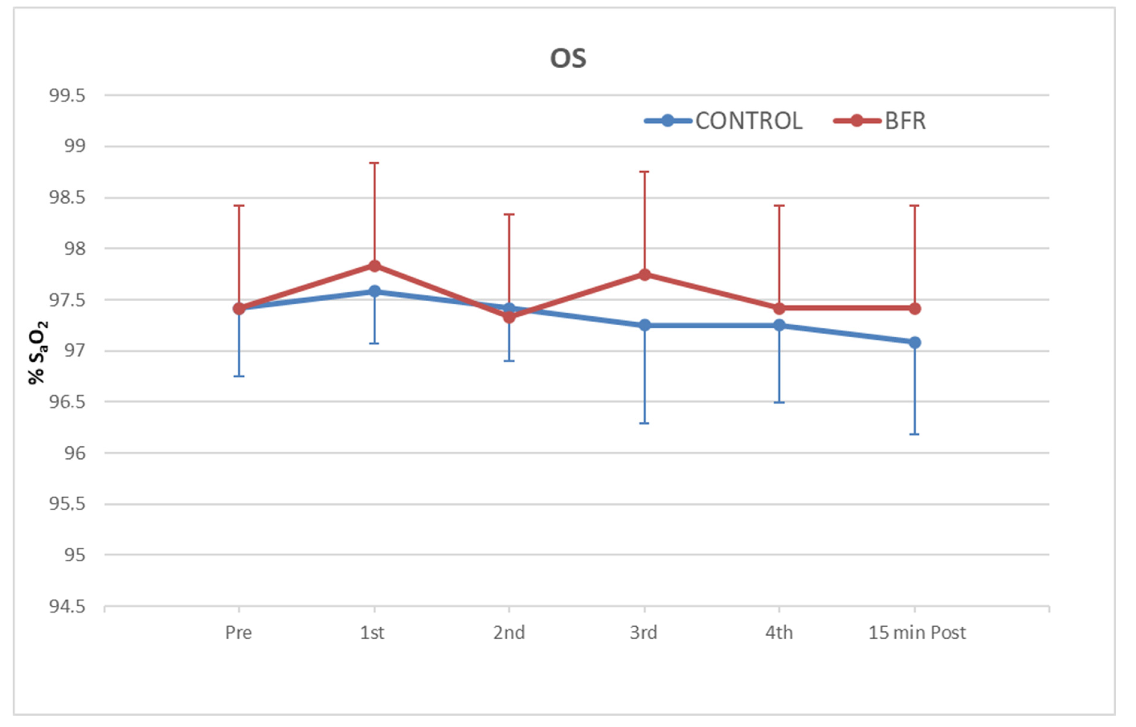
3.4. Oxygen Saturation (SpO2)

The two-way ANOVA analysis did not display a significant “group” × “time” interaction effect (p > 0.05) for the SpO2 value, whereas a main effect on the “time” factor (p < 0.05) was observed (Figure 4). The results showed that the SpO2 did not change significantly in comparison to resting values, and no statistically significant differences between the two exercise conditions were found at any time of measurement.

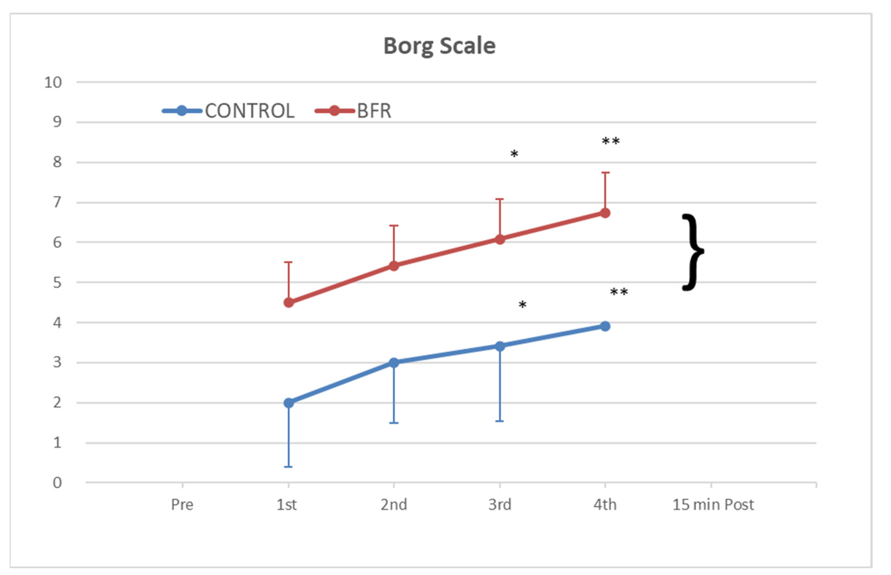
3.5. Rating of Perceived Exertion with the Borg Scale

The two-way ANOVA analysis displayed a significant “group” × “time” interaction effect (p < 0.05) for the Borg scale score, whereas a main effect on the “time” factor was observed (p < 0.001) (Figure 5). Tukey’s (HSD) post hoc test displayed a significant difference between groups in the HR score in the third (p < 0.05, 95% CI) and fourth break (p < 0.001, 95% CI). The results showed that exercise with BFR caused participants to experience higher levels of fatigue than exercise without BFR. The difference was statistically significant from the third break onwards and, in fact, the fatigue was greater in the fourth break compared to the third. The biggest difference between the two exercise conditions appeared immediately after the fourth break.

4. Discussion

The aim of this study was to investigate the effect of an isometric handgrip submaximal exercise protocol with simultaneous BFR on the hemodynamic responses of the cardiovascular system and muscle fatigue of apparently healthy adults. The effects of an isometric handgrip protocol on 30% and 50% of isometric MVC in the cardiovascular system have been studied by other researchers in both apparently healthy individuals [33] and older adults [34]. However, this is the first crossover trial to study the effect of a submaximal isometric protocol, while restricting blood flow.
The results of our study showed that there were no statistically significant changes in the hemodynamic responses of the cardiovascular system between the two exercise conditions (with and without BFR). During exercise without BFR, we observed that the HR slightly increased in its value fluctuations compared to exercise with restriction. The upward trend continued from one break to the next without, however, a statistically significant difference between the two exercise conditions at any point in time. The submaximal intensity of the exercise protocol, which could not induce strong hemodynamic responses, as well as the intermittent application of BFR from one set to the next, are possible explanations. Corresponding studies in which exercise protocols were applied with continuous application of BFR showed an increase in both HR and BP [47,48]. On the other hand, in other studies [49] with intermittent BFR, no increase in HR was observed. Therefore, it appears that the intermittent application of BFR in our study contributed to keeping HR close to resting values. The results of our research reinforce the opinion that intermittent BFR is safer regarding its effect on hemodynamic changes.
Our findings were similar regarding the effects of our protocol on systolic and diastolic pressure. The SBP and DBP values revealed an upward trend in both exercise conditions, which gradually increased from one break to the next. The increase was greater, albeit statistically insignificant, when exercising with BFR at any time of measurement. The SBP and DBP values slightly increased in exercise with BFR compared to without BFR in all intermediate measurements (first to fourth set). After 15 min, they returned close to resting values. Consequently, we concluded that the protocol did not affect BP, the increase in which may have been caused by an increase in HR. Recent research has shown that central hemodynamic load increases during exercise, with and without BFR, are more related to the effect on cardiac function (i.e., parasympathetic withdrawal) than to changes in peripheral vascular resistance [50]. However, the width of the cuff appears to affect BP. A large cuff width (13.5 cm) appears to increase HR, SBP, and DBP more compared to a smaller width (5.0 cm) [51]. Loenneke et al. [52] report that a wider cuff (≥13.5 cm) can cause a relatively total blockage, whereas a smaller cuff may require higher pressure to block blood flow. Therefore, the dimensions of the cuff we used in our research (57 × 9 cm) may be recommended for the safe application of isometric submaximal handgrip exercise protocols. The results of our research contradict those of Bonorino et al. [53], who detected an increase in both HR and BP (SBP and DBP) after a 30% exercise protocol of the elbow flexor muscles. However, their study did not include an isometric exercise protocol, but a protocol with eccentric and concentric contractions. Spranger et al. [54] report that the increase in BP during concentric and eccentric muscle contraction may be a result of muscle hypoxia, increased levels of circulating catecholamines, and hyper-reactive stress response. Our study reinforces the results of other research, such as that of de Araújo et al. [34], according to whom the isometric type of exercise affects BP less. Therefore, it may be a safer way of exercising by restricting blood flow in cases where strong hemodynamic changes are not desirable (e.g., in vulnerable older adults or the chronically ill). Additionally, the results of our research agree with those of Creudeur et al. [50], who also did not detect changes in the values of SBP and DBP after the application of a rhythmic squeeze-relax handgrip exercise protocol. They also agree with the results of other studies in which both the short-term [3] and the long-term effect of BFRT [55] on BP was investigated using different exercise protocols.
Regarding the variation of SpO2, our results showed no significant differences, both in exercise with BFR and without BFR. However, SpO2 slightly increased during the execution of a program with BFR, which is contradictory, as BFR is expected to lower SpO2. The better oxygenation of the restricted limb could be explained because of compensatory vasodilation peripheral to the restricting point, due to limited blood flow. Creudeur et al. [50] studied the tissue saturation index (TSI%) of a handgrip exercise protocol with BFR and also found an increase in tissue oxygenation immediately after exercise. These results contradicted their original opposite prediction that peripheral vascular resistance would probably be greater during BFRT. In any case, the changes in SpO2 in our study were negligible, so we do not consider further analysis necessary.
A statistically significant increase in muscle fatigue occurred during BFRT, which is confirmed by the decrease in the Borg scale score. Our protocol included four two-minute periods of isometric contraction at 30% of the isometric MVC. In contrast to other protocols, such as that of Cerqueira et al. [36], in which isometric exercise was applied until exhaustion, our intervention did not in itself justify the increased fatigue. Consequently, increased fatigue after exercise with BFR can be attributed to the BFR. Based on the RPE, the highest values recorded at the end of the fourth set without BFR corresponded to a level of exercise characterized as “easy”, while at the same time, in exercise with BFR, they corresponded to “hard” [45].
Research has shown that muscle fatigue due to BFRT is normal and is caused by limited oxygen supply [56]. Pearson and Hussain [20] report that fatigue by BFRT can lead to faster and more efficient training adjustments related to muscle hypertrophy and increased strength and endurance. On the other hand, an important factor that also causes muscle fatigue is exercise dosage (intensity, duration, and type of muscle contraction). It seems that the protocol in our research can cause adjustments in muscle strength and endurance without causing strong hemodynamic responses. The results of our study are in line with those of other studies in which increased fatigue was also found after a combination of exercise with BFR. Hussman et al. [57] observed increased fatigue levels after a low-intensity isotonic resistance exercise protocol of the knee extensors, and Copithorne and Rise [58] found increased fatigue in the elbow flexors.
We believe that the results of our study will provide useful information on the appropriateness of this combination in vulnerable groups, as well as guidance for conducting more extensive research in the future.
A number of limitations that need to be considered when interpreting the results are as follows. First, the volunteers were apparently healthy women and men, which makes it difficult to relate the results to other populations that could benefit from isometric exercise with a hand dynamometer, such as patients with a chronic disease. Due to reduced access to participants, no sample size calculation was performed. The level of fitness of the volunteers was not assessed, but none of them had any experience in isometric exercise with a hand dynamometer. Two of the 12 participants were former smokers and had abstained from smoking for nine months and one year, respectively. Additionally, the BFR pressure was the same for everyone, which could be equal to the ischemic value for participants with a low resting BP and only obstructive for those with a high or elevated BP. However, the criteria for exclusion from the study were resting SBP and DBP values ≤ 120 mmHg and 80 mmHg, respectively. Moreover, we investigated the effect of BFRT in the short term, and it remains to be studied in the long term. Another concern is whether the results would be similar or even the same if both upper limbs or lower limbs were exercised. The participants were young, which raises the question about the change in responses in older people with the same exercise protocol, which remains to be studied in future research. The same question applies to the clinical population.

5. Conclusions

Exercise with BFR seems to be a promising method in the field of exercise research, as it has significant effects on both muscle and hemodynamic levels. Ιn our study, we tried to understand how the hemodynamic system is affected in apparently healthy volunteers under specific parameters. The application of an isometric handgrip exercise protocol, with BFR at 140 mmHg and intensity at 30% of the isometric MVC, did not hemodynamically affect the cardiovascular system. However, it caused greater intense fatigue in the flexor muscles of the wrist and fingers. This is something we expected, as the hypoxic environment and the stress that the muscles receive during training could be responsible for the occurrence of fatigue. The combination of exercise with BFR could lead to faster and more intense training adjustments by increasing muscle strength in vulnerable groups, where intense hemodynamic changes are not desirable, as an alternative type of resistance training. For this reason, we consider it important to conduct further research to determine the effects of this protocol when applied to vulnerable population groups. It seems that the intermittent application of BFRT, as well as the appropriate cuff dimensions, significantly affect the presence of hemodynamic responses and muscle fatigue.

Author Contributions

Conceptualization, S.F., P.I. and D.L.; methodology, S.F. and P.I.; software, F.S.; validation, P.I., D.L. and K.K.; formal analysis, S.F.; investigation, S.F.; resources, S.F., F.S. and C.K.; data curation, S.F.; writing—original draft preparation, S.F.; writing—review and editing, D.L.; visualization, F.S. and C.K.; supervision, P.I.; project administration, P.I. All authors have read and agreed to the published version of the manuscript.

Funding

This research received no external funding.

Institutional Review Board Statement

The study was conducted in accordance with the Declaration of Helsinki and approved by the Ethics Committee of the International Hellenic University (EC-02/2023, 2 February 2023).

Informed Consent Statement

Informed consent was obtained from all subjects involved in the study.

Data Availability Statement

The data that support the findings of this study are available on request from the corresponding author. The data are not publicly available due to their containing information that could compromise the privacy of the research participants.

Acknowledgments

The authors gratefully acknowledge all participants of this study.

Conflicts of Interest

The authors declare no conflict of interest.

References

  1. World Health Organization. Physical Activity. Available online: https://www.who.int/news-room/fact-sheets/detail/physical-activity (accessed on 5 October 2022).
  2. Hughes, L.; Paton, B.; Rosenblatt, B.; Gissane, C.; Patterson, S. Blood flow restriction training in clinical musculoskeletal rehabilitation: A systematic review and meta-analysis. Br. J. Sport. Med. 2017, 51, 1003–1011. [Google Scholar] [CrossRef]
  3. Neto, G.R.; Sousa, M.S.C.; Silva, G.V.C.E.; Gil, A.L.S.; Salles, B.F.; Novaes, J.S. Acute resistance exercise with blood flow restriction effects on heart rate, double product, oxygen saturation and perceived exertion. Clin. Physiol. Funct. Imaging 2016, 36, 53–59. [Google Scholar] [CrossRef] [PubMed]
  4. Patterson, S.D.; Hughes, L.; Warmington, S.; Burr, J.; Scott, B.; Owens, J.; Abe, T.; Nielsen, J.; Libardi, C.A.; Laurentino, G.; et al. Blood Flow Restriction Exercise position stand: Considerations of Methodology, Application, and Safety. Front. Physiol. 2019, 10, 1–15. [Google Scholar] [CrossRef]
  5. Vanwye, W.R.; Weatherholt, A.M.; Mikesky, A.E. Blood Flow Restriction Training: Implementation into Clinical Practice. Int. J. Exerc. Sci. 2017, 10, 649–654. [Google Scholar] [PubMed]
  6. Wortman, R.J.; Brown, S.M.; Savage-Elliott, I.; Finley, Z.J.; Mulcahey, M.K. Blood Flow Restriction Training for Athletes: A Systematic Review. Am. J. Sport. Med. 2020, 49, 1938–1944. [Google Scholar] [CrossRef] [PubMed]
  7. Ferreira, M.L.V.; Sardeli, A.V.; De Souza, G.V.; Bonganha, V.; Santos, L.D.C.; Castro, A.; Cavaglieri, C.R.; Chacon-Mikahil, M.P.T. Cardiac autonomic and haemodynamic recovery after a single session of aerobic exercise with and without blood flow restriction in older adults. J. Sport. Sci. 2017, 35, 2412–2420. [Google Scholar] [CrossRef]
  8. Shimizu, R.; Hotta, K.; Yamamoto, S.; Matsumoto, T.; Kamiya, K.; Kato, M.; Hamazaki, N.; Kamekawa, D.; Akiyama, A.; Kamada, Y.; et al. Low-intensity resistance training with blood flow restriction improves vascular endothelial function and peripheral blood circulation in healthy elderly people. Eur. J. Appl. Physiol. 2016, 116, 749–757. [Google Scholar] [CrossRef]
  9. Bocalini, D.S.; Olher, R.D.R.V.; Bacurau, R.F.; Junior, A.F.; Junior, F.L.P.; Navarro, F.; Simoes, H.G.; Araujo, R.; de Moraes, M.R.; Rodriguez, D. Isometric handgrip does not elicit cardiovascular overload or post-exercise hypotension in hypertensive older women. Clin. Interv. Aging 2013, 8, 649–655. [Google Scholar] [CrossRef]
  10. Pinto, R.R.; Karabulut, M.; Poton, R.; Polito, M.D. Acute resistance exercise with blood flow restriction in elderly hypertensive women: Haemodynamic, rating of perceived exertion and blood lactate. Clin. Physiol. Funct. Imaging 2018, 38, 17–24. [Google Scholar] [CrossRef]
  11. Biazon, T.M.P.C.; Ugrinowitsch, C.; Soligon, S.D.; Oliveira, R.M.; Bergamasco, J.G.; Borghi-Silva, A.; Libardi, C.A. The Association between Muscle Deoxygenation and Muscle Hypertrophy to Blood Flow Restricted Training Performed at High and Low Loads. Front. Physiol. 2019, 10, 446. [Google Scholar] [CrossRef]
  12. Curran, M.T.; Bedi, A.; Mendias, C.L.; Wojtys, E.M.; Kujawa, M.V.; Palmieri-Smith, R.M. Blood Flow Restriction Training Applied with High-Intensity Exercise Does Not Improve Quadriceps Muscle Function after Anterior Cruciate Ligament Reconstruction: A Randomized Controlled Trial. Am. J. Sport. Med. 2020, 48, 825–837. [Google Scholar] [CrossRef] [PubMed]
  13. Counts, B.R.; Dankel, S.J.; Barnett, B.E.; Kim, D.; Mouser, J.G.; Allen, K.M.; Thiebaud, R.S.; Abe, T.; Bemben, M.G.; Loenneke, J.P. Influence of relative blood flow restriction pressure on muscle activation and muscle adaptation. Muscle Nerve 2016, 53, 438–445. [Google Scholar] [CrossRef]
  14. Abe, T.; Fujita, S.; Nakajima, T.; Sakamaki, M.; Ozaki, H.; Ogasawara, R.; Sugaya, M.; Kudo, M.; Kurano, M.; Yasuda, T.; et al. Effects of Low-Intensity Cycle Training with Restricted Leg Blood Flow on Thigh Muscle Volume and VO2MAX in Young Men. J. Sport. Sci. Med. 2010, 9, 452–458. [Google Scholar]
  15. Araújo, J.P.; Neto, G.R.; Loenneke, J.P.; Bemben, M.G.; Laurentino, G.C.; Batista, G.R.; Silva, J.C.G.; Freitas, E.; Sousa, M.S.C. The effects of water-based exercise in combination with blood flow restriction on strength and functional capacity in post-menopausal women. Age 2015, 37, 110. [Google Scholar] [CrossRef] [PubMed]
  16. Clarkson, M.; Conway, L.; Warmington, S.A. Blood flow restriction walking and physical function in older adults: A randomized control trial. J. Sci. Med. Sport 2017, 20, 1041–1046. [Google Scholar] [CrossRef]
  17. Barbalho, M.; Rocha, A.C.; Seus, T.L.; Raiol, R.; Del Vecchio, F.B.; Coswig, V.S. Addition of blood flow restriction to passive mobilization reduces the rate of muscle wasting in elderly patients in the intensive care unit: A within-patient randomized trial. Clin. Rehabil. 2019, 33, 233–240. [Google Scholar] [CrossRef]
  18. Jeffries, O.; Waldron, M.; Pattison, J.R.; Patterson, S. Enhanced Local Skeletal Muscle Oxidative Capacity and Microvascular Blood Flow following 7-Day Ischemic Preconditioning in Healthy Humans. Front. Physiol. 2018, 9, 463. [Google Scholar] [CrossRef]
  19. De Freitas, M.C.; Gerosa-Neto, J.; Zanchi, N.E.; Lira, F.S.; Rossi, F.E. Role of metabolic stress for enhancing muscle adaptations: Practical applications. World J. Methodol. 2017, 7, 46–54. [Google Scholar] [CrossRef]
  20. Pearson, S.J.; Hussain, S.R. A Review on the Mechanisms of Blood-Flow Restriction Resistance Training-Induced Muscle Hypertrophy. Sport. Med. 2015, 45, 187–200. [Google Scholar] [CrossRef]
  21. Soucek, M.; Frana, P.; Kara, T.; Sitar, J.; Halamek, J.; Jurak, P.; Rihacek, I.; Spinarova, L.; Oral, I. The Effect of Short-term Isometric Muscle Contraction and the Valsalva Maneuver on Systemic and Pulmonary Hemodynamics in Patients with Severe Heart Failure. Clin. Cardiol. 2009, 32, E31–E38. [Google Scholar] [CrossRef]
  22. Hartog, R.; Bolignano, D.; Sijbrands, E.; Pucci, G.; Mattace-Raso, F. Short-term vascular hemodynamic responses to isometric exercise in young adults and in the elderly. Clin. Interv. Aging 2018, 13, 509–514. [Google Scholar] [CrossRef]
  23. Perry, B.G.; De Hamel, T.; Thomas, K.N.; Wilson, L.C.; Gibbons, T.D.; Cotter, J.D. Cerebrovascular haemodynamics during isometric resistance exercise with and without the Valsalva manoeuvre. Eur. J. Appl. Physiol. 2020, 120, 467–479. [Google Scholar] [CrossRef]
  24. Jin, Y.Z.; Yan, S.; Yuan, W.X. Effect of isometric handgrip training on resting blood pressure in adults: A meta-analysis of randomized controlled trials. J. Sport. Med. Phys. Fit. 2017, 57, 154–160. [Google Scholar] [CrossRef] [PubMed]
  25. Loaiza-Betancur, A.F.; Chulvi-Medrano, I. Is Low-Intensity Isometric Handgrip Exercise an Efficient Alternative in Lifestyle Blood Pressure Management? A Systematic Review. Sport. Health 2020, 12, 470–477. [Google Scholar] [CrossRef]
  26. Farah, B.Q.; Germano-Soares, A.H.; Rodrigues, S.L.C.; Santos, C.X.; Barbosa, S.S.; Vianna, L.C.; Cornelissen, V.A.; Ritti-Dias, R.M. Acute and Chronic Effects of Isometric Handgrip Exercise on Cardiovascular Variables in Hypertensive Patients: A Systematic Review. Sports 2017, 5, 55. [Google Scholar] [CrossRef]
  27. Maga, M.; Schönborn, M.; Wachsmann-Maga, A.; Śliwka, A.; Krężel, J.; Włodarczyk, A.; Olszewska, M.; Nowobilski, R. Stimulation of the Vascular Endothelium and Angiogenesis by Blood-Flow-Restricted Exercise. Int. J. Environ. Res. Public Health 2022, 19, 15859. [Google Scholar] [CrossRef]
  28. Horiuchi, M.; Okita, K. Blood Flow Restricted Exercise and Vascular Function. Int. J. Vasc. Med. 2012, 2012, 543218. [Google Scholar] [CrossRef] [PubMed]
  29. Larkin, K.A.; Macneil, R.G.; Dirain, M.; Sandesara, B.; Manini, T.M.; Buford, T.W. Blood Flow Restriction Enhances Post–Resistance Exercise Angiogenic Gene Expression. Med. Sci. Sport. Exerc. 2012, 44, 2077–2083. [Google Scholar] [CrossRef]
  30. Ferguson, R.A.; Hunt, J.E.A.; Lewis, M.P.; Martin, N.R.W.; Player, D.J.; Stangier, C.; Taylor, C.W.; Turner, M.C. The Acute An-giogenic Signalling Response to Low-Load Resistance Exercise with Blood Flow Restriction. Eur. J. Sport Sci. 2018, 18, 397–406. [Google Scholar] [CrossRef]
  31. Montgomery, R.; Paterson, A.; Williamson, C.; Florida-James, G.; Ross, M.D. Blood Flow Restriction Exercise Attenuates the Exercise-Induced Endothelial Progenitor Cell Response in Healthy, Young Men. Front. Physiol. 2019, 10, 447. [Google Scholar] [CrossRef] [PubMed]
  32. Hudlicka, O.; Brown, M.D. Adaptation of Skeletal Muscle Microvasculature to Increased or Decreased Blood Flow: Role of Shear Stress, Nitric Oxide and Vascular Endothelial Growth Factor. J. Vasc. Res. 2009, 46, 504–512. [Google Scholar] [CrossRef] [PubMed]
  33. Silva, I.M.; Sobrinho, M.F.L.; Ritti-Dias, R.M.; Sobral, B.P.S.V.; Pirauá, A.L.T.; De Oliveira, L.M.F.T.; Farah, B.Q. Cardiovascular responses after isometric handgrip exercise at different intensities in healthy men. J. Phys. Educ. 2019, 30, 3020. [Google Scholar] [CrossRef]
  34. Araújo, C.G.S.D.; Duarte, C.V.; Gonçalves, F.D.A.; Medeiros, H.; Lemos, F.A.; Gouvêa, A.L. Hemodynamic responses to an isometric handgrip training protocol. Arq. Bras. Cardiol. 2011, 97, 413–419. [Google Scholar] [CrossRef]
  35. Cayot, T.E.; Lauver, J.; Silette, C.R.; Scheuermann, B.W. Effects of blood flow restriction duration on muscle activation and microvascular oxygenation during low-volume isometric exercise. Clin. Physiol. Funct. Imaging 2014, 36, 298–305. [Google Scholar] [CrossRef] [PubMed]
  36. Cerqueira, M.S.; Pereira, R.; de Mesquita, G.N.; Rocha, T.; de Moura Filho, A.G. Influence of Blood Flow Restriction Level on Muscle Fatigue during an Intermittent Isometric Exercise Taken to Failure. Muscle Ligaments Tendons J. 2020, 10, 408. [Google Scholar] [CrossRef]
  37. World Health Organization. Obesity and Overweight. Available online: https://www.who.int/news-room/fact-sheets/detail/obesity-and-overweight (accessed on 27 December 2022).
  38. American Heart Association. Understanding Blood Pressure Readings. Available online: https://www.heart.org/en/health-topics/high-blood-pressure/understanding-blood-pressure-readings (accessed on 10 January 2023).
  39. Stergiou, G.S.; Yiannes, N.G.; Rarra, V.C. Validation of the Omron 705 IT oscillometric device for home blood pressure measurement in children and adolescents: The Arsakion School Study. Blood Press. Monit. 2006, 11, 229–234. [Google Scholar] [CrossRef]
  40. Vera-Cala, L.M.; Orostegui, M.; Valencia-Angel, L.I.; López, N.; Bautista, L.E. Accuracy of the Omron HEM-705 CP for blood pressure measurement in large epidemiologic studies. Arq. Bras. Cardiol. 2011, 96, 393–398. [Google Scholar] [CrossRef]
  41. Essner, A.; Sjöström, R.; Ahlgren, E.; Lindmark, B. Validity and reliability of Polar® RS800CX heart rate monitor, measuring heart rate in dogs during standing position and at trot on a treadmill. Physiol. Behav. 2013, 114–115, 1–5. [Google Scholar] [CrossRef] [PubMed]
  42. Louie, A.; Feiner, J.R.; Bickler, P.E.; Rhodes, L.; Bernstein, M.; Lucero, J. Four Types of Pulse Oximeters Accurately Detect Hypoxia during Low Perfusion and Motion. Anesthesiology 2018, 128, 520–530. [Google Scholar] [CrossRef]
  43. Borg, G.A. Psychophysical bases of perceived exertion. Med. Sci. Sport. Exerc. 1982, 14, 377–381. [Google Scholar] [CrossRef]
  44. Williams, N. The Borg Rating of Perceived Exertion (RPE) scale. Occup. Med. 2017, 67, 404–405. [Google Scholar] [CrossRef]
  45. American College of Sports Medicine. ACSM’s Guidelines for Exercise Testing and Prescription, 9th ed.; Lippincott Williams & Wilkins: Baltimore, MD, USA, 2013. [Google Scholar]
  46. Centers for Disease Control and Prevention (CDC). Perceived Exertion (Borg Rating of Perceived Exertion Scale). Available online: https://www.cdc.gov/physicalactivity/basics/measuring/exertion.htm (accessed on 15 December 2022).
  47. Poton, R.; Polito, M.D. Hemodynamic response to resistance exercise with and without blood flow restriction in healthy subjects. Clin. Physiol. Funct. Imaging 2014, 36, 231–236. [Google Scholar] [CrossRef]
  48. Vieira, P.J.; Chiappa, G.R.; Umpierre, D.; Stein, R.; Ribeiro, J.P. Hemodynamic Responses to Resistance Exercise with Restricted Blood Flow in Young and Older Men. J. Strength Cond. Res. 2013, 27, 2288–2294. [Google Scholar] [CrossRef] [PubMed]
  49. Figueroa, A.; Vicil, F. Post-exercise aortic hemodynamic responses to low-intensity resistance exercise with and without vascular occlusion. Scand. J. Med. Sci. Sport. 2011, 21, 431–436. [Google Scholar] [CrossRef] [PubMed]
  50. Credeur, D.P.; Jones, R.; Stanford, D.; Stoner, L.; McCoy, S.; Jessee, M. Central cardiovascular hemodynamic response to unilateral handgrip exercise with blood flow restriction. Eur. J. Appl. Physiol. 2019, 119, 2255–2263. [Google Scholar] [CrossRef] [PubMed]
  51. Rossow, L.M.; Fahs, C.A.; Loenneke, J.P.; Thiebaud, R.S.; Sherk, V.D.; Abe, T.; Bemben, M.G. Cardiovascular and perceptual responses to blood-flow-restricted resistance exercise with differing restrictive cuffs. Clin. Physiol. Funct. Imaging 2012, 32, 331–337. [Google Scholar] [CrossRef]
  52. Loenneke, J.P.; Thiebaud, R.S.; Fahs, C.A.; Rossow, L.M.; Abe, T.; Bemben, M.G. Effect of cuff type on arterial occlusion. Clin. Physiol. Funct. Imaging 2013, 33, 325–327. [Google Scholar] [CrossRef]
  53. Bonorino, S.L.; Sá, C.A.D.; Corralo, V.D.S.; Olkoski, M.M.; Silva-Grigoletto, M.E.D.; Saretto, C.B.; Resende, A.G.D. Hemodynamic responses to strength exercise with blood flow restriction in small muscle groups. Rev. Bras. Cineantropometria Desempenho Hum. 2019, 21, e56258. [Google Scholar] [CrossRef]
  54. Spranger, M.D.; Krishnan, A.C.; Levy, P.D.; O’Leary, D.S.; Smith, S.A. Blood flow restriction training and the exercise pressor reflex: A call for concern. Am. J. Physiol. Circ. Physiol. 2015, 309, H1440–H1452. [Google Scholar] [CrossRef]
  55. Ozaki, H.; Yasuda, T.; Ogasawara, R.; Sakamaki-Sunaga, M.; Naito, H.; Abe, T. Effects of high-intensity and blood flow-restricted low-intensity resistance training on carotid arterial compliance: Role of blood pressure during training sessions. Eur. J. Appl. Physiol. 2012, 113, 167–174. [Google Scholar] [CrossRef]
  56. Cerqueira, M.S.; Pereira, R.; Rocha, T.; Mesquita, G.; de Paiva Lima, C.R.O.; Raposo, M.C.F.; De Moura Filho, A.G. Time to failure and neuromuscular response to intermittent isometric exercise at different levels of vascular occlusion: A randomized crossover study. Int. J. Appl. Exerc. Physiol. 2017, 6, 55–70. [Google Scholar] [CrossRef]
  57. Husmann, F.; Mittlmeier, T.; Bruhn, S.; Zschorlich, V.; Behrens, M. Impact of Blood Flow Restriction Exercise on Muscle Fatigue Development and Recovery. Med. Sci. Sport. Exerc. 2018, 50, 436–446. [Google Scholar] [CrossRef] [PubMed]
  58. Copithorne, D.B.; Rice, C.L. The effect of blood flow occlusion during acute low-intensity isometric elbow flexion exercise. Eur. J. Appl. Physiol. 2019, 119, 587–595. [Google Scholar] [CrossRef] [PubMed]
Figure 1. Changes in heart rate (HR) at baseline, between the sets, and 15 min after exercise with blood flow restriction (BFR) and without (control).
Physiologia 03 00018 g001
Figure 2. Changes in systolic blood pressure (SΒP) at baseline, between the sets, and 15 min after exercise with blood flow restriction (BFR) and without (control).
Physiologia 03 00018 g002
Figure 3. Changes in diastolic blood pressure (DΒP) at baseline, between the sets, and 15 min after exercise with blood flow restriction training (BFR) and without (control).
Physiologia 03 00018 g003
Figure 4. Changes in oxygen saturation (OS) at baseline, between the sets, and 15 min after exercise with blood flow restriction training (BFR) and without (control).
Physiologia 03 00018 g004
Figure 5. Changes in Borg scale score between the sets with blood flow restriction training (BFR) and without (control), with significant differences between the two exercise conditions after the third and fourth set. * Between groups significant comparisons in the post hoc testing. ** Between sets significant comparisons.
Physiologia 03 00018 g005
Table 1. Demographic characteristics of the participants.
Demographic CharacteristicsMean ± SE
Age (years)23.27 ± 0.54
BMI (value)24.86 ± 1.78
Gender (%)Women 50% n = 12
ComorbiditiesNone
MedicationNone
Smokersn = 2 (former)
Mean values and standard errors for quantitative variables and frequencies (%) for categorical variables.
Table 2. Outcome measures mean score (SD) for each time point of measurement in the two participant groups.
Baseline1st Set2nd Set3rd Set4th Set15 min after
HR pulses/minute (SD)
BFRT *74 (8)78 (7)77 (8)72 (9) 74 (10)74(10)
Control74 (7)78 (6)79 (5)75 (7)81 (4)79 (6)
SBP mmHg (SD)
BFRT106.4 (10.5)106.8 (12.4)110.8 (16.8) 109.8 (12.0)108.7 (11.1)107.0 (9.1)
Control107.1 (10.1)106.1 (10.2)104.7 (7.9)104.8 (7.7)106.9 (10.5)106.0 (11.0)
DBP mmHg (SD)
BFRT68.3 (7.1)71.7 (6.0)68.8 (7.8)73.6 (12.2)70.7 (6.7)69.0 (5.4)
Control66.9 (5.2)66.3 (7.5)66.7 (7.1)66.4 (7.2)66.6 (6.1)67.7 (8.2)
OS SpO2 (SD)
BFRT97.4 (0.8)97.8 (0.8)97.3 (1.0)97.8 (0.5)97.4 (0.9)97.4 (0.9)
Control97.4 (0.7)97.6 (0.5)97.4 (0.5)97.3 (1.0)97.3 (0.8)97.1 (0.9)
Borg scale points (SD)
BFRT-4.5 (1.8)5.4 (1.8)6.1 (2.1)6.8 (2.3) **-
Control *-2.0 (1.4)3.0 (1.6)3.4 (1.5)3.9 (1.9) **-
* Between groups significant comparisons in the post hoc testing. ** Between sets significant comparisons. BFRT: blood flow restriction training; Control: conduction of exercise with no blood restriction; HR: heart rate; SBP: systolic blood pressure; DBP: diastolic blood pressure; OS: oxygen saturation.
Disclaimer/Publisher’s Note: The statements, opinions and data contained in all publications are solely those of the individual author(s) and contributor(s) and not of MDPI and/or the editor(s). MDPI and/or the editor(s) disclaim responsibility for any injury to people or property resulting from any ideas, methods, instructions or products referred to in the content.

