Review

Dental Stem Cell-Based Therapy for Glycemic Control and the Scope of Clinical Translation: A Systematic Review and Meta-Analysis

1
Private Dental Practice, Falls Church, VA 22043, USA
2
Private Dental Practice, Mumbai 400020, India
3
Academic Affairs, Rutgers School of Dental Medicine, Newark, NJ 07101, USA
4
Department of Periodontics, Rutgers School of Dental Medicine, Newark, NJ 07103, USA
*
Author to whom correspondence should be addressed.
Int. J. Transl. Med. 2024, 4(1), 87-125; https://doi.org/10.3390/ijtm4010005
Submission received: 3 September 2023 / Revised: 6 October 2023 / Accepted: 27 October 2023 / Published: 15 January 2024

Abstract

:
Background: The tooth is a repository of stem cells, garnering interest in recent years for its therapeutic potential. The aim of this systematic review and meta-analysis was to test the hypothesis that dental stem cell administration can reduce blood glucose and ameliorate polyneuropathy in diabetes mellitus. The scope of clinical translation was also assessed. Methods: PubMed, Cochrane, Ovid, Web of Science, and Scopus databases were searched for animal studies that were published in or before July 2023. A search was conducted in OpenGrey for unpublished manuscripts. Subgroup analyses were performed to identify potential sources of heterogeneity among studies. The risk for publication bias was assessed by funnel plot, regression, and rank correlation tests. Internal validity, external validity, and translation potential were determined using the SYRCLE (Systematic Review Center for Laboratory Animal Experimentation) risk of bias tool and comparative analysis. Results: Out of 5031 initial records identified, 17 animal studies were included in the review. There was a significant decrease in blood glucose in diabetes-induced animals following DSC administration compared to that observed with saline or vehicle (SMD: −3.905; 95% CI: −5.633 to −2.177; p = 0.0004). The improvement in sensory nerve conduction velocity (SMD: 4.4952; 95% CI: 0.5959 to 8.3945; p = 0.035) and capillary-muscle ratio (SMD: 2.4027; 95% CI: 0.8923 to 3.9132; p = 0.0095) was significant. However, motor nerve conduction velocity (SMD: 3.1001; 95% CI: −1.4558 to 7.6559; p = 0.119) and intra-epidermal nerve fiber ratio (SMD: 1.8802; 95% CI: −0.4809 to 4.2413; p = 0.0915) did not increase significantly. Regression (p < 0.0001) and rank correlation (p = 0.0018) tests indicated the presence of funnel plot asymmetry. Due to disparate number of studies in subgroups, the analyses could not reliably explain the sources of heterogeneity. Interpretation: The direction of the data indicates that DSCs can provide good glycemic control in diabetic animals. However, methodological and reporting quality of preclinical studies, heterogeneity, risk of publication bias, and species differences may hamper translation to humans. Appropriate dose, mode of administration, and preparation must be ascertained for safe and effective use in humans. Longer-duration studies that reflect disease complexity and help predict treatment outcomes in clinical settings are warranted. This review is registered in PROSPERO (number CRD42023423423).

1. Introduction

In 1957, when Thomas et al. published their seminal report on allogeneic hematopoietic stem cell (HSC) transplants in humans [1], they opened the door to a revolutionary therapy for leukemia. Since then, stem cell (SC) research in diseases such as retinopathy, dementia, and diabetes mellitus has been advancing by leaps and bounds [2].
It wasn’t until the 1960s, when Friedenstein et al. identified a group of cells providing a scaffold for HSCs within bone marrow, that mesenchymal stem cells came to light [3]. These stromal cells, termed as mesenchymal stem cells (MSCs) by Caplan in 1991, are plastic, possessing the ability to differentiate into various mesenchymal cell lines and regenerate injured tissues [4]. They migrate to sites of injury and stimulate the proliferation and differentiation of native progenitor cells [5]. MSCs have relatively low immunogenicity [5,6]. Human leucocyte antigen (HLA) molecules are a group of antigen-presenting proteins which are responsible for initiating allogeneic graft rejection [7]. MSC transplants express low levels of HLA class I molecules, do not express HLA class II molecules [7], and remain inconspicuous to cytotoxic T and natural killer (NK) cells [5,8]. In addition, they stimulate the production of regulatory T and B cells [9], a group of specialized lymphocytes that suppress immune response, thereby maintaining homeostasis and immunological tolerance [10].
It is estimated that by 2030, diabetes mellitus will be prevalent in about 643 million adults worldwide [11]. Aside from diseases such as ischemic heart conditions and COVID-19, it was a leading cause of death in 2021, and the global cost for diabetes-related health care was at least USD 966 billion [11]. Uncontrolled diabetes is of grave concern, especially in the post pandemic era, as it complicates recovery and affects the prognosis of almost every infection and disease. Type 1 diabetes (T1DM) is caused by autoimmune destruction of islets of Langerhans, resulting in endogenous insulin deficiency [12]. Its pathogenesis is influenced by genetic as well as environmental factors and is most commonly diagnosed in adolescents and young adults [12]. Type 2 diabetes (T2DM) is characterized by insulin resistance and the inability of islet cells to produce sufficient insulin to compensate, leading to relative insulin deficiency [12]. It is most commonly diagnosed in middle aged adults and is often associated with obesity [12].
Approximately 50% of diabetic adults are eventually afflicted with polyneuropathy (DPN) [11]. It is characterized by axonal degeneration [13,14] caused by pro-inflammatory cytokine-releasing metabolic cascades, which result in structural and functional changes in endoneurial and microvascular tissues [15,16]. DPN is associated with a high risk for foot ulcers and lower limb amputations [16]. Treatment involves regular screening, stringent glycemic control, and pain alleviation [16].
Islet cell transplantation is an alternative therapy for diabetes mellitus, but is associated with the risk of graft rejection and paucity of donor sources [14]. Bone marrow mesenchymal stem cells (BMMSCs) are capable of differentiating into insulin secreting cells [8]. However, retrieval is often accompanied by pain [8], nerve injury [17], and low cell yield upon harvest [8]. Hence, the umbilical cord, placenta, and teeth, which are discarded after removal, have gained ground as alternate MSC reserves [8,17].
The primitive tooth organ and supporting structures are formed by complex interactions of the neural crest with epithelial and mesodermal components [18]. The dental follicle and papilla formed in turn give rise to gingiva, periodontal ligament, and dental pulp [8]. Hence, these ectomesenchymal tissues retain the stemness of neural crest cells [8,18]. There are six types of stem cells of dental origin (DSCs) that have been isolated and characterized so far: dental pulp stem cells (DPSCs), stem cells from the pulps of human exfoliated deciduous teeth (SHED), dental follicle precursor cells (DFPCs), stem cells from apical papilla (SCAP), periodontal ligament stem cells (PDLSCs), and gingival mesenchymal stem cells (GMSCs) [8,9,18]. SCs harvested from these tissues are able to transdiffferentiate into neuronal-like and pancreatic β cells [8,9,17], and this ability can be traced back to the neural crest and to similarities between neuronal and pancreatic β cells [8].
Following extraction, exfoliation, or gingival surgery, ideally under sterile conditions and minimal trauma, the tooth or periodontal tissue is preserved to maintain the viability of stem cells until their retrieval [19]. After the surface of the tooth is cleansed, the tissue is retrieved from the tooth, minced, and digested in collagenase [19]. The cells undergo neutralization and centrifugation [19]. They are then trypsinized and can differentiate into insulin releasing islet-like cells after undergoing several passages in culture [19]. The stem cell activity in a tooth varies, and is contingent on factors such as donor age, tooth morphology, DSC type, tissue health, and conditions during retrieval [20]. For instance, in one study, six lines of SCs were obtained from the pulps of six deciduous teeth of children aged 4–8 years, whereas two DPSC lines were obtained from six permanent teeth of donors aged 55–67 years [20].
DSCs have more robust population doubling rates than do BMMSCs [8]. They are also able to differentiate into odontogenic, osteogenic, chondrogenic, adipogenic, and epithelial cell lineages under specific conditions, thus possessing the potential for application in the regeneration and repair of tissues that arise from all three germ layers [19,21]. There is ongoing research for their use in pulp and dentin regeneration, osteoporosis, rheumatoid arthritis, and liver fibrosis, among other diseases [8,18,20,21].
DSCs can serve as replenishable sources of islet cells; hence, they hold allure in diabetology. Studies have been done to elucidate their effects; however, to our knowledge, a study which systematically reviews and quantifies existing data on the use of various DSCs in diabetes mellitus has not yet been undertaken. Hence, we conducted a systematic review and meta-analysis to appraise the extent of evidence in relation to the focused question: “Do dental stem cells reduce blood glucose and alleviate polyneuropathy in diabetic animals compared to animals administered with saline or vehicle?” In addition to testing this hypothesis, we discern the scope of translation of this novel therapy to humans.

2. Methods

This systematic review was reported in accordance with the guidelines outlined in the PRISMA 2020 statement [22], an updated version of Preferred Reporting Items for Systematic Reviews and Meta-Analysis, which is a set of guidelines used to improve the transparency and quality of systematic reviews (PRISMA 2020 checklists are available as Supplementary Tables S1 and S2). The review is registered in PROSPERO, an international database of systematic reviews (registration number CRD42023423423).

2.1. Search Strategy

The search was defined to identify studies that evaluated the effects of stem cells of dental origin (DSCs) on blood glucose or DPN parameters, when administered in diabetic animals. The search was conducted in multiple stages. In the first step, a search was performed using the Cochrane Library (Issue 12, 2015), PubMed (MEDLINE—1996), Scopus (1990), Ovid (Embase-1974), and Web of Science (1996) electronic databases. A search was also carried out for grey literature in OpenGrey (openSIGLE 2007). There were no restrictions regarding publication date, and the last search was performed in July 2023. The titles and abstracts were read to determine if they potentially fit the inclusion criteria. Duplicate articles were removed. If the full text was not available from the databases for review or additional information was deemed necessary for selection, the corresponding authors of potential studies were contacted via email. Following this, the full text of potential studies was read to evaluate if they should be included in the review. In the next stage, during July 2023, reference lists of all articles included in the initial step were manually searched. There were no language restrictions in our search strategy.
The search was conducted using MeSH (medical subject heading) terms belonging to the categories of disease, intervention, and population in different permutations and combinations-“Diabetes” [MeSH] OR “Diabetes Mellitus” [MeSH] OR “glucose” [MeSH] OR “insulin” [MeSH]) AND “dental stem cells” [MeSH] OR “Periodontal stem cells” [MeSH] OR “gingival stem cells” [MeSH] OR “glycemic control” [MeSH]. Free text was also used, which included “diabetes”, “diabetes mellitus”, “diabetes mellitus type 1”, “diabetes mellitus type 2”, “glucose”, “insulin”, and “dental stem cells”, “periodontal stem cells”, “gingival stem cells”, and “glycemic control”. In addition, search filters, as described by Hooijmans et al. (2010) [23], were employed for a more comprehensive retrieval of animal studies from the databases. The full version of the search filters used is presented in Supplementary Table S3.

2.2. Selection Process

The titles and abstracts of all records were screened by all authors (P.T., V.T., S.J., and G.Y.) independently. In all cases, disagreements among the reviewers regarding which articles to read through full text were resolved through discussions. All authors then read through full text articles to determine inclusion. The reference lists of selected articles were read by the authors independently. The final selection of studies was discussed, and in case of disagreements regarding inclusion of articles, a consensus was reached among the authors. Automation tools were not used at any stage of the defined search protocol.

2.3. Inclusion Criteria

  • Animal studies, regardless of species, age, and gender, published in or before July 2023, were included.
  • Studies in which the disease model was induced diabetes mellitus (Type 1 or Type 2), with any manner of disease induction, were included.
  • Studies which included control groups with animals administered with saline or vehicle for comparison were included in the review.
  • Studies which used dental stem cells, i.e., stem cells of dental origin, as the intervention were included, regardless of dose, timing, frequency, preparation, and route of administration. There were no restrictions regarding the source or portion of the tooth or its supporting tissues from which the DSCs were isolated.
  • Studies using blood glucose and/or verifiable parameters of diabetic polyneuropathy, such as sensory and motor nerve conduction velocity (SNCV and MNCV), as outcome variables were included.
  • Grey literature, such as preprints, dissertations, theses, unpublished manuscripts, and conference papers was also reviewed to determine if it met the inclusion criteria mentioned above.
There were no restrictions in regards to the language of the articles.

2.4. Exclusion Criteria

  • Animal studies which did not include diabetic models were not included in the review. In addition, animal studies which did not include diabetic controls administered with saline or vehicle for comparison with diabetic animals administered with DSCs were excluded.
  • Studies in which diabetes mellitus was induced after DSC administration were excluded.
  • Studies which did not use stem cells of dental origin were not included in the review.
  • Studies which did not measure blood glucose and/or DPN parameters, or in which these variables were not measured using valid methods, were not included in the review.
  • In vitro studies, surveys, and questionnaires were not included.
  • Reviews and duplicate articles were excluded.
The complete list of studies that were excluded after reading the full text is available upon reasonable request from the corresponding author.

2.5. Data Extraction

The data extracted from selected studies regarding the study design included country of origin, method of conducting the experiment, and study duration. Data regarding the animal model included species, age of animals at the start of the experiment, method of diabetes induction, and time of sacrifice. Information regarding the type of DSCs used in the study, source, preparation, dose, and route of administration was extracted. Changes in primary outcome variables, i.e., fasting and/or random blood glucose, and DPN parameter values, such as SNCV and MNCV, from baseline to the end of the experiment following DSC administration, were included. Other outcome measures were also noted, if present.
All authors were involved in data extraction. In case of unreported or missing data, the authors of the selected studies were contacted via email. WebPlotDigitizer, a web-based tool (https://automeris.io/WebPlotDigitizer (accessed on 3 August 2023)) was used if data was presented graphically.

2.6. Assessment of Internal Bias in Articles

The internal bias in each study was assessed using the SYRCLE (Systematic Review Center for Laboratory Animal Experimentation) tool by Hooijmans et al. (2014) [24], which in turn is based on Cochrane’s RoB (risk of bias) tool [25]. The assessment of internal bias was conducted by each of the authors (P.T., V.T., S.J., and G.Y.) individually.

2.7. Assessment of External Validity

The external validity of the studies was analyzed using a table that examined data from included studies to determine the scope of translation of DSC therapy to humans.

2.8. Meta-Analysis

Review Manager (RevMan), version 5.4 (The Cochrane Collaboration, 2020) [26], and MAJOR with jamovi statistical software (version 2.3) [27] were used to perform quantitative analyses when more than two studies provided data for each outcome of interest.
Due to anticipated differences in design and effect sizes between studies, random effects model was used for calculating summary effect estimates [28]. The inverse variance method was used to combine results across studies because with this method, more weight is assigned to studies with smaller standard errors; hence, there is greater precision in the overall summary effect [28]. Standardized mean difference (SMD) was used as the outcome measure due to inter study differences in units and methods for measuring outcomes [29]. Hedges’ adjusted g method [30] was used to compute SMD, as it adjusts for small sample bias. The SMD was presented with 95% confidence interval (CI), which represented the range in which one can be 95% certain that the true value of the SMD lies [31]. A p-value of less than 0.05 was considered statistically significant for the pooled summary effects.
DerSimonian–Laird estimator was used to calculate the extent of heterogeneity (i.e., tau2) among the studies [32]. Knapp–Hartung method [33] was used to reduce the risk of Type 1 error (i.e., incorrect rejection of a null hypothesis that is actually true), particularly in analyses that constituted a small number of studies. In addition, Cochran’s Q-test and its p value were calculated to determine potential heterogeneity [34]. A p-value of less than 0.05 was indicative of presence of heterogeneity, which cannot be attributed only to chance [28]. I2 statistic was used to examine the degree of heterogeneity [28]. I2 values of 50% or more were deemed as indicative of substantial heterogeneity [28]. Further, the prediction interval, which helps demonstrate the range of true effects that may be expected in future similar studies [35,36], was also calculated for each outcome.
Studentized residuals were calculated to identify potential outliers, i.e., studies with 95% confidence intervals that were outside those of the pooled effect [37]. The equation 100 × (1 − 0.05/(2 × k)) th percentile of a standard normal distribution, i.e., Bonferroni correction [38] with 2 sided alpha = 0.05 for k number of studies, was calculated for each analysis, and if the studentized residual range of a study exceeded this value, it was considered an outlier in the model. Studies that may have excessively leveraged the pooled effect were identified using Cook’s distances [39]. If the Cook’s distance of a study was larger than the median plus six times the interquartile range of the Cook’s distance, it was considered to be influential in the context of the analysis [39].
Multiple reports of the same study describing different outcome measures were identified during data collection. In such cases, data from all reports was collected and presented as a single study in the systematic review, and relevant quantitative data from only one report per study was used in each meta analysis to avoid unit of analysis errors [40]. For multiple-arm studies, all relevant intervention groups were combined to create one group for a single paired comparison with the control group to avoid ‘double -counting’ of animals [41]. For studies with a cross-over design, each paired analysis was estimated by calculating mean difference and standard error of the mean difference and entering the data in the analysis in the form of generic inverse variance outcome [41].

2.8.1. Subgroup Analysis

Studies included in the meta-analysis for DSC effect on blood glucose were split into subgroups to examine potential sources of heterogeneity among studies [42] and to measure various treatment effect estimates. Species differences and DSC type were used as variables to check for possible interactions among subgroups, since it was anticipated that these aspects may be potential sources of heterogeneity.
A test for significance which determines heterogeneity across subgroups was conducted, as described by Borenstein et al. (2009) [43]. The I2 statistic was used to calculate heterogeneity as a percentage in the subgroup analysis.

2.8.2. Publication Bias

If 10 or more studies provided data in the meta-analysis, the studies were assessed by funnel plot inspection for potential publication bias. Using standard error of the observed outcomes, Egger’s regression [44] and Begg and Mazumdar rank correlation [45] tests were performed to provide quantitative assessments of funnel plot asymmetry.

3. Results

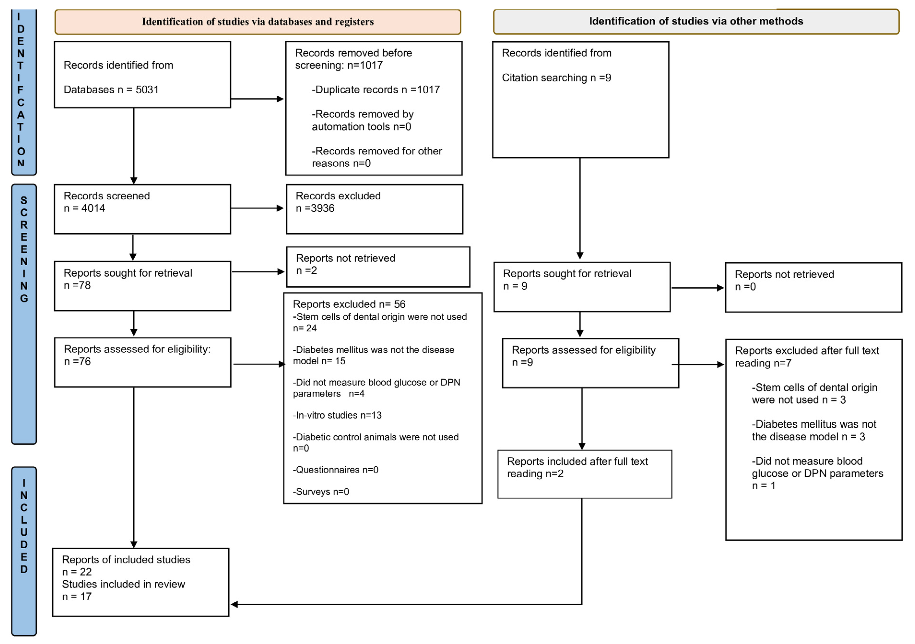
3.1. Study Selection

In total, the database search yielded 5031 records (Figure 1). After removing duplicate records, the abstracts of 4014 articles were screened for potential eligibility, and 76 studies were selected for full text reading. Two articles were retrieved from the reference lists of included studies. In all, 17 animal studies were included in the review.
Following a broad search using MeSH terms and free text, as described previously, and when Hooijimans’ search filters for animal studies [23] were not used, a single clinical study [6] in which DSCs were used to treat diabetic patients was retrieved. This is included in the review for the purpose of comparative analysis to determine the scope of clinical translation to humans.

3.2. Characteristics of Animal Studies

The descriptive analysis of included animal studies is presented in Table 1. Seven studies were conducted in Japan [13,46,47,48,49,50,51,52,53,54]; four in China [9,14,55,56,57]; three in Egypt [58,59,60,61]; two in India [17,62]; and one in Brazil [63].

3.2.1. Animal Model Characteristics

Streptozotocin (STZ) was used to induce T1DM in 14 studies [9,13,17,46,48,50,51,52,54,58,60,61,62,63]; T2DM was induced using a high-fat diet in 2 studies [14,55], while in 1 study, T2DM was induced in rats which were given a high-fat diet and then administered with STZ [57]. Spraque Dawley (SD) rats were used in six studies [13,46,52,57,58,61]; Goto-Kakizaki (GK) rats were used in two [14,55]; Wistar rats in two [17,60]; Fischer 344 rats (F344-NJcl-rnu/rnu) in one study [54]; four studies used C57BL/6 mice [9,50,51,63]; and two used Bagg Albino (BALB/c) mice [48,62]. In 1 study, female mice were used [63], whereas 16 studies used male rats or mice. Animals were sacrificed at a timepoint ranging from 20 days to 16 weeks after the transplantation of DSCs, and 4 to 52 weeks from the induction of diabetes.

3.2.2. Dental Stem Cell Selection in Animal Studies

SHED were used in 6 studies [14,50,51,55,57,62], and human DPSCs (hDPSCs) were used in five [17,48,54,58,60]. In one study, MSCs from human gingiva (hGMSCs) were used [9], whereas in another, insulin-producing cells (IPCs) derived from human periodontal ligament stem cells (hPDLSCs) and hDPSCs were used [61]. Rat derived DPSCs (rDPSCs) were used in three studies [13,46,52]; one study used DPSCs derived from the incisors of mice (mDPSCs) [63]. Rodent derived DPSCs were from the same species as the animals used to study DSC effects, although autotransplantation was not performed in any study. Whole SCs were used in 14 studies [9,13,14,17,46,48,54,55,57,58,60,61,62,63], and 3 used serum-free conditioned medium containing secretory factors of SHED (SHED-CM) [50,51] or DPSCs (DPSC-CM) [52].
Intravenous (IV) route was used to administer DSCs in seven studies [14,55,57,58,60,61,63], and intramuscular (IM) route was used in five [13,46,48,50,52]. In one study, IPCs obtained from SHED were transplanted subcutaneously [62], while in another [54], IPCs were placed under renal capsules of diabetic animals. Intraperitoneal route was used in one study [9], and in another, IV route was followed by use of intraperitoneal route [51]. In two studies, IV route was compared with other methods of DSC administration such as IM [17] and intrapancreatic routes [59]. One study reported the use of tacrolimus as an immunosuppressant [54]. There were no incidents of graft rejection reported in any study.

3.3. Effect of DSCs in Animals

Following the induction of diabetes, blood glucose increased in T1DM and T2DM animals. A decline in blood [9,17,51,54,55,56,57,58,59,60,61,62,63] and urinary glucose [62,63] occurred after DSC administration. Serum insulin and C-peptide levels improved [59,61]. Insulin resistance decreased in one T2DM study [57], whereas in another, it increased [56]. Body weight, which had markedly reduced with STZ administration, increased following DSC administration [17,57,62,63].
DPN parameters such as SNCV, MNCV, and sciatic nerve blood flow (SNBF) improved after DSC administration [13,46,48,52]. Intra-epidermal nerve fiber density (IENFD) increased [13,14,46,52], and current perception threshold (CPT) values improved [13,48]. Hyperalgesia, measured by paw withdrawal mechanical threshold (PWMT), Von Frey hairs on hind paw test (VFH) [14] and tail flick test [63] reduced with DSC administration. Twelve weeks after T1DM induction, hypoalgesia, observed in small nerve fibers, was ameliorated following hDPSC transplantation [49]. Thus, early onset hyperalgesia and late onset hypoalgesia were reversed [14,49,63]. Thermal sensitivity, evaluated using the tail immersion test, as well as grip strength improved after hDPSCs administration [17]. In one study, rats which had been induced with T1DM 48 weeks prior to rDPSC administration, showed improvement in DPN parameters [13].
A restoration of islet structure was observed [9,57,59,61,62,63]. Insulitis in T1DM animals reduced after administration of hGMSCs [9]. DSCs engrafted and transdifferentiated into insulin secreting cells in the pancreas following various routes of administration [9,51,56,63]. Ki67, a biomarker for cell differentiation, revealed an increase in the proliferation of islet cells [57,59]. The ratio of insulin secreting β cells to total pancreatic cell mass increased [9,59]. hDPSC transplantation resulted in the downregulation of caspase-3, a protease which mediates cell apoptosis, and there was an increase in beta cell mass [58].
In T2DM rats, glycogenesis and glycogen storage increased, and glycolysis reduced following SHED administration [56]. T2DM induced renal tubular dilatation and glomerulosclerosis [55], and T1DM-induced loss of the renal tubular epithelial brush border was reversed [63] following IV administration of DSCs. Inada et al. (2022) reported the presence of engrafted IPCs in the kidneys, 4 weeks after transplantation underneath the renal capsule; however, kidney function was not affected [54]. hDPSCs homed into STZ-induced injured parotid gland tissue after administration via the tail vein [58]. Parotid gland weight and salivary flow increased [58].
DPSCs differentiated into vascular endothelial cells [46]. The capillary-muscle ratio improved in muscles in which DSCs had been administered [13,14,46,49,50,52]. Myelin thickness and area increased [13], and the axonal circularity of sural nerves [47] improved. Intriguingly, hGMSCs engrafted mainly in the mesenteric and pancreatic lymph nodes, and to a lesser extent, in the pancreas, 4 weeks following intraperitoneal administration [9].
A reduction in C-reactive protein (CRP), TNF-α, IL1, IL6, IL17, and interferon-γ was observed after DSC administration [9,17,47,55,57]. Arachidonic acid [17], transforming growth factor-β (TGF-β) [17], and IL10 [47,55] increased. Vascular endothelial growth factor (VEGF) [47,49,58], nerve growth factor (NGF), and basic fibroblast growth factor (bFGF) levels improved [13,49,53].
Compared to IV, repeat IM doses were more effective in improving DPN measures in one study [17]. The effects on blood glucose were similar in intrapancreatic and IV administrations of hDPSCs in another study [59]. Improvements in DPN parameters were comparable with DPSCs and SFs of DPSCs [53]. More effective glycemic control was observed with human DSCs than with human BMMSC treatment [51,54].

3.4. Characteristics of the Clinical Study and Effects of DSCs in Humans

The clinical study was a proof of concept study conducted in China. SHED, which had been isolated from exfoliated teeth of donors, were used in T2DM patients [6]. A total of 24 patients, 45–65 years of age, were enrolled [6]. Daily insulin requirements reduced during the 6-week treatment period and the 12 months of follow up. Fasting blood glucose levels were significantly lower than those at baseline during treatment, but not at the end of follow up [6]. Post-prandial serum C-peptide significantly increased after the treatment period compared to baseline, but the increase was not statistically significant at the end of follow up.

3.5. Internal Validity of Animal Studies

The SYRCLE tool, by Hooijmans et al. (2014) [24], consists of six categories: selection, performance, detection, attrition, and reporting biases. The sixth category includes other potential sources of bias, such as pooling of drugs and unit of analysis errors. In general, details regarding study design, such as randomization during allocation, were not mentioned or were unclear (Table 2). Further, the health status of the teeth used to harvest DSCs should be reported, as carious pulpal involvement and history of invasive procedures may affect DSC viability [20,64].
In some studies [14,48,51,53,56,57,58], the author(s) who designed the protocol also conducted the experiments and interpreted the results. According to Hooijmans et al. [24], this may create a risk for performance bias, due to inadequate blinding of investigators from knowing which intervention was provided to each animal. Some studies assessing the effects of DSCs on blood glucose in T1DM as an outcome measure [54,58,60,61] did not report changes in body weight during and after treatment. It is imperative that body weight is measured, particularly in T1DM animal models, to ensure that glucose does not decrease because of toxic effects of the intervention or loss of appetite from stress [12]. In some studies, the hind limb on one side was used for administration of DSCs, and the contralateral hind limb of the same animal was used as a control [13,47,48]. This could have led to unit of analysis errors, as potential systemic effects were not considered, and it was uncertain whether or not DSCs affected the side of the animal in which their effects were not intended.
Insulin was administered through the course of the experiments in two studies to prevent excessive hyperglycemia and simulate long-term diabetes mellitus occurring in humans [13,17]. This could have confounded the true effects of DSCs on blood glucose. For instance, in another study [54], insulin implants were placed in animals and were removed 2 weeks before the end of the experiment. While on extrinsic insulin, blood glucose levels decreased in all SC treated diabetic groups and were comparable to those in the normal control group [54]. However, after insulin implant removal, blood glucose increased in all SC treated diabetic groups, except in the hDPSC group which had undergone a 3D differentiation protocol prior to administration [54].

3.6. External Validity of Animal Studies

The extent to which data from animal studies can be reliably applied in humans was analyzed (available as Supplementary Table S4).
It is important that studies represent heterogeneity in human populations [65,66]. Rodents of identical strain, species, age, and gender were used in the animal studies. In the clinical study [6], the exclusion criteria precluded the inclusion of patients that diabetes often manifests in, such as pregnant women and patients with co-morbidities [11,12].
The use of an appropriate study population better reflects the disease in humans [65,66]. The age of animals used in T1DM studies ranged from 5 to 16 weeks at the time of disease induction; the age of animals used in T2DM studies ranged from 10–12 weeks. In addition, only one animal study [63] used female rodents. On the other hand, it was unclear how many females and males were enrolled in the clinical study [6].
Omi et al. (2017) established a diabetic rodent model 48 weeks prior to rDPSC administration [13]. Since the lifespan of SD rats ranges between 2.5 and 3.5 years [67,68], this study can be considered to be a long-term diabetic rodent model. The clinical study [6] population was an appropriate representation of a chronic form of diabetes, as the patients had been diagnosed with T2DM for more than 5 years and were using insulin for not less than one year.
It is imperative that DSC therapy is not toxic and is, at the same time, effective for glycemic control and DPN. The definitions for diabetes are different for mice, since they tend to have higher blood glucose concentrations than humans [69]. Furthermore, organs such as the pancreas, liver, kidneys, brain and muscles were removed for histological study after sacrifice in the animal studies. In contrast, postmortem tests are not routinely performed in humans to verify the results of treatment [24].

3.7. Meta-Analysis

3.7.1. Forest Plot Analysis

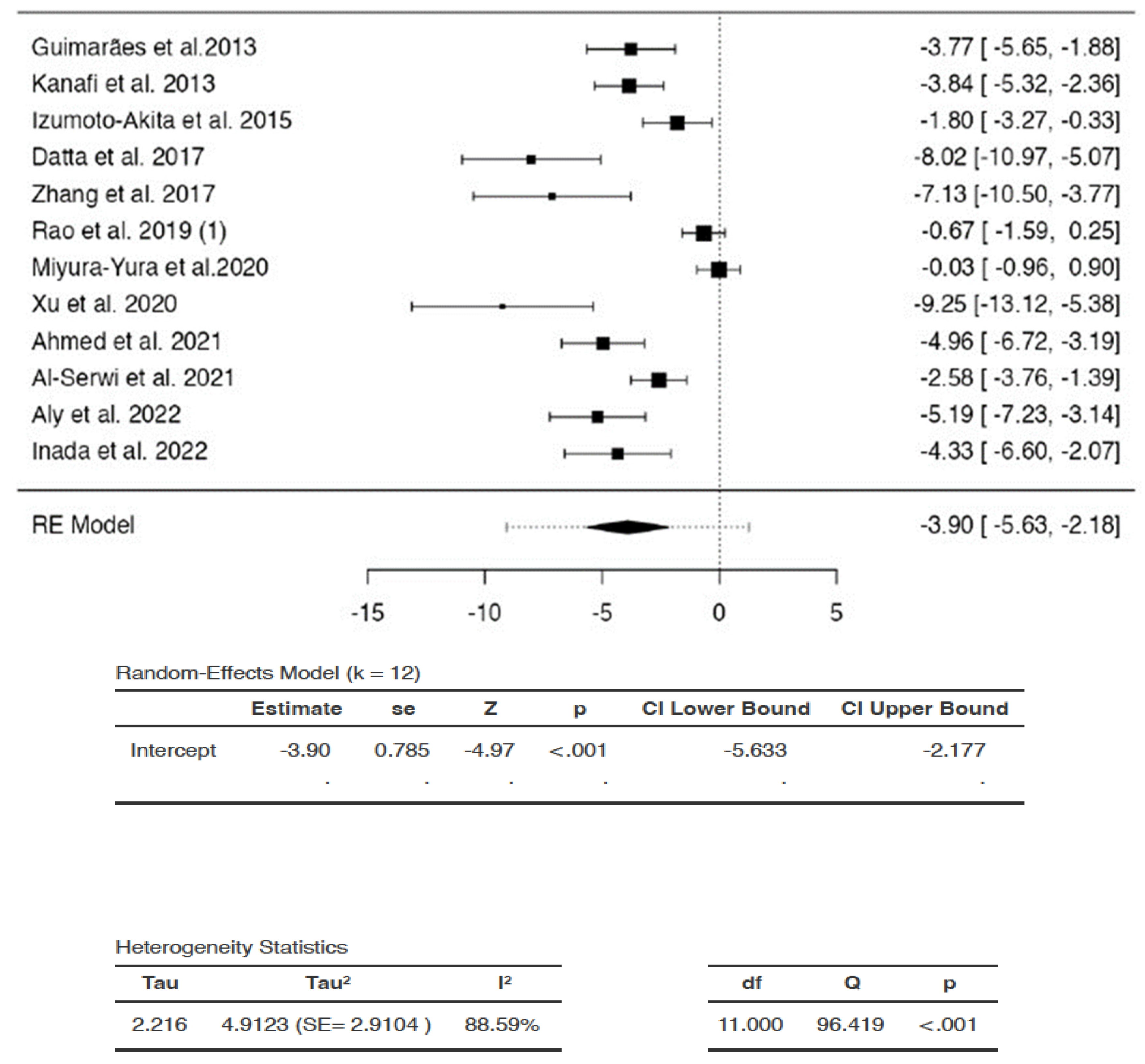
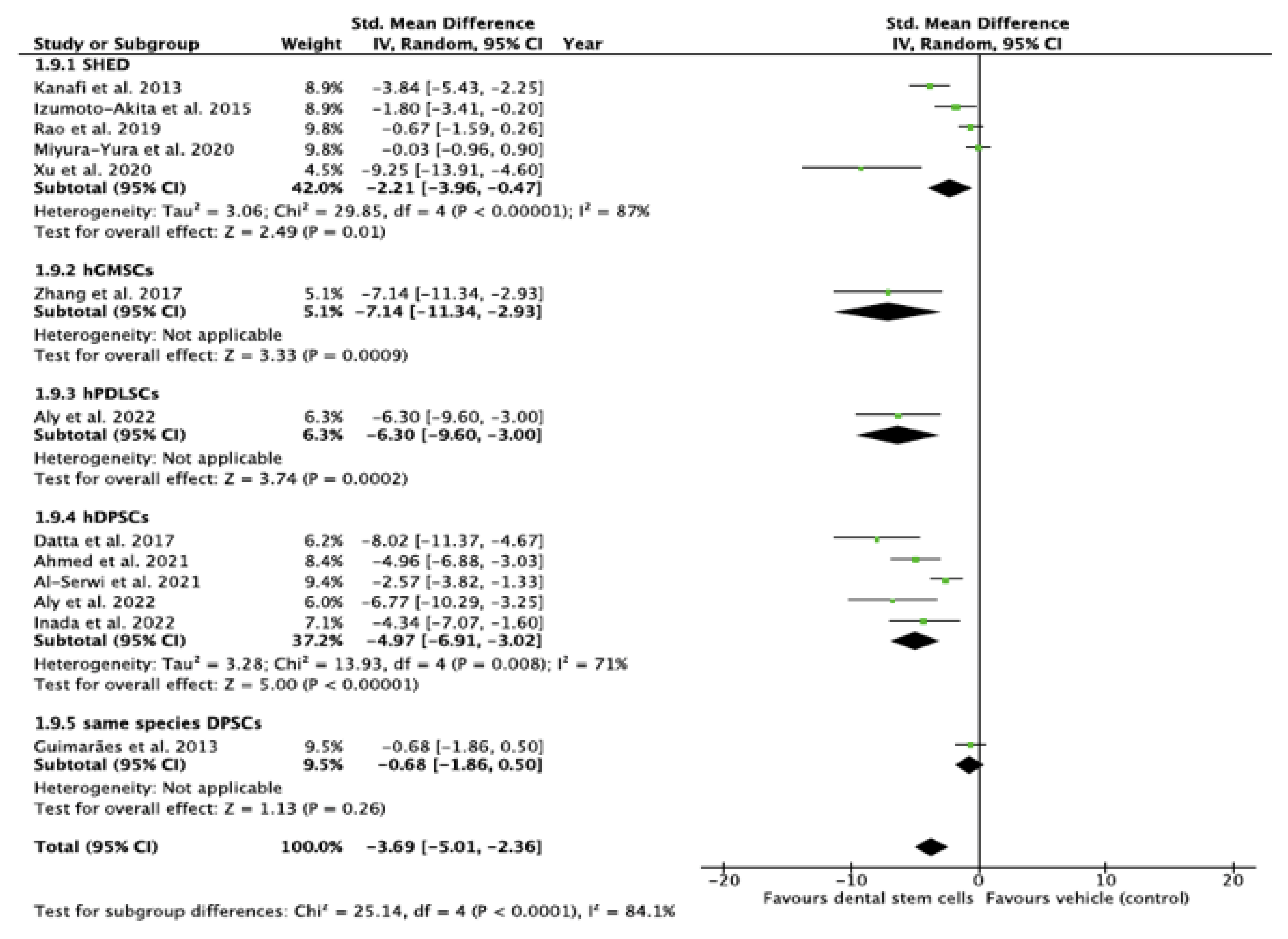
Effect of DSCs on Blood Glucose

The SMDs of individual studies were within the range of -9.2479 to −0.0270 (Figure 2). The average SMD of the analysis was observed to be −3.905 (95% CI: −5.6330 to −2.177). The average outcome differed significantly from zero (Z = −4.9737, p = 0.0004). Hence, the analysis indicated that there was a significant reduction in blood glucose in favor of DSC administration compared to saline/vehicle.
There was substantial heterogeneity between studies (Q = 96.4187, p < 0.0001, tau2 = 4.9123, I2 = 88.5914%). A 95% prediction interval for true effects was estimated to be −9.0802 to 1.2702. Hence, the 95% range of true effects contained values that were less than zero, as well as values that were more than or equal to zero. This means that even though the average outcome (Z = −4.9737) and summary point estimate (SMD = −3.905) were negative, indicating a reduction in blood glucose, DSCs may in fact, have no effect or may even increase blood glucose in similar settings, with the greatest increase in blood glucose represented as SMD of 1.2702 in the analysis.
The studentized residuals showed that none of the studies could be considered to be outliers in the context of the analysis. In addition, none of the studies were overly influential according to Cook’s distances (data for studentized residuals and Cook’s distance analyses are available as supplementary data).

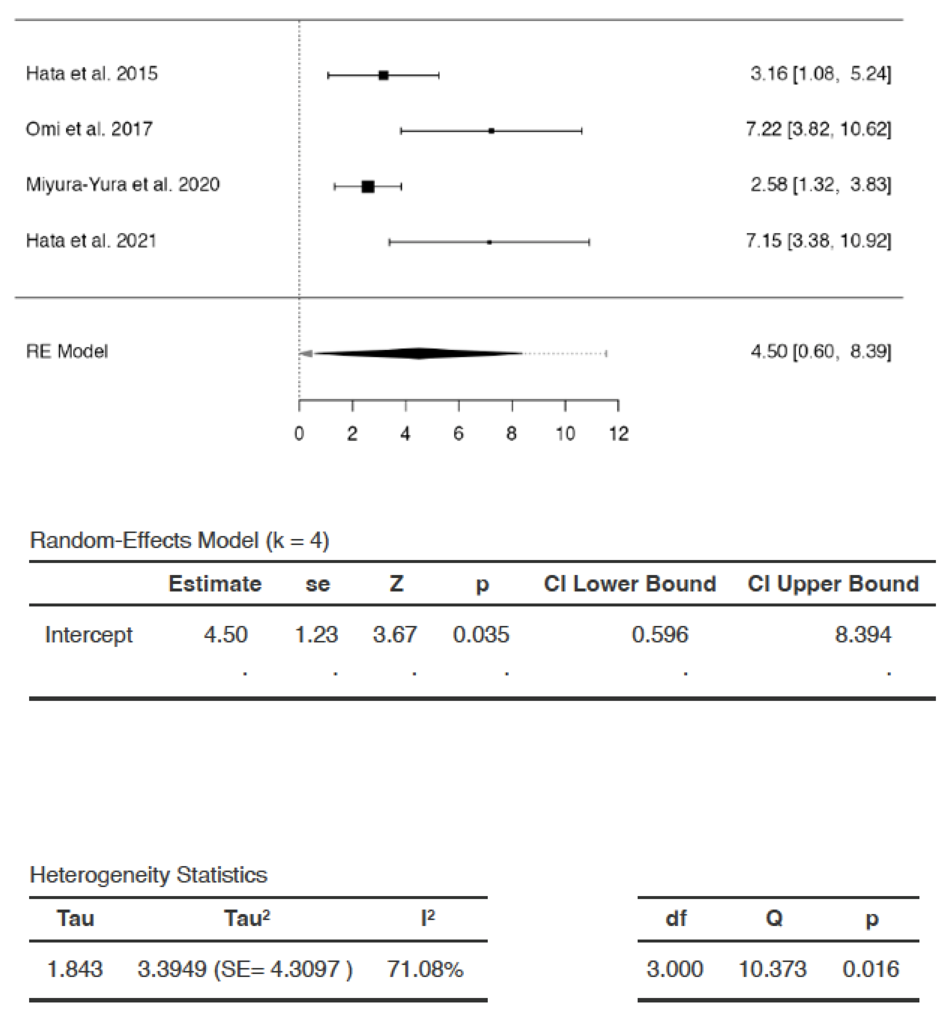
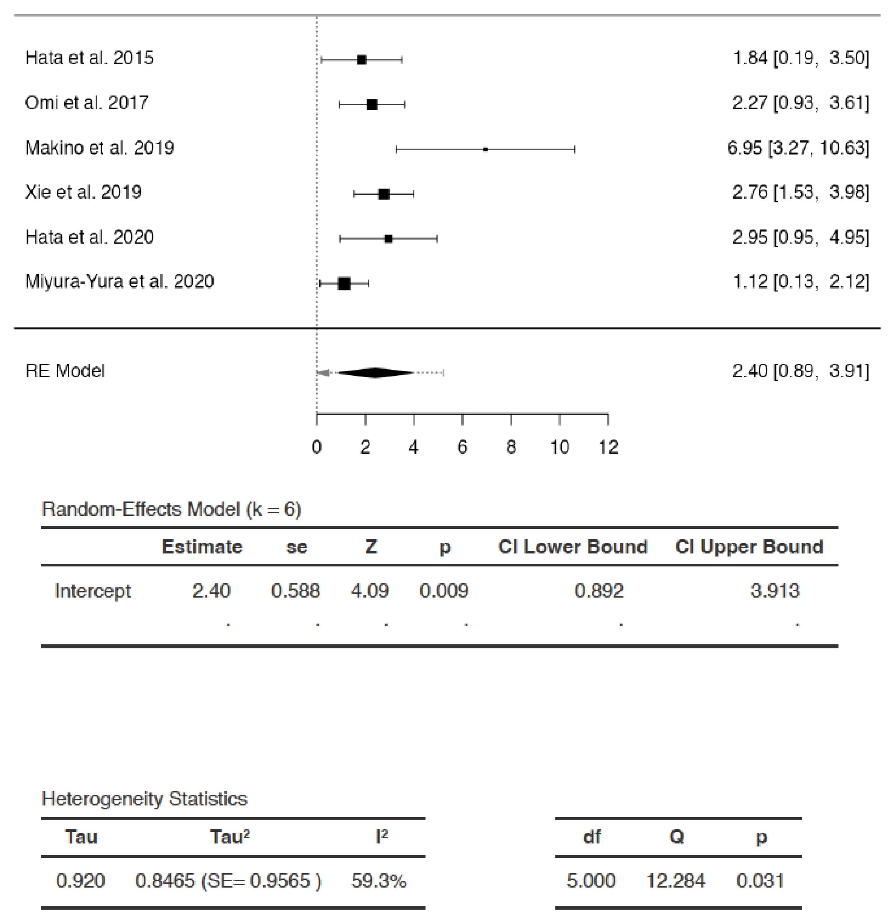
Effects of DSCs on SNCV

The SMDs ranged from 2.5757 to 7.2217 (Figure 3). The estimated average SMD was 4.4952 (95% CI: 0.5959 to 8.3945). The average outcome differed significantly from zero (Z = 3.6688, p = 0.0350). Hence, following DSC administration, there was a statistically significant increase in SNCV compared to saline/vehicle.
The analysis revealed significant heterogeneity (Q = 10.3734, p = 0.0156, tau2 = 3.3949, I2 = 71.0799%). A 95% prediction interval for true effects was estimated to be −2.5467 to 11.5371. Hence, although the average outcome (Z = 3.6688) and summary point estimate (SMD = 4.4942) were positive, indicating an increase in SNCV after DSC administration, the SNCV may remain unchanged or may even decrease following DSC administration in a future study in a similar setting, with the greatest reduction in SNCV represented as SMD of −2.5467. The studentized residuals revealed that there were no outliers in the context of this model. According to Cook’s distances, none of the studies could be considered to be overly influential.

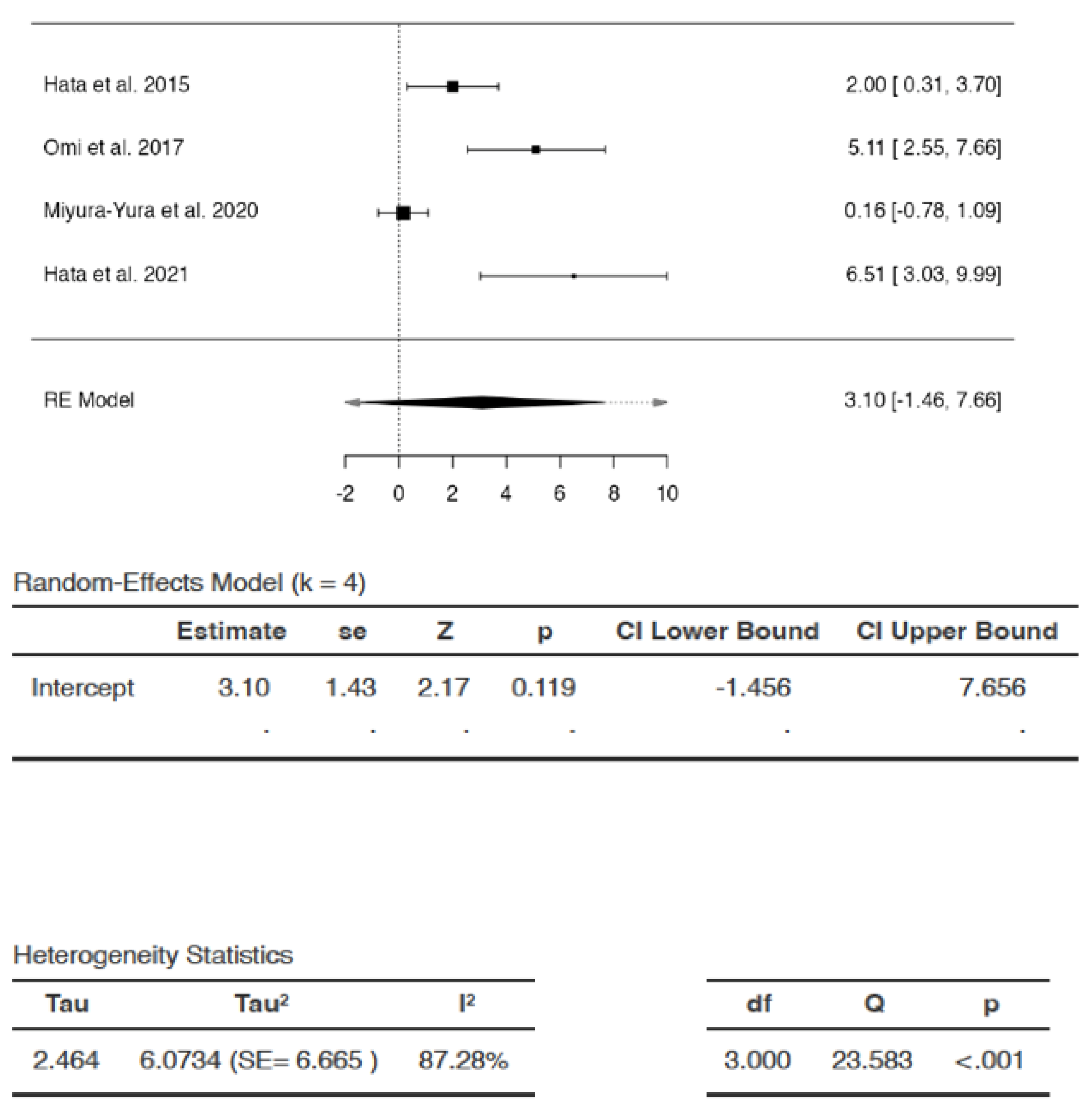
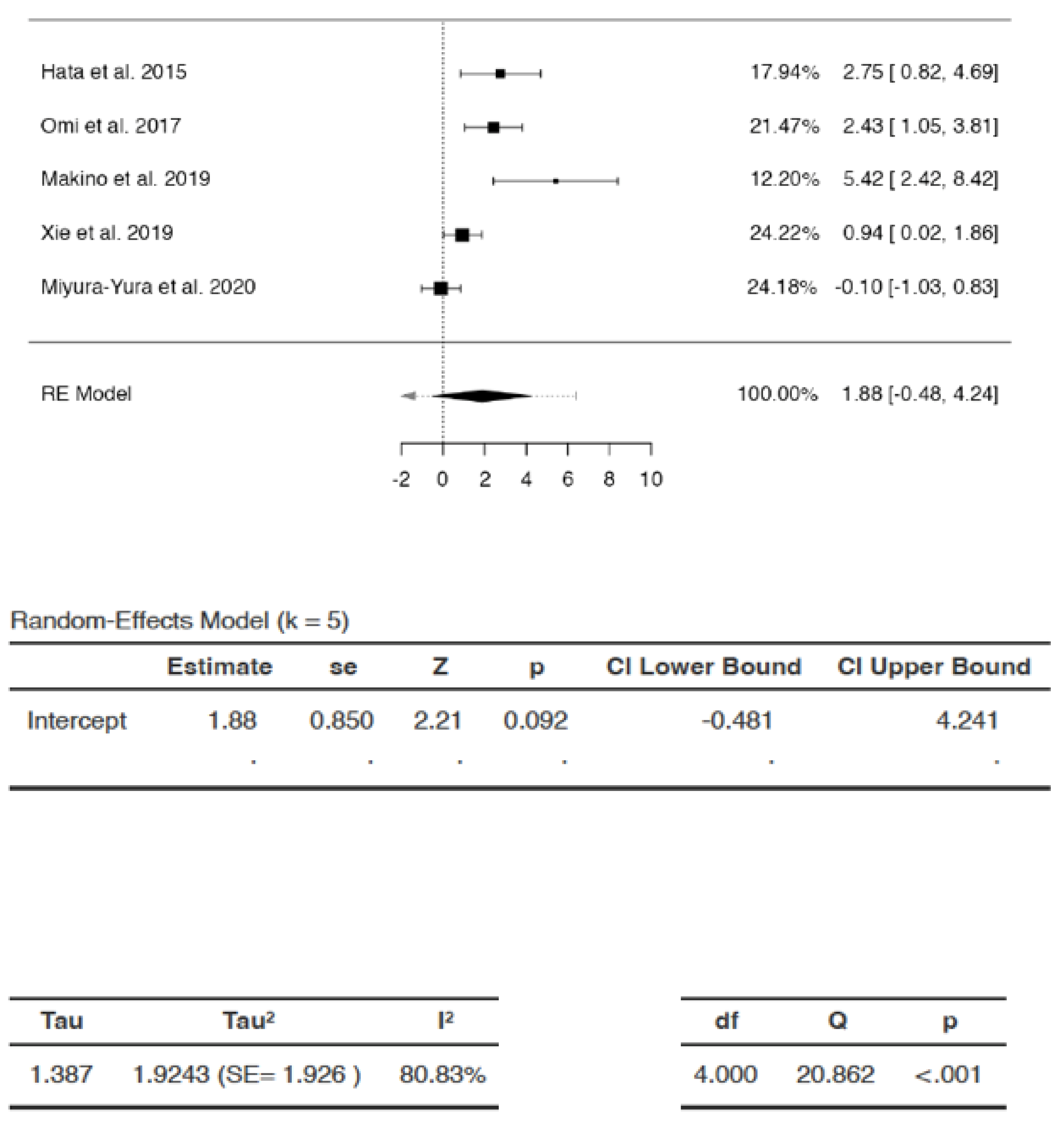
Effects of DSCs on MNCV

The SMDs of studies ranged from 0.1556 to 6.5147 (Figure 4). The average SMD was 3.1001 (95% CI: −1.4558 to 7.6559). The average outcome did not differ significantly from zero (Z = 2.1655, p = 0.119). Hence, the increase in MNCV after DSC treatment compared to that following saline/vehicle administration was not significant.
There appeared to be substantial heterogeneity (Q = 23.5832, p < 0.0001 tau2 = 6.0734, I2 = 87.2791%). A 95% prediction interval for true effects was estimated to be −5.9701 to 12.1702. Hence, although the average outcome (Z = 2.1655) and summary point estimate (SMD = 3.1001) were positive, indicating improvement in MNCV after DSC administration, the MNCV might remain unchanged or may even reduce following DSC administration in similar settings. In some cases, the result of DSC treatment may even be the exact opposite of the summary point estimate SMD, i.e., −3.1001 instead of 3.1001, with the greatest reduction in MNCV represented as SMD of −5.9701 in the analysis. The studentized residuals revealed that none of the studies could be considered as outliers in the analysis. According to Cook’s distances, none of the studies were overly influential.

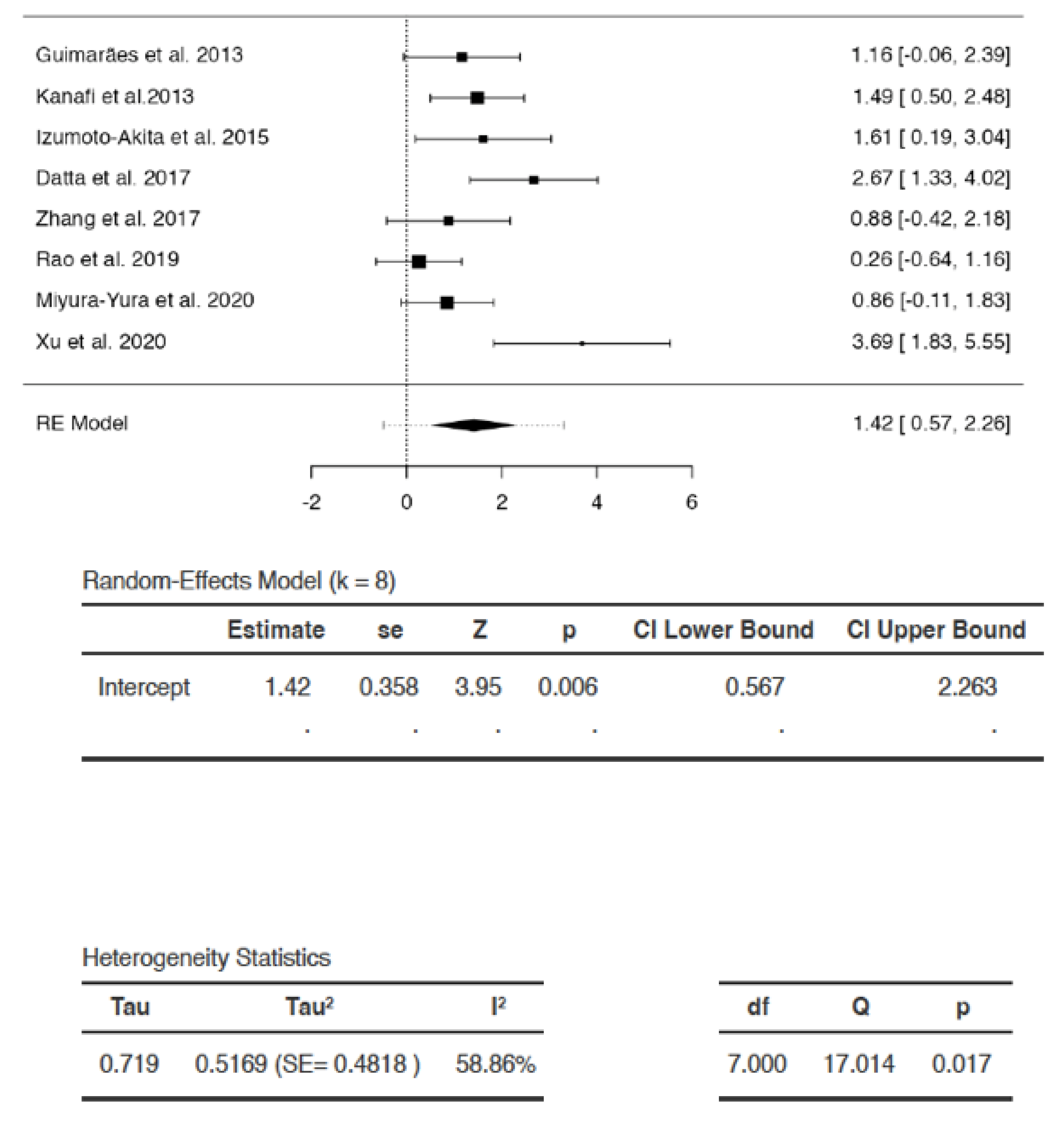
Effects of DSCs on Capillary–Muscle Ratio

The SMDs of the studies ranged from 1.1249 to 6.9490 (Figure 5). The estimated average SMD was 2.4027 (95% CI: 0.8923 to 3.9132). The average outcome was significantly different from zero (Z = 4.0891, p = 0.0095) (Figure 4). Hence, the analysis indicated that the capillary–muscle ratio increased significantly in favor of DSC treatment compared to saline/vehicle administration.
There appeared to be heterogeneity among studies (Q = 12.2844, p = 0.0311, tau2 = 0.8465, I2 = 59.2979%). A 95% prediction interval for true effects was −0.4035 to 5.209. Hence, although the average outcome (Z = 4.0891) and summary point estimate (SMD = 2.4027) were positive, indicating that capillary-muscle ratio improved with DSC administration, it may in fact remain unchanged or even reduce following DSC administration in similar settings, with the greatest reduction in capillary–muscle ratio represented as SMD of −0.4035.The studentized residuals revealed that none of the studies could be considered as outliers in the context of the analysis. According to Cook’s distances, none of the studies were overly influential.

Effects of DSCs on IENFD

The SMDs ranged from −0.0994 to 5.4206 (Figure 6). The average SMD was calculated to be 1.8802 (95% CI: −0.4809 to 4.2413). The average outcome did not differ significantly from zero (Z = 2.211, p = 0.0915). Hence, the increase in IENFD was not statistically significant following DSC administration compared to saline/vehicle.
There appeared to be substantial heterogeneity (Q = 20.8617, p = 0.0003, tau2 = 1.9243, I2 = 80.8261%). A 95% prediction interval for true effects was estimated to be −2.6374 to 6.3978. Hence although the average outcome (Z = 2.211) and summary point estimate (SMD = 1.8802) were positive, indicating an increase in IENFD after DSC administration, it may remain unchanged, or may even reduce following DSC administration in similar settings. In some cases, DSC treatment may even result in the exact opposite of the summary estimate, i.e., −1.8802 instead of 1.8802, with the greatest possible reduction in IENFD represented as −2.6374 in the analysis. The studentized residuals revealed that none of the studies could be considered outliers in the context of this model. According to Cook’s distances, none of the studies were overly influential in the analysis.

Effect of DSCs on Body Weight

The SMDs of the individual studies ranged from 0.2578 to 3.691 (Figure 7). The estimated average SMD was 1.415 (95% CI: 0.5674 to 2.2627). The average outcome differed significantly from zero (Z = 3.9475, p = 0.0056). Hence, the increase in body weight with DSC administration was statistically significant compared to saline/vehicle.
There appeared to be heterogeneity (Q = 17.0143, p = 0.0173, tau2 = 0.5169, I2 = 58.8583%). A 95% prediction interval for true effects was estimated to be −0.4846 to 3.3146. Hence, although the average outcome (Z = 3.9475) and summary point estimate (SMD = 1.415) were positive, indicating that the body weight of the diabetic animals increased with DSC administration, it may not be affected, or may even decrease with DSC administration in some studies in similar settings, with the greatest decrease in body weight observed to be SMD of −0.4846. None of the studies could be considered to be outliers or to be overly influential, according to studentized residuals and Cook’s distances.

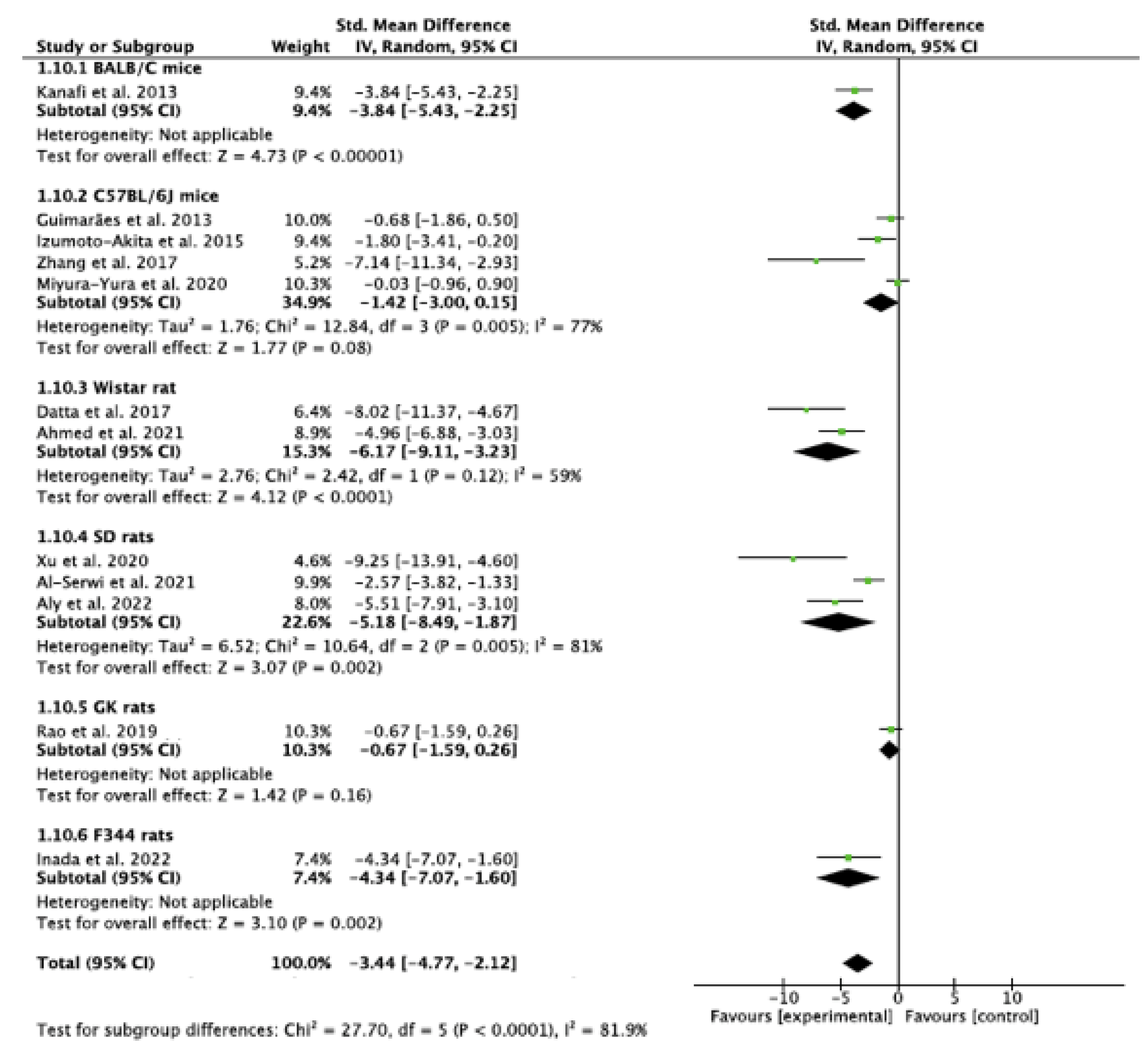
3.7.2. Subgroup Analysis

Subgroup Analysis of the Effect of DSC Type Used in the Study

The test indicated that there is a statistically significant subgroup effect (p < 0.0001), suggesting that DSC type may alter its effects on blood glucose (Figure 8). In all subgroups, DSCs were more beneficial in lowering blood glucose than saline/vehicle. Since the effect was seen more in some groups than in others, the subgroup effect was deemed quantitative. However, fewer studies were present in the hGMSCs, hPDLSC, and same species DPSC subgroups (Figure 8). In addition, there was substantial heterogeneity within the SHED (I2 = 87%) and hDPSC (I2 = 71%) subgroups. Due to the disparate number of studies and heterogeneity within the subgroups, the results of the analysis may not be reliable for interpretation.

Subgroup Analysis of the Effect of Species Variations

The analysis indicated that there is a statistically significant subgroup effect (p < 0.0001), suggesting that differences in species may influence the effect of DSCs on blood glucose (Figure 9). In all subgroups i.e., among all strains of rodents used, DSCs were more beneficial in lowering blood glucose than saline/vehicle. Since the effect was seen more in some groups than in others, the subgroup effect is quantitative. However, there was disparity in the number of studies in subgroups. In addition, there was substantial heterogeneity within the C57BL/6 (I2 = 77%), SD (I2 = 81%), and Wistar rat (I2 = 59%) subgroups. Hence, the findings may not be considered reliable.

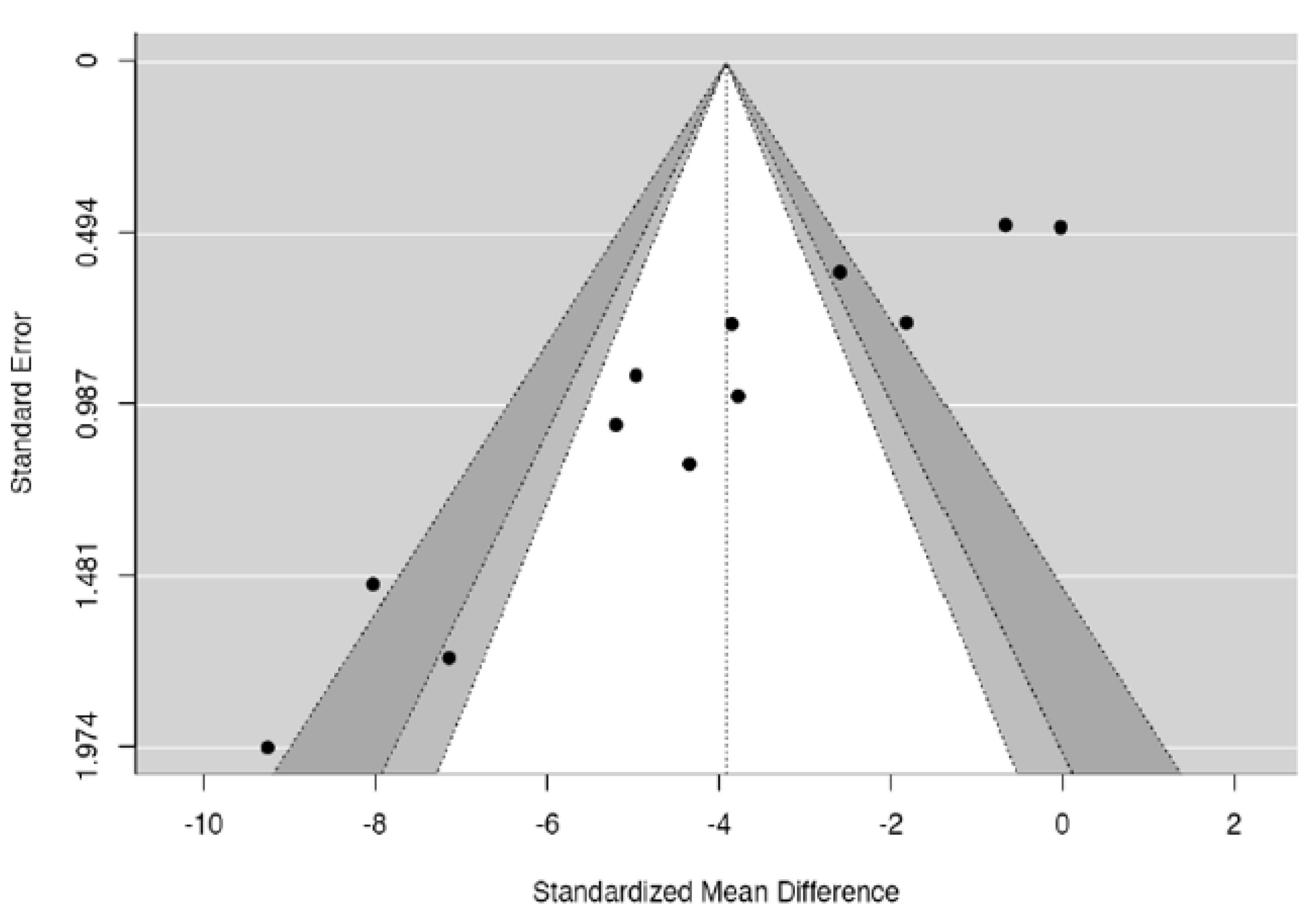
3.7.3. Funnel Plot Analysis and Publication Bias

Upon visual assessment, the funnel plot for DSC effects on blood glucose appeared asymmetrical around the vertical line representing the summary effect (Figure 10). Both regression and rank correlation tests indicated the presence of funnel plot asymmetry (p < 0.0001 and p = 0.0018, respectively) (Table 3). The asymmetry could be present due to publication bias, heterogeneity among the studies, methodological design of the studies, or chance [28,40].

4. Discussion

To our knowledge, this is the first systematic review and meta analysis that explicates the scope of various dental stem cells in the management of hyperglycemia and DPN. Quantitative analysis in the present review indicated that there was a significant reduction in blood glucose (p = 0.0004) in diabetes-induced animals following DSC administration.
The underlying mechanisms of DSCs have not been fully established. Following administration, they induce differentiation of pancreatic progenitor cells [59], promote transdifferentiation of α into β cells [57], and modulate β cell function [55,57]. Emerging evidence indicates that extracellular apoptotic vesicles (apoVs) containing proteomes are pivotal in mediating MSC functions [70]. DSCs act by paracrine signaling, secreting VEGF and bFGF, which are important for tissue growth and regeneration [13,46,53]. They also release neurotrophin-3 (NT-3) and NGF, which are essential in neuronal development and repair [13,49,50,53].
The pooled effects showed that SNCV (p = 0.035) and capillary–muscle ratio (p = 0.0095) improved significantly following DSC administration, but IENFD (p = 0.0915) and MNCV (p = 0.119) did not. IEFND is used to measure neuropathy of small fibers, such as unmyelinated C and Aδ fibers. The findings are consistent with a study in which, 16 weeks after T1DM was induced and 4 weeks after SHED-CM was administered, there was neither any improvement in IEFND, nor in thermal and tactile sensitivity tests, indicating that DSCs were unable to reverse advanced stages of hypoalgesia in DM [50]. In addition, due to the larger diameter of motor nerves, changes in MNCV occur later than those in SNCV [50]. In the same study, 4 weeks after SHED-CM was administered, MNCV did not improve, suggesting that the duration of the experiment may have been insufficient to detect changes in MNCV [50]. Hence, the review reflects that studies with longer follow-up periods, using long-term diabetes-induced animals, may be most appropriate to demonstrate the effects of DSCs on IENFD and MNCV in advanced stages of DPN.
Human derived DPSCs were able to transdifferentiate into insulin-producing β cells in the murine pancreas following IV and intrapancreatic administration [59]. Interestingly, the new cells were morphologically identical to those of the recipient animals, and the levels of human, as well as murine insulin increased in these animals [59].
A reduction in TNF-α and an increase in IL10 [47] was observed in the DSC-administered muscle on one side, compared to the contralateral side of the same animal. This is connotative of the local effects of DSCs. On the other hand, IV administration also resulted in significant improvement in hyperalgesia and late onset hypoalgesia, indicating a systemic effect by reducing blood glucose [63]. DSC engraftment in injured organs and the subsequent amelioration of DPN [63], liver disease [56], and impaired salivary flow [58] following IV administration may be suggestive of both local and systemic effects.
The IV route of DSC administration is congruent with MSC-based therapy for controlling hyperglycemia and systemic inflammation [14]. On the other hand, a single IM dose of hDPSCs resulted in improvement in DPN parameters which lasted for 16 weeks in T1DM induced rats [48]. Irrespective of the route, repeat doses may be more effective in long-term diabetes due to sustained effects on cytokines [17,55,63]. Li et al. (2021) reported that the effective rate of three IV doses of SHED, at the end of a 12 month follow up for diabetic patients was 68.18%, suggesting that therapy may be effective for at least one year [6].
The viability of SCs is affected by aging and disease; hence, it is important to isolate them from healthy teeth at a young age [13,46,48]. Cryopreservation may hold the key to effective autologous DSC therapy, as they can be isolated from healthy teeth which have been extracted at a young age, thawed, and expanded in culture when needed [13,46,48]. Cryopreserved rDPSCs demonstrated proliferative capacities that were comparable to freshly isolated rDPSCs in T1DM rats, and were able to maintain their viability for at least 6 months before administration [46]. However, cryopreservation should be studied further before clinical application with DSCs due to the risk of solution effect injury, toxicity, arrhythmia, and hypotension [71].

4.1. Scope of Translation of Dental Stem Cell-Based Therapy in Diabetes Mellitus

For research to be effectively translated, studies should accurately predict the clinical course in humans. In the presence of substantial heterogeneity among studies, the prediction interval in each analysis demonstrates how different the true effect in a new study might be from those observed in previous similar studies. It thus, also provides insight into the uncertainty in predicting the effect in settings that may be different from those included in the analysis, thereby pointing to the challenges in translation to clinical settings.
It was unclear whether adequate randomization, allocation, concealment, and blinding were followed in the animal studies included in the review (Table 2). Failure to adhere to these principles may have led to overestimating the efficacy of DSCs.
In order to optimize their contribution to clinical practice, studies should reflect the disease as well as the population for which the intervention is intended. The homogeneity in animal samples may render them unrepresentative of heterogeneous human populations [65,66]. Further, the selection of healthy animals with no prior disease, as well as shorter duration of most of the studies, do not reflect the complexity of diabetes, nor recapitulate its slow and progressive nature [65,66]. The timing of the intervention should also model the delay between the onset of symptoms and treatment that often occurs in humans. Another factor is that many organ systems are still developing in the age range of the animals used in the studies, and the changes that occur in this phase could have affected the variables that were being measured [67,68]. Moreover, diabetes, particularly T2DM, typically manifests in older individuals. In addition, although sex effects are present in diabetes, it is widely prevalent in both males and in females [72], whereas only one animal study demonstrated the effects of DSCs in female mice [63]. These factors may impede clinical translation.
The most intractable aspect of animal studies undermining their external validity is the inherent interspecies differences [65,66]. Rodents are used in biomedical research due to their genetic and physiological similarities to humans [12]. Their life stages mimic those in humans [12]. However, modern rodents have adapted to their own unique environment and have evolved to respond to diseases and interventions differently than humans [65]. Moreover, genetic variations exist, even within individual strains of the same species, such as C57BL/6 mice and GK rats, which must be considered while designing a study and interpreting its results [12].
Similar to the animal studies, the follow-up phase in the clinical study was adequate to monitor immediate and early adverse effects, but may not have been sufficient to determine long-term complications. Sometimes, certain properties of a drug may go unnoticed, even after it has passed safety checks in preclinical and short-term clinical studies. An example is the antiviral drug fialuridine, which had potential use in the treatment of hepatitis B [73]. It had passed preclinical investigations, and pilot studies with 2 and 4 week courses of the drug. However, during the 13th week of a phase 2 clinical trial, following administration of doses which were several hundred times smaller than the dose deemed safe in laboratory animals, patients suffered severe hepatotoxicity, resulting in the deaths of five patients [73]. The lesson to be learned from this tragedy is that not only are species variations underestimated, but that the duration of studies is also often insufficient to assess the risk of chronic toxicity. Even if a drug is deemed safe in animals at much higher doses, it may exhibit vastly different pharmacological properties in humans.

4.2. Publication Bias

Both Egger’s regression (p < 0.0001) and Begg and Majumdar rank correlation (p = 0.0018) tests indicated funnel plot asymmetry. One of the reasons for this asymmetry may be publication bias. This means that studies which reported amelioration of hyperglycemia were more likely to be published than studies that reported no change, or reported an increase in blood glucose. Such selective publication can hinder effective translation because the interpretation and implementation of data will be based only on partial evidence [66]. It results in the wastage of animals and resources used in unpublished studies.

4.3. Graft Rejection, Tumorigenesis, and Other Adverse Reactions

MSCs are not completely immunoprivileged and sometimes undergo graft rejection [5]. Although all the studies including the clinical study [6], used allogeneic or xenogeneic sources for DSCs, only one animal study [54] described the use of tacrolimus. Most MSCs become trapped in the lungs at some point [5,6,57] or undergo apoptosis after administration [6]. However, their fate is still obscure. Engraftment occurred in organs such as the pancreas, liver, kidneys, and muscles following various routes of administration [9,14,46,48,55,58,63]; however, in one study, it was reported that after intraperitoneal administration, very few DSCs engrafted in the lungs and brain [9]. Hypoxia, hyperglycemia, and inflammation may contribute to the homing tendencies of MSCs [55]. In diabetes, there may be multiple organs with varying degrees of inflammation [74]. Hence, potential interactions in the local environment and the risk for tumorgenicity in every organ and tissue should be studied. Another factor to consider is the risk of teratoma, a phenomenon observed when embryonic stem cells are injected in mice, and a hallmark of pluripotent stem cells [75]. Although isolated DSCs are specifically induced in the laboratory to become committed to the desired cell phenotype in vivo, their broad spectrum differentiation potential must not be ignored. Common side effects of MSC therapy in DM, such as hypoglycemia, headache, fever, and rash, should also be considered [6].
Animal studies provide the foundation for testing new therapies and help to increase our understanding of an intervention. The present systematic review aimed to provide insight into the applicability of teeth and supporting tissues as potential sources of stem cells in the treatment of diabetes mellitus. The review also underscores the importance of methodological quality of animal studies to reliably inform clinical translation. In addition, the reviewers hope to contribute to implementing the 3 Rs, i.e., replacement, refinement, and reduction, by encouraging transparent reporting, responsible use of animals, and preventing replication of flawed study designs. The review has some limitations. Substantial heterogeneity was observed, which could be attributed to factors such as differences in species, the type and preparation of DSCs, the duration of the experiments, study design, and statistical differences in results between studies [28,40]. Meta-analysis was nevertheless performed to provide quantitative assessment of the effects of DSCs and to demonstrate the differences in effects among studies. Furthermore, the disparate number of studies in the subgroups reduces the reliability of the subgroup analyses. Nonetheless, the analyses were included in the review due to the putative effects of SC type and species differences on experiment outcomes [5,8,12]. Lastly, the most crucial caveat for translation to humans is that all preclinical data should be interpreted with caution due to the irrevocable issue of interspecies differences.

5. Conclusions

The prospect of rapid and enduring glycemic control is exciting, as conventional drugs do not have lasting effects [63]. Within the limitations of this review, DSCs appear to be beneficial for glycemic control; however, the potential risk of graft rejection and tumorigenesis must not be ignored. It behooves researchers performing animal studies to adopt standards similar to those used in clinical trials, while considering methodological design and reporting. Studies with longer follow-up periods and greater sample power should be undertaken to determine long-term effects and track the fate of DSCs.

Supplementary Materials

The following supporting information can be downloaded at: https://www.mdpi.com/article/10.3390/ijtm4010005/s1, Table S1: PRISMA 2020 Abstract checklist; Table S2; PRISMA 2020 checklist; Table S3: Full version of search filters; Table S4: Comparative analysis of included studies to determine external validity; Figure S1: Outliers and residuals for the analyses.

Author Contributions

Conceptualization, P.T.; methodology, P.T.; software, P.T. and S.J.; literature search, P.T., V.T., S.J. and G.Y.; validation, P.T., V.T. and G.Y.; data analysis, P.T., V.T., S.J. and G.Y.; investigation, P.T., V.T., S.J. and G.Y.; data curation, P.T., V.T., S.J. and G.Y.; meta-analysis: P.T. and S.J.; writing—original draft preparation, P.T.; writing—review and editing, P.T., V.T., S.J. and G.Y.; visualization, P.T., V.T., S.J. and G.Y. All authors have read and agreed to the published version of the manuscript.

Funding

This research received no external funding.

Data Availability Statement

The data presented in this article is available upon reasonable request from the corresponding author.

Acknowledgments

The authors would like to thank Smt. Leela Idgunji, without whose unwavering support, the study would not have come to fruition.

Conflicts of Interest

The authors declare no conflict of interest.

References

  1. Thomas, E.D.; Lochte, H.L., Jr.; Lu, W.C.; Ferrebee, J.W. Intravenous infusion of bone marrow in patients receiving radiation and chemotherapy. N. Engl. J. Med. 1957, 257, 491–496. [Google Scholar] [CrossRef]
  2. Henig, I.; Zuckerman, T. Hematopoietic stem cell transplantation—50 years of evolution and future perspectives. Rambam Maimonides Med. J. 2014, 5, e0028. [Google Scholar] [CrossRef]
  3. Friedenstein, A.J.; Petrakova, K.V.; Kurolesova, A.I.; Frolova, G.P. Heterotopic of bone marrow. Analysis of precursor cells for osteogenic and hematopoietic tissues. Transplantation 1968, 6, 230–247. [Google Scholar] [CrossRef]
  4. Caplan, A.I. Mesenchymal Stem Cells. J. Orthop. Res. 1991, 9, 641–650. [Google Scholar] [CrossRef]
  5. Battiwalla, M.; Hematti, P. Mesenchymal stem cells in hematopoietic stem cell transplantation. Cytotherapy 2009, 11, 503–515. [Google Scholar] [CrossRef] [PubMed]
  6. Li, W.; Jiao, X.; Song, J.; Sui, B.; Guo, Z.; Zhao, Y.; Li, J.; Shi, S.; Huang, Q. Therapeutic potential of stem cells from human exfoliated deciduous teeth infusion into patients with type 2 diabetes depends on basal lipid levels and Islet function. Stem Cells Transl. Med. 2021, 10, 956–967. [Google Scholar] [CrossRef] [PubMed]
  7. Kot, M.; Baj-Krzyworzeka, M.; Szatanek, R.; Musiał-Wysocka, A.; Suda-Szczurek, M.; Majka, M. The Importance of HLA Assessment in “Off-the-Shelf” Allogeneic Mesenchymal Stem Cells Based-Therapies. Int. J. Mol. Sci. 2019, 20, 5680. [Google Scholar] [CrossRef] [PubMed]
  8. Huang, G.T.-J.; Gronthos, S.; Shi, S. Mesenchymal stem cells derived from dental tissues vs. those from other sources: Their biology and role in regenerative medicine. J. Dent. Res. 2009, 88, 792–806. [Google Scholar] [CrossRef] [PubMed]
  9. Zhang, W.; Zhou, L.; Dang, J.; Zhang, X.; Wang, J.; Chen, Y.; Liang, J.; Li, D.; Ma, J.; Yuan, J.; et al. Human gingiva-derived mesenchymal stem cells ameliorate streptozoticin-induced T1DM in mice via suppression of T effector cells and up-regulating Treg subsets. Sci. Rep. 2017, 7, 15249. [Google Scholar] [CrossRef] [PubMed]
  10. Crotty, S. A brief history of T cell help to B cells. Nat. Rev. Immunol. 2015, 15, 185–189. [Google Scholar] [CrossRef]
  11. International Diabetes Federation. Diabetes Factors and Figures. 2021. Available online: https://www.idf.org/aboutdiabetes/what-is-diabetes/facts-figures.html (accessed on 9 July 2023).
  12. King, A.J. The use of animal models in diabetes research. Br. J. Pharmacol. 2012, 166, 877–894. [Google Scholar] [CrossRef] [PubMed]
  13. Omi, M.; Hata, M.; Nakamura, N.; Miyabe, M.; Ozawa, S.; Nukada, H.; Tsukamoto, M.; Sango, K.; Himeno, T.; Kamiya, H.; et al. Transplantation of dental pulp stem cells improves long-term diabetic polyneuropathy together with improvement of nerve morphometrical evaluation. Stem Cell Res. Ther. 2017, 8, 279. [Google Scholar] [CrossRef] [PubMed]
  14. Xie, J.; Rao, N.; Zhai, Y.; Li, J.; Zhao, Y.; Ge, L.; Wang, Y. Therapeutic effects of stem cells from human exfoliated deciduous teeth on diabetic peripheral neuropathy. Diabetol. Metab. Syndr. 2019, 11, 38. [Google Scholar] [CrossRef] [PubMed]
  15. Sugimoto, K.; Yasujima, M.; Yagihashi, S. Role of advanced glycation end products in diabetic neuropathy. Curr. Pharm. Des. 2008, 14, 953–961. [Google Scholar] [CrossRef]
  16. Hicks, C.W.; Selvin, E. Epidemiology of Peripheral Neuropathy and Lower Extremity Disease in Diabetes. Curr. Diabetes Rep. 2019, 19, 86. [Google Scholar] [CrossRef] [PubMed]
  17. Datta, I.; Bhadri, N.; Shahani, P.; Majumdar, D.; Sowmithra, S.; Razdan, R.; Bhonde, R. Functional recovery upon human dental pulp stem cell transplantation in a diabetic neuropathy rat model. Cytotherapy 2017, 19, 1208–1224. [Google Scholar] [CrossRef]
  18. Honda, M.J.; Imaizumi, M.; Tsuchiya, S.; Morsceck, C. Dental follicle stem cells and tissue enjineering. J. Oral Sci. 2010, 52, 541–552. [Google Scholar] [CrossRef]
  19. Govindasamy, V.; Ronald, V.; Abdullah, A.; Nathan, K.G.; Aziz, Z.A.; Abdullah, M.; Musa, S.; Abu Kasim, N.; Bhonde, R. Differentiation of dental pulp stem cells into islet-like aggregates. J. Dent. Res. 2011, 90, 646–652. [Google Scholar] [CrossRef]
  20. Wu, W.; Zhou, J.; Xu, C.-T.; Zhang, J.; Jin, Y.-J.; Sun, G.-L. Derivation and growth characteristics of dental pulp stem cells from patients of different ages. Mol. Med. Rep. 2015, 12, 5127–5134. [Google Scholar] [CrossRef]
  21. Shi, X.; Mao, J.; Liu, Y. Pulp stem cells derived from human permanent and deciduous teeth: Biological characteristics and therapeutic applications. Stem Cells Transl. Med. 2020, 9, 445–464. [Google Scholar] [CrossRef]
  22. Page, M.J.; McKenzie, J.E.; Bossuyt, P.M.; Boutron, I.; Hoffmann, T.C.; Mulrow, C.D.; Shamseer, L.; Tetzlaff, J.M.; Akl, E.A.; Brennan, S.E.; et al. The PRISMA 2020 Statement: An Updated Guideline for Reporting Systematic Reviews. BMJ 2021, 372, n71. Available online: http://www.prisma-statement.org/ (accessed on 7 June 2023). [CrossRef] [PubMed]
  23. Hooijmans, C.R.; Tillema, A.; Leenaars, M.; Ritskes-Hoitinga, M. Enhancing search efficiency by means of a search filter for finding all studies on animal experimentation in PubMed. Lab. Anim. 2010, 44, 170–175. [Google Scholar] [CrossRef]
  24. Hooijmans, C.R.; Rovers, M.M.; de Vries, R.B.M.; Leenaars, M.; Ritskes-Hoitinga, M.; Langendam, M.W. SYRCLE’s risk of bias tool for animal studies. BMC Med. Res. Methodol. 2014, 14, 43. [Google Scholar] [CrossRef] [PubMed]
  25. Higgins, J.P.; Altman, D.G.; Gotzsche, P.C.; Juni, P.; Moher, D.; Oxman, A.D.; Savovic, J.; Schulz, K.F.; Weeks, L.; Sterne, J.A.; et al. The Cochrane Collaboration’s tool for assessing risk of bias in randomised trials. BMJ 2011, 343, d5928. [Google Scholar] [CrossRef] [PubMed]
  26. Review Manager (RevMan), Version 5.4; The Cochrane Collaboration: London, UK, 2020.
  27. The Jamovi Project. Jamovi [Computer Program]. Version 2.3. 2021. Available online: http://www.jamovi.org(accessed on 1 January 2020).
  28. Deeks, J.; Higgins, J.P.T.; Altman, D.G.; on be half of the Cochrane Statistical Methods Group. Chapter 10: Analysing data and undertaking meta-analyses. In Cochrane Handbook for SystematicReviews for Interventions Version 6.4; Cochrane: London, UK, 2023. [Google Scholar]
  29. Hedges, L.V.; Olkin, I. Statistical Methods for Meta-Analysis; Hedges, L.V., Ed.; Academic Press: Cambridge, MA, USA, 1985. [Google Scholar]
  30. Hedges, L.V. Distrubution theory for Glass’ Estimator of effect sizes and related estimators. J. Educ. Stat. 1981, 6, 107–128. [Google Scholar] [CrossRef]
  31. Neyman, J. On the problem of confidence intervals. Ann. Math. Stat. 1935, 6, 111–116. [Google Scholar] [CrossRef]
  32. DerSimonian, R.; Laird, N. Meta-analysis in clinical trials. Control. Clin. Trials 1986, 7, 177–188. [Google Scholar] [CrossRef]
  33. Knapp, G.; Hartung, J. Improved tests for a random effects meta-regression with a single covariate. Stat. Med. 2003, 22, 2693–2710. [Google Scholar] [CrossRef]
  34. Cochran, W.G. The combination of estimates from different experiments. Biometrics 1954, 10, 101–129. [Google Scholar] [CrossRef]
  35. Skipa, G. The inclusion of the estimated inter-study variation into forest plots for random effects meta analysis a suggestion for a graphical representation (abstract). In Proceedings of the XIV Cochrane Colloquium, Program and Abstract Book, Dublin, Ireland, 23–26 October 2006; p. 134. [Google Scholar]
  36. IntHout, J.; Ioannidis, J.P.A.; Rovers, M.M.; Goeman, J.J. Plea for routinely presenting prediction intervals in meta-analysis. BMJ Open 2016, 6, e010247. [Google Scholar] [CrossRef]
  37. Pope, A.J. The Statistics of Residuals and the Detection of Outliers; U.S. Dept. of Commerce, National Oceanic and Atmospheric Administration, National Ocean Survey, Geodetic Research and Development Laboratory: Miami, FL, USA, 1976; 136p. [Google Scholar]
  38. Dunn, O.J. Multiple comparisons among means. J. Am. Stat. Assoc. 1961, 56, 52–64. [Google Scholar] [CrossRef]
  39. Cook, R.D. Detection of Influential Observation in Linear Regression. Technometrics 1977, 19, 15. [Google Scholar] [CrossRef]
  40. Lefebvre, C.; Glanville, J.; Briscoe, S.; Featherstone, R.; Littlewood, A.; Metzendorf, M.I.; Noel-Storr, A.; Paynter, R.; Rader, T.; Thomas, J.; et al. Chapter 4. Searching for and selecting studies. In Cochrane Handbook for Systematic Reviews of Interventions Version 6.4; Cochrane: London, UK, 2023; Available online: https://www.training.cochrane.org/handbook (accessed on 1 August 2023).
  41. Higgins, J.P.T.; Eldridge, S.; Li, T. Chapter 23. Including variants on randomized trials. In Cochrane Handbook for Systematic Reviews of Interventions Version 6.4; (updated August 2023); Cochrane: London, UK, 2023; Available online: https://www.training.cochrane.org/handbook (accessed on 1 August 2023).
  42. Richardson, M.; Garner, P.; Donegan, S. Interpretation of subgroup analyses in systematic reviews: A tutorial. Clin. Epidemiol. Glob. Health 2019, 7, 192–198. [Google Scholar] [CrossRef]
  43. Borenstein, M.; Hedges, L.V.; Higgins, J.P.; Rothstein, H.R. Introduction to Meta-Analysis, 2nd ed.; John Wiley & Sons: Chichester, UK, 2009. [Google Scholar] [CrossRef]
  44. Egger, M.; Smith, G.D.; Schneider, M.; Minder, C. Bias in meta-analysis detected by a simple, graphical test. BMJ 1997, 315, 629–634. [Google Scholar] [CrossRef] [PubMed]
  45. Begg, C.B.; Mazumdar, M. Operating Characteristics of a Rank Correlation Test for Publication Bias. Biometrics 1994, 50, 1088–1101. [Google Scholar] [CrossRef] [PubMed]
  46. Hata, M.; Omi, M.; Kobayashi, Y.; Nakamura, N.; Tosaki, T.; Miyabe, M.; Kojima, N.; Kubo, K.; Ozawa, S.; Maeda, H.; et al. Transplantation of cultured dental pulp stem cells into the skeletal muscles ameliorated diabetic polyneuropathy: Therapeutic plausibility of freshly isolated and cryopreserved dental pulp stem cells. Stem Cell Res. Ther. 2015, 6, 162. [Google Scholar] [CrossRef]
  47. Omi, M.; Hata, M.; Nakamura, N.; Miyabe, M.; Kobayashi, Y.; Kamiya, H.; Nakamura, J.; Ozawa, S.; Tanaka, Y.; Takebe, J.; et al. Transplantation of dental pulp stem cells suppressed inflammation in sciatic nerves by promoting macrophage polarization towards anti-inflammation phenotypes and ameliorated diabetic polyneuropathy. J. Diabetes Investig. 2016, 7, 485–496. [Google Scholar] [CrossRef]
  48. Hata, M.; Omi, M.; Kobayashi, Y.; Nakamura, N.; Miyabe, M.; Ito, M.; Ohno, T.; Imanishi, Y.; Himeno, T.; Kamiya, H.; et al. Sustainable Effects of Human Dental Pulp Stem Cell Transplantation on Diabetic Polyneuropathy in Streptozotocine-Induced Type 1 Diabetes Model Mice. Cells 2021, 10, 2473. [Google Scholar] [CrossRef] [PubMed]
  49. Hata, M.; Omi, M.; Kobayashi, Y.; Nakamura, N.; Miyabe, M.; Ito, M.; Makino, E.; Kanada, S.; Saiki, T.; Ohno, T.; et al. Transplantation of human dental pulp stem cells ameliorates diabetic polyneuropathy in streptozotocin-induced diabetic nude mice: The role of angiogenic and neurotrophic factors. Stem Cell Res. Ther. 2020, 11, 236. [Google Scholar] [CrossRef] [PubMed]
  50. Miura-Yura, E.; Tsunekawa, S.; Naruse, K.; Nakamura, N.; Motegi, M.; Nakai-Shimoda, H.; Asano, S.; Kato, M.; Yamada, Y.; Izumoto-Akita, T.; et al. Secreted factors from cultured dental pulp stem cells promoted neurite outgrowth of dorsal root ganglion neurons and ameliorated neural functions in streptozotocin-induced diabetic mice. J. Diabetes Investig. 2020, 11, 28–38. [Google Scholar] [CrossRef]
  51. Izumoto-Akita, T.; Tsunekawa, S.; Yamamoto, A.; Uenishi, E.; Ishikawa, K.; Ogata, H.; Iida, A.; Ikeniwa, M.; Hosokawa, K.; Niwa, Y.; et al. Secreted factors from dental pulp stem cells improve glucose intolerance in streptozotocin-induced diabetic mice by increasing pancreatic β-cell function. BMJ Open Diabetes Res. Care 2015, 3, e000128. [Google Scholar] [CrossRef] [PubMed]
  52. Makino, E.; Nakamura, N.; Miyabe, M.; Ito, M.; Kanada, S.; Hata, M.; Saiki, T.; Sango, K.; Kamiya, H.; Nakamura, J.; et al. Conditioned media from dental pulp stem cells improved diabetic polyneuropathy through anti-inflammatory, neuroprotective and angiogenic actions: Cell-free regenerative medicine for diabetic polyneuropathy. J. Diabetes Investig. 2019, 10, 1199–1208. [Google Scholar] [CrossRef]
  53. Kanada, S.; Makino, E.; Nakamura, N.; Miyabe, M.; Ito, M.; Hata, M.; Yamauchi, T.; Sawada, N.; Kondo, S.; Saiki, T.; et al. Direct Comparison of Therapeutic Effects on Diabetic Polyneuropathy between Transplantation of Dental Pulp Stem Cells and Administration of Dental Pulp Stem Cell-Secreted Factors. Int. J. Mol. Sci. 2020, 21, 6064. [Google Scholar] [CrossRef] [PubMed]
  54. Inada, R.; Mendoza, H.Y.; Tanaka, T.; Horie, T.; Satomi, T. Preclinical study for the treatment of diabetes mellitus using β-like cells derived from human dental pulp stem cells. Regen. Med. 2022, 17, 905–913. [Google Scholar] [CrossRef]
  55. Rao, N.; Wang, X.; Xie, J.; Li, J.; Zhai, Y.; Li, X.; Fang, T.; Wang, Y.; Zhao, Y.; Ge, L. Stem Cells from Human Exfoliated Deciduous Teeth Ameliorate Diabetic Nephropathy In Vivo and In Vitro by Inhibiting Advanced Glycation End Product-Activated Epithelial-Mesenchymal Transition. Stem Cells Int. 2019, 2019, 2751475. [Google Scholar] [CrossRef] [PubMed]
  56. Rao, N.; Wang, X.; Zhai, Y.; Li, J.; Xie, J.; Zhao, Y.; Ge, L. Stem cells from human exfoliated deciduous teeth ameliorate type II diabetic mellitus in Goto-Kakizaki rats. Diabetol. Metab. Syndr. 2019, 11, 22. [Google Scholar] [CrossRef]
  57. Xu, Y.; Chen, J.; Zhou, H.; Wang, J.; Song, J.; Xie, J.; Guo, Q.; Wang, C.; Huang, Q. Effects and mechanism of stem cells from human exfoliated deciduous teeth combined with hyperbaric oxygen therapy in type 2 diabetic rats. Clinics 2020, 75, e1656. [Google Scholar] [CrossRef]
  58. Al-Serwi, R.H.; El-Khersh, A.O.F.O.; El-Akabawy, G. Human dental pulp stem cells attenuate streptozotocin-induced parotid gland injury in rats. Stem Cell Res. Ther. 2021, 12, 577. [Google Scholar] [CrossRef]
  59. El-Kersh, A.O.F.O.; El-Akabawy, G.; Al-Serwi, R.H. Transplantation of human dental pulp stem cells in streptozotocin-induced diabetic rats. Anat. Sci. Int. 2020, 95, 523–539. [Google Scholar] [CrossRef]
  60. 60Ahmed, H.H.; Aglan, H.A.; Mahmoud, N.S.; Aly, R.M. Preconditioned human dental pulp stem cells with Cetrium and yttrium oxide nanoparticles effectively ameliorate diabetes hyperglycemia while combating hypoxia. Tissue Cell 2021, 73, 101661. [Google Scholar]
  61. Aly, R.M.; Aglan, H.A.; Eldeen, G.N.; Mahmoud, N.S.; Aboul-Ezz, E.H.; Ahmed, H.H. Efficient generation of functional pancreatic β cells from dental-derived stem cells via laminin-induced differentiation. J. Genet. Eng. Biotechnol. 2022, 20, 85. [Google Scholar] [CrossRef]
  62. Kanafi, M.M.; Rajeshwari, Y.B.; Gupta, S.; Dadheech, N.; Nair, P.D.; Gupta, P.K.; Bhonde, R.R. Transplantation of islet-like cell clusters derived from human dental pulp stem cells restores normoglycemia in diabetic mice. Cytotherapy 2013, 15, 1228–1236. [Google Scholar] [CrossRef]
  63. Guimarães, E.T.; da Silva Cruz, G.; De Almeida, T.F.; de Freitas Souza, B.S.; Kaneto, C.M.; Vasconcelos, J.F.; Dos Santos, W.L.C.; Ribeiro-Dos-Santos, R.; Villarreal, C.F.; Soares, M.B.P. Transplantation of stem cells obtained from murine dental pulp improves pancreatic damage, renal function, and painful diabetic neuropathy in diabetic type 1 mouse model. Cell Transplant. 2013, 22, 2345–2354. [Google Scholar] [CrossRef]
  64. Tsai, A.I.; Hong, H.-H.; Lin, W.-R.; Fu, J.-F.; Chang, C.-C.; Wang, I.-K.; Huang, W.-H.; Weng, C.-H.; Hsu, C.-W.; Yen, T.-H. Isolation of Mesenchymal Stem Cells from Human Deciduous Teeth Pulp. BioMed Res. Int. 2017, 2017, 2851906. [Google Scholar] [CrossRef] [PubMed]
  65. Pound, P.; Ritskes-Hoitinga, M. Is it possible to overcome issues of external validity in preclinical animal research? Why most animal models are bound to fail. J. Transl. Med. 2018, 16, 304. [Google Scholar] [CrossRef]
  66. van der Worp, H.B.; Howells, D.W.; Sena, E.S.; Porritt, M.J.; Rewell, S.; O’Collins, V.; Macleod, M.R. Can animal models of disease reliably inform human studies? PLoS Med. 2010, 7, e1000245. [Google Scholar] [CrossRef]
  67. Mitchell, S.J.; Scheibye-Knudsen, M.; Longo, D.L.; de Cabo, R. Animal models of aging research: Implications for human aging and age-related diseases. Annu. Rev. Anim. Biosci. 2015, 3, 283–303. [Google Scholar] [CrossRef]
  68. Jackson, S.J.; Andrews, N.; Ball, D.; Bellantuono, I.; Gray, J.; Hachoumi, L.; Holmes, A.; Latcham, J.; Petrie, A.; Potter, P.; et al. Does age matter? The impact of rodent age on study outcomes. Lab. Anim. 2017, 51, 160–169. [Google Scholar] [CrossRef] [PubMed]
  69. Tesch, G.H.; Allen, T.J. Rodent Models of Streptozotocin-Induced Diabetic Nephropathy. Nephrology 2007, 12, 261–266. [Google Scholar] [CrossRef] [PubMed]
  70. Fu, Y.; Sui, B.; Xiang, L.; Yan, X.; Wu, D.; Shi, S.; Hu, X. Emerging understanding of apoptosis in mediating mesenchymal stem cell therapy. Cell Death Dis. 2021, 12, 596. [Google Scholar] [CrossRef] [PubMed]
  71. Gao, D.; Baust, J.M. Cryopreservation: An emerging paradigm change. Organogenesis 2009, 5, 90–96. [Google Scholar] [CrossRef]
  72. Gale, E.A.; Gillespie, K.M. Diabetes and gender. Diabetologia 2001, 44, 3–15. [Google Scholar] [CrossRef] [PubMed]
  73. McKenzie, R.; Fried, M.W.; Sallie, R.; Conjeevaram, H.; Di Bisceglie, A.M.; Park, Y.; Savarese, B.; Kleiner, D.; Tsokos, M.; Luciano, C.; et al. Hepatic Failure and Lactic Acidosis Due to Fialuridine (FIAU), an Investigational Nucleoside Analogue for Chronic Hepatitis B. N. Engl. J. Med. 1995, 333, 1099–1105. [Google Scholar] [CrossRef] [PubMed]
  74. Lontchi-Yimagou, E.; Sobngwi, E.; Matsha, T.E.; Kengne, A.P. Diabetes mellitus and inflammation. Curr. Diabetes Rep. 2013, 13, 435–444. [Google Scholar] [CrossRef]
  75. Hentze, H.; Soong, P.L.; Wang, S.T.; Phillips, B.W.; Putti, T.C.; Dunn, N.R. Teratoma formation by human embryonic stem cells: Evaluation of essential parameters for future safety studies. Stem Cell Res. 2009, 2, 198–210. [Google Scholar] [CrossRef]
Figure 1. Flow diagram for selection of studies. Flow diagram template from: Page, M.J.; McKenzie, J.E.; Bossuyt, P.M.; Boutron, I.; Hoffmann, T.C.; Mulrow, C.D.; et al. The PRISMA 2020 statement: an updated guideline for reporting systematic reviews. BMJ 2021, 372, n71. doi: 10.1136/bmj.n71. For more information, visit: http://www.prisma-statement.org/ (accessed on 9 July 2023) [22].
Ijtm 04 00005 g001
Figure 2. Effects of dental stem cells on blood glucose. (RE—random-effects, k—number of studies in analysis, SE—standard error, Z—test for overall effect, p—level of statistical significance, CI—confidence interval, Tau2—absolute value of variance i.e., heterogeneity among effect sizes, I2—statistic for degree of heterogeneity, df—degrees of freedom, Q—Cochran’s Q-test value).
Ijtm 04 00005 g002
Figure 3. Effect of dental stem cells on sensory nerve conduction velocity (SNCV). (RE—random-effects, k—number of studies in analysis, SE—standard error, Z—test for overall effect, p—level of statistical significance, CI—confidence interval, Tau2—absolute value of variance i.e., heterogeneity among effect sizes, I2—statistic for degree of heterogeneity, df—degrees of freedom, Q—Cochran’s Q-test value).
Ijtm 04 00005 g003
Figure 4. Effects of dental stem cells on motor nerve conduction velocity (MNCV). (RE—random-effects, k—number of studies in analysis, SE—standard error, Z—test for overall effect, p—level of statistical significance, CI—confidence interval, Tau2—absolute value of variance i.e., heterogeneity among effect sizes, I2—statistic for degree of heterogeneity, df—degrees of freedom, Q—Cochran’s Q-test value).
Ijtm 04 00005 g004
Figure 5. Effects of dental stem cells on capillary–muscle ratio (RE—random-effects, k—number of studies in analysis, SE—standard error, Z—test for overall effect, p—level of statistical significance, CI—confidence interval, Tau2—absolute value of variance i.e., heterogeneity among effect sizes, I2—statistic for degree of heterogeneity, df—degrees of freedom, Q—Cochran’s Q-test value).
Ijtm 04 00005 g005
Figure 6. Effects of dental stem cells on intra-epidermal nerve fiber density (IENFD). (RE—random-effects, k—number of studies in analysis, SE—standard error, Z—test for overall effect, p—level of statistical significance, CI—confidence interval, Tau2—absolute value of variance i.e., heterogeneity among effect sizes, I2—statistic for degree of heterogeneity, df—degrees of freedom, Q—Cochran’s Q-test value).
Ijtm 04 00005 g006
Figure 7. Effects of dental stem cells on body weight. (RE—random-effects, k—number of studies in analysis, SE—standard error, Z—test for overall effect, p—level of statistical significance, CI—confidence interval, Tau2—absolute value of variance i.e., heterogeneity among effect sizes, I2—statistic for degree of heterogeneity, df—degrees of freedom, Q—Cochran’s Q-test value).
Ijtm 04 00005 g007
Figure 8. Subgroup analysis with dental stem cell type as the variable. (Tau2—value of variance among effect sizes, Chi2—value of chi-squared test for heterogeneity, df—degrees of freedom, I2—degree of heterogeneity, Z—test for overall effect, p—level of statistical significance).
Ijtm 04 00005 g008
Figure 9. Subgroup analysis with species as the variable. (Tau2—value of variance among effect sizes, Chi2—value of chi-squared test for heterogeneity, df—degrees of freedom, I2—degree of heterogeneity, Z—test for overall effect, p—level of statistical significance).
Ijtm 04 00005 g009
Figure 10. Funnel plot.
Ijtm 04 00005 g010
Table 1. Descriptive analysis of animal studies.
StudyAnimal Used in Study/
Method of Disease Induction/Source of Stem Cells
Age of Animal at Start of ExperimentTime of Sacrifice Route/Mode of Stem Cell AdministrationParameters AssessedResultsAdditional Comments
SUBSET 1
Stem cells from human exfoliated deciduous teeth (SHED)
Kanafi et al., 2013
[62]
Male BALB/c
mice.

T1DM was induced by
intraperitoneal dose of STZ.

SHED from deciduous teeth.

Dental pulp stem cells (DPSCs) from extracted teeth of human adults for in vitro experiments.
6–8 weeks
old.
10 weeks
after transplantation of
islet-like
cell clusters (ICCs)
from SHED;
2 weeks after
graft removal.
Subcutaneous (SC) transplantation
of macro capsules.

8 weeks following transplantation, macro capsules
were removed to assess effect
of removal.

1000 SHED cells packed in each macro capsule.
In vitro: flow cytometry
following isolation of
DPSCs and SHED from
human teeth;
adipogenic and
osteogenic
differentiation, rate of
proliferation, insulin
release.
Immunohistochemistry,
reverse transcriptase
polymerase chain
reaction.

In vivo: blood and urine glucose, body
weight (BW) every 48
hours;
histopathology of
pancreas following
sacrifice.
In vitro: Both DPSCs, SHED showed adipogenic and osteogenic differentiation; positive for stromal markers
(CD34, CD90 etc.).
Proliferative ability
greater in SHED than DPSC.
Increased expression of insulin on 10th day.

In vivo: No graft
rejection in any mouse. Diabetic mice
with islet-like cell
(ICC) transplantation
returned to
normoglycemia
and normal level of
glucose in urine by
second week of
transplantation,
which was maintained
for 10 weeks
after transplantation
and 2 weeks after graft removal.

BW improved. Morphology of islets of Langerhans cells improved. Diabetic mice without SHED
transplantation showed hyperglycemia and
reduced BW.
In vitro experiment
demonstrated that SHED
showed greater
differentiation and
proliferative ability
than DPSCs.

In vivo experiment was
conducted with ICCs from
SHED only.
Izumoto-Akita T et
al., 2015 [51]
Male C57BL/6J mice
induced T1DM by daily
intraperitoneal injection of
Streptozotocin (STZ) for 5 days,or a single high
dose of STZ.

SHED from
6–12-year-old
patients.

Bone marrow mesenchymal stem cells (BMMSCs) from 20–22-year-old
patients.
10 weeks
old.
3 weeks after treatment
ended, 5 weeks from 1st STZ dose.
Intravenous (IV) administration
of 1 mL twice daily dose
of conditioned medium, i.e.,
SHED-CM (or
1 mL BMMSC-CM
or
24 nmol/kg
exendin-4 (Ex-4)
-type of incretin that
reduces glucose and
stimulates insulin
production) for
5 days during STZ
administration;
Thereafter,
intraperitoneal
administration
for
9 days (14 days
total) or
3 days following a
single high dose of STZ.
In vitro:
immunofluorescence
studies on the effect of
SHED-CM on mouse.
pancreatic B cells;
insulin secretion assay.


In vivo: random plasma
glucose at the same
time every day for 14 days.

Intraperitoneal
glucose tolerance test (IPGTT) on 27th day
after16 h fasting; BW,
histopathology of
pancreas following
sacrifice 3 weeks after treatment.
In vitro: number of necrotic pancreatic cells reduced
when treated with SHED-CM;
significantly
greater effect
noted in SHED-CM
compared to that of BMMSC-CM
and Ex-4.

In vivo: random plasma glucose was reduced on
12th day of treatment; IPGTT showed insulin increased in mice treated
with SHED-CM.

No change in BW.

Number of insulin-producing β-cells
increased with
SHED-CM
administration.
Despite the high dose of
SHED-CM administered,
no hematuria or animal deaths caused by administration were reported to occur.

The authors suggested that secreted factors of SHED resulted in fewer complications than transplantation of whole SHED cells and required no immunosuppressive agents.
Rao et al., 2019
[55]
Male Goto-Kakizaki
(GK) rats.
as test group;
Male Wistar
rats as
controls.

T2DM induced by high fat diet for 2–4 weeks.

SHED from
6–8-year-old
patients.

BMMSCs from 16–20-year-old
patients from bone marrow aspirate following 3rd molar extractions.

6 rats were transplanted with green fluorescence protein (GFP) GFP-SHED or GFP-BMMSCs for cell tracking in kidneys.
12 weeks old.For GK rats: 8 weeks after
administration
of stem cells
(i.e.,
8–10 weeks
from start of
experiment);
for GFP-SHED
and
GFP-BMMSC
rats:
2, 4, and 8
weeks after administration
of SHED or BMMSCs;
for Wistar rats: 10 weeks from baseline.
Administered 4 × 10⁶ cells (SHED or BMMSCs) per
animal via
tail vein.
In vitro: flow
cytometry
for surface marker
profiles; adipogenic
and
osteogenic
differentiation.
Effect of
SHED and
BMMSCs
on
epithelial—
mesenchymal
transition
(EMT) caused by
advanced
glycation
end
products (AGE).


In vivo: BW, fasting,
and non-fasting
glucose every
week; serum
triglycerides, IL1,
IL10,
TNF-α,
Hepatocyte growth
factor (HGF); renal
function tests,
renal
histological and
immunohistochemistry
following
sacrifice.
In vitro: SHED and BMMSCs showed
fibroblast-like
morphology.
Both showed
adipogenic and
osteogenic
differentiation.

In vivo: fasting
glucose decreased
with SHED
and BMMSCs;
non-fasting reduced
at
2 weeks with
BMMSCs, and at 2,3,
and 7 weeks
in SHED group.

Serum triglycerides,
urinary albumin, and
kidney to body weight ratio remained stable
with SHED and
BMMSCs as compared
to diabetic group with
no SC treatment.

IL1,TNF-α reduced in both the SHED and
BMMSC group; IL10,
and HGF increased in both
groups compared to the no treatment group; improved
renal morphology such as reduced glomerulosclerosis
and tubular dilatation seen in treatment with both types of SCs.
Results suggest that both BMMSCs and SHED are effective in treating diabetic nephropathy, although SHED appeared to have more sustained long-lasting effects than BMMSCs.


Authors attributed the improvement in renal
morphology and local
inflammation to the
local
engraftment (homing) of
SHED in kidneys.

Study used GK rats, a lean Type II DM model characterized by glucose intolerance.
└ Rao et al., 2019
[56]
Male GK rats as
test group,
8 Male Wistar
rats as
controls.


T2DM induced
by high
-fat diet.

SHED from 6–8-
year-old
patients. Study
does not mention
if teeth were
carious/infected
and
extracted.

Human BMMSCs
from commercially
available source.
12 weeks old.8 weeks after SC administration.

12 weeks from start of experiment.
Administered 4 × 10⁶ cells (SHED/BMMSCs) per
animal via tail vein.
In vitro: adipogenic
and osteogenic
differentiation.

In vivo: BW, fasting,
and non-fasting blood glucose, insulin release test (IRT), homeostatic model assessment for
insulin
resistance
(HOMA-IR).

Pancreatic histology
and
immunohistochemistry.

Liver histology and
presence of SCs in
liver.

Quantitative real time (RT-PCR) and Western blotting of liver.

SHED differentiated into adipogenic and
osteogenic cells.

BW increased during
and after treatment but was less than that in
normal rats.

FBG and non-FBG were less in treatment groups than in PBS group but higher than in normal (non-diabetic) group.
No difference b/w
SHED and BMMSC groups.
HOMA-IR increased in SHED and BMMSC groups.

Morphology of
pancreatic islet cells improved with SHED as
well as
BMMSC treatment as compared to vehicle group.

Glycogen storage improved in liver with stem cell treatment, whereas in vehicle group, glycogen reduced as compared
to normal hepatic cell morphology.

PBS group had lower pancreatic beta function values than normal group, which improved in SC groups. SHED and human BMMSCs found in liver in SC groups.

RT-PCR showed that T2DM-induced increase in enzymes was reversed with SC administration.


Homing of SCs were found in liver but not in pancreas, suggesting that IV form of administration and impaired liver may cause migration of SCs to distant organs other than the pancreas.

Authors suggest that DSCs may cause improvement in parameters by improving B-cell function.
Xie et al., 2019
[14]
GK male rats as test
group,
male Wistar rats as
controls.

T2DM induced by
high-fat diet.

Retained deciduous
teeth
extracted from
6–10
year
old patients.
10 weeks old.12 weeks after administration
of SHED, about 20 weeks from
start of experiment.
1 × 10⁷ SHED
transplanted
into caudal vein by IV
infusion, and
repeated once
after 2 weeks.
In vitro: flow cytometry
for surface markers;
differentiation into
multiple cell lineage.

In vivo: mechanical
hyperalgesia by
calculating Paw
Withdrawal Mechanical
Threshold (PWMT)
using Dixon’s up and
down method and Von
Frey hairs (VFH) on
hind paw; capillary-
muscle ratio in soleus
muscle, intra-epidermal
fiber density (IENFD)
in foot
pads. Protein
expression in skeletal
muscle after sacrifice.
In vitro: lack of CD45 surface marker (characteristic of
hematopoietic cells),
differentiated into
osteoblasts,
chondroblasts, and
odontoblasts.

In vivo: SHED found around skeletal muscle
bundles. PWMT values increased after 6 weeks
in SHED group and
were sustained for 2 weeks thereafter;
IENFD
and morphology
of sciatic nerve fibers
improved; increase in
skeletal muscle
capillary density with SHED treatment
compared to saline administration group.


Source of stem cells were extracted deciduous
teeth.


Authors attributed amelioration of diabetic
neuropathy to IV
infusion for successful
homing in skeletal
muscles.
Miyura-
Yura et al.,
2020 [50]
Male C57BL/6
Mice.


Induced
T1DM by 150 mg/kg
intraperitoneal
injection of
STZ.


SHED from
6–12
-year-old
patients.
5 weeks old
for
induced
DM
study.

Dorsal
root
ganglions
from
healthy
4–6-week-old
mice.
4 weeks after
SHED-CM
treatment, 16
weeks after
induction of
DM.
100 μL
SHED-CM
administered
into unilateral
soleus
muscle
12 weeks after
DM induction,
twice a week
for 4 weeks.
In vitro: neurite outgrowth in
dorsal root ganglion of
mice;
SHED effect on cell
viability of human
umbilical vein
endothelial cells
(HUVECs from
cell bank).

In vivo: BW, blood
glucose; thermal plantar
test, motor and sensory nerve conduction
velocity
(MNCV, SNCV),
intra-epidermal nerve fiber ratio (IENFD),
blood flow sciatic
nerve (SNBF),
capillary
-muscle fiber
ratio.
Dorsal root ganglion
neurites were longer
with SHED in vitro.

SHED-CM did not
affect glucose levels or body
weight of diabetic
mice. Von Frey tests showed that thermal
sensitivity did not
reduce with SHED-CM. SNCV was ameliorated, but
MNCV, IENFD
did not
improve with
SHED-CM.

SHED-CM increased
capillary-muscle
density
ratio and improved
blood flow in
treated side as
compared to
untreated side
of diabetic mice.
Study suggests soluble
factors from SHED caused
neurite outgrowths,
increased number of
capillaries, and blood flow
in skeletal muscle, which
improved neural function.

Treatment with
SHED started after
12 weeks of DM
with advanced
stages
of neuropathy,
resulting in no improvement in
thermal sensitivity,
MNCV and
IENFD,
indicating that
early
intervention
might be
more
effective.

Study suggests that stem cells may not be as effective in animals of a
different species.
Xu et al., 2020
[57]
Male Sprague Dawley (SD) rats.

T2DM was induced in
rats which were fed
with high-fat diet for
8 weeks,
and
then
administered with a
single intraperitoneal
dose of STZ.

Rats were divided into
five groups: stem cell therapy only (SC);
stem cells and
hyperbaric oxygen
(SC+ HBO); hyperbaric oxygen only (HBO); diabetic control group (DM); and non-diabetic normal rats (NC).
Age of rats at start of experiment was unclear.6 weeks after treatment with either SHED or hyperbaric oxygen (HBO) or combined SHED and HBO treatment.Rats administered 0.5 mL SHED through caudal
veins in first and third
week after induction of T2DM. Fourth group
consisted of normal
rats
on a normal diet. Rats
in normal control and
HBO groups were
transfused with equal
volumes of sodium
chloride.

HBO treatment with
pure oxygen
administered for
1 hour daily for 28 days
to
rats in HBO or
combined SC and
HBO groups.
BW, blood glucose
serum
insulin,
HOMA-IR,
serum
lipid panel.

Insulin and glucagon
in pancreatic islets
following treatment.

Inflammation;
apoptosis in
pancreatic cells.

Mental state of rats.
Frequency of urination reduced, BW increased in SC+HBO
and SC groups.

Blood glucose reduced and serum insulin increased in SC+HBO and SC groups.

Serum LDL, serum TNF-alpha reduced in these groups as
compared to DM
control group.


Inflammation and apoptosis was reduced in pancreatic cells in SC+HBO and SC groups.

Mental state of rats was better in SC+HBO and SC groups than in DM groups.

Study showed that SHED caused a decrease in serum lipids, and the change was demonstrated to be earlier in combination with HBO.
SUBSET 2 DPSCs derived from humans (hDPSCs)
Datta et
al., 2017
[17]
Male Wistar
rats induced
with DM by
single intraperitoneal
injection with
55 mg/kg STZ.

hDPSCs
from healthy
third molars
from 18–40-year-old
patients.
16 weeks old.4 weeks and 8 weeks after
hDPSC
treatment; 10 weeks and 14
weeks after STZ administration.
6 weeks after
DM induction,
1 × 10⁶ hDPSCs
administered IV
via lateral tail
vein or IM
through soleus
muscle once or
with a
second
dose 4
weeks after
the
first dose.
In vitro: adipogenic
and
osteogenic
differentiation.

In vivo: blood glucose,
BW, thermal
hyperalgesia by tail
immersion test; grip strength; sciatic nerve
conduction; immunohistochemistry
following sacrifice;
plasma proteins.
Positive for stromal cell markers; adipogenic
and osteogenic
differentiation.

In vivo: decrease in
BW in diabetic rats,
BW increased with
hDPSC treatment, but decreased after 6 weeks
of IV treatment; blood glucose levels were reduced to normal with
IM and IV
administrations of
hDPSCs.

Tail flick test showed
thermal sensitivity was reduced with treatment across groups treated
with hDPSCs. Grip
strength improved
more
in IV than IM groups, although with IM
repeat doses, the grip strength increased more than with a
single dose at 8 weeks.
DPSCs caused a reduction
of IL-6, TNF-α and
increased
TGF-β. Repeat IM doses of hDPSCs caused an increase in arachidonic acid.

The study suggests that repeat doses of IM transplantation are a more effective long-term option to treat DPN, which may be attributed to the presence of SCs within the soleus muscle.
Al-Serwy et al.,
2021 [58]
Male Spraque Dawley
(SD) rats.

T1DM induced by
STZ
by intraperitoneal
route.

hDPSCs from adult
human impacted
third molars.
6–8 weeks old.28 days after hDPSC transplantation.Group 1: normal
control rats.

Group 2: untreated
diabetes
induced rats.

Group 3: diabetic
rats treated with
1x 10⁶ DPSCs by
IV administration.
Fasting blood
glucose, glucose
tolerance
test,
parotid gland
weight and
histology,
salivary flow
rate,
caspase-3.
Blood glucose reduced with DPSC transplantation.

Parotid gland weight
and salivary flow improved with DPSCs.

Caspase-3 reduced
and VEGF upregulated with DPSCs.
Presence of transplanted DPSCs in STZ-injured parotid gland demonstrates the migratory ability
of stem cells towards injured tissue.
└ El-Kersh
et al., 2020
[59]
Male SD rats.

Healthy impacted
third molars from
adult patients.

T1DM induced by
intraperitoneal
injection of STZ.
6 weeks old.4 weeks after transplantation;
5 weeks after
DM
induction.
Rats were divided
into four
groups:
non diabetic rats,
diabetic rats that
were given buffer;
DM rats that
were treated with
1 × 10⁶ DPSCs by IV;
and
DM rats that were
treated
with 1 × 10⁶ DPSCs
via
intrapancreatic
administration.

For IV group, DPSCs
administered into
tail vein; for
intrapancreatic
group
DPSC suspension
administered
into pancreas.
Insulin and C-peptide assay,
blood glucose.

Pancreatic
immuno
-histochemical
and histological
analyses.
Blood glucose levels
in IV group reduced
by 7th day following transplantation;
in intrapancreatic
group, the blood
glucose levels were
reduced by 14th day
after DPSC
treatment.

Glucose homeostasis
was maintained as
established
by glucose tolerance
tests after 4 weeks
of DPSC treatment
in both IV and
intrapancreatic
groups.

Insulin and C-peptide levels were higher in
the DPSC treated
groups than in the non-treated group.

Pancreatic islet
morphology
and
angiogenesis
improved
in
both
intrapancreatic
and
IV groups .
The study demonstrated that pancreatic function was re-
established and maintained for 4 weeks following DPSC treatment.

The study showed that intrapancreatic and IV administrations have comparable therapeutic results in an experimental T1DM model.
Hata et al.,
2021 [48]
BALB/cAJcl-nu/nu nude male mice.

hDPSCs from impacted third molars from
human adults aged
13–23 years.

T1DM induced by single dose of
intraperitoneal
injection of STZ.
Mice that were not
treated with STZ
were normal controls.
6 weeks old.16 weeks
following
transplantation
of hDPSCs;
24 weeks
after induction
of DM.
Intramuscular (IM)
transplantation of
hDPSCs in saline
injected
in 10 separate sites
in the
hind-
limb on one
side. Saline was
injected into the
opposite hind limb
on the
control side.
Neurite outgrowth of mouse dorsal root ganglion (DRG) in
vitro.

Blood glucose;
body weight (BW).

4 and 16 weeks after transplantation, SNCV, sciatic blood flow,
current perception
threshold (CPT).

Location of
transplanted
hDPSCs in
gastrocnemius
muscle.
BW was reduced in DM mice than in normal control mice, blood
glucose
was significantly increased in DM mice than in
normal
control mice.

hDPSCs promoted DRG neurite outgrowth in
vitro.

MNCV and SNCV
and sciatic blood flow
reduced in DM mice
in saline injected sides; however,
they significantly improved in hDPSC
sides at 4 weeks, and
were maintained upto
16
weeks
post- transplantation.

CPT significantly improved in hDPSC injected side at 4 weeks up to 16 weeks post-transplantation.

hDPSCs were found
around muscle bundles
of gastrocnemius muscle in
hDPSC side and not
in saline injected sides
16 weeks post-
transplantation.
Longer duration effects of DSCs on DPN were highlighted in this study.
└ Hata et al.,
2020 [49]
Male nude mice (BALB/cAJcl-nu/nu).

hDPSCs from impacted third molars extracted from humans
13–23 years
of age.

DM induced by
intraperitoneal
injection
of STZ. Mice
that
did not receive
STZ were
normal
controls.
6 weeks old.4 weeks
following
hDPSC
trans
plantation,
12 weeks
after
induction
of DM.
8 weeks after STZ
administration,
hDPSCs
in saline
injected in
10 separate sites in
unilateral right hind
limb
skeletal
muscle of
all mice.

Saline was injected
into
the left
hind limb muscle
on the opposite
side as controls.


Body weight (BW),
blood
glucose.

MNCV, SNCV,
SNBF, CPT

Characterization
of
transplanted
hDPSCs.

Capillary-muscle
density ratio in
gastrocnemius
muscle.

Effect of human
VEGF
and NGF
antibodies
on hDPSC
treatment.

CPT increased in
saline injected sides
and improved in
hDPSC
injected sides of mice.

All diabetic mice
showed reduced BW
and
increased
blood
glucose
at the
end of
experiment.

MNCV, SNCV,
and SNBF reduced
in saline
-injected sides of
DM
mice compared to
normal mice, but
were significantly improved
in hDPSC-treated sides
of diabetic mice.

hDPSCs were found
localized around
muscles 4 weeks
after transplantation, whereas
the saline injected
sides did not
show any hDPSCs.

Human VEGF, NGF expressed in hDPSC sides only in normal and diabetic mice. VEGF and NGF antibodies suppressed hDPSC effects
in MNCV and SNCV
in hDPSC transplanted sides of mice.

Capillary-muscle
ratio reduced in
DM mice in
vehicle side, but
improved significantly
in hDPSC
side
of diabetic
mice.
Suppressing effect of human VEGF and NGF antibodies on hDPSCs in DM mice shows that angiogenic and neurotrophic factors are important for hDPSC treatment.


Potential cross effects of human VEGF and NGF antibodies on mouse VEGF and NGF may be present.
Ahmed et al.,
2021 [60]
Adult male Wistar rats.

Insulin producing cells (IPCs) from hDPSCs
derived from human
adult teeth.

T1DM induced by
single dose of SC
injection of STZ.
Normal controls
were not treated
with STZ.
Age of rats unclear.28 days
following IPC transplantation.
Group 1: 10 rats—
normal controls.

Group 2: 10 rats—
untreated diabetic rats.

Group 3: 10 rats–
diabetic rats treated
with IPCs from DPSCs generated in the
presence of cerium
nanoparticles by
IV administration
(tail vein).

Group 4: 10 rats—diabetic
rats treated with IPCs
from DPSCs generated
in the presence of yttrium nanoparticles
by IV.
Blood glucose, serum insulin (INS), hepatic hexokinase, glucose 6 phosphate dehydrogenase (G6PD), location of labeled IPCs in body following transplantation.Blood glucose reduced and INS increased following IPC
administration in
both treated diabetic groups compared to
diabetic controls.

Hepatic hexokinase and G6PD, which
were reduced in induced diabetes,
increased in Group 3 compared to untreated diabetic rats. Group 3
and 4 showed increase
in G6PD in diabetic rats; however,
Group 3 generated greater G6PD activity than Group 4.
Transplanted IPCs were located in the pancreas and improved pancreatic morphology.
Conditioned IPCs underwent hypoxia prior to transplantation.
Inada et al.,
2022 [54]
40 Male nude
F344-NJCl-rnu/rnu
rats.


T1DM induced by
STZ
via
intraperitoneal route.

hDPSCs from adult teeth.

Human BMMSCs
(hBMMSCs)
obtained from
SC bank.


Tacrolimus was injected in diabetic group rats, along with transplants.

Insulin implants were placed in diabetic rats at
12 weeks of age and
removed at 16 weeks
of age.
9 weeks old.4 weeks after hDPSC transplantation
and at
18 weeks of
age.
Group 1: normal control rats.

Group 2: diabetic control rats.

Group 3: rats transplanted with hBMMSCs.

Group 4: rats transplanted with 2D hBMMSCs.

Group 5: 3D
hBMMSCs transplanted rats.

Group 6: hDPSC transplanted rats.

Group 7: 2D hDPSC transplanted rats.

Group 8: 3D hDPSC transplanted rats.

Transplants of IPCs from SCs were placed in capsules of the left kidney of each animal; the cells were 5 × 10⁶ per animal.
Non-fasting blood glucose, glucose tolerance test.

Water consumption.

Immunohistochemistry
of kidneys.

Serum human
and rat insulin,
serum urea,
and creatinine.
After insulin implant
was removed, water
consumption increased
in all except the
3D hDPSC group.
All groups except
for the 3D HDPSC
group showed
an
increase in blood
glucose after insulin
implants
were removed.

Human as well as
rat insulin increased
in the
3D-hDPSC group.
After measuring serum urea and creatinine levels, it was found that there were no differences between diabetic rats and normal controls. The authors concluded that there were no effects of stem cells on the kidneys.

Since rat as well as human insulin increased in the 3D hDPSC group, it was determined that transplanted IPCs performed an
endocrine function and also aided
in
regeneration of
host islet cells.
SUBSET 3
human
gingival
stem
cells
(hGMSCs)
Zhang et al.,
2017 [9]
Wild type
C57BL/6-foxgfb male
mice.

Induced
with T1DM by
multiple
doses of STZ
40 mg/kg via
intraperitoneal
route
for 5 days.


Healthy
gingival tissue
from third
molar
extraction
sites of Asian
male or
female patients aged
20–30 yrs.
old.
6–8 weeks old.10 days and
30 days after
STZ
administration.
1 × 10⁶ GMSCs
(test group) or dermal
fibroblasts
(group 2)
administered
via intraperitoneal
route at
0, 7, 14, 21, and
28 days
after STZ
administration.
In vitro: suppressive
assay; murine CD4+T
cell
differentiation.

In vivo: non-fasting
blood glucose twice a week for 30 days
following STZ
administration.

Histology and
Immunohistochemistry
of pancreas following
sacrifice.
GMSCs have similar morphology to
fibroblasts.
CD4+T cell
differentiation
was
reduced;
GMSCs
expressed CD39
and CD73
molecules.

DM was delayed with GMSC administration; blood
glucose was reduced
more than with
fibroblasts; DM was
not prevented
completely with
GMSCs.

More islet cells stained positive for insulin in
the GMSC
group, and insulitis
was reduced
significantly more
than
with fibroblasts.
IL-17
and interferon-γ
reduced after GMSC
administration.
The study suggests the
immunomodulatory
mechanism of GMSCs is
through CD39 or CD73
signals.

T1DM was suppressed
due to IL17 inhibition by
GMSCs.

GMSCs were found
homed in pancreas and
pancreatic lymph nodes,
possibly attributed to intraperitoneal
route.
SUBSET 4
Stem cells derived from human PDL
(hPDLSCs)
Aly et al.,
2022 [61]
Adult male
Spraque Dawley
(SD) rats.

IPCs from PDLSCs
from healthy
impacted human
third molars
and from human
DPSCs from
third molars (aged 16–24 years).

Differentiation into
IPCs was induced
by Laminin 411.

T1DM induced
with single SC
dose of STZ.
Age of rats
unclear.
28 days after transplantation.Group 1: normal
controls.

Group 2: untreated
diabetic controls.

Group 3: diabetic rats treated with 5 × 10⁶
IPCs from human
DPSCs
by
IV route.

Group 4: diabetic rats treated with 5 × 10⁶
IPCs from human PDLSCs by
IV route.
Blood glucose, serum
insulin (INS),
C-Peptide
(CP)
Blood glucose
reduced with IPC
transplantation
from both human
DPSCs and PDLSCs.

INS and CP
increased with IPCs
from both sources.
The study demonstrates the efficacy of IPCs
from PDLSCs, as well as DPSCs in regulating glucose and insulin in diabetic rats.
SUBSET 5
DPSCs
derived
from
teeth of
rats (rDPSCs)
Hata et al.,
2015 [46]
Male SD
rats induced
with T1DM
by a
single dose of
60 mg/kg STZ
intraperitoneal
injection.


DPSCs from
incisors of
6-week-old
male SD rats
or green
fluorescent
protein (GFP)
SD rats.
6 weeks old.4 weeks after
transplantation
of rDPSCs;
12 weeks
from
time of DM
induction.
1 × 10⁶ cells
per
limb (either
freshly isolated
or partly frozen
for 6 months)
of DPSCs injected
into the unilateral
skeletal muscle
of hind limb
while 1 mL
saline
administered
similarly in
the other hind limb,
8 weeks
following
induction of
T1DM.
In vitro: assessment of
cell surface markers
CD34, CD49d,
and CD45.
Differentiation of
fresh
and frozen DPSCs.

In vivo: 4 weeks after
DPSC administration,
MNCV, SNCV
SNBF; IENFD,
capillary -muscle
fiber ratio, and
location
of
DPSCs
from GFP rats.
DPSCs were spindle shaped and expressed CD29 and CD90. Both fresh and cryopreserved DPSCs showed
adipogenic and
osteogenic
differentiation.
DPSCs
expressed VEGF
and bFGF.

MNCV, SNCV,
SNBF and IENFD increased in fresh
as well
as in cryopreserved
DPSC
injected hind limbs; vascular endothelial cell - muscle ratio increased with DPSCs; DPSC engrafted around skeletal muscle and did not differentiate into adipocytes or osteoblasts.
Transplanted DPSCs
differentiated into
PECAM-1 positive vascular endothelial cells, as indicated
by GFP stained cells.


The study showed
that
the proliferative
ability of frozen DPSCs was similar
to that of freshly
isolated DPSCs.
└ Omi
et al., 2016
[47]
Male SD rats
induced with
T1DM by
intraperitoneal
injection of
60 mg/kg STZ.

DPSCs from
mandibular
incisors of 6-
week-old
normal male
SD rats or
GFP
transgenic SD
rats.
6 weeks old.4 weeks after
transplantation
of DPSCs; 12 weeks from
induction of
DM.
8 weeks
following
induction of DM,
administration
into unilateral
hind limb with
1 × 10⁶
DPSCs and
1 mL saline in
opposite hind limb.
In vitro: morphology
and characterization of DPSCs; adipogenic and osteogenic
differentiation.

In vivo: MNCV, SNCV,
SNBF; histology
following sacrifice; no
monocytes or
macrophages in sciatic
nerves; IL10, VEGF,
bFGF.
In vitro: DPSCs
expressed
angiogenic factors
such as
VEGF, BFGF, NGF.

In vivo: BW, blood
glucose was not
affected by
DPSC treatment.
Monocytes/
macrophage
numbers
were reduced;
delayed MNCV,
SNCV, and SNBF
was
improved with
DPSCs
in treated hind limb; TNF-α was reduced and
IL10 increased in
sciatic nerve in treated side.
The study suggests that
DPN could
be related to inflammatory processes,
as reduction of
macrophages and pro inflammatory
factors TNF-α, and an
increase in 1L-10
occurred with
concomitant amelioration
of DPN following DPSC
administration.
The study showed that
angiogenic factors may
also play a role in the
treatment of DPN.
Omi et al.,
2017 [13]
Male SD rats
induced with
T1DM by intra-
peritoneal
injection of
STZ 60 mg/kg.

DPSCs
harvested
from incisors
of 6-week-old
SD rats.

Dorsal root
ganglions
(DRG) from 8-week-
old
mice.
6 weeks old.4 weeks after
DPSC administration;
52 weeks after
induction of
DM.
48 weeks
following DM
induction, 1 ×10⁶
DPSCs
administered
into unilateral
hind limb;
saline was
injected on
opposite hind
limb as the control.
In vitro:
differentiation
potential, neurite
outgrowth of DRG,
Schwann cell
viability.

In vivo: MNCV,
SNCV,
SNBF, current
perception threshold
(CPT); capillary
density,
nerve fiber density
following
sacrifice.
DPSCs differentiated
into adipocytes and
osteocytes and chondrocytes, promoted neurite outgrowth of DRG
Schwann cell viability
and myelin growth.

MNCV, SNCV, SNBF, IENFD, and CPT improved in DPSC
treated limb, DPSCs
caused increase in the expression of NGF,
bFGF; capillary density and myelin thickness of sural nerve was
increased in treated
side.
The experiment was conducted in rats which
were induced with long-term DM (48 weeks).


The study suggests that
DPN was
reduced due to
the effects of DPSCs
on
myelin
thickness of nerves.
Makino
et al., 2019 [52]
Male SD rats
induced with
T1DM by intra-
peritoneal
injection of STZ
60 mg/kg.

Conditioned medium
of DPSCs (DPSC-CM)
derived from
incisors of 6-week
-old
SD rats after sacrifice.
6 weeks old.4 weeks after
DPSC
administration;
12 weeks after
DM induction.
8 weeks after
DM induction
by STZ, 1 × 10⁶
DPSC-CM
administered in
unilateral hind
limb; 1.0 mL/rat
saline injected
in opposite hind
limb.
In vitro: cell
proliferation assay with
human umbilical vein
endothelial cells
(HUVEC);
differentiation
potential.

In vivo:
Body weight
(BW),
blood glucose,
MNCV,
SNCV, SNBF,
IENFD,
and
immune-
histochemistry
following sacrifice.
Increased proliferation
of HUVECs.

No changes in BW and blood glucose. MNCV, SNCV, SNBF, nerve
density increased on DPSC-CM injected hind limb; number of macrophages reduced in
sciatic nerve of
DPSC-CM
treated limb; capillary density in skeletal
muscle
increased with
DPSC-CM, but was
unaffected in sciatic nerves in hind limb.
DPSC-CM contains VEGF; hence, its use may be contra-indicated in patients with risk of diabetic retinopathy.
└ Kanada
et al., 2020
[53]
Male SD rats.

DPSCs harvested from incisors of 6-week-old male green fluorescent protein GFB transgenic
SD rats.

Intraperitoneal injection
of STZ used to induce
DM.
6 weeks old.4 weeks after DPSC administration; 12 weeks after DM induction.DM induced rats were
either injected with
saline, DPSCs or
secretory factors of
DPSCs (DPSC-SF) in the skeletal muscles of
unilateral hind limb.

Rats that were not
induced with DM were
included in
the normal control
group.
BW, blood glucose.

SNCV, MNCV SNBF, intraepidermal nerve
fiber density (IEFND).

Capillary-muscle ratio
in skeletal hind limbs.

Characterization of
secretory factors of
DPSCs
used in the study.
DM-induced rats
showed lower BW
and higher
blood
glucose
levels than
non-DM rats.
Neither DPSCs
nor DPSC-SF showed
significant
improvement in
the BW or
blood
glucose
values in
DM rats.

DPSCs and DPSC-SF
improved SNCV,
MNCV, sciatic
nerve blood flow and
IENFD values in
the hind limbs that
were treated as
compared to saline
injected DM
rats.

Muscle volume and capillary-muscle ratio improved with DPSC and DPSC-SF administration.
No significant difference between the effects of DPSCs and DPSC-SF administration.

The study design demonstrated that SC administration results were limited to at or near the site of administration.

VEGF, NGF, and IL-1 β were identified as some of the secretory factors of DPSCs.
SUBSET 6 DPSCs
derived
from
teeth of
mice (mDPSCs)
Guimarães
et al., 2013
[63]
Female
C57BL/6
mice
induced
T1DM by
3 daily
intraperitoneal
injections of
80mg/kg STZ.

DPSCs from
mandibular
incisors of
male
enhanced
GFP C57BL/6
mice.
8 weeks old.30 days and
90 days
after
first
STZ dose.
10 days after
first STZ
induction of
DM,
administration
of 1 × 10⁶
DPSCs
in each
mouse
via orbital
plexus injection.

BW,
weekly
blood glucose,
proteinuria,
glycosuria,
urea,
histopathological
assessment of
pancreas
and kidneys
following
sacrifice 30 days
after
DM induction;
tail flick
test up to 90 days
after
1st STZ dose.
At 21 days after DPSC treatment, blood glucose levels were reduced in diabetic mice; BW was normalized.

Increase in insulin producing pancreatic cells was seen with DPSC treatment. Engraftment
of
stem cells in the pancreas was observed.
Reduced
glucose,
protein
and increased urea
levels were found in
the urine. Morphologic changes in the kidneys, which were found in non-treatment diabetic mice, such as the loss of the epithelial brush border, were not seen in DPSC-treated mice, and
less deposition of glycogen in the tubules was noted in DPSC-treated mice.

Mice developed nociceptive values comparable to those of non-diabetic mice 3 days after DPSC treatment, a result that was
maintained throughout the study.
The study showed that DPSCs
improved kidney function.

C57BL/6 mice are known to be relatively resistant to nephropathy.

Blood glucose levels increased
gradually, in spite of DPSC
treatment, suggesting
that repeat doses of DPSCs
may be required.
Donor mice stem cells
secreting insulin were
found engrafted in the
pancreas.
Table 2. Internal validity in animal studies. The table is based on the SYRCLE (Systematic Review Center for Laboratory Animal Experimentation) tool by Hooijmans et al. (2014) [24], which in turn is based on the Cochrane RoB (Risk of bias) tool [25], to assess the risk of internal bias. The tool includes six main categories of bias including selection, performance, detection, attrition, reporting, and other sources of bias, such as unit of analysis errors and pooling drugs or contamination. Each category consists of components or ‘domains’ (ten in all). Hooijmans et al. formulated a series of questions under each domain that helps reviewers to ascertain the risk of bias in the studies. If a signaling question is answered with a ’no’, it is indicative of high risk of bias; if it is answered with a ’yes’, it indicates a low risk of bias, and an answer of ’unclear’ indicates an unclear risk of bias in that domain. In this table, ‘Low’ means that that all the signaling questions in that domain were answered with a ‘yes’ and hence, the risk in that domain is deemed low by the reviewers. ‘High’ means that at least one signaling question related to the domain was answered with a ’no’ and is hence deemed to have a high risk of bias in that domain and category. ‘U’ signifies that the risk of bias in that category is unclear, because some or all answers to the signaling questions in that domain are unclear.
StudySelection Bias—
Sequence Generation
Selection
Bias—
Baseline
Characteristics
Selection Bias—
Allocation Concealment
Performance
Bias—
Random
Housing
Performance
Bias—
Blinding
Detection
Bias—
Random
Outcome
Assessment
Detection
Bias—
Blinding
Attrition
Bias—
Incomplete
Outcome
Data
Reporting
Bias—
Selective
Outcome
Reporting
Other—
Other Types of Bias
Guimerães et al., 2013 [63]ULowUUUUUULowU
Kanafi et al., 2013 [62]ULowUUUUUUUU
Hata et al., 2015 [46]
Omi et al., 2016 [47]UUUUUUUUUHigh ***
Izumoto-Akita et al., 2015 [51]ULowLowUHigh *UULowUU
Omi et al., 2017 [13]ULowUUUUUUUHigh ^***
Datta et al., 2017 [17]ULowUUUUULowUHigh ^
Zhang et al., 2017 [9]ULowUUUUUULowLow
Rao et al., 2019 [55]
Rao et al., 2019 [56]ULowUUHigh *UULowLowLow
Makino et al., 2019 [52]
Kanada et al., 2020 [53]ULowUUHigh *UUUUU
Xie et al., 2019 [14]LowLowUUHigh *UULowULow
El-Kersh et al., 2020 [59]
Al-Serwy et al., 2021 [58]ULowUUHigh *UULowHigh **U
Miyura Yura et al., 2020 [50]UUUUUUUUUU
Hata et al., 2021 [48]ULowUUHigh *UUUUHigh ***
Hata et al., 2020 [49]
Xu et al., 2020 [57]ULowUUHigh *UUUUU
Ahmed et al., 2021 [60]ULowUUUUULowHigh **U
Aly et al., 2022 [61]ULowUUUUUUHigh **U
Inada et al., 2022 [54]UUUUUUUUHigh **U
* The author(s) who designed the experiment also conducted and analyzed the results of the experiment. According to the SYRCLE tool, this could potentially lead to inadequate blinding, and hence, these studies were deemed to be at high risk for performance bias. ** Study did not report changes in body weight corresponding to changes in blood glucose during the experiment, which reviewers ascertained to be a key outcome measure for a T1DM animal model. *** Studies which used one hind limb of each diabetic animal to test the intervention and the opposite hind limb of the same animal as a diabetic control were deemed to be at risk for unit of analysis errors. ^ Insulin was subcutaneously administered throughout the course of the experiment to simulate sustained diabetes mellitus in humans. This may have potentially influenced the results of the study (pooling drugs/contamination based on SYRCLE). ^*** Insulin was subcutaneously administered once per month 8 weeks after STZ administration to simulate long-term diabetes and prevent excessive hyperglycemia. Additionally, the study used one hind limb of each animal for the intervention and the contra lateral hind limb of the same animal to administer saline as diabetic control. This may have led to contamination/pooling drug and unit of analysis errors. Multiple reports of a single study are presented together. The analysis was performed for all reports. Assessments of reports which demonstrated greater risk of bias in any category than other report(s) of the same study are presented in the table to represent each study. If all reports of the same study demonstrated similar risk of bias, the most recent report was used to represent the study.
Table 3. Egger’s regression and Begg and Mazumdar rank correlation tests for funnel plot.
TestValuep
Egger’s regression−7.831p < 0.0001
Begg and Mazumdar rank correlation−0.667p = 0.0018
Disclaimer/Publisher’s Note: The statements, opinions and data contained in all publications are solely those of the individual author(s) and contributor(s) and not of MDPI and/or the editor(s). MDPI and/or the editor(s) disclaim responsibility for any injury to people or property resulting from any ideas, methods, instructions or products referred to in the content.

