Article

Characterization of Microplastics and Mesoplastics and Presence of Biofilms, Collected in the Gualí Wetland Cundinamarca, Colombia

by
Maria Alejandra Porras-Rojas
1,
Cristina Charry-Vargas
2,
Jorge Leonardo Muñoz-Yustres
3,
Paula Martínez-Silva
4 and
Luis David Gómez-Méndez
5,*
1
Programa de Microbiología Industrial, Facultad de Ciencias, Pontificia Universidad Javeriana, Cra. 7 No. 43-82, Bogotá 110221, Colombia
2
Independent Researcher, Neiva 410010, Colombia
3
Facultad de Ciencias Exactas y Naturales, Universidad Surcolombiana, Av. Pastrana Borrero—Carrera 1, Neiva 410010, Colombia
4
Programa de Ingeniería Ambiental, Facultad de Ingeniería, Corporación Universitaria del Huila-CORHUILA, Calle 21 No. 6-01, Neiva 410010, Colombia
5
Grupo de Investigaciones en Biotecnología Ambiental e Industrial (GBAI), Departamento de Microbiología, Facultad de Ciencias, Pontificia Universidad Javeriana, Cra. 7 No. 43-82, Bogotá 110221, Colombia
*
Author to whom correspondence should be addressed.
Microplastics 2023, 2(3), 255-267; https://doi.org/10.3390/microplastics2030021
Submission received: 18 May 2023 / Revised: 21 July 2023 / Accepted: 29 July 2023 / Published: 1 August 2023
(This article belongs to the Collection Current Opinion in Microplastics)

Abstract

:
Wetlands are being contaminated by housing developments, effluents, industrial areas, and poor sanitation, resulting in the presence of plastic polymers and the development of biofilms on these materials, which represent an elevated risk to freshwater fauna and flora. The objective of this study was to characterize mesoplastics and microplastics, collected in the Gualí Wetland, Colombia, as well as to verify the presence of biofilms on such polymers. Nine water samples (36 L per sample) were evaluated at three points of the wetland; the size of the particles was determined by image analysis, the type of polymer through FTIR, and the presence of biofilms by microscopy. A total of 79 items/0.135 m3 were collected, 2 macroplastic items, 53 mesoplastic items, and 24 microplastic items. The presence of fragments (70%) and pellets (41%), with transparent (40%) and white (30%) being the predominant ones, was outstanding. Among the polymers, high-density polyethylene (HDPE) dominated, followed by expanded polystyrene. The results of SEM demonstrated the presence of diatoms on the surface of the plastic polymers. Furthermore, the results showed a greater amount of HDPE mesoplastics and microplastics in the shape of fragments and pellets. In addition, the presence of biofilms on these plastic particles can increase the adsorption of contaminants, negatively affecting this ecosystem. The outcome of this study can be used to identify bacteria that reside in biofilms associated with microplastics and mesoplastics.

1. Introduction

According to the report by [1], wetlands are areas of marshes, swamps, and peatlands or covered surfaces, natural or artificial, permanent, or temporary, with static or flowing water, be it fresh, brackish, or salty, the depth of which at low tide does not exceed six meters. These ecosystems are especially relevant due to their high number of ecosystem services, which result in the improvement of air and water quality [2] and the maintenance of biodiversity, being a habitat for wildlife [3] and a refuge for a wide variety of birds [4]. Additionally, in Colombia, wetlands strongly influence the culture, economy, and quality of life of the human populations near them [5]. The Gualí Wetland is of regional importance, since, due to its location, it constitutes the space where rainwater from the highlands arrives, evidencing its function of buffering, flood prevention, and water storage, and it is used for the agricultural and livestock activities of the municipalities of Funza and Mosquera, coordinated by the Ramada Irrigation and Drainage District [6].
There are many threats and factors resulting in the loss and degradation of these ecosystems, such as the extraction of water, the increase in invasive species, the removal of fauna, and the processes of urbanization and industrialization, which result in sedimentation and the accumulation of polluting organic material [7], including plastics.
Plastics have played a significant role in the technological development and daily lives of human beings, generating an increasingly higher demand and, consequently, greater production throughout the world. Plastic materials, which have a long-lasting durability, buoyancy, widespread use, omnipresence, and accumulation in large proportions, represent a major source of waste since about 70% of the plastic waste generated is improperly managed, being deposited in landfills [8] and aquatic ecosystems, including the open sea, coasts, rivers, beaches, and wetlands [9], having severe impacts on the ecosystems and public health at a global level [10].
A major problem is the presence of microplastic particles (MPs) [11], with regular and irregular shapes, whose sizes range between 1 μm and 5 mm and which are insoluble in water and classified as primary and secondary microplastics [12]. Primary microplastics are manufactured as micrometric-sized plastic particles, fibers, or powders, used in personal care products, synthetic textiles, detergents, and paints [13], while secondary microplastics are derived from the breaking of larger plastics, such as containers or bags [14], which are then released into the environment under physical, chemical, and biological activities [15]. MPs have toxic effects on aquatic organisms, due to their size, which is like that of their food sources [16], being absorbed or accidentally eaten, generating representative changes in several biomarkers, at the cellular level [17]; on the other hand, MPs tend to accumulate in sediments, affecting benthic species [14].
Through long-term exposure and migration by water flow in aquatic environments, MPs allow the creation of an ideal niche for the colonization of microorganisms (biofilms) [17] and are a more-stable substrate, compared to the natural substrates. The microbial communities, present on MPs, are called the “plastisphere” [13] and have been shown to be potential vectors of aquatic pathogens and harmful algae [18]. For this reason, various methods are currently used for their reliable identification, including Raman spectroscopy and FTIR spectroscopy [15], as well as optical and electron microscopy. The objective of this study was to characterize mesoplastics (plastics with sizes between 5 mm and 25 mm) and microplastics (1 mm and 4 mm) and to verify the presence of biofilms on these materials obtained in the Gualí Wetland in the Cundinamarca Department of Colombia, due to the impact that these have on the municipality and due to the wetland being considered as a protected ecosystem and conservation area for fauna and flora species.

2. Materials and Methods

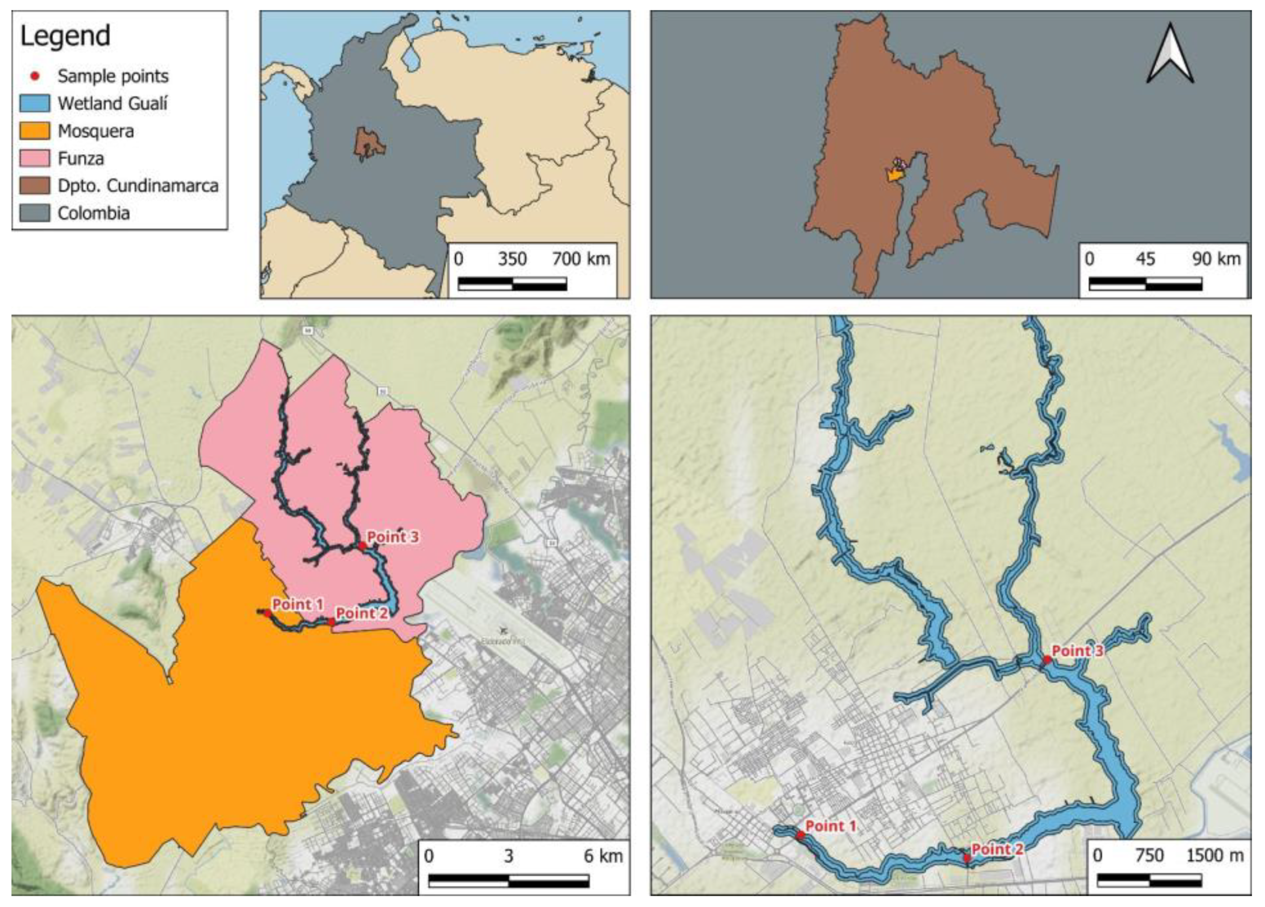
2.1. Study Site and Experimental Design

The study was conducted in the Gualí Wetland (4°42′23.5″ N–74°10′46.4″ W) [4], located between the municipalities of Funza and Mosquera, in the Department of Cundinamarca, Colombia (Figure 1). The Gualí Wetland has a total area of 141.09 ha. It is surrounded by farmland and industries [19].
Three samplings were carried out (between March and October 2021) at three different points in the wetland, for a total of nine samples (Figure 1). The first and third samplings were conducted in the rainy season and the second in the dry season. The sampling points were: Point 1 (4°42′19.2″ N and 74°13′22.3″ W), near an industrial and urban area and the roads that connect the municipalities of Funza and Mosquera; Point 2 (4°42′5.4″ N and 74°12′5.5″ W), close to the Gualí Wetland Linear Park, a school, and the municipality’s exit roundabout; and Point 3 (4°43′34.7″ N and 74°11′29.8″ W), close to the Funza wastewater treatment plant (WWTP) and industrial areas that receive currents from the Bogotá River.
The sampling protocol of [20] was followed with modifications. A metal bucket with a capacity of 12 L was used, performing 3 launches to obtain a total of 36 L per sampling point, with a depth between 0 and 1.5 m. Then, the collected water was poured directly through sieves with a 4.75 mm and 25 µm hole size, to separate the organic matter from the plastic pieces [21]. To avoid cross-contamination, sterile metal tweezers were used to remove and store the plastics, separating the plastic material from the minerals (sand, silt, and clay), shells, and organic matter, by visual inspection, and using the hot needle method [22], which were retained in the 4.75 mm sieve. All the material retained in the 25 µm sieve and selected from the first sieve were placed in sterile 50 mL Falcon® tubes, with a 0.85% (w/v) saline solution. The samples were taken to the Microbiology Laboratory of the Pontificia Universidad Javeriana and stored at room temperature (14 °C) until analysis.

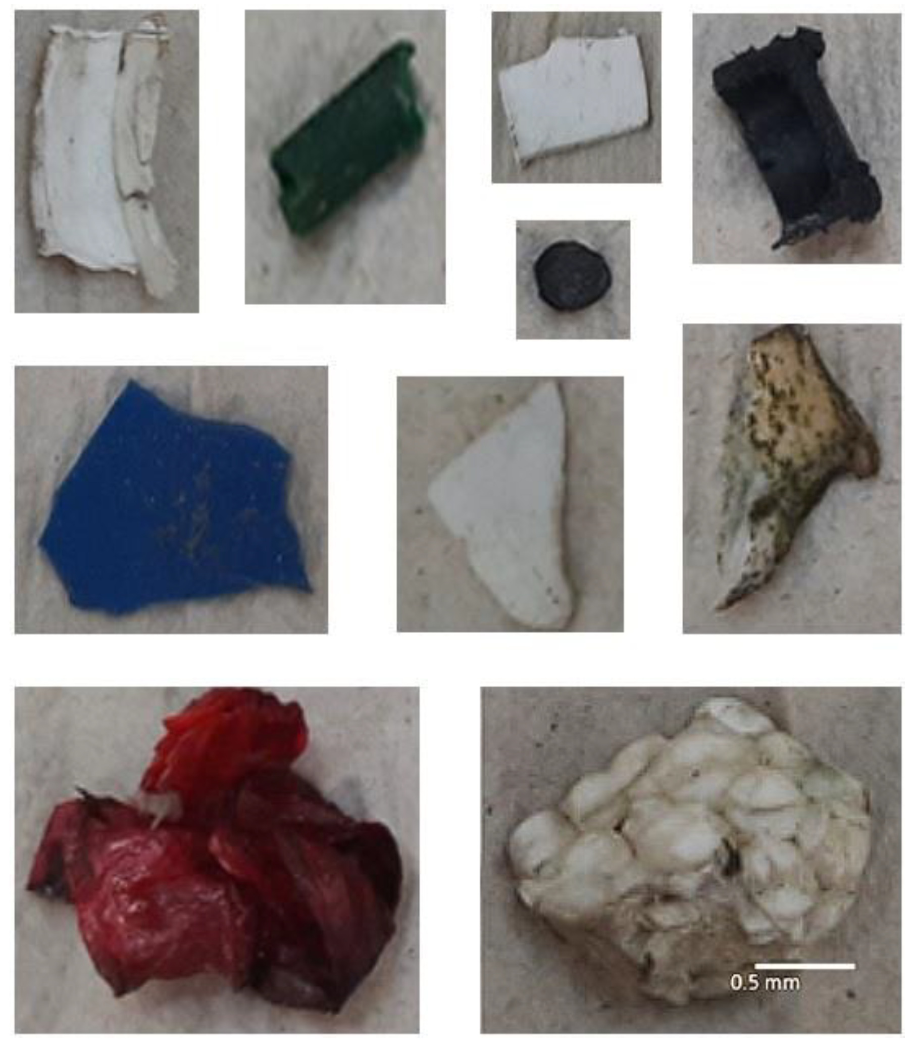
2.2. Observation and Identification of Microplastics and Mesoplastics

The collected plastic samples were observed through a Zeiss® Stemi 305 stereomicroscope, between 8× and 40×, classified according to color, size, and morphology using the microplastic categorization system [22]. To confirm that the collected samples were plastic, a hot needle was used to observe the melting or deformation of the object [23]. The size determination was performed using the Image J Version 1.52 Fiji program, classifying the plastics as microplastics (≥1 mm and <5 mm) or mesoplastics (>5 mm and <25 mm). The polymer type was determined through Fourier transform infrared spectroscopy coupled with attenuated total reflectance (FTIR-ATR), in the Shimadzu® MIRacle-10, IRTracer-100 equipment, following the parameters of [24] (measurement mode: % transmittance, apodization: Happ-Genzel, No. of scans: 15, resolution: 4.0, range (cm−1): 400–4000). Each spectrum was compared with the “OpenSpecy” spectra library and the apparatus database [25,26].

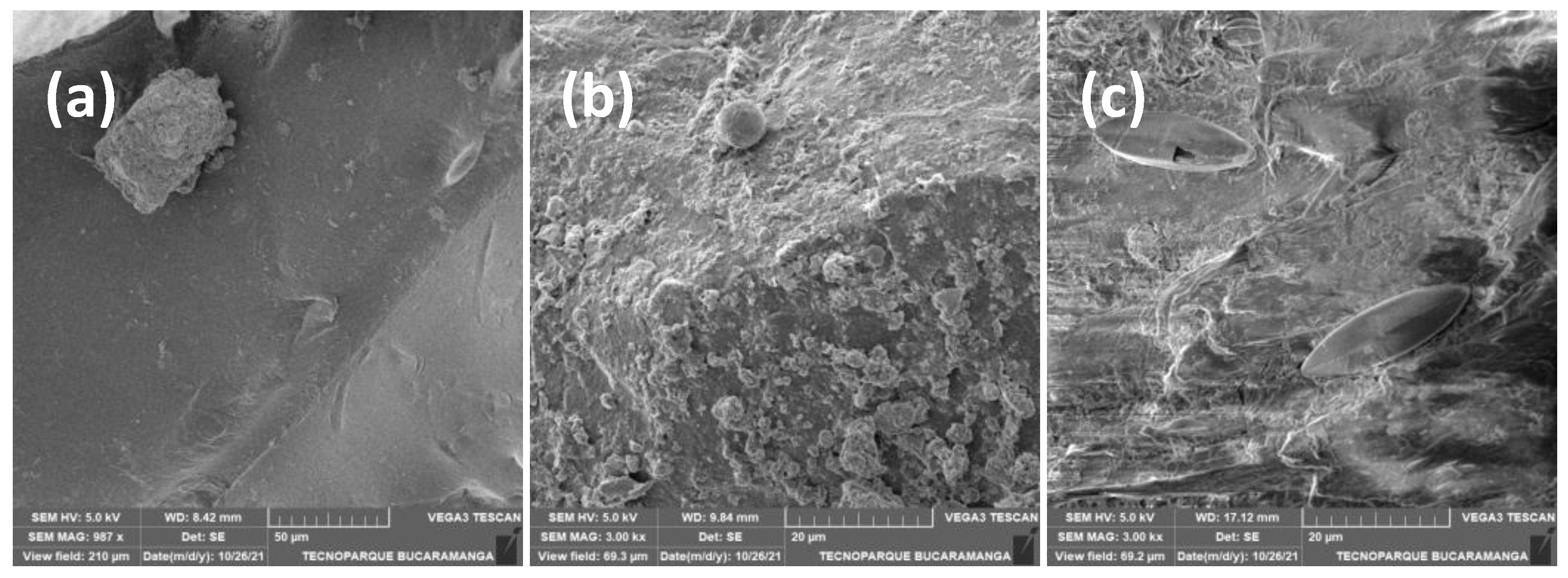
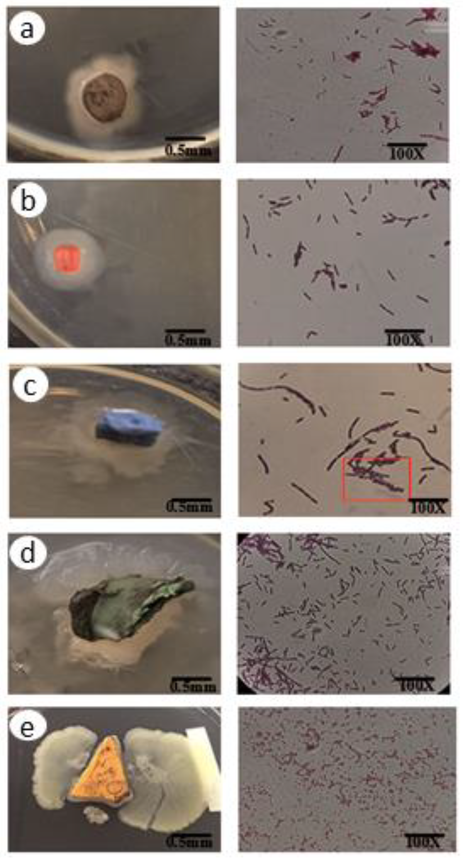
2.3. Detecting Biofilms in Microplastics and Mesoplastics (Determining Their Presence)
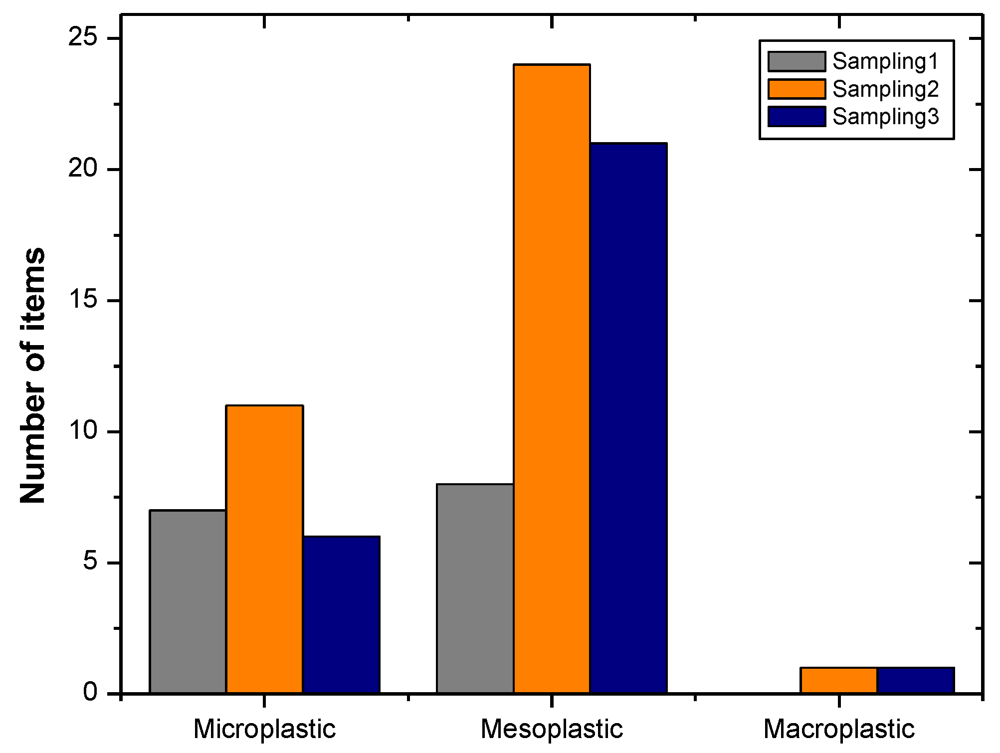
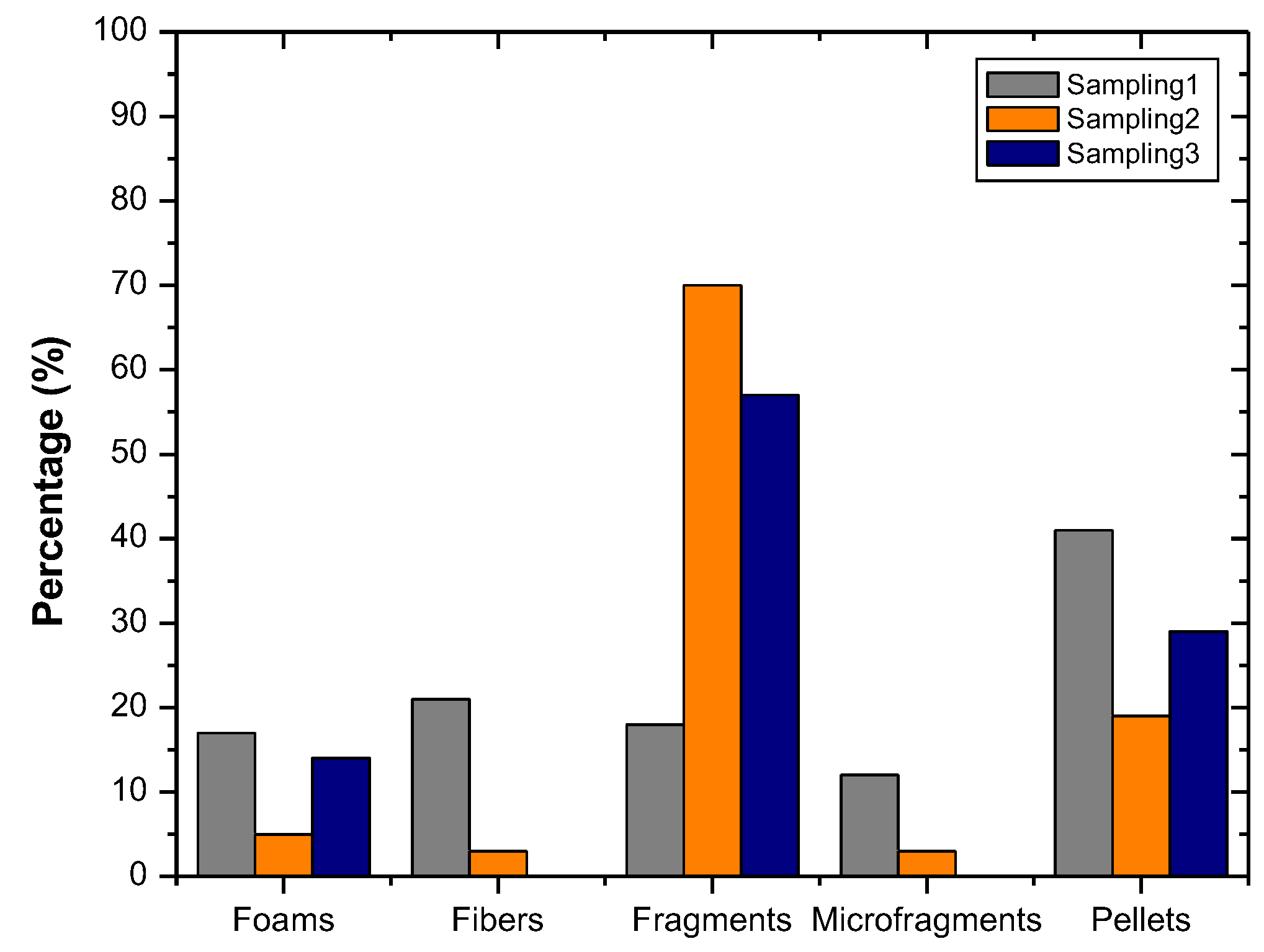
Biofilm detection was performed according to the previously published modified method [27]. Each plastic polymer was washed three times with 2 mL of sterile water and dried in the open air for 45 min in sterile Petri dishes; then, they were immersed in 0.5 mL of crystal violet dye (1% w/v) for 45 min at room temperature (14 °C), washed again with 5 mL of sterile water, and dried at room temperature for 45 min. Each piece was placed in a 25 mL beaker, with 1 mL of ethanol (95% v/v) for 10 min, to remove possible biofilms from the plastic. Finally, the ethanol was transferred to a spectrophotometric cell, and the presence of biofilms was determined by optical density at 595 nm in a Thermo Scientific GENESYS™ 20 visible spectrophotometer.
The most-frequent polymers were placed into agar and nutrient broth for 48 h. Macroscopic and microscopic descriptions of the recovered colonies were made. Additionally, eleven plastic polymers were randomly chosen and scanned with scanning electron microscopy (SEM) using a TESCAN® Vega 3 electron microscope, following the parameters of [28].

3. Results

A total of 79 plastic items were collected during the sampling procedures (79 items/0.135 m3) (Table 1).
Microplastics of 5 mm were the longest length observed and 0.2 mm the shortest length (Figure 2 and Figure 3).
The largest number of items, in each sample, was dominated by the presence of mesoplastics, with a total of 24 and 21 items in the second and third sampling procedures, respectively, followed by microplastics with 11 and 6 items, in the same sampling procedures (Figure 3).
On the other hand, a categorization by morphology was made, obtaining fragments, micro fragments, fibers, pellets, and foams. The most-abundant category was fragments, followed by pellets and foams (Figure 4).
Regarding the categorization by color, the presence of the tones of blue, white, beige, brown, orange, black, red, pink, transparent, and green stood out within the three sampling procedures carried out (Figure 5). Likewise, the FTIR (Figure 6) allowed the identification of the polymer types that were collected at the three sampling points (Figure 7).
For the association of biofilms on the plastics, the technique based on the measurement of the optical density was used. Optical density was proportional to the amount of biofilm per surface area in the sample. The modified crystal violet (CV) staining method was used to obtain the absorbance readings of the polymers.
To corroborate what was described above and to carry out a characterization of the topography of the plastics, scanning electron microscopy (SEM) was performed, and the images are presented in Figure 8.
Finally, the plastic pieces that were placed into nutrient agar for culture allowed the development of colonies around them, a phenomenon that is described as the “plastisphere” by authors such as [29] (Figure 8).

4. Discussion

In this study, 0.6 items/L were obtained, ten-times more than was obtained by Almeida et al. (2023), who collected 0.06 items/L in the Lalu urban wetland, in Tibet, during August, but fewer items than those obtained by the same authors in January (3.05 items/L) [30]. The results obtained in the present investigation were also lower compared to other studies carried out in wetlands, such as the one carried out by Ziajahromi et al. (2020) [31], at the entrance and exit of the ecosystem, finding values of 0.9 ± 0.3 and 4, 0 ± 2.4 microplastic particles/L, respectively, and by the study by Xia et al. (2022) [32], which showed an average abundance of 41.8 ± 4.8 items/L.
There is a direct relationship between the presence of plastic pieces and urbanized areas [33,34]. In this study, Points 3 and 1 were close to industrial zones, urbanized areas, and roads highly traveled by heavy-duty vehicles, cars, and pedestrians; this agrees with the high presence of plastic parts, especially a high incidence of mesoplastics with values of 47%, 65% and 75% for the first, second, and third samplings, respectively. On the other hand, Sampling Point 2, despite being close to a school and a linear park, showed a lower content of plastic pieces, probably due to the reduced transit of school children and pedestrians caused by the COVID-19 pandemic.
Townsend et al. (2019), in their study carried out in urban wetlands, highlighted that there was a high presence of fragments, with the most-common type of plastics generated coming from the fragmentation of larger plastic items. These findings agree with the results obtained in this study (Figure 6). It should be noted that the wetland, as an open-air ecosystem, is strongly linked to factors such as ultraviolet radiation, changes in temperature, the presence of native microorganisms, and the pH of the water, which favor the fragmentation of these polymers into secondary microplastics and mesoplastics.
Additionally, the presence of pellets is associated with the industrial production of primary microplastics, which are used as a basic raw material in various processes that generate commercial products of high importance and consumption. However, during their production and transportation, many of these microplastic pieces can accidentally end up in drains and easily travel to various ecosystems. Consequently, they have been associated with industrial effluents and nearby WWTP outlets [35,36], for example the case of the Siberian point that is close to the WWTP of the municipality of Funza, which is, therefore, exposed to a high frequency of this type of microplastic. A study conducted in the surface waters of the Laurentian Lakes (USA and Canada) found significant amounts of contamination from pellets derived from personal care products [34]. Another study of primary microplastics carried out in Humber Bay (Canada) established that the accumulation rate of this material was concentrated in the form of pellets from industrial sources, and its presence depended on seasonal conditions such as rainfall. Therefore, periods of high rainfall resulted in a lower predisposition of these throughout the bay, attributing this to the increase in water flows through the tributaries [37]; this agrees with the results obtained in this study. On the other hand, the presence of fibers was due to the proximity to textile industries [38].
The presence of a high proportion of transparent and white items is caused by the high frequency of pellets and disposable plastics, especially single-use plastics, which have this color, or due to bleaching processes caused by environmental factors, especially ultraviolet radiation [39].
The color effect can greatly influence the probability of the ingestion of these pollutants by different organisms. It has been shown that especially white and blue pieces are commonly consumed by fish [40,41]. Therefore, it is highly likely that the plastic pieces present in wetlands are causing damage to the fauna that lives there, entering the food chain, until they reach humans [22].
From the spectra obtained by FTIR-ATR for each item, a high frequency of high-density polyethylene (HDPE) was observed (Figure 7) [42], representing 41% of the total samples. The analysis with FTIR-ATR also showed that some polymers contain silicates, as seen in Figure 6 and Figure 7. This occurs because silicates are an essential element for plants, especially when they grow with water as a substrate, as in this case, due to the high presence of Eichhornia crassipes [42]. Moreover, the presence of silicates may occur since the wetland is close to soils used for agricultural. Silicate particles may be transported by runoff and end up in water bodies, adhering to the plastic pieces [43].
Among the most produced and commonly used plastic polymers, we can find polyethylene (PE) and high-density polyethylene (HDPE). These types of polymers have different uses at an industrial level, such as the production of plastic pipes, food and detergent containers, and the production of primary microplastics, especially pellets. They have uses in household items [44]. A study conducted on the banks of the Humber River in Canada determined that 95% of the plastics collected were PE, while only 5% were polypropylene (PP) [45]. Similarly, in the Tamar Estuary (U.K.), a study found that the most-ubiquitous types of microplastics recovered were PE, polystyrene, and PP [46]. These studies can be compared and are consistent with the data obtained, where HDPE was found more frequently (Figure 8).
The increase in the frequency of PE could be explained based on the sedimentology hypothesis, where the different densities of each type of plastic could lead to different deposits and distributions. Since PE has a lower density than water compared to other polymers such as PP, it is generally found floating on the surface of bodies of water. Additionally, it should be noted that, since these pieces have an indefinite exposure time and are constantly influenced by environmental factors, this can contribute to a change in the hydrophobicity of the polymer, making it more hydrophilic [47].
In this study, both Sampling 1 and Sampling 3 were carried out in the rainy seasons, resulting in a lower number of items compared to Sampling 2, which was conducted during the dry season. The temporal distribution of the contamination by microplastics and mesoplastics may be limited in different environmental situations, such as the seasons. Therefore, during rainy seasons, there are effects on the runoff flow at the entrance and exit of plastic pieces, which affects the relative quantity of these items. This information is consistent with what authors such as Jang Y. et al. (2020) have described, where they characterized the abundance of microplastics in the South Yellow Sea (China) in January (6.5 ± 2.1 items/L), which was higher than in April (4.9 ± 2.1 items/L) and August (4.5 ± 1.8 items/L). The latter is associated with hot or dry currents in the study area. Similarly, a study conducted in the province of Phuket (Thailand) determined the abundance of microplastics in sediments, ranging between 300 and 900 items/kg of dry weight in the dry seasons and 33 and 400 items/kg of dry weight in the rainy seasons [47]
Coinciding with optical density, a study in the Bay of Bengal showed that biofilm formation on plastic can vary over time, depending on the productivity of the surrounding water. An increase in the concentration of salts or poorly soluble organic matter causes fouling. Therefore, increasing the organic matter surrounding plastics enhances the formation of biomass. In other words, a decrease in the hydrophobicity of the plastic items, due to exposure, easily leads to the absorption of organic and inorganic nutrients from the water, which can quickly attract microorganisms to the plastic surface and initiate the formation of the plastisphere [48].
As for the absorbances recorded in Table 2, they were not comparable because each one corresponded to a different plastic piece. However, an inversely proportional relationship was observed, as described by [29], where the greater the surface area of the plastic and the less roughness and hydrophobicity it has, the greater the amount of associated biofilm will be, resulting in a greater absorbance. However, this relationship can be influenced by factors such as physical properties (color, shape, density, hydrophobicity, specific surface area) [27].
This confirmed that the plastic samples not only presented a deterioration in their surface roughness, fractures, cracks, and morphological changes, but also some conglomerates, spherical particles, and ovoid particles. This confirmed that the exposure of plastics in the water samples over long periods of time causes microbial colonization and the production of biofilms on the surface of microplastics and mesoplastics [29].
Invasive species can use microplastics and mesoplastics as a floating “raft” that allows them to traverse distances and find new habitats. However, as the size of the part decreases, its surface-area-to-volume ratio increases. For this reason, large concentrations of microplastics can provide a considerable surface area for microorganisms to adhere to. Additionally, these polymers, when exposed to the environment, have organic matter on their surface, serving as a substrate for the development of microorganisms. Figure 8 shows the presence of bacterial groups and diatoms in this case. Similar findings were described by [22] in their studies on both primary and secondary microplastics, collected from the marine environment.
This once again confirmed what was previously described, where when the plastic has a greater surface area, the development of the plastisphere around it will be greater, compared to smaller pieces. The results illustrated how bacterial growth spreads from each fragment (Figure 9) on the agarized culture medium, indicating the presence of the plastisphere on these items. Likewise, growth (red box) and the presence of sporulated bacilli and Gram-positive and -negative cocci through microscopy (Gram staining), were frequently evidenced.

5. Conclusions

The research indicated that the predominant characteristics of plastic polymers in the Gualí Wetland in Colombia were high-density polyethylene microplastics and mesoplastics, in the form of transparent and white fragments and pellets, associated with industrial and urban waste. Likewise, the presence of biofilms that transport bacterial groups and diatoms on the plastic particles tended to increase the adsorption of toxic contaminants, which impact the flora and fauna of wetlands.
This study is the baseline for future research related to the characterization of mesoplastics and microplastics in Colombian wetlands and their associated biofilms.

Author Contributions

Conceptualization: M.A.P.-R. and L.D.G.-M.; investigation: M.A.P.-R. and C.C.-V.; methodology: M.A.P.-R., J.L.M.-Y., C.C.-V. and L.D.G.-M.; writing, draft: M.A.P.-R. and C.C.-V.; drawing graphics: J.L.M.-Y. and L.D.G.-M.; writing and editing: M.A.P.-R., C.C.-V., J.L.M.-Y., P.M.-S. and L.D.G.-M. All authors have read and agreed to the published version of the manuscript.

Funding

This research received no external funding.

Institutional Review Board Statement

Not applicable.

Informed Consent Statement

Not applicable.

Data Availability Statement

Not applicable.

Acknowledgments

We thank the Vortex Company, who provided the scanning electron microscope for the “Red tecnoparque Colombia” located in Bucaramanga city, Santander, Colombia.

Conflicts of Interest

The authors declare no conflict of interest.

References

  1. Peck, D. The Ramsar Convention Manual: A Guide to the Convention on Wetlands (Ramsar, Iran, 1971); Ramsar Convention Bureau: Gland, Switzerland, 1997. [Google Scholar]
  2. Estupiñán-Torres, S.; Cepeda, A.; Hurtado, A.; Vega, K. Calidad bacteriológica del agua del humedal Gualí-Tres Esquinas, Funza, Cundinamarca. Nova 2020, 18, 113–122. [Google Scholar] [CrossRef]
  3. Patiño, J.E.; Estupiñán-Suarez, L.M. Hotspots of Wetland Area Loss in Colombia. Wetlands 2016, 36, 935–943. [Google Scholar] [CrossRef]
  4. Rodriguez-Linares, J.C.; Chaparro-Herrera, S.; Sua-Becerra, A.; Echeverry-Galvis, M.A. Estado poblacional del cucarachero de pantano, Cistothorus apolinari (Passeriformes: Troglodytidae) en siete humedales de la Sabana de Bogotá, Colombia. Rev. Biol. Trop. 2019, 67, 1257–1268. [Google Scholar] [CrossRef]
  5. Garcés-Ordóñez, O.; Castillo, V.; Rueda, R.; Ríos, M.; Bayona, M.; Molina, F.; Escobar, M. Diagnóstico de residuos microplásticos en las zonas marinas de Colombia. INVEMAR MinAmbiente 2017, 108–166. [Google Scholar]
  6. CAR, C.A. Humedales del Territorio CAR. Consolidación de los Humedales de jurisdicción CAR; Imprenta Nacional de Colombia: Bogotá, Colombia, 2011; pp. 15–18. [Google Scholar]
  7. Flórez, C.; Estupiñán-Suárez, L.M.; Rojas, S.; Aponte, C.; Quiñones, M.; Acevedo, Ó.; Vilardy, S.; Jaramilo, Ú. Identificación espacial de los sistemas de humedales continentales de Colombia. Biota Colomb. 2016, 17, 44–62. [Google Scholar]
  8. Li, W.; Tse, H.F.; Fok, L. Plastic waste in the marine environment: A review of source, occurrence, and effects. Sci. Total Environ. 2016, 566, 333–349. [Google Scholar] [CrossRef] [PubMed]
  9. Rios, L.M.; Moore, C.; Jones, P.R. Persistent organic pollutants carried by synthetic polymers in the ocean environment. Mar. Pollut. Bull. 2007, 54, 1230–1237. [Google Scholar] [CrossRef] [PubMed]
  10. Geyer, R.; Jambeck, J.R.; Law, K.L. Production, use, and fate of all plastics ever made. Sci. Adv. 2017, 3, e1700782. [Google Scholar] [CrossRef] [Green Version]
  11. Lai, K.P.; Tsang, C.F.; Li, L.; Yu, R.M.K.; Kong, R.Y.C. Microplastics act as a carrier for wastewater-borne pathogenic bacteria in sewage. Chemosphere 2022, 301, 134692. [Google Scholar] [CrossRef]
  12. Wu, X.; Hou, H.; Liu, Y.; Yin, S.; Bian, S.; Liang, S.; Wan, C.; Yuan, S.; Xiao, K.; Liu, B.; et al. Microplastics affect rice (Oryza sativa L.) quality by interfering metabolite accumulation and energy expenditure pathways: A field study. J. Hazard. Mater. 2022, 422, 126834. [Google Scholar] [CrossRef] [PubMed]
  13. Pisani, X.G.; Lompré, J.S.; Pires, A.; Greco, L.L. Plastics in scene: A review of the effect of plastics in aquatic crustaceans. Environ. Res. 2022, 212, 113484. [Google Scholar] [CrossRef]
  14. Hanif, M.A.; Ibrahim, N.; Dahalan, F.A.; Ali, U.F.M.; Hasan, M.; Jalil, A.A. Microplastics and nanoplastics: Recent literature studies and patents on their removal from aqueous environment. Sci. Total Environ. 2022, 810, 152115. [Google Scholar] [CrossRef] [PubMed]
  15. Kniggendorf, A.K.; Wetzel, C.; Roth, B. Microplastics detection in streaming tap water with Raman spectroscopy. J. Sens. 2019, 19, 1839. [Google Scholar] [CrossRef] [PubMed] [Green Version]
  16. Mukhopadhyay, P.; Arkkakadavil Valsalan, S. Presence of Microplastics in Freshwater Ecosystems: An Unheeded Emerging Concern—A Global Review. Pollution 2022, 8, 69–104. [Google Scholar]
  17. Battulga, B.; Kawahigashi, M.; Oyuntsetseg, B. Characterization of biofilms formed on polystyrene microplastics (PS-MPs) on the shore of the Tuul River, Mongolia. Environ. Res. 2022, 212, 113329. [Google Scholar] [CrossRef] [PubMed]
  18. Song, Y.; Zhang, B.; Zou, L.; Xu, F.; Wang, Y.; Xin, S.; Wang, Y.; Zhang, H.; Ding, N.; Wang, R. Comparative analysis of selective bacterial bolonization by polyethylene and polyethylene terephthalate microplastics. Front. Microbiol. 2022, 13, 836052. [Google Scholar] [CrossRef]
  19. Rosselli, L.; Stiles, F. Wetland habitats of the Sabana de Bogotá Andean Highland Plateau and their birds. Aquat. Conserv. 2012, 22, 303–317. [Google Scholar] [CrossRef]
  20. Ramírez, A. Métodos de recolección. Int. J. Trop. Biol. 2010, 58, 41–50. [Google Scholar]
  21. Feng, S.; Lu, H.; Yao, T.; Xue, Y.; Yin, C.; Tang, M. Spatial characteristics of microplastics in the high-altitude area on the Tibetan Plateau. J. Hazard. Mater. 2021, 417, 126034. [Google Scholar] [CrossRef]
  22. Beckingham, B.; Apintiloaiei, A.; Moore, C.; Brandes, J. Hot or not: Systematic review and laboratory evaluation of the hot needle test for microplastic identification. Microplast. Nanoplast. 2023, 3, 8. [Google Scholar] [CrossRef]
  23. Crawford, C.; Quinn, B. Microplastic Pollutants; Elsevier: Amsterdam, The Netherlands, 2017; Volume 10, pp. 219–267. [Google Scholar]
  24. Gómez-Méndez, L.D.; Moreno-Bedoya, D.A.; Poutou-Piñales, R.A.; Salcedo-Reyes, J.C.; Pedroza-Rodríguez, A.M.; Vargas, A.; Bogoyá, J. Biodeterioration of plasma pretreated LDPE sheets by Pleurotus ostreatus. PLoS ONE 2018, 13, e0203786. [Google Scholar] [CrossRef] [Green Version]
  25. Dehaut, A.; Hermabessiere, L.; Duflos, G. Current frontiers and recommendations for the study of microplastics in seafood. Trends Anal. Chem. 2019, 116, 346–359. [Google Scholar] [CrossRef]
  26. INVEMAR—Instituto de Investigaciones Marinas y Costeras. Protocolo de Muestreo Y análisis de microplásticos en Aguas Marinas Superficiales, Sedimentos de Playas Y Tracto Digestivo de Peces. Componente 5: Diagnóstico de Microplásticos en Zonas Costeras de Colombia. Resolución 646 MinAmbiente; INVEMAR: Santa Marta, Colombia, 2017; 21p. [Google Scholar]
  27. Lobelle, D.; Cunliffe, M. Early microbial biofilm formation on marine plastic debris. Mar. Pollut. Bull. 2011, 62, 197–200. [Google Scholar] [CrossRef]
  28. Pivokonsky, M.; Cermakova, L.; Novotna, K.; Peer, P.; Cajthaml, T.; Janda, V. Occurrence of microplastics in raw and treated drinking water. Sci. Total Environ. 2018, 643, 1644–1651. [Google Scholar] [CrossRef]
  29. Miao, L.; Jou, J.; Young, G.; Liu, Z.; Liu, S.; Li, T.; Mo, Y.; Guo, S.; Qu, H. Acute effects of nanoplastics and microplastics on periphytic biofilms depending on particle size, concentration, and surface modification. Environ. Pollut. 2019, 255, 113300. [Google Scholar] [CrossRef] [PubMed]
  30. Almeida, C.; Marisa, R.; Sáez-Zamacona, I.; Silva, D.; Rodrigues, S.; Pereira, R.; Ramos, S. The Role of Estuarine Wetlands (Saltmarshes) in Sediment Microplastics Retention. Water 2023, 15, 1382. [Google Scholar] [CrossRef]
  31. Xia, F.; Liu, H.; Zhang, J.; Wang, D. Migration characteristics of microplastics based on source-sink investigation in a typical urban wetland. Water Res. 2022, 213, 118154. [Google Scholar] [CrossRef] [PubMed]
  32. Ziajahromi, S.; Drapper, D.; Hornbuckle, A.; Rintoul, L.; Leusch, F.D.L. Microplastic pollution in a stormwater floating treatment wetland: Detection of tyre particles in sediment. Sci. Total Environ. 2020, 713, 136356. [Google Scholar] [CrossRef] [PubMed]
  33. Townsend, K.; Cheng-Hsuan, L.; Sharley, D.; Pettigrove, V. Associations between microplastic pollution and land use in urban wetland sediments. Environ. Sci. Pollut. Res. 2019, 26, 22551–22561. [Google Scholar] [CrossRef]
  34. Eriksen, M.; Mason, S.; Wilson, S.; Zellers, A.; Amato, S. Microplastic pollution in the Surface waters of the Laurentian Great Lakes. Mar. Pollut. Bull. 2013, 77, 177–182. [Google Scholar] [CrossRef]
  35. Talvitie, J.; Heinonen, M.; Pääkkönen, J.P.; Vahtera, E.; Mikola, A.; Setälä, O.; Vahala, R. Do wastewater treatment plants act as a potential point source of microplastics? Preliminary study in the coastal Gulf of Finland, Baltic Sea. Water Sci. Technol. 2015, 72, 1495–1504. [Google Scholar] [CrossRef] [PubMed]
  36. Murphy, F.; Ewins, C.; Carbonnier, F.; Quinn, B. Wastewater treatment works (WwTW) as a source of microplastics in the aquatic environment. Environ. Sci. Technol. 2016, 50, 5800–5808. [Google Scholar] [CrossRef] [PubMed] [Green Version]
  37. Corcoran, P.; Norris, T.; Ceccanese, T.; Walzak, M.; Helm, P.; Marvin, C. Hidden plastics of Humber Bay, Canada, and their potential preservation in the sediment record. Environ. Pollut. 2015, 204, 17–25. [Google Scholar] [CrossRef]
  38. Free, C.; Jensen, O.; Mason, A.; Eriksen, M.; Williamson, N.; Boldgiv, B. High levels of microplastic pollution in a large, remote mountain lake. Mar. Pollut. Bull. 2014, 85, 156–163. [Google Scholar] [CrossRef]
  39. Xiong, X.; Zhang, K.; Chen, X.; Shi, H.; Wang, Z.; Wu, C. Microplastics in freshwater sediment; A review on methods, occurrence, sources. Sci. Total Environ. 2020, 754, 141948. [Google Scholar]
  40. Barboza, L.; Lopes, C.; Oliveira, P.; Bessa, F.; Otero, V.; Henriques, B.; Raimundo, J.; Caetano, M.; Vale, C.; Guilhermino, L. Microplastics in wild fish from Northeast Atlantic Ocean and its potential for causing neurotoxic effects, lipid oxidative damage, and human health risks associated with ingestion exposure. Sci. Total Environ. 2020, 717, 134625. [Google Scholar] [CrossRef]
  41. Bray, L.; Digka, N.; Tsangaris, C.; Camedda, A.; Gambaiani, D.; Giuseppe, L.; Matiddi, M.; Miaud, C.; Palazzo, L.; Olmo, A.; et al. Determining suitable fish to monitor plastic ingestion trends in the Mediterranean Sea. Environ. Pollut. 2019, 247, 1071–1077. [Google Scholar] [CrossRef] [PubMed]
  42. Zhang, B.; Yang, X.; Lingchen, L.; Chen, L.; Teng, J.; Zhu, X.; Zhao, J.; Wang, Q. Spatial and seasonal variations in biofilm formation on microplastics in coastal waters. Sci. Total Environ. 2021, 770, 145303. [Google Scholar] [CrossRef]
  43. Pereira, A.P.D.S.; Silva, M.H.P.D.; Lima, É.P.; Paula, A.D.S.; Tommasini, F.J. Processing and characterization of PET composites reinforced with geopolymer concrete waste. Mater. Res. 2017, 20, 411–420. [Google Scholar] [CrossRef] [Green Version]
  44. Scopetani, C.; Chelazzi, D.; Martellini, T.; Pellinen, J.; Ugolini, A.; Sarti, C.; Cinicinelli, A. Occurrence and characterization of microplastic and mesoplastic pollution in the Migliarino San Rossore, Massaciuccoli, Nature Park (Italy). Mar. Pollut. Bull. 2021, 171, 112712. [Google Scholar] [CrossRef]
  45. Ballent, A.; Cocoran, P.; Madden, O.; Longstaffe, F. Sources and Sinks of Microplastics in Canadian Lake Ontario Nearshore, tributary and beach sediments. Earth Sci. Publ. 2016, 110, 383–395. [Google Scholar] [CrossRef] [Green Version]
  46. Sadri, S.; Thompson, R. On the quantity and composition of floating plastic debris entering and leaving the Tamar Estuary, Southwest England. Mar. Pollut. Bull. 2014, 81, 55–60. [Google Scholar] [CrossRef] [PubMed]
  47. Jiwarungrueangkul, T.; Phaksopa, J.; Sompongchaiyakul, P.; Tipmanee, D. Seasonal microplastic variations in estuarine sediments from urban canals on the west coast of Thailand: A case study in Phuket province. Mar. Pollut. Bull. 2021, 168, 112452. [Google Scholar] [CrossRef] [PubMed]
  48. Sunitha, T.; Monisha, V.; Sivanesan, S.; Vasanthy, M.; Omine, K.; Sivasankar, V. Micro-plastic pollution along the Bay of Bengal coastal strech of Tamil Nadu, South India. Sci. Total Environ. 2020, 756, 144073. [Google Scholar] [CrossRef] [PubMed]
Figure 1. Location of the study area in the Gualí Wetland and the municipalities of Funza, Mosquera, and Cundinamarca, Colombia.
Microplastics 02 00021 g001
Figure 2. Plastic items collected in the Gualí Wetland. White bar = 0.5 mm.
Microplastics 02 00021 g002
Figure 3. Number of items collected in each sampling procedure according to their size.
Microplastics 02 00021 g003
Figure 4. Percentage of collected items according to their morphology.
Microplastics 02 00021 g004
Figure 5. Number of collected items according to their color.
Microplastics 02 00021 g005
Figure 6. FTIR-ATR of four collected items: (a) Polyethylene + silicate (PES). (b) Chlorinated polyvinyl chloride (CPVC). (c) Polyethyleneterephtalate (PET). (d) High-density polyethylene (HDPE).
Microplastics 02 00021 g006aMicroplastics 02 00021 g006b
Figure 7. Percentage of collected items according to their polymer composition. Polypropilene + silicate (PPS), polyethylene + inorganic silicate (PE + IS), polyethylene + silicate + BIO (PE + SBIO), chlorinated polyethylene (PE + Cl), polyethylene (PE), high-density polyethylene (HDPE), expanded polystyrene (EPS), chlorinated polyvinyl chloride (CPVC).
Microplastics 02 00021 g007
Figure 8. Scanning electron microscopy of microplastics (a) and mesoplastics (b,c).
Microplastics 02 00021 g008
Figure 9. Plastisphere development in microplastics (ac) and mesoplastics (d,e) obtained at the three sampling points. Bacterial growth (red box) and presence observed from plastic particles and under microscope (Gram staining).
Microplastics 02 00021 g009
Table 1. Number of items collected per sample at each sampling point.
PointSampling 1
(28 March 2021)
Sampling 2
(25 June 2021)
Sampling 3
(9 October 2021)
Total
Point 15241140
Point 22338
Point 3891431
Total15362879
Table 2. Absorbances of items collected with the CV technique.
IdentificationPolymerClassification (Size)Absorbance (595 nm)
1PE + Clmesoplastic1.901
2PETmesoplastic0.894
3HDPEmesoplastic1.019
4CPVCmicroplastic0.572
5PEmicroplastic0.522
6EPSmesoplastic2.276
7HDPEmicroplastic0.541
8PPSmicroplastic0.451
9HDPEmesoplastic0.977
10HDPEmicroplastic0.442
11PE + Clmesoplastic2.098
12EPSmesoplastic1.342
Disclaimer/Publisher’s Note: The statements, opinions and data contained in all publications are solely those of the individual author(s) and contributor(s) and not of MDPI and/or the editor(s). MDPI and/or the editor(s) disclaim responsibility for any injury to people or property resulting from any ideas, methods, instructions or products referred to in the content.

