Review

Virus-like Particles (VLPs) as Important Tools for Flavivirus Vaccine Development

by
Leda R. Castilho
1,2,*,
Nathalia R. Mattos
1,
Wallace S. Abreu
1,3 and
Melissa L. E. Gutarra
3,4
1
Cell Culture Engineering Lab, PEQ, COPPE, Universidade Federal do Rio de Janeiro (UFRJ), Rio de Janeiro 21941-598, RJ, Brazil
2
Biochemistry Graduate Program, IQ, Universidade Federal do Rio de Janeiro (UFRJ), Rio de Janeiro 21941-909, RJ, Brazil
3
EPQB Graduate Program, EQ, Universidade Federal do Rio de Janeiro (UFRJ), Rio de Janeiro 21941-909, RJ, Brazil
4
NUMPEX-Bio, Duque de Caxias Campus, Universidade Federal do Rio de Janeiro (UFRJ), Duque de Caxias 25240-005, RJ, Brazil
*
Author to whom correspondence should be addressed.
Biologics 2022, 2(4), 226-242; https://doi.org/10.3390/biologics2040018
Submission received: 28 July 2022 / Revised: 29 October 2022 / Accepted: 31 October 2022 / Published: 31 October 2022
(This article belongs to the Section Vaccines)

Abstract

:
Flaviviruses, such as dengue, zika, yellow fever, West Nile, and Japanese encephalitis virus, are RNA viruses belonging to the Flaviviridae family (genus Flavivirus). They represent an important global health concern, since most areas of the world are endemic for at least one of these viruses. Although vaccines for five flaviviruses currently exist, there is a need for new vaccines to protect from established, emerging, and reemerging flaviviruses. Yellow fever vaccine shortages experienced in the last decade, combined with the risk of YFV spread to Asia and the restrictions of vaccine administration to certain population segments, show that even when a highly efficacious vaccine is available, new and improved vaccines might be needed. Virus-like particles (VLPs) are multiprotein structures that mimic the virus, but do not contain its genetic material. As such, VLPs have an excellent track record of strong immunogenicity and high safety, dating back to the introduction of the first recombinant hepatitis B vaccine in the 1980s. Flavivirus-like particles (FVLPs) have been extensively studied, especially for DENV, JEV, and ZIKV, and could give rise to next-generation recombinant subunit flavivirus vaccines based on VLPs incorporating molecular features intended to ensure high efficacy and minimize the risk of antibody-dependent enhancement (ADE) upon infection with other flaviviruses.

1. Introduction

Flaviviruses present a global threat, since most areas of the world are endemic for at least one of these viruses [1]. Examples of well-known flaviruses are dengue viruses of serotypes 1-4 (DENV 1-4), West-Nile virus (WNV), Zika virus (ZIKV), Japanese encephalitis virus (JEV), and yellow fever virus (YFV), among others. Dengue viruses alone infect over 400 million people worldwide per year, and more than a quarter of the world’s population lives in areas where DENV is endemic [2].
The genus Flavivirus of the Flaviviridae family comprises more than 70 enveloped viruses, which are transmitted mainly by arthropods (especially mosquitoes and ticks) and can cause severe illnesses in humans [2]. The flavivirus genome consists of a positive-sense, single-stranded RNA that encodes a polyprotein [3]. After translation of the viral RNA, the polyprotein is cleaved by host- and virus-encoded proteases, so that seven non-structural proteins and three structural proteins are generated [4]. These structural proteins are known as capsid (C), premembrane (prM), and envelope (E) proteins.
Currently, licensed vaccines only exist against five flaviviruses: JEV, tick-borne encephalitis virus (TBEV), Kyasanur forest disease virus (KFDV), DENV, and YFV [5,6,7,8,9]. All these vaccines are based on production technologies based on whole viruses (whole native viruses or whole chimeric viruses) as active pharmaceutical ingredients, containing either the inactivated pathogen (TBEV, KFDV, and JEV vaccines) or live-attenuated viruses (YFV, DENV, and also JEV vaccines). In the case of DENV, the licensed vaccine is tetravalent (against all four dengue serotypes) and based on four chimeric viruses. The four chimeras were obtained by engineering the live-attenuated 17D yellow-fever vaccine strain in order to have the YFV prM and E genes replaced by those of DENV1, DENV2, DENV3, or DENV4, respectively.
However, new flavivirus vaccines are urgently needed for different reasons. First of all, new vaccines against more flaviviruses are needed in order to prevent disease caused by other established, emerging, or reemerging flavivirus threats, such as West-Nile, Zika, and other viruses [2]. Regarding the existing licensed vaccines, industrial-scale propagation of the whole viruses requires high biosafety level facilities for vaccine manufacturing. Furthermore, while they are protective, for most of them, there is the need for frequent iterative boosting to maintain protective immunity [2]. This is not the case for the live-attenuated JEV and yellow-fever vaccines, which give lifelong protection. However, the YFV vaccine is produced in embryonated chicken eggs, which makes it difficult to ramp up production to respond to outbreaks. YFV vaccine shortages have been experienced during recent outbreaks, such as those that occurred in Africa in 2016 and in Brazil in 2017–2018, resulting in the use of fractional (1/5) vaccine doses as an attempt to meet demand and control these outbreaks [5,10]. Moreover, the life-attenuated YFV vaccine is not recommended to several population groups, and rare, but serious adverse effects can occur, which become more apparent in the setting of mass vaccination campaigns. The risk of YFV spreading in the near future to Asia and other regions may result in severe vaccine shortage and a large number of deaths, making it evident that, even in the case that a safe and highly efficacious vaccine exists, there is a need for a new yellow fever vaccine.
Virus-like particles (VLPs) are three-dimensional particles that mimic the organization and conformation of native viruses but lack the viral genome [11]. They result from the self-assembly of structural viral proteins and present the antigen in a repetitive way, enhancing the immune response. Subunit vaccines containing particles made of viral proteins produced by recombinant DNA technology are an already proven vaccine platform. Their immunogenicity can be further enhanced by the use of adjuvants [12,13]. Therefore, VLP-based subunit vaccines represent a promising alternative for the development of new safe and effective flavivirus vaccines.

2. Viral Vaccine Types

In the last several years, the range of types of vaccines adopted in viral vaccines that have reached approval for human use has increased (Table 1). Traditional viral vaccine production technologies have been mostly based on whole viruses (either inactivated or live-attenuated). Recombinant subunit viral vaccines were an innovation enabled by the development of recombinant DNA technology, and the first human vaccine of this type was approved in 1986 [14,15]. However, intensive research in the field of vaccinology in the last few decades paved the path for the approval of new vaccine technologies for human use, such as viral vectored and nucleic acid vaccines [16,17,18,19,20]. These new technologies, which had been in development for many years for a range of different diseases, have proven very useful to help control public health emergencies.
Subunit vaccines contain one or more fragments of the pathogen and can be of different classes, depending on the type of antigen it contains: (i) toxoid; (ii) polysaccharide conjugate; (iii) outer membrane vesicle; and (iv) recombinant protein/VLP [25]. Whereas the three first types form the basis of several bacterial vaccines approved to date, the latter type is of great significance in the field of viral vaccines.
Based on recombinant DNA technology developed in the 1970s, a new era of viral vaccines based on recombinant viral proteins started in 1986, when the first recombinant hepatitis B vaccine containing the surface antigen produced in engineered yeast was licensed for human use [14,15]. However, it was just in the last two decades that further recombinant subunit viral vaccines were licensed for human use: two HPV vaccines (Gardasil®, Cervarix®) in 2006 and 2007 [26], a hepatitis E vaccine in 2012 [27], a flu vaccine in 2013 [28], a malaria vaccine (positively reviewed by the European Medicines Agency in 2015 and recommended for widespread use by the World Health Organization in 2021) [29,30,31], a zoster vaccine in 2017 [32], and COVID-19 subunit vaccines in 2021–2022 [33,34].
In most of these approved vaccines, the recombinant proteins self-assemble to form 3D particles, and particulate antigens enable enhanced activation of B and T cells [35]. Recombinant protein and VLP-based subunit vaccines have a large and consolidated track record of being safe and efficacious in all population segments, ranging from newborns (hepatitis B) to the elderly (flu), and the production of flavivirus-like particles (FVLPs) has been widely investigated for flavivirus vaccine development. Therefore, this will be the focus of this review.
Chimeric and viral vectored vaccines were also enabled by the development of recombinant DNA technology and use viruses as vaccine vectors for delivery of antigen-encoding gene(s). These two vaccine technologies are frequently referred to as a single vaccine platform [36,37]. By 2010, when several vaccine candidates of these types were in Phase I, II, or III of clinical development, a flavivirus vaccine was the first chimeric vaccine to be approved for human use [36]. IMOJEV®, which consists of the premembrane and envelope sequences of an attenuated JEV strain replacing the corresponding genes in the attenuated 17D yellow fever strain, was first approved in Australia in August 2010 [17]. A similar approach was used for the development of Dengvaxia®, a tetravalent dengue vaccine that was first approved for human use in Mexico in 2015 [38,39].
Viral vectored vaccines rely on the use of a virus to deliver a key immunogenic sequence of an unrelated virus, and this approach has been under investigation in the last few decades for a wide range of virus families [40]. For example, the live-attenuated measles virus has been used as a vector for the development of several viral vaccines, including vaccines for flaviviruses such as JEV, dengue, and zika [41,42,43,44], the latter having reached clinical phase 1 [45]. The rVSV-ZEBOV vaccine against the Ebola virus was the first viral vectored vaccine to be approved for human use. The pivotal clinical trial data originated from a ring vaccination controlled trial carried out in Guinea during the 2013–2016 Ebola outbreak in West-Africa [46]. The vaccine proved to be a very useful tool to control the public health emergency, and was prequalified by the WHO and approved in USA and Europe in 2019 [20]. However, perhaps the most prominent examples of viral vectored vaccines are those based on adenoviral vectors. Adenovirus-based vaccines had been in development for over 30 years [47,48], and clinical trials had been carried out for several pathogens, including the MERS coronavirus and Ebola virus [49,50]. The previous large clinical experience with those vaccines certainly contributed to accelerate the development of COVID-19 adenovirus-based vaccines: by May 2021, four different adenovirus-based COVID-19 vaccines (Oxford Astrazeneca, Janssen, Gamaleya and Cansino vaccines) had been authorized for human use [18].
Nucleic acid based vaccines rely either on plasmid DNA or mRNA encoding the antigen of choice, and as such were also enabled by recombinant DNA technology. Upon cellular uptake, the transgene encoded by the vaccine is expressed in target cells of the organism, mimicking protein synthesis and post-translational modifications (e.g., glycosylation) that occur during an infection [37]. Because DNA vaccines need to cross two different membranes (the plasma and the nuclear membranes) in order to achieve protein expression, doses are usually high, and many efforts since the 1990s have focused on improving the DNA uptake into the cell, in order to enhance expression of the encoded antigen [37,51]. Several DNA vaccines have been developed up to a clinical phase for a range of viral pathogens, such as flaviviruses (zika and West Nile virus), HIV, influenza, HPV, malaria, MERS-COV, and Ebola, among others [16,52,53], but the first DNA vaccine to be approved for human use was ZyCoV-D®, which received emergency use authorization for COVID-19 in India in 2021 [51].
The use of mRNA to in vivo express proteins was initially hampered by its intrinsic instability and inflammatory potential [51]. However, the development of nucleoside-modified, highly purified mRNA produced by in vitro transcription allowed these shortcomings to be overcome, resulting in a new class of biologics for therapy and vaccine development [19,54,55,56]. Lipid nanoparticles that protect mRNA and deliver it into the cells represent a further innovation that was crucial for the development of mRNA vaccines. The first clinical trial of an mRNA prophylactic vaccine against an infectious disease started in 2013 for a rabies vaccine candidate [57,58]. By 2019, preclinical and clinical trials had demonstrated safety and immunogenicity of several mRNA vaccine candidates in animals and humans, and over 15 mRNA vaccine candidates targeting 10 different infectious diseases had entered clinical trials, including a zika mRNA vaccine currently undergoing a Phase 2 trial [59,60,61]. Investigations on mRNA vaccination against MERS-COV as a prototype pathogen, aiming at preparedness against betacoronaviruses as possible pandemic threats, were ongoing before the emergence of SARS-CoV-2 [62,63]. All of this decade-long research on mRNA immunotherapies and vaccines paved the path for a timely development and authorization of the first two human mRNA vaccines in 2020, both targeting COVID-19 [64,65].

3. Virus-like Particles (VLPs) and Flavivirus-like Particles (FVLPs)

Virus-like particles are an important and proven subclass of subunit vaccines with several advantages over traditional vaccine technologies: they are non-replicating and contain purified antigens of choice, thus minimizing the risk of adverse effects as compared to traditional whole-virus vaccines; and they are usually produced by modern, scalable biotechnological techniques, allowing production scale-up in the case of outbreaks. From the immunological point of view, VLPs have been shown to be strong activators of dendritic cells, which are the most potent antigen presenting cells and whose activation primes B and T cell responses [66].
VLPs consist of three-dimensional nanometric structures formed by recombinant viral structural proteins. VLPs provide a powerful tool for the development of safe and effective vaccines against viral pathogens, due to their ability to present conformational epitopes and to elicit neutralizing antibody responses. This is of relevance, since neutralizing antibody titers are a very reliable correlate of protection for many virus families, including flaviviruses. As shown for DENV 1-4 VLPs, highly conformational and quaternary structure-dependent antibody epitopes found on native viruses can be efficiently displayed on VLP surfaces [67]. This is an advantage, e.g., over inactivated viruses for vaccine development, since chemical inactivation can have deleterious effects on conformational neutralizing epitopes of the E protein, as shown when comparing a ZIKV VLP-based and an inactivated vaccine candidate [12]. As a whole, VLPs offer a safe approach to vaccine development as compared to traditional whole-virus approaches, from early in vitro/in vivo studies of the vaccine candidate up to its large-scale production.
For flaviviruses, VLPs with antigenic and functional properties similar to those of infectious virions are generated by expression of the genes encoding the (pre)membrane (prM/M) and envelope (E) proteins alone [68,69], or in the presence of the capsid (C) gene [70]. Figure 1 illustrates the differences between an infectious virion and VLPs containing or not the capsid protein.
Previous research has demonstrated the possibility of enhanced immune response with VLPs expressing engineered antigens and presenting conformational epitopes that are usually absent in other vaccine types (e.g., formalin-inactivated viruses and monomeric subunit vaccines), eventually allowing lower dose sizes to elicit a robust response [11]. Moreover, VLPs can be tailor-made to incorporate various molecular features, not only with the aim of providing better immunogens, but also of helping to elucidate the role of these molecular features in immune protection and providing customized probes to interrogate the antibody repertoire and to discover candidate therapeutic antibodies [71].

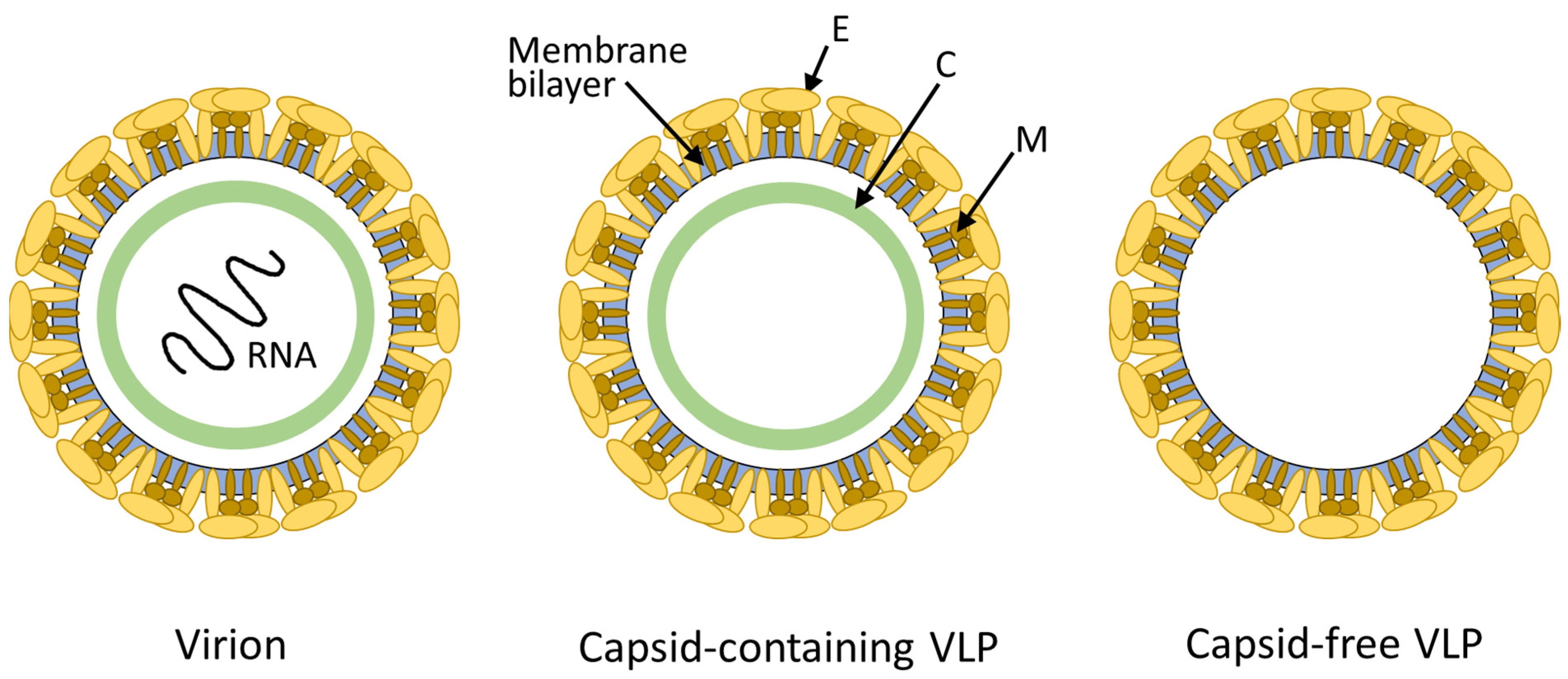
3.1. Molecular Features of FVLPs

FVLPs assemble into membrane vesicles, are secreted, and retain the morphology and antigenicity of live infectious virions [72,73]. Thus, the vast knowledge about flaviviruses serves as a basis to design improved FVLPs. This is of particular interest because VLPs can be easily engineered to obtain customized molecular features that can influence both clinical and manufacturing aspects related to their use as vaccines, such as immune response, antibody-dependent enhancement of infection, stability, and VLP expression level and secretion.
The signal peptide of the prM protein influences processing of the structural proteins prM and E, and has several implications on biological properties of flaviviruses [74,75]. According to Chang and colleagues, also for plasmid DNA vaccines encoding the prM-E genes for in vivo VLP expression, the signal sequence located at the N-terminus of prM was crucial for an adequate biosynthesis and protein processing, preserving the native conformation and glycosylation profiles [76,77]. The use of the signal peptide of JEV has been used by several authors for the expression of prM-E of other flaviviruses, both for in vivo (e.g., DNA and mRNA vaccines) and for in vitro (VLP production in cell culture) expression [69,77,78,79,80]. Alvim et al. [78] studied the production of zika VLPs in stably transfected HEK293 cells and observed that four signal peptides designed in-house led to higher levels of secreted VLPs than zika wild-type signal peptides, indicating that improved FVLP production might be achieved by further signal peptide optimization efforts.
The construction of chimeric VLPs has also been proposed as a way to improve VLP expression and secretion, both in vivo and in vitro. The replacement of the stem and transmembrane regions of the E protein with those of another flavivirus might increase the release of VLPs, both in vitro [81] or in vivo [69]. Chang et al. [81] compared the production of DENV2 VLPs in transfected COS-1 cells using three different constructs: one based on DENV2 wild-type prM-E, the second one replacing the 10% C-terminal end with that of JEV, and the third one replacing the 20% C-terminal end with that of JEV. Efficient secretion was observed just for the latter, indicating that the region starting in residue 397 of the E protein is important for prM-E secretion. Purdy and Chang [82] further investigated, for all DENV serotypes, the expression of wild-type and chimeric (DENV-JEV) VLPs, and observed different effects depending on the serotype. In DENV2, three residues in the E-H1 α-helix domain were shown to be responsible for the intracellular retention of E protein in this serotype. Chang et al. [81] verified that the introduction of the 20% JEV stem and transmembrane regions had no effects on recognition by anti-DENV2 monoclonal antibodies, so that the authors postulated this to be an antigenically inert region.
An important aspect of flavivirus biology is related to the maturation of virions along the flavivirus lifecycle. Immature virions that bud into the endoplasmic reticulum display 60 trimers of prM-E heterodimers arranged with icosahedral symmetry on its surface, but after furin-mediated cleavage of the “pr” portion of prM during egress from the cell, mature infectious virions are formed. These display 90 antiparallel M-E heterodimers that expose distinct surfaces of E, as compared to immature virions [83]. However, due to an inefficient maturation process, flavivirus-infected cells often secrete a mix of mature, partially immature, and fully immature particles into the extracellular space [84]. This structural heterogeneity can impact the antigenic and functional characteristics of flaviviral virions and VLPs [85,86].
Virus-like particles open the possibility of modulating the maturation state of viral particles. More mature VLPs can be obtained by coexpressing the gene of a furin-like protease in the host cell line, analogously to a strategy that has been shown to enable the production of more homogenous mature dengue virus populations [86]. On the other hand, immature FVLPs can be generated by introducing a mutation in the furin recognition site to prevent cleavage at the pr/M junction. Such immature FVLPs are important tools for investigations on the maturation state dependency of immune response, and can inform a more rational design of flavivirus vaccines.
Data gathered over the last years for zika mRNA and DNA vaccine candidates confirm the influence of structural features on the quality of the immune response elicited by vaccination. When DNA and mRNA vaccines are developed, studies are conducted using the nucleic acid construct to in vitro express the antigen and to characterize this antigen [69,83] because it will be the antigen that in vivo will be responsible for eliciting the immune response. However, learning from literature data obtained for mRNA or DNA vaccine candidates that are based on the same genes that encode a flavivirus VLP (e.g., prM-E) helps with understanding molecular features that should or should not be contained in the nucleic acid constructs used to in vitro express VLPs.
When a zika DNA vaccine candidate containing the JEV stem and transmembrane regions (named VRC5288) and a candidate based on the wild-type zika prM-E sequence (named VRC5283) underwent Phase I clinical evaluation, the wild-type vaccine candidate showed more robust neutralizing antibody and T-cell responses [52]. Similarly, when mRNA versions of VRC5283 and VRC5288 were tested as mRNA vaccine candidates, VRC5283 showed a higher neutralizing antibody response in mice immunized with two vaccine doses, three weeks apart from each other. When mice were challenged with infectious zika virus, 90% of mice immunized with VRC5283 presented undetectable viral load, whereas, in just 20% of mice immunized with VRC5288, the viral load was undetectable [83]. Maciejewski et al. [87] used samples from non-human primate (NHP) and human trials conducted with the DNA versions of VRC5283 and VRC5288 vaccine candidates to further investigate what the implications of the introduction of the JEV stem and transmembrane regions were in the VRC5288 construct. The authors found out that there were qualitative differences among antibodies elicited by both vaccine candidates: VRC5283 generated relatively more antibodies binding to mature zika virions, whereas antibodies generated by VRC5288 were more sensitive to the maturation state of virions. According to Dowd et al. [69], when expressed in vitro to generate VLPs, a VRC5288 DNA construct had shown more efficient release of VLPs into cell culture supernatants than VRC5283 DNA construct, which was in agreement with earlier studies that had shown that chimeric prM-E constructs bearing the JEV stem and transmembrane regions improved VLP secretion. Thus, the quantity of the recombinant immunogen should not be the only criterium for selection of the optimal immunogen.
Although these observations arise from nucleic acid-based vaccines that induce the in vivo production of VLPs, the knowledge gained can certainly be extrapolated to in vitro produced VLPs, highlighting that immunogen structure should be carefully designed to prioritize a high quality of the immune response to be elicited rather than the quantity or yield achieved during the VLP production process.
A further aspect related to the quality of the immune response, which is of foremost importance for flavivirus vaccine development, is the need to design vaccine candidates that minimize the risk of antibody-dependent enhancement (ADE) of infection, considering flaviviruses for which ADE has been reported as relevant. Antibodies that bind to virions, but do not neutralize them, facilitate infection of cells that express Fc-γ receptors, and thus lead to ADE, with increased viral load and more severe pathogenesis [2,88]. Although the occurrence of ADE in vivo has been mainly reported for DENV, in vitro assays using Fc-γ expressing cells have shown ADE potential for a range of flaviviruses [2].
ADE is of foremost relevance to DENV pathogenesis, but also of high relevance for vaccine development, since any risk of disease enhancement, not only a lack of protection, is of concern [89]. Clinical development of the only licensed dengue vaccine (Dengvaxia, Sanofi) showed differences in efficacy among originally seronegative and originally seropositive clinical trial participants, and long-term monitoring of subjects raised the possibility that in originally seronegative young children vaccination acted like a first infection that made them more susceptible to severe dengue upon exposure to dengue virus [90]. Halstead [91] argued against the use of Dengvaxia, claiming that antibody-dependent enhanced dengue disease occurred in seronegatives who were sensitized by vaccine, regardless of age. Thus, ADE is widely recognized as a challenge for developing flavivirus vaccines, e.g., for dengue [92] and zika viruses [93].
Therefore, a rational development of a flavivirus VLP-based vaccine should take the large knowledge gathered around flaviviral structure and ADE into consideration. Studies with monoclonal antibodies have provided structural insights that allowed the identification of sites in the flaviviral envelope protein that give rise to ADE-prone antibodies and those that induce potent, protective antibodies [94]. In mature flaviviruses, where the 180 E proteins are organized as dimers in a herringbone pattern, the exposed surface of the dimers is mostly virus-specific, whereas more conserved regions, such as the fusion loop, are normally buried into the dimer. However, there is an exposed region known as “E dimer epitope” (EDE), which is relatively conserved among flaviviruses and is targeted by antibodies that potently neutralize a broader spectrum of viruses, such as all four DENV serotypes and, in the case of mAbs classified as EDE1 (which do not require the glycan on the 150 loop to bind to EDE), also ZIKV. Using a heterologous prime-and-boost strategy for immunogens from different flaviviruses (e.g., ZIKV and a DENV serotype) should efficiently elicit the formation of EDE antibodies, thus resulting in a broadly neutralizing response, with the added advantage that EDE antibodies have been shown to bind partially mature and fully mature virions equally well [94]. Thus, VLPs of different flaviviruses, presenting stabilized dimers on their surface, could represent an alternative for a safe and efficient heterologous primary vaccination series.
Maturation state of virions also plays a role in ADE. It has been shown that a particular monoclonal antibody (E53), which recognizes the highly conserved fusion loop peptide of flaviviruses, preferentially binds to immature virus particles. Although fully immature virions are non-infectious due to the presence of prM inhibiting virus attachment and fusion, Rodenhuis-Zybert et al. [84] have shown that the E53 antibody bound to fully immature WNV and DENV particles was able to render the immature virions infectious by facilitating cell entry followed by prM cleavage by the endosomal furin.
A similar phenomenon occurs with anti-prM antibodies, which bind to immature virus particles, enable viral entry and maturation, and render these virions infectious. Anti-prM antibodies are especially cross-reactive among dengue virus serotypes, and their role in ADE among DENV serotypes was demonstrated by Dejniratiisai et al. [95]. This indicates that one way of developing improved flavivirus vaccines is to maximize production of fully mature FVLPs in order to minimize cross-reactive anti-prM antibodies.
Since most pathogenic flaviviruses share the same mosquito vector, co-circulation of multiple flaviviruses increases the probability of multiple infections [96]. Depending on the antigenic properties of the flaviviruses to which a person is exposed, flavivirus cross-reactivity can be either beneficial or could potentially promote disease enhancement [97]. The concerns about ADE due to non-neutralizing cross-reactive antibodies have led to efforts to identify mutations in the prM-E sequence with the aim of knocking out cross-reactivity in flavivirus vaccine candidates. It has been shown that few mutations can have great impacts on flavivirus structure. Goo et al. [98] identified a single mutation at residue 198 of the envelope protein that regulates structural flexibility in flaviviruses, influencing virus stability, sensitivity to neutralization by antibodies and pathogenicity. Berneck et al. [99] applied four point mutations in and near the fusion loop peptide of a recombinant E protein of ZIKV and showed that the engineered antigen showed the same ability to elicit neutralizing antibodies of the wild-type counterpart, but induced significantly fewer cross-reactive antibodies and showed no signs of ADE in vitro. Thus, designing mutations in cross-reactive domains of prM-E can lead to flavivirus VLP-based vaccines with an increased safety profile.
Another approach to generate better flavivirus immunogens that has been proposed by Rey et al. [94] consists of mutating the E protein to remove the N-glycosylation site on the 150 loop in order to avoid inducing antibodies classified as EDE2, which are those requiring the glycan to bind to EDE and that have been shown in the case of ZIKV to have ADE potential [94].
Dengue vaccine development has shown that vaccines targeting all four serotypes should simultaneously elicit a balanced neutralizing response against all four to minimize the risk of more severe disease from subsequent natural DENV infection [2]. Since this remains a challenge, especially for live-attenuated vaccines, another possible approach is to develop a monovalent “universal” dengue vaccine candidate containing a single antigen with the ability to elicit broadly neutralizing, protective antibodies against the four serotypes of DENV [100]. Uno and Ross [101] used a computationally optimized broadly reactive antigen (COBRA) methodology to design engineered E antigens that were included in prM-E constructs expressed in HEK293 cells. The resulting COBRA VLPs were tested in animals and elicited antibodies that neutralized strains across all four serotypes, paving the way to a monovalent DENV vaccine that elicits protective immunity against all four serotypes.
When a set of more diverse flaviviruses is the target, multivalent vaccine formulations remain a popular choice. Garg et al. [102] reported the development of a tetravalent vaccine candidate containing VLPs of JEV, YFV, ZIKV, and of the alphavirus Chikungunya. Alvim et al. [78], Lima et al. [103], and Alvim et al. [68] have also been working on virus-like particles of zika, yellow fever, and other flaviviruses, with the aim of using them in a multivalent vaccine formulation. Multivalent formulations of FVLPs represent a very useful approach, since the optimal composition of the VLP mix can be determined after careful evaluation of the immune response elicited by the different VLPs, when tested individually and then combined in different proportions.

3.2. Technologies Proposed for the Production of Flavivirus-like Particles

Different expressions systems have been proposed for the production of FVLPs (Table 2). Although mammalian cells are mostly used to express the flaviviral structural protein genes, insect cells, plant cells and microbial systems have also been used. Among mammalian cells, expression of FVLPs has been reported mostly in Chinese hamster ovary (CHO), baby hamster kidney (BHK-21) and especially human embryonic kidney (HEK293) cells [68,70,78,82,104,105,106]. These three cell lines are prone to adaptation to suspension growth, which facilitates scalability of the VLP production process as compared to adherent cell growth, but starting already from a suspension-adapted cell line, such as HEK293F used by Urakami et al. [107] or HEK293SF-3F6 used by Alvim et al. [68,78], facilitates full development of the vaccine production process without major changes in the production cell line and in the cell cultivation process.
Yamaji et al. [108] tested different insect cell lines for the production of JE-VLPs and selected Spodoptera frugiperda (Sf9) cells as the cell substrate for infection with the recombinant baculovirus bearing the prM-E construct. Particles carrying the E antigen were secreted to the supernatant and detected in comparable levels when the wild-type JEV prM signal sequence or the Drosophila BiP signal sequence were used. Other insect cell signal sequences gave lower yields of secreted VLPs. Additionally, a novel baculovirus/mosquito cell (BacMos) expression system for the production of JE-VLPs was proposed by Chang et al. [109] and resulted in secreted prM-E VLPs with densities that ranged from 30% to 55% across a sucrose gradient.
When plant cells were used for dengue VLP expression, prM-E constructs did not result in proper VLP expression. However, the co-expression of the DENV structural proteins with a truncated version of the non-structural proteins (lacking NS5 that contains the RNA-dependent RNA polymerase) led to the assembly of DENV VLPs in plants, which were comparable in appearance and size to VLPs produced in mammalian cells [110].
Microbial systems have also been used, but limitations have been observed. Hirsch et al. [111] have expressed the E protein of DENV-1 in E. coli bacteria and found E protein-rich particles in the inclusion bodies inside the bacterial cells. The particles showed a size comparable to the viral particles, but did not show a proper virus-like particle structure, thus being named by the authors “virus-sized particles” (VSPs). When rabbits were immunized with VSPs, no neutralizing antibodies were elicited.
On the other hand, Shanmugam et al. [112] used the yeast Pichia pastoris to produce VLPs bearing the zika envelope domain III (EDIII) antigen. However, it was not a true zika VLP, since the yeast was genetically modified to express a 4:1 ratio of the hepatitis B surface antigen (HBsAg) to the zika EDIII fused in frame with HBsAg. The resulting particles were shown to elicit zika neutralizing antibodies in BALB/c mice, and not to enhance infection in immunodeficient Ag129 mice sub-lethally challenged with DENV-2. Ramasamy et al. [113] also used P. pastoris to produce a VLP based on HBsAg, but bearing the EDIII of all four DENV serotypes fused in-frame with HBsAg. Mosaic virus-like particles (VLPs) were obtained, which elicited neutralizing antibodies against all four DENV serotypes in mice and macaques.
Urakami et al. [107] and Thompson et al. [106] evaluated expressing prM-E constructs of dengue and zika, respectively, with an F108A mutation in the fusion loop structure of E to increase the production of VLPs. When this mutation was adopted to express DENV VLPs, increases of over 10-fold in VLP secretion to the supernatant were observed. The mutated VLPs were used either individually or as a tetravalent antigen mix, and strong neutralization activity against each DENV serotype was elicited in mice [107]. However, the same strategy proved to be deleterious for zika VLPs: the F108A mutation resulted in a modest (2-fold) increase in VLP secretion, and the F108A mutated VLPs presented an immature surface, did not elicit neutralizing antibodies and did not decrease RNAemia to a significant extent as compared to the control group [106].
Another modification at the molecular level that was made aiming at maximizing VLP secretion was to replace the carboxy-terminal 20% region of DENV-1 E protein gene with the corresponding sequence of Japanese encephalitis virus (JEV) [82]. This resulted in a 16-fold increase in ELISA titers of secreted DENV-1 VLPs as compared to constructs containing the full-length DENV-1 E protein gene. However, as discussed in the previous section, it has been observed for certain flaviviruses that the introduction of mutations or chimeric regions may lead to changes in the maturation state of the VLPs and may affect neutralizing titers and ADE potential.
Although most reports use transient transfections to produce each lot of VLPs, when aiming at production scalability and lower costs, the generation of stable cell lines that constitutively express the VLPs should be favored. Hua et al. [105] generated a stable BHK-21 cell line secreting prM-E based Japanese encephalitis virus-like particles (JE-VLPs), attaining an antigen concentration in the supernatant in the range of 15–20 mg/L. JE-VLPs intended for diagnostic test development were also produced by a stable BHK cell line [114]. Our group has generated stable HEK293 cell lines secreting ZIKV, YFV, and DENV VLPs, out of a panel of 12 flaviviruses for which we have already produced VLPs by transient transfection. Powers et al. [115] generated HEK293 stable cell lines to express virus-like particles of 13 different flaviviruses, aiming at diagnostic use, whereas Garg et al. [70] used HEK293T cells to generate a stable cell line constitutively secreting C-prM-E zika VLPs. However, the inclusion of the capsid (C) required the co-expression of the non-structural protein NS2B-3 by means of a bicistronic cassette.
Alvim et al. [68,78] investigated different ways to improve FVLP production levels. FACS-aided selection of high-producing cells out of the stable cell pool, combined to intermittent perfusion culture in shake flasks and continuous perfusion culture in instrumented bioreactors allowed considerable gains in productivity. Daily medium exchange (intermittent perfusion) enhanced VLP production by approximately 4-fold and 9-fold over batch cultures for zika and yellow fever VLPs, respectively [68,78]. Continuous perfusion cultivation in instrumented bioreactors allowed continuous harvest of VLPs, with an inclined settler providing better results than an ATF2 filtration system, which caused retention of YF-VLPs inside the bioreactor [68].
Finally, traditionally sucrose cushion or gradient ultracentrifugation techniques have been used to purify VLPs to high purity levels. More recent works have been adopting chromatographic techniques instead. Thompson et al. [106] and Lima et al. [103] have used the multimodal resin CaptoCore 700 for the purification of zika VLPs. On the other hand, Alvim et al. [68] investigated the purification of yellow fever VLPs by a one-step steric exclusion chromatography (SXC), which allowed significant removal of impurities. Further advances in the downstream processing of VLPs, possibly combining chromatography techniques and membrane processes, will be key to allowing VLPs to be produced at scale, to be obtained in high purity and at costs compatible with vaccines that should be affordable for vaccine access in any country in the world.
Table 2. Examples of expression systems used for production of flavivirus-like particles or other biological particles bearing flaviviral antigen(s).
Expression SystemCell Type UsedFVLPs or Particles Containing a Flaviviral AntigenReferences
Mammalian cellsChinese hamster ovary (CHO) cellsDENV[82]
Baby hamster kidney (BHK) cellsJEV[105]
Human embryonic
kidney (HEK293) cells
ZIKV[70,78,106]
YFV[68]
DENV[107]
Insect cellsSf-9JEV[108]
High FiveJEV[108]
Mosquito cellsJEV[109]
Plant cellsNicotiana benthamianaDENV[110]
Microbial cellsE. coliDENV “virus-sized particles”[111]
P. pastorisHBsAg *-ZIKV EDIII[112]
HBsAg *-DENV EDIII[113]
* HBsAg: hepatitis B surface antigen.

4. Conclusions

There is a concrete need for new safe and effective vaccines for established, emerging, and reemerging flaviviruses. Dengue viruses (serotypes 1-4), for example, are endemic in vast areas of the globe and represent a threat to a large portion of the world’s population. The high mortality of yellow fever in unvaccinated individuals, vaccine shortages experienced in recent outbreaks and the risk of the virus spreading to Asia are motivations for the development of new YF vaccines, in spite of the long-term existence of a safe and efficacious vaccine. Zika virus emergence in 2015 and the absence of a vaccine until today is evidence of the risk posed by emerging and reemerging flavivirus, making the need for flavivirus preparedness clear. Virus-like particles mimicking the virus but lacking the genetic material of the virus represent non-replicative, safe and immunogenic alternatives for flavivirus vaccine development. The possibility of incorporating specific molecular features in VLPs allows a better fine-tuning regarding the maturation state of the flaviviral particles and enables minimizing the risk of antibody-dependent enhancement (ADE) of infection, which is a concern in flavivirus vaccine development. Several works in literature have shown that scalable processes for flavivirus-like particle production can be established, indicating that, when pre-clinical and clinical data demonstrating safety and effectiveness of new flavivirus vaccines based on VLPs is gathered, future large-scale production should not be a concern.

Author Contributions

Writing—original draft preparation, L.R.C., N.R.M., W.S.A.; writing—review and editing, L.R.C., W.S.A., M.L.E.G. All authors have read and agreed to the published version of the manuscript.

Funding

The authors wish to thank the Brazilian research funding agencies Fundação Carlos Chagas Filho de Amparo à Pesquisa do Estado do Rio de Janeiro (FAPERJ), Conselho Nacional de Desenvolvimento Científico e Tecnológico (CNPq) and Coordenação de Aperfeiçoamento de Pessoal de Nível Superior (Capes) for research grants supporting the Cell Culture Engineering Laboratory of COPPE/UFRJ.

Conflicts of Interest

The authors declare no conflict of interest.

References

  1. Collins, M.H.; Metz, S.W. Progress and works in progress: Update on flavivirus vaccine development. Clin. Ther. 2017, 39, 1519–1536. [Google Scholar] [CrossRef] [PubMed] [Green Version]
  2. Pierson, T.C.; Diamond, M.S. The continued threat of emerging flaviviruses. Nat. Microbiol. 2020, 5, 796–812. [Google Scholar] [CrossRef] [PubMed]
  3. Payne, S.L. Family Flaviviridae. In Viruses: From Understanding to Investigation; Payne, S.L., Ed.; Academic Press: Cambridge, MA, USA, 2017; Chapter 15; pp. 129–139. [Google Scholar] [CrossRef]
  4. Lobigs, M. Flavivirus premembrane protein cleavage and spike heterodimer secretion require the function of the viral proteinase NS3. Proc. Natl. Acad. Sci. USA 1993, 90, 6218–6222. [Google Scholar] [CrossRef] [PubMed] [Green Version]
  5. Hansen, C.A.; Barrett, A.D.T. The present and future of yellow fever vaccines. Pharmaceuticals 2021, 14, 891. [Google Scholar] [CrossRef]
  6. Hegde, N.R.; Gore, M.M. Japanese encephalitis vaccines: Immunogenicity, protective efficacy, effectiveness, and impact on the burden of disease. Hum. Vaccin. Immunother. 2017, 13, 1320–1337. [Google Scholar] [CrossRef] [Green Version]
  7. Prompetchara, E.; Ketloy, C.; Thomas, S.J.; Ruxrungtham, K. Dengue vaccine: Global development update. Asian Pac. J. Allergy Immunol. 2020, 38, 178–185. [Google Scholar] [CrossRef]
  8. Rendi-Wagner, P. Advances in vaccination against tick-borne encephalitis. Expert Rev. Vaccines 2008, 7, 589–596. [Google Scholar] [CrossRef]
  9. Shah, S.Z.; Jabbar, B.; Ahmed, N.; Rehman, A.; Nasir, H.; Nadeem, S.; Jabbar, I.; Rahman, Z.U.; Azam, S. Epidemiology, Pathogenesis, and Control of a Tick-Borne Disease-Kyasanur Forest Disease: Current Status and Future Directions. Front. Cell Infect. Microbiol. 2018, 8, 149. [Google Scholar] [CrossRef] [Green Version]
  10. Montalvo Zurbia-Flores, G.; Rollier, C.S.; Reyes-Sandoval, A. Re-thinking yellow fever vaccines: Fighting old foes with new generation vaccines. Hum. Vaccin. Immunother. 2022, 18, 1895644. [Google Scholar] [CrossRef]
  11. Roldão, A.; Mellado, M.C.; Castilho, L.R.; Carrondo, M.J.; Alves, P.M. Virus-like particles in vaccine development. Expert Rev. Vaccines 2010, 9, 1149–1176. [Google Scholar] [CrossRef]
  12. Boigard, H.; Alimova, A.; Martin, G.R.; Katz, A.; Gottlieb, P.; Galarza, J.M. Zika virus-like particle (VLP) based vaccine. PLoS Negl. Trop. Dis. 2017, 11, e0005608. [Google Scholar] [CrossRef] [PubMed] [Green Version]
  13. Vang, L.; Morello, C.S.; Mendy, J.; Thompson, D.; Manayani, D.; Guenther, B.; Julander, J.; Sanford, D.; Jain, A.; Patel, A.; et al. Zika virus-like particle vaccine protects AG129 mice and rhesus macaques against Zika virus. PLoS Negl. Trop. Dis. 2021, 15, e0009195. [Google Scholar] [CrossRef] [PubMed]
  14. Hilleman, M.R. Yeast recombinant hepatitis B vaccine. Infection 1987, 15, 3–7. [Google Scholar] [CrossRef] [PubMed]
  15. Huzair, F.; Sturdy, S. Biotechnology and the transformation of vaccine innovation: The case of the hepatitis B vaccines 1968-2000. Stud. Hist. Philos. Biol. Biomed. Sci. 2017, 64, 11–21. [Google Scholar] [CrossRef]
  16. Gary, E.N.; Weiner, D.B. DNA vaccines: Prime time is now. Curr Opin Immunol. 2020, 65, 21–27. [Google Scholar] [CrossRef]
  17. Halstead, S.B.; Thomas, S.J. New Japanese encephalitis vaccines: Alternatives to production in mouse brain. Expert Rev. Vaccines 2011, 10, 355–364. [Google Scholar] [CrossRef]
  18. Mendonça, S.A.; Lorincz, R.; Boucher, P.; Curiel, D.T. Adenoviral vector vaccine platforms in the SARS-CoV-2 pandemic. NPJ Vaccines 2021, 6, 97. [Google Scholar] [CrossRef]
  19. Sahin, U.; Karikó, K.; Türeci, Ö. mRNA-based therapeutics--developing a new class of drugs. Nat. Rev. Drug Discov. 2014, 13, 759–780. [Google Scholar] [CrossRef]
  20. Wolf, J.; Jannat, R.; Dubey, S.; Troth, S.; Onorato, M.T.; Coller, B.A.; Hanson, M.E.; Simon, J.K. Development of pandemic vaccines: ERVEBO case study. Vaccines 2021, 9, 190. [Google Scholar] [CrossRef]
  21. FDA. Vaccines Licensed for Use in the United States. 2022. Available online: https://www.fda.gov/vaccines-blood-biologics/vaccines/vaccines-licensed-use-united-states (accessed on 25 July 2022).
  22. WHO. Prequalified Vaccines. 2022. Available online: https://extranet.who.int/pqweb/vaccines/prequalified-vaccines (accessed on 25 July 2022).
  23. EMA. Vaxzevria (Previously COVID-19 Vaccine AstraZeneca). 2022. Available online: https://www.ema.europa.eu/en/medicines/human/EPAR/vaxzevria-previously-covid-19-vaccine-astrazeneca (accessed on 25 July 2022).
  24. Ministry of Science and Technology of India. DBT-BIRAC supported ZyCoV-D developed by Zydus Cadila Receives Emergency Use Authorization. 2021. Available online: https://pib.gov.in/PressReleasePage.aspx?PRID=1747669 (accessed on 25 July 2022).
  25. Oxford University, Vaccine Knowledge Project. Types of Vaccine. 2021. Available online: https://vk.ovg.ox.ac.uk/vk/types-of-vaccine (accessed on 24 July 2022).
  26. Cheng, L.; Wang, Y.; Du, J. Human papillomavirus vaccines: An updated review. Vaccines 2020, 8, 391. [Google Scholar] [CrossRef]
  27. Li, S.W.; Zhao, Q.; Wu, T.; Chen, S.; Zhang, J.; Xia, N.S. The development of a recombinant hepatitis E vaccine HEV 239. Hum. Vaccin. Immunother. 2015, 11, 908–914. [Google Scholar] [CrossRef] [PubMed] [Green Version]
  28. Cox, M.M.J. Innovations in the insect cell expression system for industrial recombinant vaccine antigen production. Vaccines 2021, 9, 1504. [Google Scholar] [CrossRef]
  29. EMA. First Malaria Vaccine Receives Positive Scientific Opinion from EMA. 2015. Available online: https://www.ema.europa.eu/en/news/first-malaria-vaccine-receives-positive-scientific-opinion-ema (accessed on 25 July 2022).
  30. Laurens, M.B. RTS,S/AS01 vaccine (Mosquirix™): An overview. Hum. Vaccin. Immunother. 2020, 16, 480–489. [Google Scholar] [CrossRef] [PubMed]
  31. WHO. WHO Recommends Groundbreaking Malaria Vaccine for Children at Risk. 2021. Available online: https://www.who.int/news/item/06-10-2021-who-recommends-groundbreaking-malaria-vaccine-for-children-at-risk (accessed on 25 July 2022).
  32. Bharucha, T.; Ming, D.; Breuer, J. A critical appraisal of ‘Shingrix’, a novel herpes zoster subunit vaccine (HZ/Su or GSK1437173A) for varicella zoster virus. Hum. Vaccin. Immunother. 2017, 13, 1789–1797. [Google Scholar] [CrossRef] [Green Version]
  33. Estrada, J.A.; Cheng, C.Y.; Ku, S.Y.; Hu, H.C.; Yeh, H.W.; Lin, Y.C.; Chen, C.P.; Cheng, S.H.; Janssen, R.; Lin, I.F. An Immunobridging Study to Evaluate the Neutralizing Antibody Titer in Adults Immunized with Two Doses of Either ChAdOx1-nCov-19 (AstraZeneca) or MVC-COV1901. Vaccines 2022, 10, 655. [Google Scholar] [CrossRef] [PubMed]
  34. Parums, D.V. Editorial: First Approval of the Protein-Based Adjuvanted Nuvaxovid (NVX-CoV2373) Novavax Vaccine for SARS-CoV-2 Could Increase Vaccine Uptake and Provide Immune Protection from Viral Variants. Med. Sci. Monit. 2022, 28, e936523. [Google Scholar] [CrossRef]
  35. Mohsen, M.O.; Augusto, G.; Bachmann, M.F. The 3Ds in virus-like particle based-vaccines: “Design, Delivery and Dynamics”. Immunol. Rev. 2020, 296, 155–168. [Google Scholar] [CrossRef] [PubMed]
  36. Draper, S.J.; Heeney, J.L. Viruses as vaccine vectors for infectious diseases and cancer. Nat. Rev. Microbiol. 2010, 8, 62–73. [Google Scholar] [CrossRef] [PubMed]
  37. Rauch, S.; Jasny, E.; Schmidt, K.E.; Petsch, B. New Vaccine Technologies to Combat Outbreak Situations. Front. Immunol. 2018, 9, 1963. [Google Scholar] [CrossRef] [PubMed]
  38. Sanofi. Dengvaxia®, World’s First Dengue Vaccine, Approved in Mexico. 2015. Available online: https://www.sanofi.com/en/media-room/press-releases/2015/2015-12-09-15-30-00-1684331 (accessed on 19 July 2022).
  39. Tully, D.; Griffiths, C.L. Dengvaxia: The world’s first vaccine for prevention of secondary dengue. Ther. Adv. Vaccines Immunother. 2021, 9, 25151355211015839. [Google Scholar] [CrossRef]
  40. Bassi, M.R.; Larsen, M.A.; Kongsgaard, M.; Rasmussen, M.; Buus, S.; Stryhn, A.; Thomsen, A.R.; Christensen, J.P. Vaccination with Replication Deficient Adenovectors Encoding YF-17D Antigens Induces Long-Lasting Protection from Severe Yellow Fever Virus Infection in Mice. PLoS Negl. Trop. Dis. 2016, 10, e0004464. [Google Scholar] [CrossRef] [PubMed]
  41. Brandler, S.; Lucas-Hourani, M.; Moris, A.; Frenkiel, M.P.; Combredet, C.; Février, M.; Bedouelle, H.; Schwartz, O.; Desprès, P.; Tangy, F. Pediatric measles vaccine expressing a dengue antigen induces durable serotype-specific neutralizing antibodies to dengue virus. PLoS Negl. Trop. Dis. 2007, 1, e96. [Google Scholar] [CrossRef] [PubMed]
  42. Higuchi, A.; Toriniwa, H.; Komiya, T.; Nakayama, T. Recombinant measles AIK-C vaccine strain expressing the prM-E antigen of Japanese Encephalitis Virus. PLoS ONE 2016, 11, e0150213. [Google Scholar] [CrossRef] [PubMed]
  43. Lin, T.H.; Chen, H.W.; Hsiao, Y.J.; Yan, J.Y.; Chiang, C.Y.; Chen, M.Y.; Hu, H.M.; Wu, S.H.; Pan, C.H. Immunodomination of Serotype-Specific CD4+ T-Cell Epitopes Contributed to the Biased Immune Responses Induced by a Tetravalent Measles-Vectored Dengue Vaccine. Front. Immunol. 2020, 11, 546. [Google Scholar] [CrossRef] [PubMed] [Green Version]
  44. Nuernberger, C.; Bodmer, B.S.; Fiedler, A.H.; Gabriel, G.; Muehlebach, M.D. A measles virus-based vaccine candidate mediates protection against Zika virus in an allogeneic mouse pregnancy model. J. Virol. 2019, 93, e01485-18. [Google Scholar] [CrossRef] [Green Version]
  45. National Library of Medicine (US). ClinicalTrials.gov. NCT02996890. Zika-Vaccine Dose Finding Study Regarding Safety, Immunogenicity and Tolerability (V186-001). 2016. Available online: https://clinicaltrials.gov/ct2/show/NCT02996890 (accessed on 19 July 2022).
  46. Henao-Restrepo, A.M.; Camacho, A.; Longini, I.M.; Watson, C.H.; Edmunds, W.J.; Egger, M.; Carroll, M.W.; Dean, N.E.; Diatta, I.; Doumbia, M.; et al. Efficacy and effectiveness of an rVSV-vectored vaccine in preventing Ebola virus disease: Final results from the Guinea ring vaccination, open-label, cluster-randomised trial (Ebola Ça Suffit!). Lancet 2017, 389, 505–518. [Google Scholar] [CrossRef] [Green Version]
  47. Prevec, L.; Campbell, J.B.; Christie, B.S.; Belbeck, L.; Graham, F.L. A recombinant human adenovirus vaccine against rabies. J. Infect. Dis. 1990, 161, 27–30. [Google Scholar] [CrossRef]
  48. Tatsis, N. Ertl HC. Adenoviruses as vaccine vectors. Mol. Ther. 2004, 10, 616–629. [Google Scholar] [CrossRef]
  49. Folegatti, P.M.; Bittaye, M.; Flaxman, A.; Lopez, F.R.; Bellamy, D.; Kupke, A.; Mair, C.; Makinson, R.; Sheridan, J.; Rohde, C.; et al. Safety and immunogenicity of a candidate Middle East respiratory syndrome coronavirus viral-vectored vaccine: A dose-escalation, open-label, non-randomised, uncontrolled, phase 1 trial. Lancet Infect. Dis. 2020, 20, 816–826. [Google Scholar] [CrossRef]
  50. Ledgerwood, J.E.; DeZure, A.D.; Stanley, D.A.; Coates, E.E.; Novik, L.; Enama, M.E.; Berkowitz, N.M.; Hu, Z.; Joshi, G.; Ploquin, A.; et al. Chimpanzee adenovirus vector Ebola vaccine. N. Engl. J. Med. 2017, 376, 928–938. [Google Scholar] [CrossRef]
  51. Sheridan, C. First COVID-19 DNA Vaccine Approved, Others in Hot Pursuit. 2021. Available online: https://www.nature.com/articles/d41587-021-00023-5 (accessed on 24 July 2022).
  52. Gaudinski, M.R.; Houser, K.V.; Morabito, K.M.; Hu, Z.; Yamshchikov, G.; Rothwell, R.S.; Berkowitz, N.; Mendoza, F.; Saunders, J.G.; Novik, L.; et al. Safety, tolerability, and immunogenicity of two Zika virus DNA vaccine candidates in healthy adults: Randomised, open-label, phase 1 clinical trials. Lancet 2018, 391, 552–562. [Google Scholar] [CrossRef] [Green Version]
  53. Ledgerwood, J.E.; Pierson, T.C.; Hubka, S.A.; Desai, N.; Rucker, S.; Gordon, I.J.; Enama, M.E.; Nelson, S.; Nason, M.; Gu, W.; et al. A West Nile virus DNA vaccine utilizing a modified promoter induces neutralizing antibody in younger and older healthy adults in a phase I clinical trial. J. Infect. Dis. 2011, 203, 1396–1404. [Google Scholar] [CrossRef] [PubMed] [Green Version]
  54. Karikó, K.; Buckstein, M.; Ni, H.; Weissman, D. Suppression of RNA recognition by Toll-like receptors: The impact of nucleoside modification and the evolutionary origin of RNA. Immunity 2005, 23, 165–175. [Google Scholar] [CrossRef] [Green Version]
  55. Karikó, K.; Muramatsu, H.; Ludwig, J.; Weissman, D. Generating the optimal mRNA for therapy: HPLC purification eliminates immune activation and improves translation of nucleoside-modified, protein-encoding mRNA. Nucleic Acids Res. 2011, 39, e142. [Google Scholar] [CrossRef] [Green Version]
  56. Karikó, K.; Muramatsu, H.; Welsh, F.A.; Ludwig, J.; Kato, H.; Akira, S.; Weissman, D. Incorporation of pseudouridine into mRNA yields superior nonimmunogenic vector with increased translational capacity and biological stability. Mol. Ther. 2008, 16, 1833–1840. [Google Scholar] [CrossRef] [PubMed]
  57. Alberer, M.; Gnad-Vogt, U.; Hong, H.S.; Mehr, K.T.; Backert, L.; Finak, G.; Gottardo, R.; Bica, M.A.; Garofano, A.; Koch, S.D.; et al. Safety and immunogenicity of a mRNA rabies vaccine in healthy adults: An open-label, non-randomised, prospective, first-in-human phase 1 clinical trial. Lancet 2017, 390, 1511–1520. [Google Scholar] [CrossRef]
  58. Dolgin, E. The Tangled History of mRNA Vaccines. 2021. Available online: https://www.nature.com/articles/d41586-021-02483-w (accessed on 24 July 2022).
  59. Chaudhary, N.; Weissman, D.; Whitehead, K.A. mRNA vaccines for infectious diseases: Principles, delivery and clinical translation. Nat. Rev. Drug Discov. 2021, 20, 817–838. [Google Scholar] [CrossRef] [PubMed]
  60. National Library of Medicine (US). ClinicalTrials.gov. NCT04917861. A Study of Zika Vaccine mrna-1893 in Adult Participants Living in Endemic and Non-Endemic Flavivirus Areas. 2021. Available online: https://clinicaltrials.gov/ct2/show/NCT04917861 (accessed on 24 July 2022).
  61. Zhang, C.; Maruggi, G.; Shan, H.; Li, J. Advances in mRNA vaccines for infectious diseases. Front. Immunol. 2019, 10, 594. [Google Scholar] [CrossRef] [Green Version]
  62. Corbett, K.S.; Edwards, D.K.; Leist, S.R.; Abiona, O.M.; Boyoglu-Barnum, S.; Gillespie, R.A.; Himansu, S.; Schäfer, A.; Ziwawo, C.T.; DiPiazza, A.T.; et al. SARS-CoV-2 mRNA vaccine design enabled by prototype pathogen preparedness. Nature 2020, 586, 567–571. [Google Scholar] [CrossRef]
  63. Graham, B.S.; Corbett, K.S. Prototype pathogen approach for pandemic preparedness: World on fire. J. Clin. Investig. 2020, 130, 3348–3349. [Google Scholar] [CrossRef]
  64. Baden, L.R.; El Sahly, H.M.; Essink, B.; Kotloff, K.; Frey, S.; Novak, R.; Diemert, D.; Spector, S.A.; Rouphael, N.; Creech, C.B.; et al. Efficacy and Safety of the mRNA-1273 SARS-CoV-2 Vaccine. N. Engl. J. Med. 2021, 384, 403–416. [Google Scholar] [CrossRef] [PubMed]
  65. Polack, F.P.; Thomas, S.J.; Kitchin, N.; Absalon, J.; Gurtman, A.; Lockhart, S.; Perez, J.L.; Pérez Marc, G.; Moreira, E.D.; Zerbini, C.; et al. Safety and Efficacy of the BNT162b2 mRNA Covid-19 Vaccine. N. Engl. J. Med. 2020, 383, 2603–2615. [Google Scholar] [CrossRef] [PubMed]
  66. Zepeda-Cervantes, J.; Ramírez-Jarquín, J.O.; Vaca, L. Interaction between virus-like particles (VLPs) and pattern recognition receptors (PRRs) from dendritic cells (DCs): Toward better engineering of VLPs. Front. Immunol. 2020, 11, 1100. [Google Scholar] [CrossRef] [PubMed]
  67. Metz, S.W.; Thomas, A.; White, L.; Stoops, M.; Corten, M.; Hannemann, H.; de Silva, A.M. Dengue virus-like particles mimic the antigenic properties of the infectious dengue virus envelope. Virol. J. 2018, 15, 60. [Google Scholar] [CrossRef]
  68. Alvim, R.G.F.; Lima, T.M.; Silva, J.L.; de Oliveira, G.A.P.; Castilho, L.R. Process intensification for the production of yellow fever virus-like particles as potential recombinant vaccine antigen. Biotechnol Bioeng. 2021, 118, 3581–3592. [Google Scholar] [CrossRef]
  69. Dowd, K.A.; Ko, S.Y.; Morabito, K.M.; Yang, E.S.; Pelc, R.S.; DeMaso, C.R.; Castilho, L.R.; Abbink, P.; Boyd, M.; Nityanandam, R.; et al. Rapid development of a DNA vaccine for Zika virus. Science 2016, 354, 237–240. [Google Scholar] [CrossRef] [Green Version]
  70. Garg, H.; Mehmetoglu-Gurbuz, T.; Ruddy, G.M.; Joshi, A. Capsid containing virus like particle vaccine against Zika virus made from a stable cell line. Vaccine 2019, 37, 7123–7131. [Google Scholar] [CrossRef]
  71. Fahad, A.S.; Timm, M.R.; Madan, B.; Burgomaster, K.E.; Dowd, K.A.; Normandin, E.; Gutiérrez-González, M.F.; Pennington, J.M.; De Souza, M.O.; Henry, A.R.; et al. Functional Profiling of Antibody Immune Repertoires in Convalescent Zika Virus Disease Patients. Front. Immunol. 2021, 12, 615102. [Google Scholar] [CrossRef]
  72. Rana, J.; Slon Campos, J.L.; Leccese, G.; Francolini, M.; Bestagno, M.; Poggianella, M.; Burrone, O.R. Role of Capsid Anchor in the Morphogenesis of Zika Virus. J. Virol. 2018, 92, e01174-18. [Google Scholar] [CrossRef]
  73. Wong, S.H.; Jassey, A.; Wang, J.Y.; Wang, W.C.; Liu, C.H.; Lin, L.T. Virus-like particle systems for vaccine development against viruses in the flaviviridae family. Vaccines 2019, 7, 123. [Google Scholar] [CrossRef] [Green Version]
  74. Lee, E.; Stocks, C.E.; Amberg, S.M.; Rice, C.M.; Lobigs, M. Mutagenesis of the signal sequence of yellow fever virus prM protein: Enhancement of signalase cleavage In vitro is lethal for virus production. J. Virol. 2000, 74, 24–32. [Google Scholar] [CrossRef] [PubMed] [Green Version]
  75. Zhang, R.; Miner, J.J.; Gorman, M.J.; Rausch, K.; Ramage, H.; White, J.P.; Zuiani, A.; Zhang, P.; Fernandez, E.; Zhang, Q.; et al. A CRISPR screen defines a signal peptide processing pathway required by flaviviruses. Nature 2016, 535, 164–168. [Google Scholar] [CrossRef] [PubMed] [Green Version]
  76. Chang, G.J.J.; Davis, B.S.; Hunt, A.R.; Holmes, D.A.; Kuno, G. Flavivirus DNA Vaccines. Ann. N. Y. Acad. Sci. 2001, 951, 272–285. [Google Scholar] [CrossRef] [PubMed]
  77. Davis, B.S.; Chang, G.J.; Cropp, B.; Roehrig, J.T.; Martin, D.A.; Mitchell, C.J.; Bowen, R.; Bunning, M.L. West Nile virus recombinant DNA vaccine protects mouse and horse from virus challenge and expresses in vitro a noninfectious recombinant antigen that can be used in enzyme-linked immunosorbent assays. J. Virol. 2001, 75, 4040–4047. [Google Scholar] [CrossRef] [Green Version]
  78. Alvim, R.G.F.; Itabaiana, I., Jr.; Castilho, L.R. Zika virus-like particles (VLPs): Stable cell lines and continuous perfusion processes as a new potential vaccine manufacturing platform. Vaccine 2019, 37, 6970–6977. [Google Scholar] [CrossRef]
  79. Wollner, C.J.; Richner, M.; Hassert, M.A.; Pinto, A.K.; Brien, J.D.; Richner, J.M. A dengue virus serotype 1 mRNA-LNP vaccine elicits protective immune responses. J. Virol. 2021, 95, e02482-20. [Google Scholar] [CrossRef]
  80. Zhang, S.; Liang, M.; Gu, W.; Li, C.; Miao, F.; Wang, X.; Jin, C.; Zhang, L.; Zhang, F.; Zhang, Q.; et al. Vaccination with dengue virus-like particles induces humoral and cellular immune responses in mice. Virol. J. 2011, 8, 333. [Google Scholar] [CrossRef] [Green Version]
  81. Chang, G.J.; Hunt, A.R.; Holmes, D.A.; Springfield, T.; Chiueh, T.S.; Roehrig, J.T.; Gubler, D.J. Enhancing biosynthesis and secretion of premembrane and envelope proteins by the chimeric plasmid of dengue virus type 2 and Japanese encephalitis virus. Virology 2003, 306, 170–180. [Google Scholar] [CrossRef] [Green Version]
  82. Purdy, D.E.; Chang, G.J. Secretion of noninfectious dengue virus-like particles and identification of amino acids in the stem region involved in intracellular retention of envelope protein. Virology 2005, 333, 239–250. [Google Scholar] [CrossRef]
  83. Luisi, K.; Morabito, K.M.; Burgomaster, K.E.; Sharma, M.; Kong, W.P.; Foreman, B.M.; Patel, S.; Fisher, B.; Aleshnick, M.A.; Laliberte, J.; et al. Development of a potent Zika virus vaccine using self-amplifying messenger RNA. Sci. Adv. 2020, 6, eaba5068. [Google Scholar] [CrossRef]
  84. Rodenhuis-Zybert, I.A.; Moesker, B.; da Silva Voorham, J.M.; van der Ende-Metselaar, H.; Diamond, M.S.; Wilschut, J.; Smit, J.M. A fusion-loop antibody enhances the infectious properties of immature flavivirus particles. J. Virol. 2011, 85, 11800–11808. [Google Scholar] [CrossRef] [PubMed] [Green Version]
  85. Dowd, K.A.; Mukherjee, S.; Kuhn, R.J.; Pierson, T.C. Combined effects of the structural heterogeneity and dynamics of flaviviruses on antibody recognition. J. Virol. 2014, 88, 11726–11737. [Google Scholar] [CrossRef] [PubMed] [Green Version]
  86. Mukherjee, S.; Sirohi, D.; Dowd, K.A.; Chen, Z.; Diamond, M.S.; Kuhn, R.J.; Pierson, T.C. Enhancing dengue virus maturation using a stable furin over-expressing cell line. Virology 2016, 497, 33–40. [Google Scholar] [CrossRef]
  87. Maciejewski, S.; Ruckwardt, T.J.; Morabito, K.M.; Foreman, B.M.; Burgomaster, K.E.; Gordon, D.N.; Pelc, R.S.; DeMaso, C.R.; Ko, S.Y.; Fisher, B.E.; et al. Distinct neutralizing antibody correlates of protection among related Zika virus vaccines identify a role for antibody quality. Sci. Transl. Med. 2020, 12, eaaw9066. [Google Scholar] [CrossRef] [PubMed]
  88. Wollner, C.J.; Richner, J.M. mRNA vaccines against flaviviruses. Vaccines 2021, 9, 148. [Google Scholar] [CrossRef] [PubMed]
  89. Katzelnick, L.C.; Gresh, L.; Halloran, M.E.; Mercado, J.C.; Kuan, G.; Gordon, A.; Balmaseda, A.; Harris, E. Antibody-dependent enhancement of severe dengue disease in humans. Science 2017, 358, 929–932. [Google Scholar] [CrossRef] [Green Version]
  90. Plotkin, S.A. Dengue vaccines: The road to failure or to success? Hum. Vaccin. Immunother. 2020, 16, 2677–2679. [Google Scholar] [CrossRef]
  91. Halstead, S.B. Dengvaxia sensitizes seronegatives to vaccine enhanced disease regardless of age. Vaccine 2017, 35, 6355–6358. [Google Scholar] [CrossRef]
  92. Shukla, R.; Ramasamy, V.; Shanmugam, R.K.; Ahuja, R.; Khanna, N. Antibody-Dependent Enhancement: A Challenge for Developing a Safe Dengue Vaccine. Front. Cell Infect. Microbiol. 2020, 10, 572681. [Google Scholar] [CrossRef]
  93. Dai, L.; Xu, K.; Li, J.; Huang, Q.; Song, J.; Han, Y.; Zheng, T.; Gao, P.; Lu, X.; Yang, H.; et al. Protective Zika vaccines engineered to eliminate enhancement of dengue infection via immunodominance switch. Nat. Immunol. 2021, 22, 958–968. [Google Scholar] [CrossRef]
  94. Rey, F.A.; Stiasny, K.; Vaney, M.C.; Dellarole, M.; Heinz, F.X. The bright and the dark side of human antibody responses to flaviviruses: Lessons for vaccine design. EMBO Rep. 2018, 19, 206–224. [Google Scholar] [CrossRef] [PubMed]
  95. Dejnirattisai, W.; Jumnainsong, A.; Onsirisakul, N.; Fitton, P.; Vasanawathana, S.; Limpitikul, W.; Puttikhunt, C.; Edwards, C.; Duangchinda, T.; Supasa, S.; et al. Cross-reacting antibodies enhance dengue virus infection in humans. Science 2010, 328, 745–748. [Google Scholar] [CrossRef] [PubMed] [Green Version]
  96. Krol, E.; Brzuska, G.; Szewczyk, B. Production and biomedical application of flavivirus-like particles. Trends Biotechnol. 2019, 37, 1202–1216. [Google Scholar] [CrossRef] [PubMed] [Green Version]
  97. Rathore, A.P.S.; St John, A.L. Cross-Reactive Immunity Among Flaviviruses. Front. Immunol. 2020, 11, 334. [Google Scholar] [CrossRef] [PubMed] [Green Version]
  98. Goo, L.; VanBlargan, L.A.; Dowd, K.A.; Diamond, M.S.; Pierson, T.C. A single mutation in the envelope protein modulates flavivirus antigenicity, stability, and pathogenesis. PLoS Pathog. 2017, 13, e1006178. [Google Scholar] [CrossRef] [Green Version]
  99. Berneck, B.S.; Rockstroh, A.; Fertey, J.; Grunwald, T.; Ulbert, S. A recombinant zika virus envelope protein with mutations in the conserved fusion loop leads to reduced antibody cross-reactivity upon vaccination. Vaccines 2020, 8, 603. [Google Scholar] [CrossRef]
  100. Galula, J.U.; Salem, G.M.; Chang, G.J.; Chao, D.Y. Does structurally-mature dengue virion matter in vaccine preparation in post-Dengvaxia era? Hum. Vaccin. Immunother. 2019, 15, 2328–2336. [Google Scholar] [CrossRef] [Green Version]
  101. Uno, N.; Ross, T.M. Universal dengue vaccine elicits neutralizing antibodies against strains from all four dengue virus serotypes. J. Virol. 2021, 95, e00658-20. [Google Scholar] [CrossRef]
  102. Garg, H.; Mehmetoglu-Gurbuz, T.; Joshi, A. Virus Like Particles (VLP) as multivalent vaccine candidate against Chikungunya, Japanese Encephalitis, Yellow Fever and Zika Virus. Sci. Rep. 2020, 10, 4017. [Google Scholar] [CrossRef]
  103. Lima, T.M.; Souza, M.O.; Castilho, L.R. Purification of flavivirus VLPs by a two-step chomatographic process. Vaccine 2019, 37, 7061–7069. [Google Scholar] [CrossRef]
  104. Garg, H.; Sedano, M.; Plata, G.; Punke, E.B.; Joshi, A. Development of Virus-Like-Particle Vaccine and Reporter Assay for Zika Virus. J. Virol. 2017, 91, e00834-17. [Google Scholar] [CrossRef] [PubMed] [Green Version]
  105. Hua, R.H.; Li, Y.N.; Chen, Z.S.; Liu, L.K.; Huo, H.; Wang, X.L.; Guo, L.P.; Shen, N.; Wang, J.F.; Bu, Z.G. Generation and characterization of a new mammalian cell line continuously expressing virus-like particles of Japanese encephalitis virus for a subunit vaccine candidate. BMC Biotechnol. 2014, 14, 62. [Google Scholar] [CrossRef] [PubMed] [Green Version]
  106. Thompson, D.; Guenther, B.; Manayani, D.; Mendy, J.; Smith, J.; Espinosa, D.A.; Harris, E.; Alexander, J.; Vang, L.; Morello, C.S. Zika virus-like particle vaccine fusion loop mutation increases production yield but fails to protect AG129 mice against Zika virus challenge. PLoS Negl. Trop. Dis. 2022, 16, e0010588. [Google Scholar] [CrossRef] [PubMed]
  107. Urakami, A.; Ngwe Tun, M.M.; Moi, M.L.; Sakurai, A.; Ishikawa, M.; Kuno, S.; Ueno, R.; Morita, K.; Akahata, W. An envelope-modified tetravalent dengue virus-like-particle vaccine has implications for flavivirus vaccine design. J. Virol. 2017, 91, e01181-17. [Google Scholar] [CrossRef] [Green Version]
  108. Yamaji, H.; Segawa, M.; Nakamura, M.; Katsuda, T.; Kuwahara, M.; Konishi, E. Production of Japanese encephalitis virus-like particles using the baculovirus-insect cell system. J. Biosci. Bioeng. 2012, 114, 657–662. [Google Scholar] [CrossRef]
  109. Chang, Y.H.; Chiao, D.J.; Hsu, Y.L.; Lin, C.C.; Wu, H.L.; Shu, P.Y.; Chang, S.F.; Chang, J.H.; Kuo, S.C. Mosquito cell-derived japanese encephalitis virus-like particles induce specific humoral and cellular immune responses in mice. Viruses 2020, 12, 336. [Google Scholar] [CrossRef] [Green Version]
  110. Ponndorf, D.; Meshcheriakova, Y.; Thuenemann, E.C.; Dobon Alonso, A.; Overman, R.; Holton, N.; Dowall, S.; Kennedy, E.; Stocks, M.; Lomonossoff, G.P.; et al. Plant-made dengue virus-like particles produced by co-expression of structural and non-structural proteins induce a humoral immune response in mice. Plant Biotechnol. J. 2021, 19, 745–756. [Google Scholar] [CrossRef]
  111. Hirsch, J.; Faber, B.W.; Crowe, J.E., Jr.; Verstrepen, B.; Cornelissen, G. E. coli production process yields stable dengue 1 virus-sized particles (VSPs). Vaccine 2020, 38, 3305–3312. [Google Scholar] [CrossRef]
  112. Shanmugam, R.K.; Ramasamy, V.; Shukla, R.; Arora, U.; Swaminathan, S.; Khanna, N. Pichia pastoris-expressed Zika virus envelope domain III on a virus-like particle platform: Design, production and immunological evaluation. Pathog Dis. 2019, 77, ftz026. [Google Scholar] [CrossRef]
  113. Ramasamy, V.; Arora, U.; Shukla, R.; Poddar, A.; Shanmugam, R.K.; White, L.J.; Mattocks, M.M.; Raut, R.; Perween, A.; Tyagi, P.; et al. A tetravalent virus-like particle vaccine designed to display domain III of dengue envelope proteins induces multi-serotype neutralizing antibodies in mice and macaques which confer protection against antibody dependent enhancement in AG129 mice. PLoS Negl. Trop. Dis. 2018, 12, e0006191. [Google Scholar] [CrossRef] [Green Version]
  114. Mali, D.N.; Bondre, V.P. Japanese encephalitis genotype I virus-like particles stably expressed in BHK-21 cells serves as potential antigen in JE IgM ELISA. Appl. Microbiol. Biotechnol. 2022, 106, 1945–1955. [Google Scholar] [CrossRef] [PubMed]
  115. Powers, J.A.; Skinner, B.; Davis, B.S.; Biggerstaff, B.J.; Robb, L.; Gordon, E.; de Souza, W.M.; Fumagalli, M.J.; Calvert, A.E.; Chang, G.J. Development of HEK-293 cell lines constitutively expressing flaviviral antigens for use in diagnostics. Microbiol. Spectr. 2022, 10, e0059222. [Google Scholar] [CrossRef] [PubMed]
Figure 1. Schematic diagrams of mature flaviviruses or mature virus-like particles (VLPs): infectious virus (virion), capsid-containing VLP and capsid-free VLP. The positive-sense RNA found in virions is shown in black. The capsid (C) protein is represented in green, the envelope (E) protein in orange, the membrane (M) protein in brown, and the lipid membrane bilayer in blue.
Biologics 02 00018 g001
Table 1. Overview of types of vaccines that form the basis of approved viral vaccines.
Viral Vaccine TypeActive Pharmaceutical IngredientExamples of Vaccines Approved for Human UseExample of Country or Region Where Approved|Reference
TargetTrade NameManufacturer
Whole virus vaccinesInactivated virusInflluenzaFluadSeqirusUSA [21]
Japanese encephalitisIxiaroValnevaUSA [21]
Live-attenuated virusYellow feverYF-Vax|StamarilSanofiUSA [21]
Measles-mumps-rubellaMMR IIMerckUSA [21]
Subunit
vaccines
Recombinant viral proteinHerpes ZosterShingrixGSKUSA [21]
Virus-like particleCervical cancer caused by certain HPV strainsGardasilMerckUSA [21]
CervarixGSKUSA [21]
Chimeric and vectored vaccinesChimeric virusJapanese encephalitisImojevSanofiWHO prequalified [22]
Virus carrying immunogenic sequence of unrelated virusEbolaErveboMerckUSA [21]
COVID-19Vaxzevria|CovishieldAstraZenecaEuropean Union [23]
Nucleic acid vaccinesDNACOVID-19ZyCoV-DZydus CadilaIndia * [24]
mRNA/lipid nanoparticleCOVID-19ComirnatyPfizer-BioNTechUSA [21]
COVID-19SpikevaxModernaUSA [21]
* emergency use authorisation.
Publisher’s Note: MDPI stays neutral with regard to jurisdictional claims in published maps and institutional affiliations.

