Article

Peripheral Blood CD8+ T-Lymphocyte Subsets Are Associated with Prognosis in Prostate Cancer Patients

by
Constantin N. Baxevanis
,
Savvas Stokidis
,
Maria Goulielmaki
,
Angelos D. Gritzapis
and
Sotirios P. Fortis
*
Cancer Immunology and Immunotherapy Center, Cancer Research Center, Saint Savas Cancer Hospital, 171 Alexandras Avenue, 11522 Athens, Greece
*
Author to whom correspondence should be addressed.
Onco 2023, 3(3), 165-174; https://doi.org/10.3390/onco3030012
Submission received: 7 July 2023 / Revised: 19 July 2023 / Accepted: 21 July 2023 / Published: 26 July 2023

Abstract

:

Simple Summary

The field of precision oncology may benefit from the discovery of reliable biomarkers for prognosis and prediction, which will help to tailor therapeutic treatments, thus avoiding overdosing, over-treatment and side effects. Such biomarkers are usually discovered in the tumor tissue, which is not an easy task given the inaccessibility of the malignant tissue in many types of cancer. In contrast, liquid biopsies, by being non-invasive procedures, can be repeatedly obtained from patients without any discomfort during their therapy, thus conferring an important advantage over tissue biopsy analysis. In this work, we could identify, for the first time, a prognostic biosignature consisting of CD8+ T-lymphocyte subsets in the peripheral blood of prostate cancer patients.

Abstract

Background: Various studies have reported associations between frequencies of total peripheral blood lymphocytes and prostate cancer prognosis, but none so far has addressed the prognostic role of CD8+ T-lymphocyte subsets. Methods: A total of 43 prostate cancer patients with metastatic disease and 81 patients with non-metastatic disease were included in this study. Flow cytometry analyses were employed for determining the frequencies of peripheral CD8+ T-lymphocyte subsets. Results: Statistically significant lower levels of terminally differentiated effector (TEMRA) cells in patients with non-metastatic disease vs. patients with metastatic disease were observed. Central memory (CM) and effector memory (EM) CD8+ subsets, were found to be significantly higher in patients with non-metastatic disease vs. patients with metastatic disease. A similar profile was revealed when these CD8+ subsets were analyzed based on the patients’ Gleason scores, as well as by combined disease stage (i.e., non-metastatic vs. metastatic disease) and Gleason score. Conclusions: Peripheral blood-derived CD8+ T-lymphocyte memory subsets could function as biomarkers for the prognosis of PCa.

1. Introduction

Tumor recurrence and metastasis are associated with deficiencies in immune lymphocyte function resulting in tumor escape from immune surveillance [1,2]. Thus, cancer evolution is directly associated with immune system functions, which govern the outcome of the continuous and dynamic interactions between tumor cells and immune lymphocytes [1]. In this scenario, robust activation of anti-tumor cytotoxic T lymphocyte activity during therapies (mostly including immunotherapies) will result in efficient eradication of the autologous tumor cells with impressive clinical outcomes [3,4]. This potential effect of the immune system on clinical outcomes is of particular importance because it leads to the identification of predictive biomarkers for various therapeutic approaches. In addition, these biomarkers may have a prognostic value for disease progression to advanced stages.
CD8+ T lymphocytes represent the dominant effector cell type with potent MHC class I—restricted antitumor cytotoxic activity, mediated by the release of perforin and granzyme B [5]. Based on their different functional programs, CD8+ T lymphocytes are grouped into phenotypically distinct subsets based on the expression of CCR7, CD45RA and CD28, including naïve (CD45RA+ CCR7+ CD28+), central memory (CM; CD45RA− CCR7+ CD28+), effector memory (EM; CD45RA− CCR7− CD28+) and terminally differentiated effector memory (TEMRA; CD45RA+ CCR7− CD28−) cells [6,7,8,9].
Previous studies have shown an association between the densities of CD8+ T lymphocytes in the peripheral blood and the prognosis of various types of cancers, including non-small cell lung, breast, colorectal, pancreatic and bladder cancers [10,11,12,13,14]. In prostate cancer (PCa), most studies have focused on the associations between intratumoral T lymphocyte subpopulations and clinical outcomes [15,16,17,18]. However, such analyses require prostatic tumor tissue, which is not always acquirable and, in many cases, the collected tissue material is not sufficient for exhaustive analyses. In a recent study, Mao et al. [19] reported an association between absolute counts of peripheral CD4+ T lymphocytes and NK cells with PCa patients’ survival rates, providing the first evidence that the assessment of peripheral immune parameters based on CD4+ T lymphocyte and NK cell absolute counts is valuable for predicting the survival status. Herein, we specifically analyzed the association between changes in CD8+ T-lymphocyte subsets and levels of malignancy in PCa patients with non-metastatic vs. metastatic disease.

2. Materials and Methods

2.1. Patients

A total of 124 patients, 81 with non-metastatic (N-MD) prostate cancer (PCa) and 43 with metastatic disease (MD), from the “Saint Savas Cancer Hospital” in Greece were enrolled between March 2017 and April 2020. Written informed consent was obtained from all patients. The study and the informed consent form were approved by the Hospital IRB (IRB-ID6777/14-06-2017) and the Ethical Committee of the University of Athens (IRB-ID1516015872/03-02-2016). Patients with additional malignancies, or with a recent blood transfusion, were excluded. Patients enrolled in this study received standard medical treatment upon diagnosis and had complete medical records, including PCa characteristics and treatments received before enrolment. The histologic type of PCa in all patients of the study was adenocarcinoma.

2.2. Blood Sample Collection

Peripheral blood at a 10 mL volume was collected from PCa patients at the time of enrollment and peripheral blood mononuclear cells (PBMCs) were isolated using standard techniques. Briefly, gradient separation was performed using Ficoll (Biochrom, Holliston, MA, USA) and the cells were subsequently washed twice with PBS. The cell density was determined using a Neubauer chamber (Poly-optik GmbH, Bad Blankenburg, Germany). Cell viability was always above 95%. Accordingly, 10 × 106 /mL cells were resuspended in a freezing medium consisting of RPMI 1640 (Thermofisher, Waltham, MA, USA) supplemented with FCS (Thermofisher, Waltham, MA, USA) and 10% DMSO (Sigma-Aldrich, St. Louis, MO, USA) at room temperature (RT), transferred to cryovials, left at −80 °C overnight and, finally, stored in liquid nitrogen until further processing.

2.3. Flow Cytometry

Frozen aliquots of PBMCs were thawed in pre-warmed culture medium (RPMI 1640, supplemented with 20% FCS, 0.5 mM L-Glutamine and antibiotic-antimycotic (all from Thermofisher, Waltham, MA, USA). The cell number was determined by microscopic observation in a Neubauer chamber. Following double wash with PBS and staining with Zombie Aqua™ (Biolegend, San Diego, CA, USA) for 20 min in the dark at RT, for the exclusion of dead cells, PBMCs were washed once with PBS supplemented with 5% FCS and incubated with the following monoclonal antibodies (Biolegend, San Diego, CA, USA): anti-CD14-BV510 (Clone: 63D3)/anti-CD19-BV510 (Clone: H1B19) for the exclusion of monocytes and B cells, respectively, and anti-CD3-PE/Cy7 (Clone: UCHT1), anti-CD8-APC/Cy7 (Clone: SK1), anti-CD28-PerCP/Cyanine5.5 (CD28.2), anti-CD45RA-Alexa Fluor 700 (Clone: HI100) and anti-CCR7- PE/Dazzle™ 594 (Clone: G043H7) for 20 min in the dark at RT, for the identification of specific cell subsets. The cells were washed twice and were immediately analyzed by flow cytometry (FACSAria III, BD, Franklin Lakes, NJ, USA). Then, 5 × 10⁴ CD8+ T lymphocytes were collected. Data analysis was performed using Infinicyt 2.0.6 software (Cytognos S.L., Salamanca, Spain). For subsequent analyses, distinct CD8+ T-lymphocyte subsets were identified as follows: naive (CD45RA+ CCR7+ CD28+), central memory (CM, CD45RA− CCR7+ CD28+), effector memory (EM, CD45RA− CCR7− CD28+) and terminally differentiated effector memory (TEMRA, CD45RA+ CCR7− CD28−).

2.4. Statistical Analysis

Statistical analysis of the data was performed using GraphPad Prism 8.0.2 software (GraphPad Software, Inc., San Diego, CA, USA). Statistically significant differences in cell subsets between different patient groups were identified by applying non-parametric Mann-Whitney (unpaired) tests. p-values below 0.05 were considered significant.

3. Results

3.1. Patient Characteristics

A total of 124 patients with PCa, including 81 patients with non-metastatic disease (N-MD) (65.3%) and 43 patients with metastatic disease (MD) (34.7%), were enrolled in this study. The demographic and clinical features are summarized in Table 1. The median age was similar for both groups. In the N-MD group, the number of patients at the various disease stages was: n = 16 for T1; n = 42 for T2; n = 22 for T3. Overall, 19 patients with MD (44.2%) had bone metastases; 11 patients (25.6%) had soft tissue metastases and the remaining 13 patients (30.2%) had both bone and soft tissue metastases. The treatments for patients with N-MD included radical prostatectomy (RP), primary radiotherapy (PRTX) and brachytherapy (Brachy), whereas those with MD received androgen deprivation therapy (ADT). Fifty-four patients with N-MD disease had a Gleason Score (GS) < 8 (66.7%) and twenty-seven patients had GS ≥ 8 (33.3%). The corresponding percentages in patients with MD were 27.9% and 67.4%, respectively. The anatomic sites for metastases are also presented in Table 1. The 33 patients in the MD group without any treatment were diagnosed with de novo metastatic disease; that is, at their first presentation, they already had metastases and blood sampling was performed before therapy initiation.

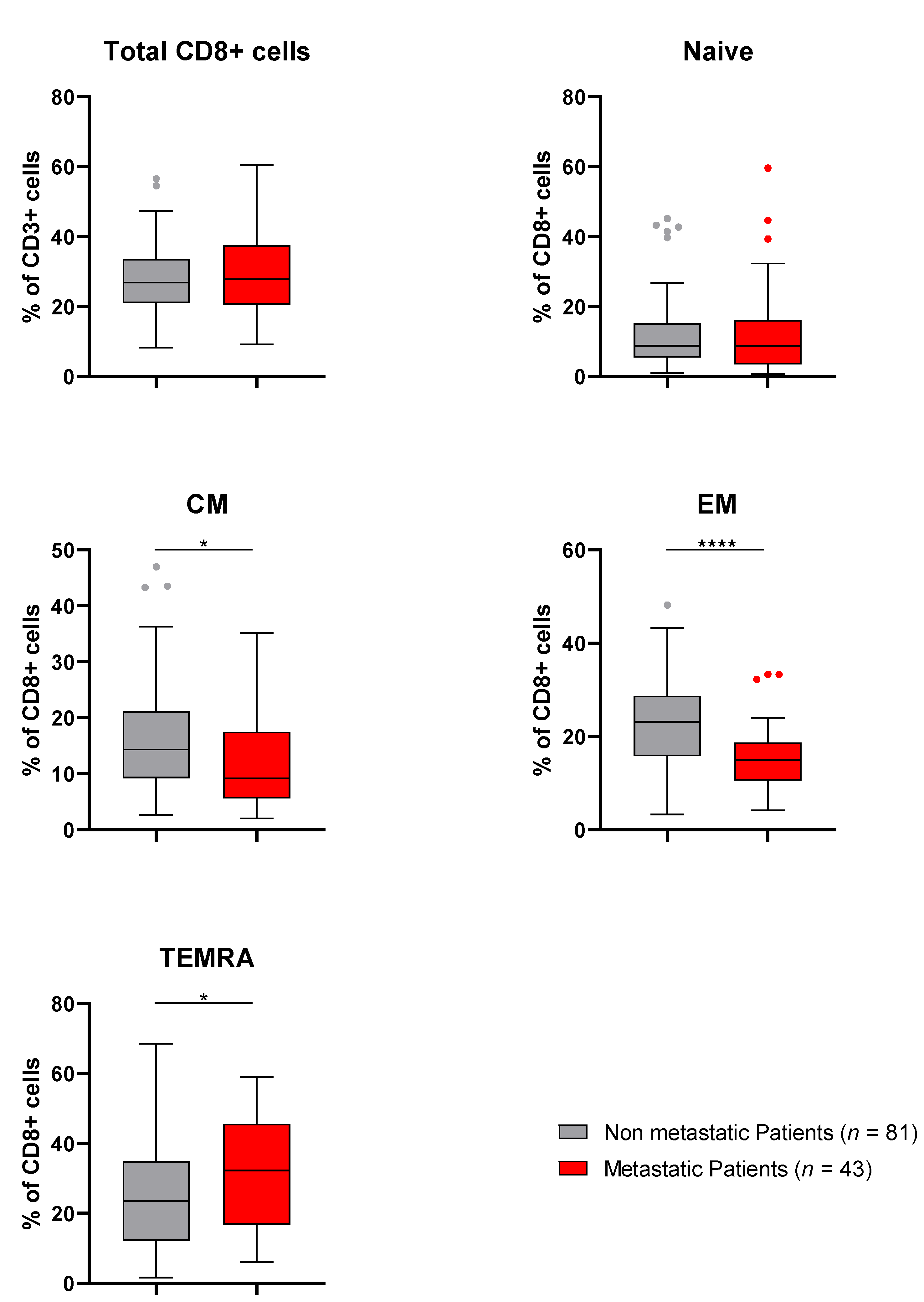
3.2. Comparison of CD8+ T-Lymphocyte Subsets between N-MD PCa and MD PCa Patients

To investigate any possible alterations in the densities of the various subsets of CD8+ T lymphocytes including naïve, CM, EM and TEMRA subsets, based on the levels of malignancy, we compared percentages of these CD8+ T subsets (among total CD8+ T lymphocytes) in our cohort of PCa patients with N-MD vs. MD. The results showed statistically significant lower levels of TEMRA cells in patients at the early stages of PCa (i.e., N-MD) vs. patients with MD suggesting increased percentages of terminally differentiated cells at higher tumor loads of PCa (Figure 1). However, as also shown in Figure 1, when analyzing the CM and EM CD8+ subsets, these were found to be significantly higher in patients with N-MD vs. patients with MD, possibly indicating that CM and EM subsets in N-MD patients have more delayed kinetics towards a terminally differentiated phenotype as compared to their counterparts in patients with MD. Total CD8+ population and naïve CD8+ cells were comparable between the two groups (Figure 1).

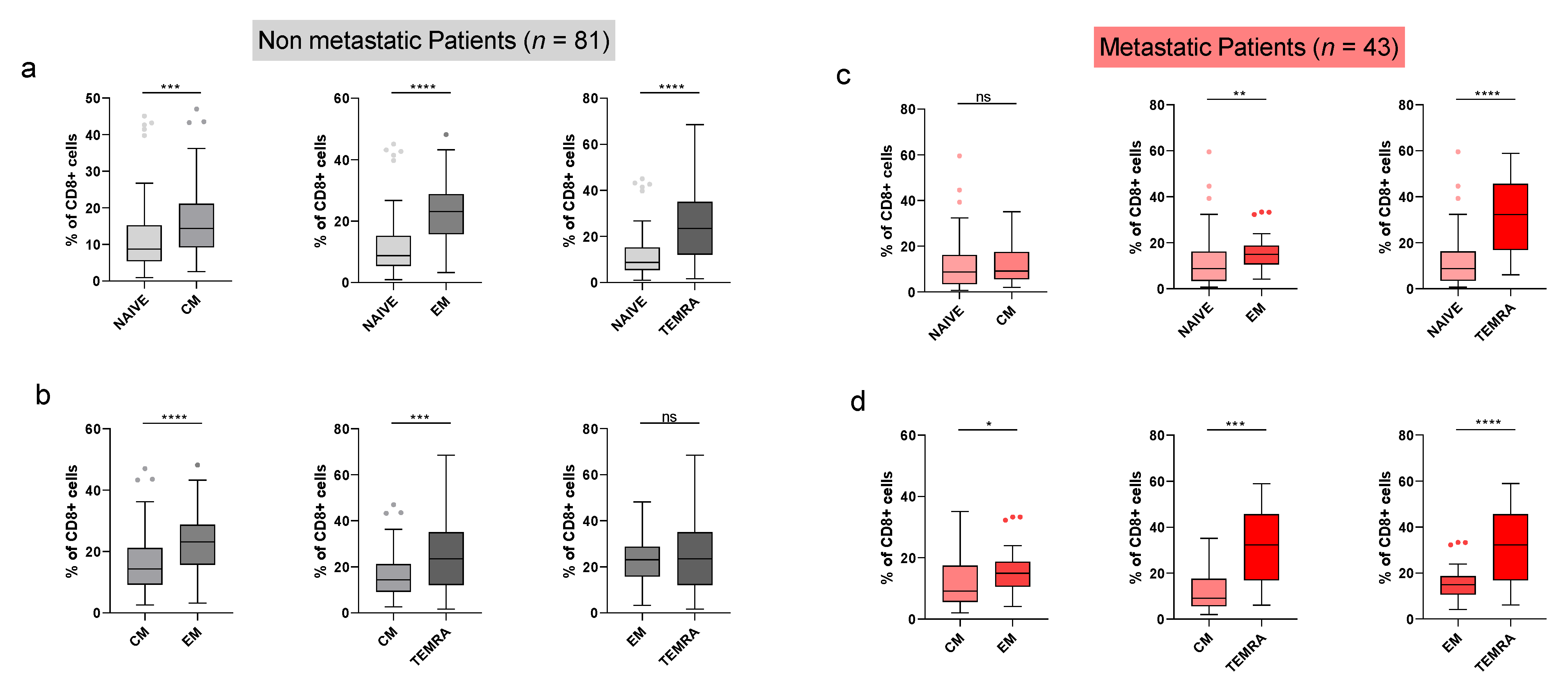
3.3. Intragroup Comparisons of CD8+ T-Lymphocyte Subsets

The densities of these CD8+ T subsets were further separately analyzed in the group of PCa patients with N-MD and in the group of patients with MD. As shown in Figure 2a, in the N-MD group naïve cells were at lower densities than any other subset, followed by CM cells, which were at significantly lower densities compared to EM and TEMRA cells (Figure 2b). TEMRA cells were at the highest densities compared to the naïve and CM subsets (Figure 2a,b). A similar subset density profile was also detected in the MD group (Figure 2c,d) with the exception of significantly higher percentages of TEMRA vs. EM cells (Figure 2d; right panel), which was not the case in the N-MD group (both subsets were determined with densities that did not statistically differ; Figure 2b; left panel).

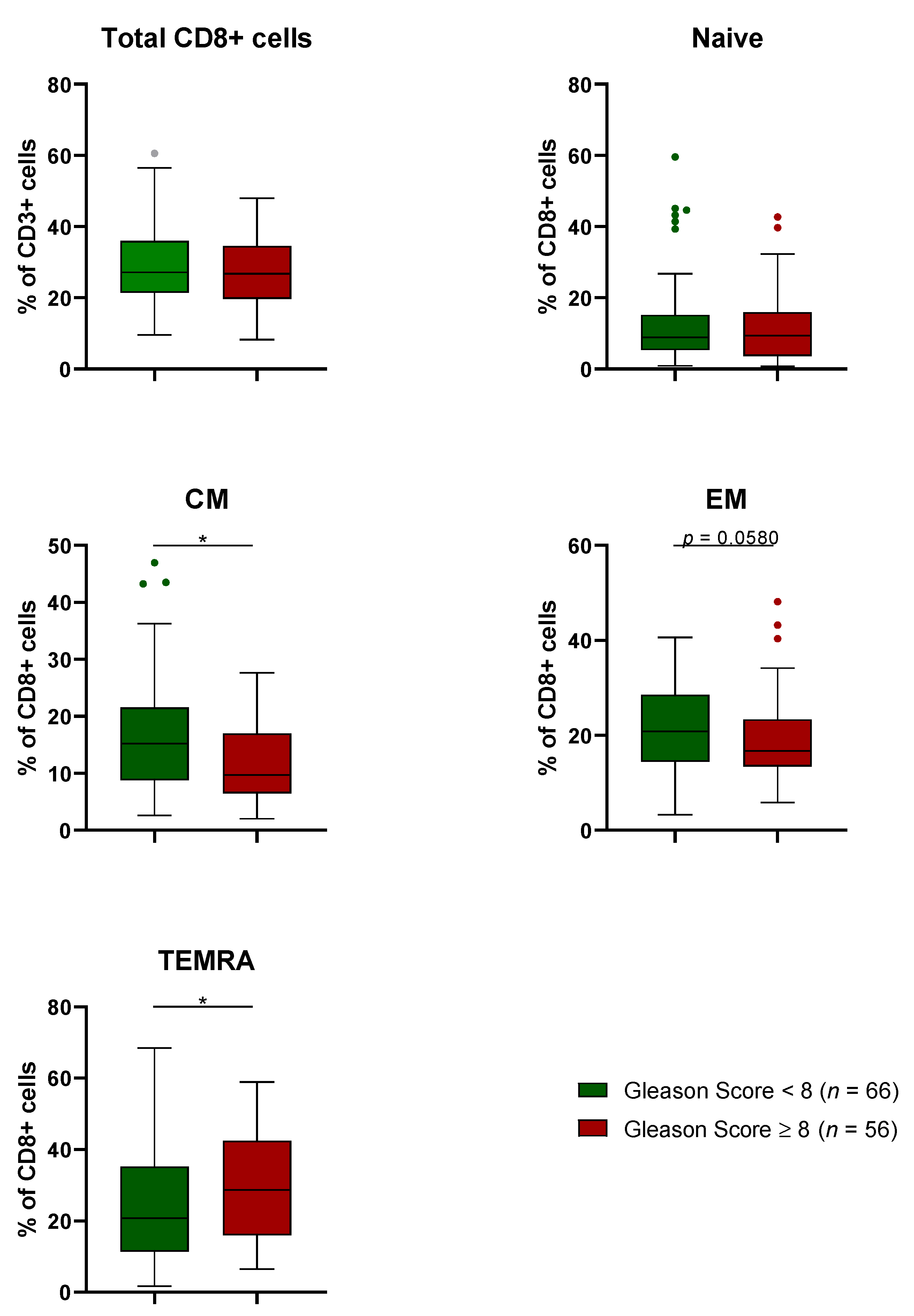
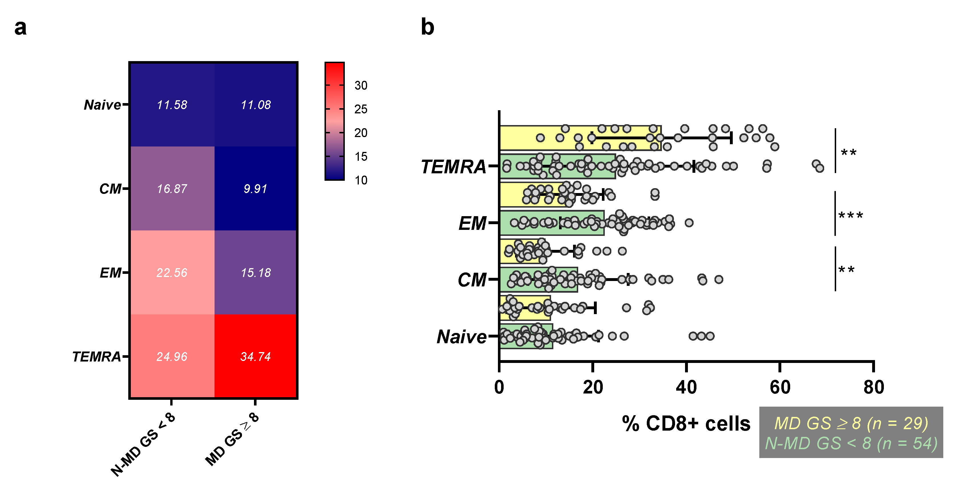
3.4. Comparison of CD8+ T-Lymphocyte Subsets based on Gleason Score (GS)

By stratifying our patients based on PCa aggressiveness (i.e., GS < 8 vs. ≥ 8), we found increased frequencies of (i) TEMRA cells in GS ≥ 8 (Figure 3; lower panel) and (ii) CM and EM cells in GS < 8 (Figure 3; middle panel), whereas no differences could be detected for naïve and total CD8+ cells between the two groups of patients (Figure 3; upper panel). Given that 81.8% of our PCa patients with GS ≤ 8 also had N-MD (54 of 66 patients), these data suggest that increased TEMRA cells are detected among patients with MD and high GS, whereas CM and EM cells are at higher frequencies in the group of patients with N-MD and lower GS. Figure 4a shows a heat map with CD8+ subset densities for the groups of PCa patients with combined N-MD and GS < 8 (n = 54) vs. patients with MD and GS ≥ 8 (n = 29). There is a statistical significance for the increased frequencies of TEMRA cells in the MD and GS ≥ 8 group vs. the group with N-MD and GS < 8 (Figure 4b). Moreover, the lower densities of CM and EM subsets in the MD and GS ≥ 8 group showed a clear significant difference when compared to those determined in the N-MD and GS < 8 group (Figure 4b).

4. Discussion

Samples taken from tumor tissue represent the main source for detecting biomarkers for cancer prognosis and prediction of therapies. However, tumor biopsies are invasive and sometimes impossible to obtain and these cannot be repeatedly collected for monitoring disease progression. On the other hand, peripheral blood collection is a non-invasive method and easy to repeatedly perform during therapies without any discomfort. For that reason, today there is an increasing tendency toward the development of peripheral blood-derived biomarkers for disease monitoring. This kind of monitoring made it possible to study dynamic alterations in the various circulating immune lymphocyte subsets at different disease stages, which could act as surrogates for the prognosis and prediction of ongoing therapies.
Upon MHC-class I—restricted recognition of their tumor antigens, CD8+ T lymphocytes, a major type of cytotoxic cells, acquire various functional activities including cytotoxicity and the production of cytokines to kill their tumor targets. A few of these tumor-specific CD8+ T lymphocytes are preserved for long periods of time as memory cells. Such effector memory CD8+ T lymphocytes either migrate to lymphoid organs (CD8+ CM) or preferentially migrate home to non-lymphoid tissues (CD8+ EM) where, upon re-stimulation by the same tumor antigen, rigorously proliferate and gain enhanced antitumor functions [6,20]. Upon repeated encounters and stimulation by their cognate antigens, some of these subsets become terminally differentiated and re-express naïve cell markers (CD8+ TEMRA) [21].
In the present study, we compared the frequencies of CD8+ T-lymphocyte subsets in the peripheral blood of PCa patients with metastatic vs. non-metastatic disease, in an effort to determine any possible associations between significant alterations in these subsets and the levels of disease malignancy and tumor load. Given the impaired T-lymphocyte immunity observed in PCa patients with advanced disease [22,23,24], the determination of such changes in CD8+ T subset frequencies could function as a surrogate for disease prognosis. In parallel, we performed similar CD8+ T subset analyses in PCa patients with aggressive vs. less aggressive disease in order to validate their prognostic value in PCa, irrespective of any previous treatments. Our studies made the following observations. At first, there was a significant difference in the percentages of CD8+ CM and EM subsets between PCa patients with MD and N-MD (lower vs. higher, respectively). Given that CD8+ EM confers protective immunity by migrating to the inflamed tissues and exerting local cytotoxic functions, while CD8+ CM readily responds to antigenic stimulation in the secondary lymphoid organs and differentiate into effector cells [6], it is conceivable that the higher frequencies of both subsets in patients with non-metastatic disease reflect a functionally active immune system representing a biomarker of favorable prognosis. We also found higher percentages of CD8+ TEMRA cells in the patient cohort with metastatic disease, suggesting that these may have a restricted capacity for performing antitumor cytotoxic activity. In line with this, in a recent report, it was found that in patients with advanced esophageal squamous cell carcinoma receiving radiotherapy combined with immunotherapy, PD-1+ CD8+ T lymphocytes exhibited higher functional and activated characteristics and had enhanced memory properties (including both EM and CM subsets) and less TEMRA cells than PD-1- CD8+ T lymphocytes [25]. Our study also revealed similar CD8+ T subset profiles for our PCa patient cohorts stratified by tumor aggressiveness. Thus, patients with GS < 8 exhibited higher frequencies of EM and CM cells and lower frequencies of TEMRA cells compared with patients having GS ≥ 8. Intratumoral analyses in localized PCa linked increased immunosuppression and epigenetic modifications leading to compromised immunity with high GS [26,27]. Thus, peripheral biomarkers comprising high frequencies of CM and EM cells, as well as low ones of TEMRA cells, seem to correlate with lower tumor load (non-metastatic disease) and lower tumor aggressiveness (lower GS levels), whereas the opposite is true for the inverse subset frequencies (i.e., links to metastatic disease and higher tumor aggressiveness by low CM, EM and high TEMRA frequencies). Importantly, these observations were confirmed when we analyzed these subset frequencies in our PCa patients with combined disease status and GS. We could show that patients with a more favorable prognosis based on non-metastatic disease combined with GS < 8 also had higher densities of CM and EM cells, as well as lower frequencies of TEMRA cells. In contrast, patients with a poor prognosis having metastatic disease and GS ≥ 8 exhibited low densities of CM and EM cells and high densities of TEMRA cells.
Undoubtedly, it should be taken into consideration that the different treatment approaches in PCa may affect the rates of immune subpopulations; however, up-to-date studies have mainly focused on the tumor microenvironment. For example, stereotactic body radiation therapy (SBRT) has been found to alter the immune environment within PCa tumors, causing increases in the relative abundance of myeloid cells and decreases in CD8+ T cells [28,29]. Nonetheless, there has not been extensive research on the effects on systemic circulation, although it is known that radiotherapy promotes immunogenic cell death by remodeling the tumor microenvironment and induces immune responses by enhancing antigen cross-presentation and CD8+ T cell response [30,31]. Activated T cells have been found to be increased after low-dose-rate brachytherapy in the peripheral blood, whereas memory CD8+ T cells are decreased after treatment [32]. The numbers of immunosuppressive CD14(+) HLA-DR(low) cells were found to be elevated in PCa patients, with their levels being normalized post-radical prostatectomy, thus indicating an association between such immune responses and reduced tumor burden [33].

5. Conclusions

Herein, we demonstrate variability in the circulating levels of CD8+ CM, EM and TEMRA T lymphocytes among PCa patients with metastatic vs. non-metastatic disease. We noted a significant increase in the percentage of CD8+ CM, EM T lymphocytes with a concomitant decrease in TEMRA cells in patients with low tumor load (N-MD) as compared to patients with MD having a higher tumor burden. This pilot study adds to the limited literature regarding the effects of high tumor burden and high GS on circulating CD8+ T subsets, particularly CM, EM and TEMRA, and ascribes them a role as surrogates for disease prognosis. Our data for similar variabilities in these subsets, when comparing PCa patients with high GS vs. low GS and in patient groups stratified by combined N-MD and low GS vs. MD and high GS, add further evidence to their role as surrogates for disease progression and suggest that their serial monitoring may be informative for the prognosis and prediction of ongoing therapies. These findings require further validation in a larger cohort of patients.

Author Contributions

Conceptualization, C.N.B. and S.P.F.; methodology, S.S., M.G. and S.P.F.; validation, C.N.B., A.D.G. and S.P.F.; formal analysis, C.N.B., S.S., M.G., A.D.G. and S.P.F.; investigation, S.S., M.G. and S.P.F.; resources, C.N.B. and S.S.; data curation, C.N.B., S.S., M.G., A.D.G. and S.P.F.; writing—original draft preparation, C.N.B. and S.P.F.; writing—review and editing, C.N.B., S.S., M.G., A.D.G. and S.P.F.; visualization, C.N.B. and S.P.F.; supervision, C.N.B. and S.P.F.; project administration, C.N.B. and S.P.F.; funding acquisition, C.N.B. All authors have read and agreed to the published version of the manuscript.

Funding

This research has been co-financed by the European Regional Development Fund of the European Union and Greek national funds through the Operational Program Competitiveness, Entrepreneurship and Innovation, under the call RESEARCH—CREATE—INNOVATE (project code: T1EDK-01404).

Institutional Review Board Statement

The study was conducted in accordance with the Declaration of Helsinki, and approved by the Hospital IRB (IRB-ID6777/14-06-2017) and the Ethical Committee of the University of Athens (IRB-ID1516015872/03-02-2016).

Informed Consent Statement

Informed consent was obtained from all subjects involved in the study.

Data Availability Statement

The data presented in this study are available upon reasonable request.

Conflicts of Interest

The authors declare no conflict of interest.

References

  1. Hanahan, D.; Weinberg, R.A. Hallmarks of cancer: The next generation. Cell 2011, 144, 646–674. [Google Scholar] [CrossRef] [PubMed] [Green Version]
  2. Schreiber, R.D.; Old, L.J.; Smyth, M.J. Cancer immunoediting: Integrating immunity’s roles in cancer suppression and promotion. Science 2011, 331, 1565–1570. [Google Scholar] [CrossRef] [Green Version]
  3. Dunn, G.P.; Old, L.J.; Schreiber, R.D. The immunobiology of cancer immunosurveillance and immunoediting. Immunity 2004, 21, 137–148. [Google Scholar] [CrossRef] [PubMed] [Green Version]
  4. Desai, R.; Coxon, A.T.; Dunn, G.P. Therapeutic applications of the cancer immunoediting hypothesis. Semin. Cancer Biol. 2022, 78, 63–77. [Google Scholar] [CrossRef] [PubMed]
  5. Baxevanis, C.N.; Voutsas, I.F.; Tsitsilonis, O.E.; Gritzapis, A.D.; Sotiriadou, R.; Papamichail, M. Tumor-specific CD4+ T lymphocytes from cancer patients are required for optimal induction of cytotoxic T cells against the autologous tumor. J. Immunol. 2000, 164, 3902–3912. [Google Scholar] [CrossRef] [Green Version]
  6. Sallusto, F.; Geginat, J.; Lanzavecchia, A. Central memory and effector memory T cell subsets: Function, generation, and maintenance. Annu. Rev. Immunol. 2004, 22, 745–763. [Google Scholar] [CrossRef]
  7. Saule, P.; Trauet, J.; Dutriez, V.; Lekeux, V.; Dessaint, J.P.; Labalette, M. Accumulation of memory T cells from childhood to old age: Central and effector memory cells in CD4(+) versus effector memory and terminally differentiated memory cells in CD8(+) compartment. Mech. Ageing Dev. 2006, 127, 274–281. [Google Scholar] [CrossRef]
  8. Buggert, M.; Price, D.A.; Mackay, L.K.; Betts, M.R. Human circulating and tissue-resident memory CD8(+) T cells. Nat. Immunol. 2023, 24, 1076–1086. [Google Scholar] [CrossRef]
  9. Sun, L.; Su, Y.; Jiao, A.; Wang, X.; Zhang, B. T cells in health and disease. Signal Transduct. Target. Ther. 2023, 8, 235. [Google Scholar] [CrossRef]
  10. Teramatsu, K.; Oono, T.; Oyama, K.; Fujimori, N.; Murakami, M.; Yasumori, S.; Ohno, A.; Matsumoto, K.; Takeno, A.; Nakata, K.; et al. Circulating CD8(+)CD122(+) T cells as a prognostic indicator of pancreatic cancer. BMC Cancer 2022, 22, 1134. [Google Scholar] [CrossRef]
  11. Larsson, A.M.; Nordström, O.; Johansson, A.; Rydén, L.; Leandersson, K.; Bergenfelz, C. Peripheral Blood Mononuclear Cell Populations Correlate with Outcome in Patients with Metastatic Breast Cancer. Cells 2022, 11, 1639. [Google Scholar] [CrossRef]
  12. Zhang, G.; Liu, A.; Yang, Y.; Xia, Y.; Li, W.; Liu, Y.; Zhang, J.; Cui, Q.; Wang, D.; Liu, X.; et al. Clinical predictive value of naïve and memory T cells in advanced NSCLC. Front. Immunol. 2022, 13, 996348. [Google Scholar] [CrossRef]
  13. Yuan, C.; Huang, J.; Li, H.; Zhai, R.; Zhai, J.; Fang, X.; Wu, Y. Association of clinical outcomes and the predictive value of T lymphocyte subsets within colorectal cancer patients. Front. Surg. 2023, 10, 1102545. [Google Scholar] [CrossRef] [PubMed]
  14. Zhang, W.; Shi, L.; Zhao, Z.; Du, P.; Ye, X.; Li, D.; Cai, Z.; Han, J.; Cai, J. Disruption of CTLA-4 expression on peripheral blood CD8 + T cell enhances anti-tumor efficacy in bladder cancer. Cancer Chemother. Pharmacol. 2019, 83, 911–920. [Google Scholar] [CrossRef]
  15. Yang, Y.; Attwood, K.; Bshara, W.; Mohler, J.L.; Guru, K.; Xu, B.; Kalinski, P.; Chatta, G. High intratumoral CD8(+) T-cell infiltration is associated with improved survival in prostate cancer patients undergoing radical prostatectomy. Prostate 2021, 81, 20–28. [Google Scholar] [CrossRef]
  16. McAllister, M.; Constâncio, V.; Patek, S.; Gan, H.W.G.; Bailey, P.; Wheadon, H.; Underwood, M.; Leung, H.; Edwards, J. Inflammatory infiltration is associated with AR expression and poor prognosis in hormone naïve prostate cancer. Prostate 2020, 80, 1353–1364. [Google Scholar] [CrossRef]
  17. McArdle, P.A.; Canna, K.; McMillan, D.C.; McNicol, A.M.; Campbell, R.; Underwood, M.A. The relationship between T-lymphocyte subset infiltration and survival in patients with prostate cancer. Br. J. Cancer 2004, 91, 541–543. [Google Scholar] [CrossRef]
  18. Andersen, L.B.; Nørgaard, M.; Rasmussen, M.; Fredsøe, J.; Borre, M.; Ulhøi, B.P.; Sørensen, K.D. Immune cell analyses of the tumor microenvironment in prostate cancer highlight infiltrating regulatory T cells and macrophages as adverse prognostic factors. J. Pathol. 2021, 255, 155–165. [Google Scholar] [CrossRef] [PubMed]
  19. Mao, F.; Yang, C.; Luo, W.; Wang, Y.; Xie, J.; Wang, H. Peripheral blood lymphocyte subsets are associated with the clinical outcomes of prostate cancer patients. Int. Immunopharmacol. 2022, 113, 109287. [Google Scholar] [CrossRef]
  20. Derksen, L.Y.; Tesselaar, K.; Borghans, J.A.M. Memories that last: Dynamics of memory T cells throughout the body. Immunol. Rev. 2023, 316, 38–51. [Google Scholar] [CrossRef] [PubMed]
  21. Golubovskaya, V.; Wu, L. Different Subsets of T Cells, Memory, Effector Functions, and CAR-T Immunotherapy. Cancers 2016, 8, 36. [Google Scholar] [CrossRef] [Green Version]
  22. Rodems, T.S.; Heninger, E.; Stahlfeld, C.N.; Gilsdorf, C.S.; Carlson, K.N.; Kircher, M.R.; Singh, A.; Krueger, T.E.G.; Beebe, D.J.; Jarrard, D.F.; et al. Reversible epigenetic alterations regulate class I HLA loss in prostate cancer. Commun. Biol. 2022, 5, 897. [Google Scholar] [CrossRef] [PubMed]
  23. Bansal, D.; Reimers, M.A.; Knoche, E.M.; Pachynski, R.K. Immunotherapy and Immunotherapy Combinations in Metastatic Castration-Resistant Prostate Cancer. Cancers 2021, 13, 334. [Google Scholar] [CrossRef] [PubMed]
  24. Sun, B.L. Immunotherapy in treatment of metastatic prostate cancer: An approach to circumvent immunosuppressive tumor microenvironment. Prostate 2021, 81, 1125–1134. [Google Scholar] [CrossRef] [PubMed]
  25. Wei, H.; Li, Y.; Guo, Z.; Ma, X.; Li, Y.; Wei, X.; Han, D.; Zhang, T.; Chen, X.; Yan, C.; et al. Comparison of dynamic changes in the peripheral CD8(+) T cells function and differentiation in ESCC patients treated with radiotherapy combined with anti-PD-1 antibody or concurrent chemoradiotherapy. Front. Immunol. 2022, 13, 1060695. [Google Scholar] [CrossRef]
  26. Adorno Febles, V.R.; Hao, Y.; Ahsan, A.; Wu, J.; Qian, Y.; Zhong, H.; Loeb, S.; Makarov, D.V.; Lepor, H.; Wysock, J.; et al. Single-cell analysis of localized prostate cancer patients links high Gleason score with an immunosuppressive profile. Prostate 2023, 83, 840–849. [Google Scholar] [CrossRef]
  27. Unger, K.; Hess, J.; Link, V.; Buchner, A.; Eze, C.; Li, M.; Stief, C.; Kirchner, T.; Klauschen, F.; Zitzelsberger, H.; et al. DNA-methylation and genomic copy number in primary tumors and corresponding lymph node metastases in prostate cancer from patients with low and high Gleason score. Clin. Transl. Radiat. Oncol. 2023, 39, 100586. [Google Scholar] [CrossRef]
  28. Kane, N.; Romero, T.; Diaz-Perez, S.; Rettig, M.B.; Steinberg, M.L.; Kishan, A.U.; Schaue, D.; Reiter, R.E.; Knudsen, B.S.; Nickols, N.G. Significant changes in macrophage and CD8 T cell densities in primary prostate tumors 2 weeks after SBRT. Prostate Cancer Prostatic Dis. 2023, 26, 207–209. [Google Scholar] [CrossRef]
  29. Nickols, N.G.; Ganapathy, E.; Nguyen, C.; Kane, N.; Lin, L.; Diaz-Perez, S.; Nazarian, R.; Mathis, C.; Felix, C.; Basehart, V.; et al. The intraprostatic immune environment after stereotactic body radiotherapy is dominated by myeloid cells. Prostate Cancer Prostatic Dis. 2021, 24, 135–139. [Google Scholar] [CrossRef]
  30. Ollivier, L.; Labbé, M.; Fradin, D.; Potiron, V.; Supiot, S. Interaction Between Modern Radiotherapy and Immunotherapy for Metastatic Prostate Cancer. Front. Oncol. 2021, 11, 744679. [Google Scholar] [CrossRef]
  31. Zhu, S.; Wang, Y.; Tang, J.; Cao, M. Radiotherapy induced immunogenic cell death by remodeling tumor immune microenvironment. Front. Immunol. 2022, 13, 1074477. [Google Scholar] [CrossRef] [PubMed]
  32. Kubo, M.; Satoh, T.; Ishiyama, H.; Tabata, K.I.; Tsumura, H.; Komori, S.; Iwamura, M.; Baba, S.; Hayakawa, K.; Kawamura, T.; et al. Enhanced activated T cell subsets in prostate cancer patients receiving iodine-125 low-dose-rate prostate brachytherapy. Oncol. Rep. 2018, 39, 417–424. [Google Scholar] [CrossRef] [PubMed] [Green Version]
  33. Brusa, D.; Simone, M.; Gontero, P.; Spadi, R.; Racca, P.; Micari, J.; Degiuli, M.; Carletto, S.; Tizzani, A.; Matera, L. Circulating immunosuppressive cells of prostate cancer patients before and after radical prostatectomy: Profile comparison. Int. J. Urol. Off. J. Jpn. Urol. Assoc. 2013, 20, 971–978. [Google Scholar] [CrossRef] [PubMed]
Figure 1. Differences in the composition of lymphocyte subsets between prostate cancer (PCa) patients with metastatic and non-metastatic disease. The Tukey whisker plots depict the percentages of CD8+ T lymphocytes within the CD3+ T subset, and accordingly, naïve, central memory (CM), effector memory (EM) and terminally differentiated effector memory (TEMRA) CD8+ T lymphocytes. The grey and red dots represent outliers (values). Statistically significant differences in the percentages of cell populations between metastatic and non-metastatic individuals were identified by performing non-parametric Mann-Whitney (unpaired) tests. p-values below 0.05 signify a statistical significance. * p < 0.05; **** p < 0.0001.
Onco 03 00012 g001
Figure 2. Variations in naïve and memory CD8+ T-lymphocyte subsets between metastatic and non-metastatic prostate cancer (PCa) patients. The Tukey whisker plots depict differences between the percentages of naïve and memory cells and between distinct memory cell populations within the CD8+ T subset in individuals with (a,b) non-metastatic and (c,d) metastatic disease. CM, central memory; EM, effector memory; TEMRA, terminally differentiated effector memory. The grey and red dots represent outliers (values). Statistically significant differences were identified by performing non-parametric Mann-Whitney (unpaired) tests. p-values below 0.05 signify a statistical significance. *, p < 0.05; **, p < 0.01; ***, p < 0.001; ****, p < 0.0001; ns, p > 0.05.
Onco 03 00012 g002
Figure 3. Differences in the composition of lymphocyte subsets between prostate cancer (PCa) patients based on their Gleason score. The Tukey whisker plots depict the percentages of CD8+ T lymphocytes within the CD3+ T subset, and accordingly, naïve, central memory (CM), effector memory (EM) and terminally differentiated effector memory (TEMRA) CD8+ T lymphocytes. The green and red dots represent outliers (values). Statistically significant differences in the percentages of cell populations between individuals with a high (≥8) and low (<8) Gleason score were identified by performing non-parametric Mann-Whitney (unpaired) tests. p-values below 0.05 signify a statistical significance. * p < 0.05.
Onco 03 00012 g003
Figure 4. Variations in naïve and memory CD8+ cell subsets between prostate cancer (PCa) patients with non-metastatic disease and a low Gleason score (<8) and patients with metastatic disease and a high Gleason score (≥8) at the time of enrolment. (a) The heatmap presents mean percentage values of naïve, central memory (CM), effector memory (EM) and terminally differentiated effector memory (TEMRA) CD8+ T lymphocytes between PCa patients with non-metastatic disease (N-MD) and a Gleason score (GS) < 8 and patients with metastatic disease (MD) and a GS ≥ 8. Color change from blue to red indicates an increase in cell abundance. (b) The scatter plots represent differences in the percentages of naïve and memory cell subsets within the CD8+ T subset, between PCa patients with N-MD and a GS < 8 and patients with MD and a GS ≥ 8. The charts show the mean values with error bars designating the standard deviations. The grey dots represent values. Statistically significant differences were identified by performing non-parametric Mann-Whitney (unpaired) tests. p-values below 0.05 signify a statistical significance. **, p < 0.01; ***, p < 0.001.
Onco 03 00012 g004
Table 1. Patients’ clinicopathological characteristics.
Clinicopathological Characteristics
Non Metastatic Disease (N-MD) at EnrolmentMetastatic Disease (MD) at Enrolment
No. of PCa patients8143
Age (Median)
68.768.6
Gleason Score
<854 (66.7%)12 (27.9%)
≥827 (33.3%)29 (67.4%)
Missing02 (4.7%)
pT stage
T1c16 (19.8%)-
T2a, T2b, T2c42 (51.9%)-
T3a, T3b22 (27.1%)-
Missing1 (1.2%)-
Site of metastases at the time of blood sampling
Bone metastases-19 (44.2%)
Soft tissue metastases-11 (25.6%)
Bone metastases and soft tissue metastases-13 (30.2%)
Type of therapy received at the time of blood sampling
RP32 (39.5%)-
PRTX9 (11.1%)-
BRACHY2 (2.5%)-
ADT-10 (23.3%)
ΝOΝΕ38 (46.9%)33 (76.7%)
RP, Radical Prostatectomy; PRTX, primary radiotherapy; BRACHY, Brachytherapy; ADT, Androgen Deprivation Therapy.
Disclaimer/Publisher’s Note: The statements, opinions and data contained in all publications are solely those of the individual author(s) and contributor(s) and not of MDPI and/or the editor(s). MDPI and/or the editor(s) disclaim responsibility for any injury to people or property resulting from any ideas, methods, instructions or products referred to in the content.

