Review

Structure-Function-Environment Relationship of the Isomers Zeaxanthin and Lutein

by
Barbara Demmig-Adams
*,
Stephanie K. Polutchko
and
William W. Adams III
Department of Ecology and Evolutionary Biology, University of Colorado, Boulder, CO 80309-0334, USA
*
Author to whom correspondence should be addressed.
Photochem 2022, 2(2), 308-325; https://doi.org/10.3390/photochem2020022
Submission received: 24 March 2022 / Revised: 14 April 2022 / Accepted: 15 April 2022 / Published: 18 April 2022
(This article belongs to the Special Issue Plant Photochemistry, Reactive Oxygen Species and Photoprotection)

Abstract

:
A synthesis is provided of the roles of the carotenoids zeaxanthin and/or lutein in opposing (i) photodamage in plants, (ii) photodamage to the human eye as well as cognitive dysfunction and a host of human diseases and disorders, and (iii) damage to extremophile microorganisms in the most inhospitable environments on earth. Selected examples are used to examine microenvironments and basic biological structures with which these xanthophylls associate as well as the effect of the organisms’ external environment. An overview is presented of the multiple principal mechanisms through which these xanthophylls can directly or indirectly impact organisms’ internal redox (oxidant/antioxidant) balance that provides input into the orchestration of growth, development, and defense in prokaryotic microorganisms, plants, and humans. Gaps in the research are identified, specifically with respect to the need for further in vivo assessment of the mechanisms.

1. Introduction

Carotenoids play a vital role in an array of physiological functions across all organisms. There are over 1100 naturally occurring carotenoids thus far described [1]. Carotenoids are widely studied, and a wealth of information is available on their natural functions (e.g., [2]) as well as their roles in human nutrition and health (e.g., [3]) but many questions remain. After touching on some common features of carotenoids below, we provide a synthesis of the literature on organisms that utilizes the closely related carotenoids, zeaxanthin, and lutein, in key roles including support for the ability of (i) plants to survive and thrive in challenging environments, (ii) humans to optimize cognitive function as well as to stave off a host of diseases and disorders, and (iii) some (zeaxanthin-accumulating) microorganisms to grow in the most inhospitable environments on earth. Information on zeaxanthin and lutein is compared across taxa with respect to multiple functions and microenvironments as well the role of the organism’s external environment. For further detail, we refer to authoritative reviews in each area.

Carotenoids in a Nutshell

Carotenoids are synthesized de novo by organisms from all three major categories (domains) of life, i.e., eukaryotes (such as plants, algae, and some fungi), bacteria, and archaea [1]. Animals, however, must consume carotenoids with their diet, except for the special case of some aphids that acquire carotenoid biosynthesis genes via transfer from fungi [4]. Carotenoids are classified as either carotenes (pure hydrocarbons without oxygen, e.g., β-carotene) or xanthophylls (that contain oxygen, e.g., lutein and zeaxanthin) and absorb the visible light of blue or blue-green wavelengths (resulting in their yellow, orange, or red color). The interaction of carotenoids, or their derivatives, with visible light gives rise to key functions in light absorption for both humans and plants. In human vision, the light-absorbing component of the protein rhodopsin is derived from carotenoids with provitamin A activity [5]. In photosynthetic organisms, carotenoids have widespread functions that support photosynthesis, with a particularly prominent ecological role of green-light absorbance by fucoxanthin, siphonoxanthin, and peridinin in some algae [6,7,8,9]. In particular, zeaxanthin and lutein have vital functions in the protection against damage by intense light in both plants (e.g., [10]) and the human eye [11].
Carotenoids also play key roles in light-independent processes, e.g., as gene regulators of human immune function. Carotenoid-derived vitamin A has a well-documented immunoregulatory role [5] and a similar role is emerging for xanthophylls. Xanthophylls may be especially important in opposing non-resolving inflammation that can trigger a plethora of associated inflammatory diseases, disorders, and dysfunctions [12,13]. Additionally, lutein and zeaxanthin are emerging as candidates for protecting cognitive function across the human lifespan, including attention, memory, learning, and executive functions [14,15].
How do carotenoids support so many functions? Pioneering work concentrated on two mechanisms. The first was the protective role in the de-excitation of singlet oxygen, a reactive oxygen species (ROS) formed under intense light in both photosynthesis [16,17] and the human eye [11]. Second, xanthophylls can act as molecular rivets in biological membranes due to their specific length and chemical structure [18]. A large body of mechanistic work ranging from carotenoids dissolved in various solvents to isolated pigment-binding complexes, isolated organelles, and mutants/transgenic organisms with altered carotenoid composition, has revealed multiple potential functions for carotenoids. A given carotenoid can apparently have different multiple functions depending on its specific microenvironment because “energetics and dynamics of carotenoid excited states” are controlled not only by factors such as conjugated-chain length and functional groups but also by, “perhaps most importantly, carotenoid interaction with the local environment” [19]. Furthermore, how much carotenoid is accumulated, and its location, is affected by an organism’s genetic makeup as well as its acclimation to the external environment in which it developed.
The following sections highlight selected examples for zeaxanthin and/or lutein of (i) the contexts in which xanthophylls are found—either within organisms or with respect to external environments/habitats, (ii) basic biological structures with which these xanthophylls associate, and (iii) the multiple ways in which these xanthophylls directly and/or indirectly impact organisms’ internal redox (oxidant/antioxidant) balance that provides input into the orchestration of growth, development, and defense in prokaryotic microorganisms [20], plants [21], and humans [22] alike.

2. Xanthophylls in High-Stress Contexts

Zeaxanthin and lutein are structural isomers with zeaxanthin possessing a slightly longer system of conjugated double bonds (11) than lutein (10; Figure 1).

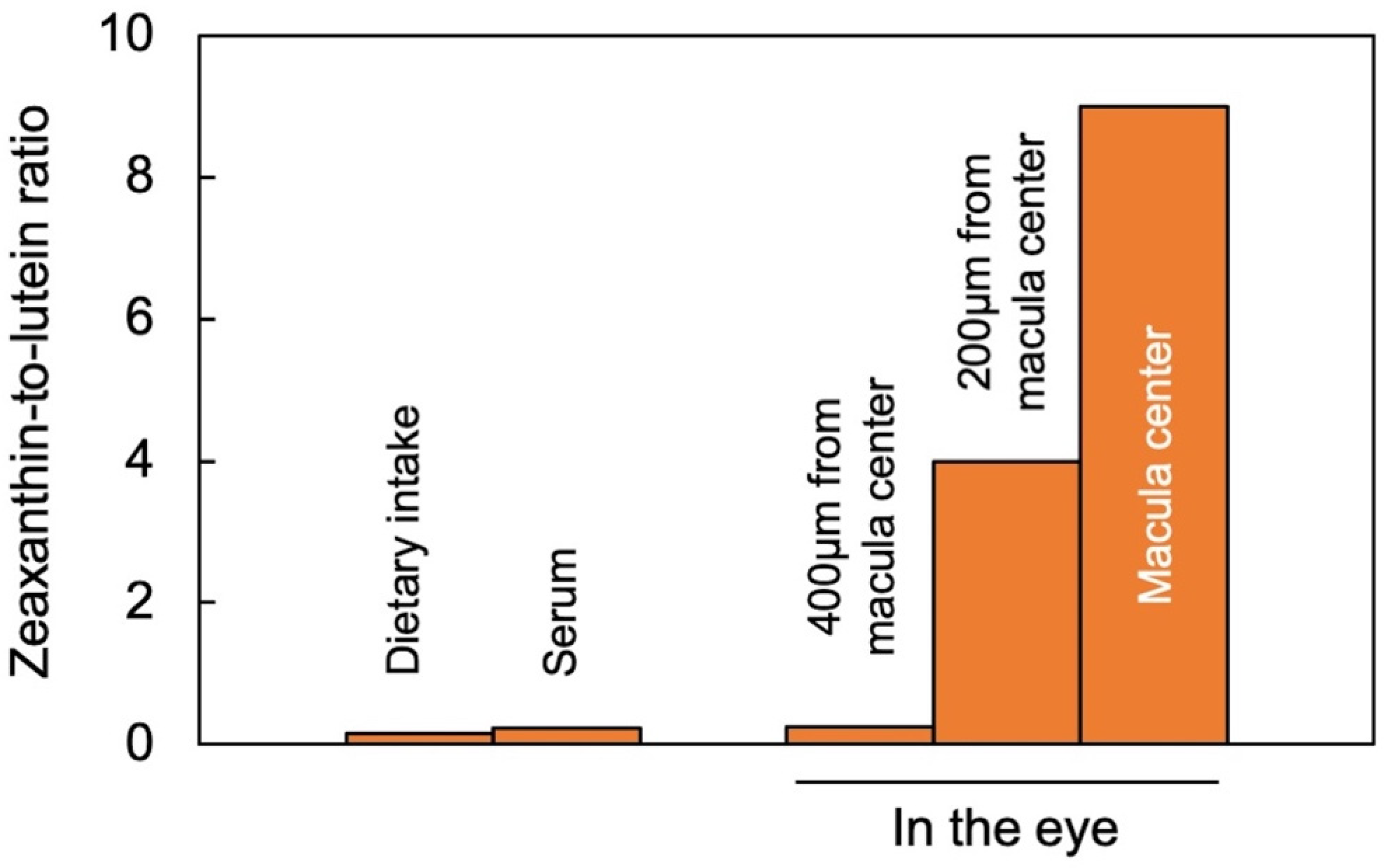
2.1. Zeaxanthin and Lutein in the Human Eye/Retina

Lutein and zeaxanthin are differentially distributed across the human retina (Figure 2). The yellow center of the eye (macula), where the brightest light is received, has the highest overall xanthophyll concentration and the highest ratio of zeaxanthin to lutein [11]. The total xanthophyll concentration is about 1 mM in the macula and declines to less than 10 μΜ in the peripheral regions of the retina [23]. In addition to zeaxanthin and lutein, meso-zeaxanthin (a zeaxanthin stereoisomer) is present in the macula and is apparently produced from dietary lutein but not from dietary zeaxanthin [24].
A recent study using confocal resonance Raman spectroscopy, validated by the biochemical characterization of carotenoid composition, described the variation in the zeaxanthin-to-lutein ratio over short distances using continuous scans of xanthophyll composition across donor retinas [25]. The zeaxanthin-to-lutein ratios were 9:1 or greater in the center of the macula; 4:1 at a short distance (200 µm) from the center; and 1:4 just outside the macula (Figure 2; [25]). This preferential placement of zeaxanthin where the brightest light is received clearly indicates the unique role of zeaxanthin in supporting the vision process in the presence of bright light. Still, it does not allow an assessment of which one(s) of the multiple possible roles of zeaxanthin is/are at work in this location. Original ideas (starting in 1861) about the function of the xanthophyll-rich macula initially centered on potential improvements in visual acuity and contrast sensitivity with reduced glare sensitivity and light scatter (see [11]). However, the subsequent rise of age-related macular degeneration in the human population shifted the focus of attention to photoprotection (see review [11]). Nevertheless, both principal roles are still discussed today, and multiple mechanisms are under consideration (see below).
Figure 2. Zeaxanthin-to-lutein ratios across a gradient from dietary intake to blood serum and different locations in the human eye, after data from [26] for dietary intake and serum, and for the areas within and around the macula from [25]. “Zeaxanthin” here represents the sum of zeaxanthin and meso-zeaxanthin (present in a 1;1 molar ratio in the macula).
Photochem 02 00022 g002

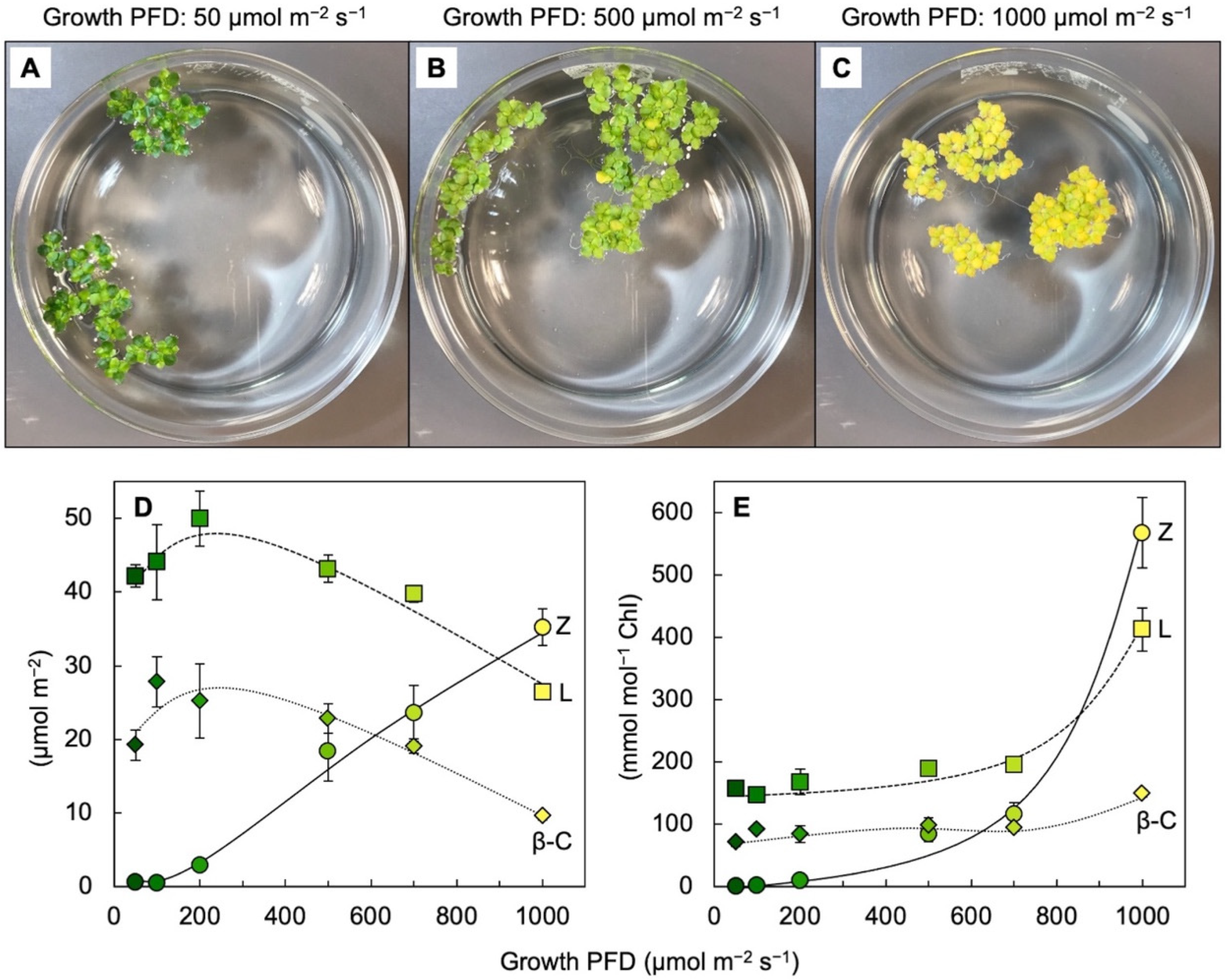
2.2. Zeaxanthin and Lutein in Leaves

Leaves of plants growing in sunny locations under conditions favorable for growth rapidly form and remove zeaxanthin as the fraction of absorbed light not utilized in photochemistry rises and falls over the course of the day (e.g., [10,27]). The characterizations of the latter functional features were enabled by the development of portable instruments to measure chlorophyll fluorescence from leaves under field conditions and in the presence of bright light [28,29]. Zeaxanthin is formed in the presence of excess absorbed light from the di-epoxide violaxanthin via the mono-epoxide antheraxanthin in the xanthophyll cycle [30,31]. Violaxanthin levels exhibit complementary decreases and increases over the course of the day (e.g., [10,27]). However, the levels of other ubiquitous leaf carotenoids (lutein, β-carotene, and neoxanthin) do not typically change over the course of the day in sun-exposed habitats [10]. In slow-growing evergreens (which utilize a low fraction of full sunlight for photochemistry), the ratio of zeaxanthin to lutein can approach unity in full sun at midday, but this ratio is much lower in most plant systems most of the time. Ample zeaxanthin for human nutrition is thus hard to come by when relying on rapidly growing leafy greens, harvested and stored before consumption. For human nutrition, crops that combine rapid growth and high zeaxanthin levels would be desirable.
We recently reported about the unusual ability of aquatic plants (Lemnaceae, or duckweeds) to simultaneously grow very rapidly and accumulate exceptionally high levels of zeaxanthin [32,33,34]. Figure 3 shows that visual appearance and carotenoid levels in Lemna grown under a wide range of photon flux densities (PFDs) in which plant growth remained high, with plant area doubling every other day. Additionally, zeaxanthin levels continued to rise when the absorbed light became increasingly excessive, whereas the levels of chlorophyll, lutein, and β-carotene declined (Figure 3D). The plants grown under the highest light intensity were bright yellow (Figure 3C), still grew very rapidly, and exhibited zeaxanthin-to-lutein ratios as high as ~1.3 (up from ~0.5 under the next lowest growth PFD). Due to the fact that the levels of chlorophyll a + b declined more sharply than those of any of the carotenoids under the highest-growth PFD, carotenoid levels increased relative to chlorophyll, and none more sharply than zeaxanthin (Figure 3E). A considerable portion of this zeaxanthin is presumably dissolved in the phospholipid portion of chloroplast membranes. A role for zeaxanthin, but not lutein, as a membrane-based antioxidant and/or membrane stabilizer was proposed for plants [35,36]. The sharp increase in the zeaxanthin-to-lutein ratio in duckweed at the highest PFD (Figure 3D) is reminiscent of the dynamics across the human eye described above.
The unusual ability of duckweeds to combine pronounced zeaxanthin accumulation with fast growth may be associated with (i) the exposure of the entire leaf cross-section to excess light due to minimal self-shading (in the absence of multi-tiered structures within leaves or plant canopies [32,33,34]) and (ii) the loss of controls that act on growth in land plants [37]. This unusual combination may also apply to other fast-growing edible aquatic plants [32], which could be of considerable interest to human nutrition and illustrates the importance of species choice. Although whole-food sources with superior zeaxanthin content are of interest to human nutrition, it should be noted that supplements (e.g., the AREDS2 formulation with 10 mg lutein and 2 mg zeaxanthin) can also provide benefits, such as the delayed progression to advanced macular degeneration [38]; for additional studies, see Section 4 below.
In addition to lending support in high-light environments, zeaxanthin also plays a role in the heat tolerance of plants. A greater heat tolerance was demonstrated for the Arabidopsis thaliana lines, which were engineered to overexpress β-hydroxylase (the enzyme catalyzing biosynthetic conversion of β-carotene to zeaxanthin). This engineered line exhibited elevated zeaxanthin levels but no enhancement of non-photochemical energy dissipation, which is consistent with the role of additional zeaxanthin in the phospholipid portion of the photosynthetic membrane [39].

2.3. Zeaxanthin and Related Xanthophylls in Extremophiles

Among the over 1100 naturally occurring carotenoids described, only seven are synthesized de novo by organisms from all three domains of life [1]. The few known carotenoids synthesized by representatives of eukaryotes, bacteria, and archaea include zeaxanthin and its biosynthetic precursors (for a detailed review of carotenoid biosynthetic pathways among the taxa of life, see [40]). Zeaxanthin is found not only in light-absorbing/photosynthetic bacteria but also in non-photosynthetic bacteria and archaea. Although the functions of zeaxanthin and related xanthophylls (Figure 4) in these organisms are yet to be elucidated, the environments in which they occur expose the organisms to high levels of stress (visible or ionizing radiation, heat, or salinity). Among eukaryotes, fungi typically do not produce lutein or zeaxanthin but can produce a variety of other carotenoids [41,42] as well as many other pigments (e.g., melanins, flavins, phenazines, quinones, monascins, violacein, and indigo; [43]).
Zeaxanthin and related xanthophylls accumulate in bacteria and archaea that occur in the most extreme environments. Prokaryotic organisms do not synthesize lutein [42] but do produce zeaxanthin. Zeaxanthin-producing microorganisms include photosynthetic cyanobacteria, such as Synechococcus (see [44]) that accumulates zeaxanthin in the high-light environment of surface ocean water, but less so in sub-surface layers where light levels are lower [45]. Cyanobacteria use other xanthophylls, such as 3′-hydroxyechinenone (Figure 4), to protect their phycobilisome chromophores [46] that harvest light in blue- and red-light-depleted zones deeper in the water column.
Zeaxanthin is also accumulated by non-photosynthetic bacteria, some of which are named after this feature (Mesoflavibacter zeaxanthinifaciens, Zeaxanthinibacter enoshimensis, Paracoccus zeaxanthinifaciens; [47,48]). A notable case of an extremophile is the bacterium Deinococcus radiodurans, isolated from a radioactive site in Japan and described as a “gold medalist” for the tolerance of ionizing radiation [49]. Deinococcus radiodurans accumulate deinoxanthin and zeaxanthin [50,51,52,53]. Ionizing radiation produces vast amounts of ROS (e.g., hydroxyl radical and superoxide) via water hydrolysis [54]. A zeaxanthin glycoside ester, thermozeaxanthin, is accumulated in non-photosynthetic microorganisms, including members of the bacterial genera Halococcus, Halobacterium, and Thermus [55,56] as well as the Archean Haloarcula japonica [57], which all exhibit robust resistance to salinity and/or extreme heat. These findings further support the role of xanthophylls, with features such as those shown in Figure 4, in supporting life in extreme environments.

3. Association with Proteins and/or Phospholipid Bilayers

3.1. Association of Lutein and Zeaxanthin with Proteins—Selected Examples across Taxa

Carotenoids bind to a wide variety of proteins, including some with chromophores that intercept light and many that do not. In photosynthetic organisms, most light-harvesting proteins bind carotenoids in addition to their primary light-collecting chromophores (see chapters on carotenoid association with light-collecting complexes in various photosynthetic organisms in [58]). Ongoing work continues to expand the list of carotenoid-binding proteins in photosynthetic organisms that either bind light-harvesting pigments or interact with light-harvesting complexes. Many of these proteins bind lutein and/or zeaxanthin (see, e.g., [59]) or other xanthophylls (see, e.g., [60] for the orange carotenoid-binding protein of cyanobacteria).
Similarly, proteins involved in human vision (such as retinoid transporter proteins that have indirect roles in the vision process) bind zeaxanthin and lutein [38]. Moreover, selective uptake of zeaxanthin and lutein into the macula of the human retina is mediated by two different proteins [61] that bind either zeaxanthin and meso-zeaxanthin (glutathione S-transferase [62]) or lutein (steroidogenic acute regulatory domain protein [63]). Additionally, there are carotenoid-binding proteins not associated with light-collecting processes. These include proteins that transport carotenoids through the bloodstream in humans, such as high-density lipoprotein (for an in-depth review of human proteins that bind carotenoids, especially lutein and zeaxanthin, see [38]).

3.2. Lutein and Zeaxanthin Localization within the Phospholipid Bilayer of Biological Membranes—Selected Examples across Taxa

Carotenoids may have first emerged in archaea as molecules that reinforced biological membranes as “molecular rivets” with just the right length and structure to span the phospholipid bilayer [18,64]. In many other organisms, carotenoids are also localized in membranes. Xanthophylls, in particular, can incorporate directly into phospholipid bilayers in a membrane-spanning orientation with no apparent association with proteins and do so in some microorganisms (see above), plants [34,36], and humans. The high levels of carotenoids in the human brain (71% of which consisted of the xanthophylls lutein, zeaxanthin, and cryptoxanthin [65]) are likely localized largely in the phospholipid bilayer of membranes. Although it seems clear that lutein is a component of the lipid bilayer portion of animal membranes, localization in the lipid bilayer portion of plant membranes has thus far been discussed mainly for zeaxanthin [35,36]. Future research should further address if lutein also plays a role in plant membranes, and if so, why.
In vitro studies demonstrated that lutein and zeaxanthin have different orientations in phospholipid bilayers. Whereas zeaxanthin was exclusively orientated in a perpendicular, membrane-spanning orientation, some of the lutein was oriented in a horizontal position parallel to the phospholipid head groups [66,67]. On the other hand, lutein may have a higher propensity to form tightly stacked aggregates that exhibit a blue shift in xanthophyll absorbance, which may affect the absorption of blue light in the retina [68]. Moreover, biological membranes are clearly heterogeneous along their axes, with some microdomains containing more polyunsaturated fatty acids (PUFAs) and others more saturated fatty acids and cholesterol; xanthophylls are concentrated in the areas enriched in PUFAs [69,70]. More work is needed to distinguish the similarities and differences in zeaxanthin and lutein localization and/or orientation in microdomains and in membranes as well as the functional significance of such differences.

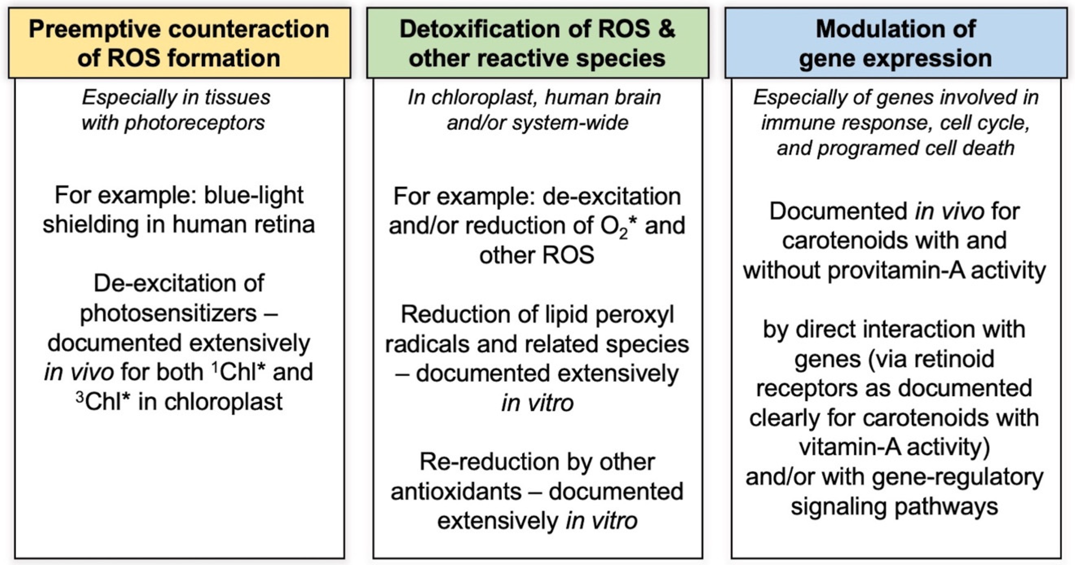
4. Multiple Functional Intersections of Xanthophylls with Reactive Oxygen Species in the Context of Gene Regulation

As touched upon in the introduction, aerobic life forms, from microorganisms to plants and humans, use redox-signaling pathways and redox-modulated gene regulators to orchestrate growth, development, and defense (see, e.g., [22]). Although the modest ROS levels formed in chloroplasts and/or mitochondria provide essential input into these regulatory circuits, elevated ROS levels formed in these organelles and other sites under certain conditions can activate responses, such as programmed cell death, which occur long before further increases in ROS levels cause uncoordinated oxidative damage to cell structures (see, e.g., [71]). Many metabolites that interact with ROS (and that are synthesized endogenously in some organisms or consumed within the diet in others) either directly eliminate ROS or interact with the signaling molecules and/or gene regulators. In humans, signaling pathways and gene regulators were modulated by supplementation with either lutein or mixes of lutein and zeaxanthin. This includes gene regulators such as NF-kB (nuclear factor kappa B) and Nfr2 (nuclear factor erythroid 2–related factor 2), both of which receive input from the cellular redox state and, in turn, contribute to shifting redox homeostasis [72]. Lutein supplementation dampened the activity of NF-kB, which is activated by ROS-stimulated signaling pathways and triggers responses that produce more ROS as part of immune defenses [72]). On the other hand, lutein supplementation activated Nfr2, which triggers the production of endogenous antioxidants [72]. Since most of these studies were conducted using lutein-only supplements, more studies are needed on the effect of zeaxanthin versus lutein.
One example of redox-modulated gene regulators in plants are the transcription factors of the C-repeat binding factor (CBF) family that play a role in coordinating plant response to changes in the external or internal environment (see, e.g., [73]). One of the inputs into this and other redox-signaling networks is the level of excess excitation energy in the chloroplast (that is, in turn, modulated by carotenoid-dependent de-excitation events; see below). The system of conjugated double bonds allows carotenoids to not only absorb visible light, but also to potentially modulate redox-responsible elements of gene-regulatory pathways via direct antioxidant effects. Alternatively, or in addition, carotenoids and/or their derivatives can modulate such pathways directly by binding to gene-regulatory proteins. Such direct binding to gene regulatory proteins is well-known for carotenoids with provitamin A activity and is increasingly proposed as a target of investigation for other carotenoids (see e.g., [72]).
Figure 5 classifies the effects of carotenoids, and especially xanthophylls with an emphasis on lutein and zeaxanthin, into three principal categories of intersection with ROS. These categories include
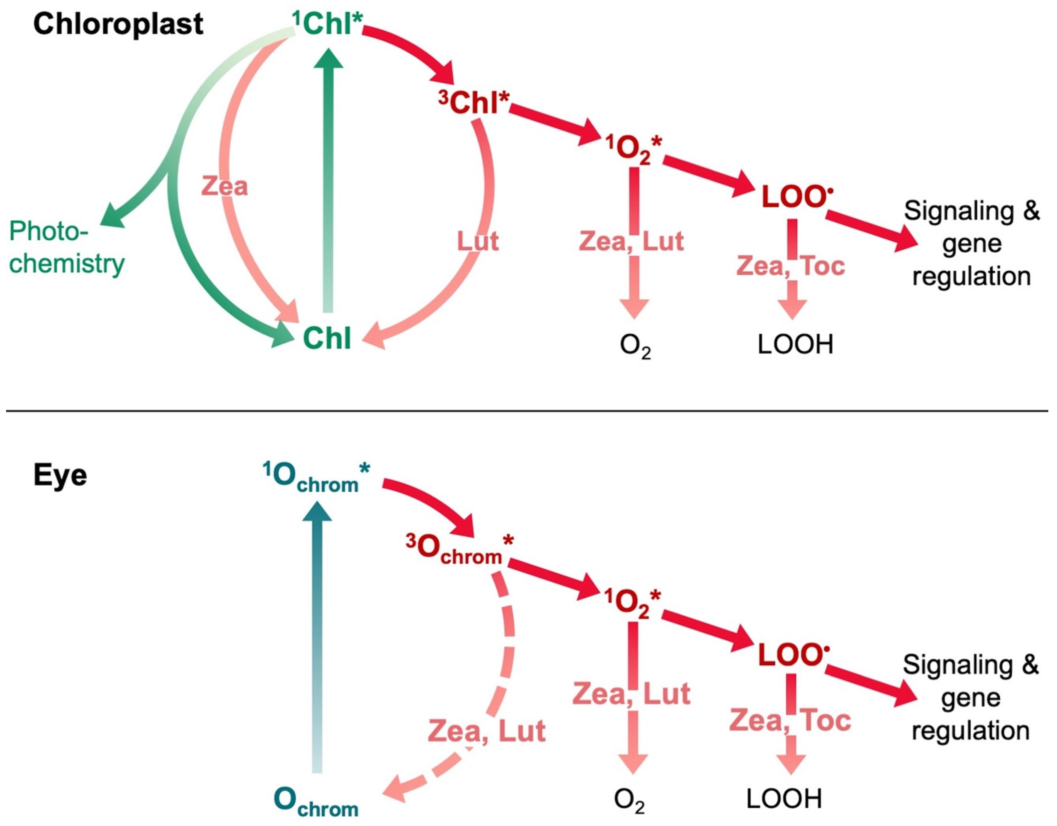
  • Pre-emptive counteraction of ROS formation by blue-light shielding in the retina and de-excitation of light-absorbing chromophores that can transfer excitation energy and/or electrons to oxygen,
  • Possible direct detoxification of ROS and other reactive species after they have been formed, followed by re-reduction by other antioxidants (well-documented in vitro but more difficult to verify in vivo), and
  • Modulation of gene expression, especially for genes responsive to the organism’s redox state, the gene products of which also affect the redox state. Such genes include those involved in defense/immune responses as well as cell cycle control and programmed cell death in plants, humans, and other organisms.
Each category of interaction will be examined more thoroughly in the following subsections of the review.

4.1. Preemptive Counteraction of ROS Formation by Photosensitizers

ROS formation by light-absorbing photoreceptors can be prevented in several ways. Particularly in the central portion of the retina, blue light is absorbed by macular xanthophylls. A model was proposed by Luchowski and his co-workers [74], in which trans-to-cis photoisomerization allows macular xanthophylls to work as “molecular blinds”, where xanthophylls in trans configurations assume a perpendicular orientation in the membrane under dim light and allow blue light to pass, thus enabling color vision and high acuity in dim light. In contrast, xanthophylls in cis configuration assume a position parallel to the membrane surface, in which they attenuate blue light under high-light exposure [74].
The absorption of more light (excitation energy) than can be processed photochemically leads to the interconversion of pigment-excited singlet states to triplet states, with a subsequent transfer of excitation energy. This sequence of events is shown in Figure 6 for chlorophyll or ocular chromophores. Although a substantial body of work on the interaction of lutein and zeaxanthin with excited states of chromophores exists for photosynthesis (see below), more work is needed to address such interactions in the human eye.
ROS and other reactive species can be formed by ocular photosensitizers in the human eye, such as the by-products of retinal or, possibly, trans-retinal itself [11,75]. Such by-product accumulation also increases with age [11]. The latter authors concluded that “zeaxanthin and lutein can protect against photooxidation by quenching the excited state sensitizer and also by intercepting and quenching singlet oxygen after it is formed.” Further details of this process should be the target of future research, with particular attention paid to which excited states of which ocular chromophores are subject to de-excitation via xanthophylls.
In the chloroplast, both triplet (3Chl*) and singlet (1Chl*) excited states of chlorophyll can be de-excited by carotenoids. Both lutein [76] and zeaxanthin [77] can de-excite 3Chl* in specific components of the light-harvesting system of plants. De-excitation of 1Chl*, which is used to drive photochemistry, is under tight metabolic control to prevent a loss of usable excitation energy. This control involves (i) proteins that activate the de-excitation process and (ii) control of xanthophyll concentration by xanthophyll cycles. There is a long-standing debate about the role of zeaxanthin as an allosteric regulator of non-photochemical energy dissipation, either instead of or in addition to a direct role in 1Chl* de-excitation (see chapters in [58]). Xanthophylls, and especially zeaxanthin, may enhance 1Chl* de-excitation indirectly by altering the microenvironment of chlorophyll complexes, e.g., [78,79]. Zeaxanthin may also aid in the integration of pigment-binding proteins into the photosynthetic membrane [80]. These different mechanisms are not mutually exclusive and may occur concomitantly in specific microenvironments, in different organisms, or in different growth environments.
In vivo measurements of time-resolved fluorescence in the intact microalgae, Nannochloropsis oceanica, indicated that de-excitation of 1Chl* can take place either via excitation-energy transfer or rapid reversible charge (electron) transfer between chlorophyll and zeaxanthin [81]. Lutein can also de-excite 1Chl* via a rapidly reversible charge transfer in certain chlorophyll-binding complexes of plants in zeaxanthin-free mutants with excess lutein (see, e.g., [82]), but only in the presence of zeaxanthin in wild-type [83]. Such added control over the removal of the excited state 1Chl* that can also drive photochemistry by a xanthophyll (zeaxanthin) that is formed only in the presence of excess light would appear attractive. In contrast, most or all lutein is already present even under light levels limiting to photosynthesis. However, a role in the de-excitation of 1Chl* was also described for a pool of lutein formed in another xanthophyll cycle (the lutein epoxide cycle) found only in certain plant species [84]. Other xanthophylls can apparently also de-excite the singlet excited state of chlorophyll or other chromophores in other organisms [58,60]. Photophysical models based on fluorescence lifetime measurements predict (i) a 10-fold higher capacity for quenching per molecule for zeaxanthin versus lutein and (ii) a low likelihood that zeaxanthin serves only as an allosteric regulator [85].
The growth environment during plant development may affect the microenvironments in which xanthophylls act within the intact organism. Observation under natural field conditions, as well as the recent application of chlorophyll fluorescence lifetime snapshots, indicate that the effects of the repeated exposure of plants to excess light—as is typical under natural conditions—differ from those of single transfers from darkness or non-excessive light to excess light and back. For example, the components of non-photochemical energy dissipation with rather rapid kinetics of onset and relaxation are modified depending on the components with slower kinetics.
Individuals of a tropical evergreen that had experienced daily exposure to excess light in their growth environment exhibited a dramatically faster onset and post-exposure relaxation of non-photochemical fluorescence quenching indicative of 1Chl* de-excitation, as well as concomitant dramatically faster formation and post-exposure removal of zeaxanthin compared to plants of the same species that were grown in the absence of excess light [86,87]. In addition, almost instantaneous onset and post-exposure relaxation of non-photochemical fluorescence quenching, indicative of 1Chl* de-excitation, was seen in the leaves of vines growing under a forest canopy where they were exposed to rapidly fluctuating light intensity (lightflecks) daily [88]. During the day, these leaves maintained considerable zeaxanthin levels that were evidently not continuously engaged in photoprotective energy dissipation. The employment of fluorescence lifetime snapshot studies to monitor nonphotochemical energy dissipation throughout repeated light-dark cycles “has the potential to reveal new insights into the complicated and overlapping nature of the various responses associated with [energy dissipation] induction and relaxation and the complex roles of the various molecular actors [involved]” [82].
The effects of a light environment during development have also been reported in animals, for example, dark-reared mice exhibit very rapid photoreceptor degeneration upon exposure to even moderate light levels [89]. Moreover, there are differences in the individual genetic propensity of humans to accumulate xanthophylls in the macula ([90] see also [91,92]). These findings suggest that both genetic variation and the external environment during development affect endogenous carotenoid localization and function in various organisms.

4.2. De-Excitation of Singlet Oxygen

Carotenoid-facilitated de-excitation of singlet oxygen has been the subject of considerable investigation and is visited here only in passing. Schalch and his co-workers concluded that “zeaxanthin and lutein can protect against photooxidation by … intercepting and quenching singlet oxygen after it is formed” and that “zeaxanthin appears to be a better photoprotectant than lutein” [11]. It is noteworthy that a mix of zeaxanthin, meso-zeaxanthin, and lutein was more effective in protecting the retina than either lutein or zeaxanthin alone. This outcome is consistent with a descending singlet-oxygen quenching efficiency in the order of all three combined > meso-zeaxanthin > zeaxanthin > lutein (summarized in [75]). Leaf carotenoids can quench singlet oxygen either by the transfer of excitation energy and subsequent loss of this energy as heat or by a chemical mechanism involving oxidation of the carotenoid [16,17].

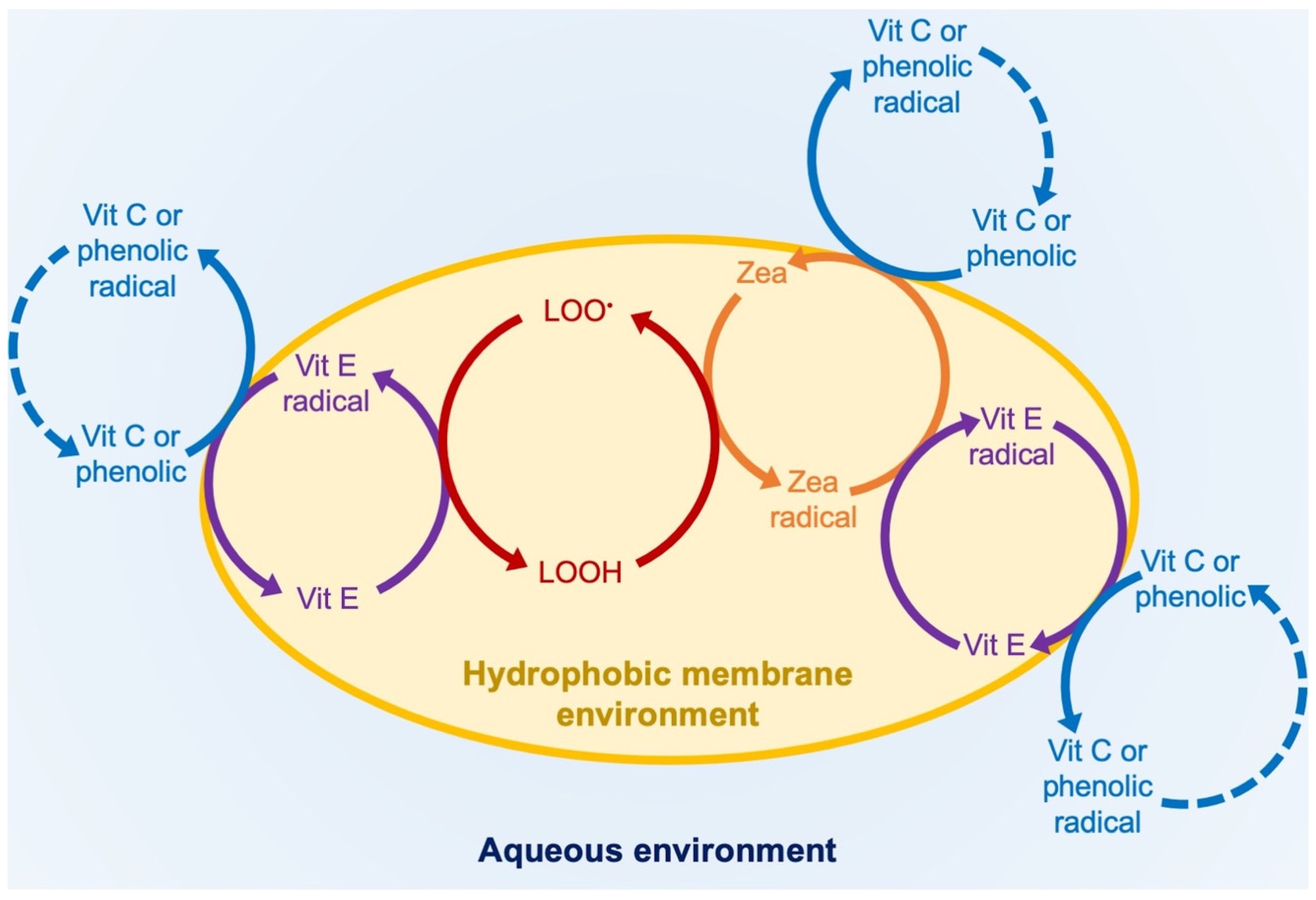
4.3. Oxidation and Re-Reduction of Zeaxanthin in Phospholipid Bilayers In Vitro

Due to their ability to donate electrons as well as their propensity to become pro-oxidants, carotenoids are principally well suited to serve as links in a chain of redox cycles, although this remains to be further verified in vivo. Numerous studies conducted in vitro have demonstrated a direct antioxidant effect, i.e., the ability to reduce lipid peroxyl radicals, for carotenoids and especially zeaxanthin (e.g., [93,94,95]). The resulting carotenoid radicals can themselves become oxidants, and such pro-oxidant effects of carotenoid radicals can be avoided via re-reduction by another antioxidant (Figure 5; [11,96]). Figure 7 outlines the process of re-reduction and recycling of zeaxanthin in membrane environments. This has been documented in vitro for vitamin E (in the lipid phase) and for ascorbate (vitamin C; Figure 7) as well as other dietary (phenolic) and endogenous (glutathione and, eventually, NADH) antioxidant metabolites at the membrane/aqueous-phase interface (see [94]). Lipid peroxidation chemically induced in liposomes was poorly opposed by lycopene, partially prevented by either β-carotene and one of several ketocarotenoids, and fully prevented by either zeaxanthin or vitamin E (tocopherol) [97]. In other studies, combinations of zeaxanthin with vitamin E or zeaxanthin with vitamin C lowered lipid peroxidation more than either alone, with the degree of protection varying with the type of hydroperoxide [95]. These interactions form multiple intertwined cycles of oxidation and reduction reactions (Figure 7).

4.4. Carotenoids and Gene Expression in Plants and Humans

As stated above, ROS have an essential role not only in biotic defense (against pathogens and other invaders), but also in the overall orchestration of growth, development, reproduction, and aging. In addition to encountering ROS in the context of light absorption, organisms produce ROS endogenously during energy metabolism (e.g., in chloroplasts and/or mitochondria) as well as for biotic defense and the elimination of other unwanted cells. Consequently, the modulation of ROS production by agents with direct or indirect antioxidant effects plays a similarly integral part in this orchestration. For example, the process of photosynthesis and its inherent role in ROS production, provides input into several signal transduction networks with “profound influence on almost every aspect of plant biology” [98]. As regulators of many key genes, ROS function “to monitor metabolic flux” [98]. The same can be expected for ROS-opposing antioxidant processes. Although there are numerous water-soluble antioxidant systems, just a handful of membrane-embedded systems—including carotenoids and vitamin E—interact with the initial events of the membrane-associated redox signaling pathways (see Figure 7).
Biological membranes are the site of the production of lipid peroxidation-based gene regulators in plants and humans. Due to their exceptional sensitivity to oxidation, PUFAs are sentinels for oxidative stress and the input from PUFA derivatives into redox-signaling pathways serves as an early-warning system that triggers changes in gene expression and defense responses. In plants, a group of stress and/or defense hormones (e.g., jasmonic acid and its precursors and derivatives) are derived from lipid peroxidation [99]. In vivo effects of vitamin E level on the level of jasmonates and/or jasmonate-dependent anatomical or functional features are evident from the use of tocopherol mutants [100]. Additionally, a possible similar role for zeaxanthin is suggested by the increased level of the jasmonic acid precursor 12-oxo-phytodienoic acid in a zeaxanthin-deficient mutant [101]. Although these mutant studies indicate in vivo roles of xanthophylls or tocopherols in signaling pathways involving lipid-peroxidation derivatives, Munné-Bosch emphasized that it is the interplay among these and other factors that determines overall outcomes, with multiple factors operating in conjunction with each other [102].
Research on xanthophylls in humans has mirrored the progression of research on vitamins from attention to topical functions (such as the function of vitamin A in vision or of vitamin D in bone health) to system-wide gene regulation. The remainder of this section focuses on gene-expression modulation by carotenoids in humans. Carotenoids with provitamin-A activity, β-carotene, β-cryptoxanthin, and α-carotene act through the formation of vitamin A (a group of retinoids including retinol, retinal, and retinoic acid) with well-characterized immuno-regulatory functions. Retinoids directly interact with genes by binding to a retinoid receptor and moving into the nucleus where they can regulate the immune response as well as cell division, cell death, and other key functions and are often activated via redox-signaling pathways in response to a shift to the oxidative side [103].
A large body of supplementary studies has established that non-provitamin-A carotenoids, such as lutein or a mix of lutein and zeaxanthin, can also modulate the expression of genes involved in the human immune response, including as master gene regulators such as NF-kB and Nfr2 [72]. A meta-analysis of randomized controlled trials on the effect of xanthophyll supplementation on inflammation reported significant effects of either lutein, lutein and zeaxanthin, cryptoxanthin, or astaxanthin in lowering inflammation markers [104]. It is thus clear that these xanthophylls have an impact on the organism’s redox state in vivo. However, more work is needed to clarify whether these xanthophylls act by modulating redox-signaling pathways or by binding directly to ligand-activated receptor proteins that function in gene regulation. The possibility has been suggested that the cleavage products of zeaxanthin and lutein [105] may bind to such ligand-activated receptors [106,107]. The oxidation products of carotenoids may also act as signaling molecules in plants [16].
Much recent research on carotenoids in humans has focused on the brain with its high concentrations of PUFAs, oxygen, and xanthophylls. Supplementation with xanthophylls that lower inflammation level [108,109,110] resulted in better cognitive function across the lifespan [108,111,112]. Cognitive performance was also enhanced by supplementation with a combination of lutein and the omega-3 PUFA docosahexaenoic acid [113,114] or a combination of vitamin E and carotenoids [115]. More studies are needed with different supplements—alone and in combination.

5. Conclusions and Recommendations about Zeaxanthin and Lutein

5.1. Summary

  • Both zeaxanthin and lutein can principally act via multiple different mechanisms as components of light-collecting systems with photosensitizers as well as in light-independent roles.
  • Both zeaxanthin and lutein can bind to multiple different proteins.
  • Zeaxanthin, but not lutein, is found in all three domains of life, ranging from extremophile microorganisms to humans.
  • Zeaxanthin and lutein assume distinguishable positions within phospholipid bilayers.
  • There is evidence that both zeaxanthin and lutein play a role in the phospholipid bilayer portion of biological membranes in animal systems, but thus far only for zeaxanthin in plant and bacterial/archaeal systems.
  • Lutein, or the combination of lutein and zeaxanthin, plays a role in human vision, as well as in modulating the human immune response (lessening inflammation), in human cognitive function, and in additional processes not related to human vision.
  • In addition to its role in plant photoprotection, zeaxanthin may have a role in the modulation of plant defenses and other functions beyond photosynthesis.

5.2. Future Research

Advances in technology are needed that allow for the non-intrusive characterization of the in vivo functions of zeaxanthin and lutein (as well as other xanthophylls) in specific endogenous microenvironments in organisms that developed under, and that are exposed to, external environments typical of natural settings. Specific questions for future research thus include the effect of genetic variation and growth environment on xanthophyll localization and function, the function of lutein and zeaxanthin in the phospholipid portions of biological membranes with respect to microdomains and between plants and animals, and the evaluation of the role of xanthophylls as direct antioxidants in biological membranes, modulators of redox-signaling pathways, or direct gene regulators.

Author Contributions

B.D.-A. wrote the first draft of the manuscript and designed the figures; S.K.P. created the figures; S.K.P. and W.W.A.III edited the manuscript. All authors have read and agreed to the published version of the manuscript.

Funding

This research received no external funding.

Data Availability Statement

Not applicable. No original data are shown (the data in Figure 2 and Figure 3 were re-graphed from the original sources cited).

Acknowledgments

This research was made possible by support from the University of Colorado.

Conflicts of Interest

The authors declare no conflict of interest.

References

  1. Yabuzaki, J. Carotenoids Database: Structures, chemical fingerprints and distribution among organisms. Database 2017, 2017, bax004. [Google Scholar] [CrossRef] [PubMed] [Green Version]
  2. Britton, G.; Liaaen-Jensen, S.; Pfander, H. Carotenoids. Natural Functions; Birkhäuser Verlag: Basel, Switzerland, 2008; Volume 4. [Google Scholar] [CrossRef]
  3. Britton, G.; Liaaen-Jensen, S.; Pfander, H. Carotenoids. Nutrition and Health; Birkhäuser Verlag: Basel, Switzerland, 2009; Volume 5. [Google Scholar] [CrossRef]
  4. Moran, N.A.; Jarvik, T. Lateral transfer of genes from fungi underlies carotenoid production in aphids. Science 2010, 328, 624–627. [Google Scholar] [CrossRef] [PubMed] [Green Version]
  5. Nishino, A.; Maoka, T.; Yasui, H. Preventive effects of β-cryptoxanthin, a potent antioxidant and provitamin A carotenoid, on lifestyle-related diseases—A central focus on its effects on non-alcoholic fatty liver disease (NAFLD). Antioxidants 2022, 11, 43. [Google Scholar] [CrossRef] [PubMed]
  6. Hofmann, E.; Hiller, R.G.; Welte, W.; Diederichs, K. Light harvesting by carotenoids: Peridinin-chlorophyll-protein (PCP) from Amphidinium carterae structural relation to proteins with globin fold. Z. Krist. Suppl. 1997, 11, 58. [Google Scholar]
  7. Nagao, R.; Yokono, M.; Teshigahara, A.; Akimoto, S.; Tomo, T. Light-harvesting ability of the fucoxanthin chlorophyll a/c-binding protein associated with photosystem II from the diatom Chaetoceros gracilis as revealed by picosecond time-resolved fluorescence spectroscopy. J. Phys. Chem. B 2014, 118, 5093–5100. [Google Scholar] [CrossRef]
  8. Polívka, T.; Hiller, R.G.; Frank, H.A. Spectroscopy of the peridinin–chlorophyll-a protein: Insight into light-harvesting strategy of marine algae. Arch. Biochem. Biophys. 2007, 458, 111–120. [Google Scholar] [CrossRef]
  9. Wang, W.; Qin, X.; Sang, M.; Chen, D.; Wang, K.; Lin, R.; Lu, C.; Shen, J.R.; Kuang, T. Spectral and functional studies on siphonaxanthin-type light-harvesting complex of photosystem II from Bryopsis corticulans. Photosynth. Res. 2013, 117, 267–279. [Google Scholar] [CrossRef]
  10. Demmig-Adams, B.; Stewart, J.J.; López-Pozo, M.; Polutchko, S.K.; Adams, W.W., III. Zeaxanthin, a molecule for photoprotection in many different environments. Molecules 2020, 25, 5825. [Google Scholar] [CrossRef]
  11. Schalch, W.; Landrum, J.T.; Bone, R.A. The eye. In Carotenoids. Nutrition and Health; Britton, G., Pfander, H., Liaaen-Jensen, S., Eds.; Birkhäuser Verlag: Basel, Switzerland, 2009; Volume 5, pp. 301–334. [Google Scholar] [CrossRef]
  12. Furman, D.; Campisi, J.; Verdin, E.; Carrera-Bastos, P.; Targ, S.; Franceschi, C.; Ferrucci, L.; Gilroy, D.W.; Fasano, A.; Miller, G.W.; et al. Chronic inflammation in the etiology of disease across the life span. Nat. Med. 2019, 25, 1822–1832. [Google Scholar] [CrossRef]
  13. Schett, G.; Neurath, M.F. Resolution of chronic inflammatory disease: Universal and tissue-specific concepts. Nat. Commun. 2018, 9, 3261. [Google Scholar] [CrossRef]
  14. Demmig-Adams, B.; López-Pozo, M.; Stewart, J.J.; Adams, W.W., III. Zeaxanthin and lutein: Photoprotectors, anti-inflammatories, and brain food. Molecules 2020, 25, 3607. [Google Scholar] [CrossRef] [PubMed]
  15. Stringham, J.M.; Johnson, E.J.; Hammond, B.R. Lutein across the lifespan: From childhood cognitive performance to the aging eye and brain. Curr. Dev. Nutr. 2019, 3, nzz066. [Google Scholar] [CrossRef] [PubMed] [Green Version]
  16. Havaux, M. Carotenoid oxidation products as stress signals in plants. Plant J. 2013, 79, 597–606. [Google Scholar] [CrossRef] [PubMed]
  17. Ramel, F.; Birtic, S.; Cuiné, S.; Triantaphylidès, C.; Ravanat, J.-L.; Havaux, M. Chemical quenching of singlet oxygen by carotenoids in plants. Plant Physiol. 2012, 158, 1267–1278. [Google Scholar] [CrossRef] [PubMed] [Green Version]
  18. Vershinin, A. Biological functions of carotenoids-Diversity and evolution. BioFactors 1999, 10, 99–104. [Google Scholar] [CrossRef] [PubMed]
  19. Polívka, T.; Frank, H.A. Spectroscopic investigation of carotenoids involved in non-photochemical fluorescence quenching. In Non-Photochemical Quenching and Energy Dissipation in Plants, Algae and Cyanobacteria. Advances in Photosynthesis and Respiration; Demmig-Adams, B., Garab, G., Adams, W.W., III, Govindjee, Eds.; Springer: Dordrecht, The Netherlands, 2014; Volume 40, pp. 203–227. [Google Scholar] [CrossRef]
  20. Sevilla, E.; Bes, M.T.; González, A.; Peleato, M.L.; Fillat, M.F. Redox-based transcriptional regulation in prokaryotes: Revisiting model mechanisms. Antioxid. Redox Signal. 2019, 30, 1651–1696. [Google Scholar] [CrossRef]
  21. Foyer, C.H.; Noctor, G. Redox regulation in photosynthetic organisms: Signaling, acclimation, and practical implications. Antioxid. Redox Signal. 2009, 11, 861–905. [Google Scholar] [CrossRef]
  22. Sies, H.; Jones, D.P. Reactive oxygen species (ROS) as pleiotropic physiological signalling agents. Nat. Rev. Mol. Cell Biol. 2020, 21, 363–383. [Google Scholar] [CrossRef]
  23. Krinsky, N.I.; Landrum, J.T.; Bone, R.A. Biologic mechanisms of the protective role of lutein and zeaxanthin in the eye. Annu. Rev. Nutr. 2003, 23, 171–201. [Google Scholar] [CrossRef] [Green Version]
  24. Johnson, E.J.; Neuringer, M.; Russell, R.M.; Schalch, W.; Snodderly, D.M. Nutritional manipulation of primate retinas, III: Effects of lutein or zeaxanthin supplementation on adipose tissue and retina of xanthophyll-free monkeys. Investig. Opthalmol. Vis. Sci. 2005, 46, 692. [Google Scholar] [CrossRef]
  25. Li, B.; George, E.W.; Rognon, G.T.; Gorusupudi, A.; Ranganathan, A.; Chang, F.Y.; Shi, L.; Frederick, J.M.; Bernstein, P.S. Imaging lutein and zeaxanthin in the human retina with confocal resonance Raman microscopy. Proc. Natl. Acad. Sci. USA 2020, 117, 12352–12358. [Google Scholar] [CrossRef] [PubMed]
  26. Widomska, J.; Zareba, M.; Subczynski, W. Can xanthophyll-membrane interactions explain their selective presence in the retina and brain? Foods 2016, 5, 7. [Google Scholar] [CrossRef] [PubMed] [Green Version]
  27. Adams, W.W., III; Demmig-Adams, B. Operation of the xanthophyll cycle in higher plants in response to diurnal changes in incident sunlight. Planta 1992, 186, 390–398. [Google Scholar] [CrossRef]
  28. Adams, W.W., III; Demmig-Adams, B. Chlorophyll fluorescence as a tool to monitor plant response to the environment. In Chlorophyll a Fluorescence: A Signature of Photosynthesis. Advances in Photosynthesis and Respiration; Papageorgiou, G.C., Govindjee, Eds.; Springer: Dordrecht, The Netherlands, 2004; Volume 19, pp. 583–604. [Google Scholar] [CrossRef]
  29. Schreiber, U.; Schliwa, U.; Bilger, W. Continuous recording of photochemical and non-photochemical chlorophyll fluorescence quenching with a new type of modulation fluorometer. Photosynth. Res. 1986, 10, 51–62. [Google Scholar] [CrossRef] [PubMed]
  30. Hager, A. The reversible, light-induced conversions of xanthophylls in the chloroplast. In Pigments in Plants; Czygan, F.-C., Ed.; Fischer: Stuttgart, Germany, 1980; pp. 57–79. [Google Scholar]
  31. Yamamoto, H.Y. A random walk to and through the xanthophyll cycle. In Photoprotection, Photoinhibition, Gene Regulation, and Environment. Advances in Photosynthesis and Respiration; Demmig-Adams, B., Adams, W.W., III, Mattoo, A.K., Eds.; Springer: Dordrecht, The Netherlands, 2008; Volume 21, pp. 1–10. [Google Scholar] [CrossRef]
  32. Demmig-Adams, B.; López-Pozo, M.; Polutchko, S.K.; Fourounjian, P.; Stewart, J.J.; Zenir, M.C.; Adams, W.W., III. Growth and nutritional quality of Lemnaceae viewed comparatively in an ecological and evolutionary context. Plants 2022, 11, 145. [Google Scholar] [CrossRef]
  33. Stewart, J.J.; Adams, W.W., III; Escobar, C.M.; López-Pozo, M.; Demmig-Adams, B. Growth and essential carotenoid micronutrients in Lemna gibba as a function of growth light intensity. Front. Plant Sci. 2020, 11, 480. [Google Scholar] [CrossRef]
  34. Stewart, J.J.; Adams, W.W., III; López-Pozo, M.; Doherty Garcia, N.; McNamara, M.; Escobar, C.M.; Demmig-Adams, B. Features of the duckweed Lemna that support rapid growth under extremes of light intensity. Cells 2021, 10, 1481. [Google Scholar] [CrossRef]
  35. Havaux, M.; Niyogi, K.K. The violaxanthin cycle protects plants from photooxidative damage by more than one mechanism. Proc. Natl. Acad. Sci. USA 1999, 96, 8762–8767. [Google Scholar] [CrossRef] [Green Version]
  36. Havaux, M.; García-Plazaola, J.I. Beyond non-photochemical fluorescence quenching: The overlapping antioxidant functions of zeaxanthin and tocopherols. In Non-Photochemical Quenching and Energy Dissipation in Plants, Algae and Cyanobacteria. Advances in Photosynthesis and Respiration; Demmig-Adams, B., Garab, G., Adams, W.W., III, Govindjee, Eds.; Springer: Dordrecht, The Netherlands, 2014; Volume 40, pp. 583–603. [Google Scholar] [CrossRef]
  37. Michael, T.P.; Ernst, E.; Hartwick, N.; Chu, P.; Bryant, D.; Gilbert, S.; Ortleb, S.; Baggs, E.L.; Sree, K.S.; Appenroth, K.J.; et al. Genome and time-of-day transcriptome of Wolffia australiana link morphological minimization with gene loss and less growth control. Genome Res. 2021, 31, 225–238. [Google Scholar] [CrossRef]
  38. Arunkumar, R.; Calvo, C.M.; Conrady, C.D.; Bernstein, P.S. What do we know about the macular pigment in AMD: The past, the present, and the future. Eye 2018, 32, 992–1004. [Google Scholar] [CrossRef] [Green Version]
  39. Davison, P.A.; Hunter, C.N.; Horton, P. Overexpression of β-carotene hydroxylase enhances stress tolerance in Arabidopsis. Nature 2002, 418, 203–206. [Google Scholar] [CrossRef] [PubMed]
  40. Alcaíno, J.; Baeza, M.; Cifuentes, V. Carotenoid distribution in nature. In Carotenoids in Nature; Stange, C., Ed.; Springer: Cham, Switzerland, 2016; Volume 79, pp. 3–33. [Google Scholar] [CrossRef]
  41. Liu, C.; Hu, B.; Cheng, Y.; Guo, Y.; Yao, W.; Qian, H. Carotenoids from fungi and microalgae: A review on their recent production, extraction, and developments. Bioresour. Technol. 2021, 337, 125398. [Google Scholar] [CrossRef] [PubMed]
  42. Nelis, H.J.; De Leenheer, A.P. Microbial sources of carotenoid pigments used in foods and feeds. J. Appl. Bacteriol. 1991, 70, 181–191. [Google Scholar] [CrossRef]
  43. Dufossé, L.; Fouillaud, M.; Caro, Y.; Mapari, S.A.; Sutthiwong, N. Filamentous fungi are large-scale producers of pigments and colorants for the food industry. Curr. Opin. Biotechnol. 2014, 26, 56–61. [Google Scholar] [CrossRef] [PubMed]
  44. Ničková, K.; Štys, D. Carotenoid pigments in three Synechococcus strains. Algol. Stud. Hydrobiol. Suppl. 2003, 109, 421–428. [Google Scholar] [CrossRef]
  45. Bemal, S.; Anil, A.C. Genetic and ecophysiological traits of Synechococcus strains isolated from coastal and open ocean waters of the Arabian Sea. FEMS Microbiol. Ecol. 2016, 92, fiw162. [Google Scholar] [CrossRef] [Green Version]
  46. Wilson, A.; Muzzopappa, F.; Kirilovsky, D. Elucidation of the essential amino acids involved in the binding of the cyanobacterial orange carotenoid protein to the phycobilisome. Biochim. Biophys. Acta BBA-Bioenerg. 2022, 1863, 148504. [Google Scholar] [CrossRef]
  47. Berry, A.; Janssens, D.; Hümbelin, M.; Jore, J.P.; Hoste, B.; Cleenwerck, I.; Vancanneyt, M.; Bretzel, W.; Mayer, A.F.; Lopez-Ulibarri, R.; et al. Paracoccus zeaxanthinifaciens sp. nov., a zeaxanthin-producing bacterium. Int. J. Syst. Evol. Microbiol. 2003, 53, 231–238. [Google Scholar] [CrossRef] [Green Version]
  48. Ram, S.; Mitra, M.; Shah, F.; Tirkey, S.R.; Mishra, S. Bacteria as an alternate biofactory for carotenoid production: A review of its applications, opportunities and challenges. J. Funct. Foods 2020, 67, 103867. [Google Scholar] [CrossRef]
  49. Ghedira, K.; Othman, H.; Saied, T.; Baccar, Z.M.; Hosni, F.; Hamzaoui, A.H.; Thamaraiselvi, K.; Abdelmelek, H.; Srairi-Abid, N.; Costa, M.C.; et al. Insights into ionizing-radiation-resistant bacteria S-layer proteins and nanobiotechnology for bioremediation of hazardous and radioactive waste. In Management of Hazardous Wastes; Saleh, H.E.-D.M., Rahman, R.O.A., Eds.; IntechOpen: London, UK, 2016. [Google Scholar]
  50. Adamec, F.; Farci, D.; Bína, D.; Litvín, R.; Khan, T.; Fuciman, M.; Piano, D.; Polívka, T. Photophysics of deinoxanthin, the keto-carotenoid bound to the main S-layer unit of Deinococcus radiodurans. Photochem. Photobiol. Sci. 2020, 19, 495–503. [Google Scholar] [CrossRef]
  51. Asker, D.; Beppu, T.; Ueda, K. Unique diversity of carotenoid-producing bacteria isolated from Misasa, a radioactive site in Japan. Appl. Microbiol. Biotechnol. 2007, 77, 383–392. [Google Scholar] [CrossRef] [PubMed]
  52. Farci, D.; Kereïche, S.; Pangeni, S.; Haniewicz, P.; Bodrenko, I.V.; Ceccarelli, M.; Winterhalter, M.; Piano, D. Structural analysis of the architecture and in situ localization of the main S-layer complex in Deinococcus radiodurans. Structure 2021, 29, 1279–1285.e3. [Google Scholar] [CrossRef] [PubMed]
  53. Tian, B.; Xu, Z.; Sun, Z.; Lin, J.; Hua, Y. Evaluation of the antioxidant effects of carotenoids from Deinococcus radiodurans through targeted mutagenesis, chemiluminescence, and DNA damage analyses. Biochim. Biophys. Acta BBA-Gen. Subj. 2007, 1770, 902–911. [Google Scholar] [CrossRef] [PubMed]
  54. Gudkov, S.V.; Grinberg, M.A.; Sukhov, V.; Vodeneev, V. Effect of ionizing radiation on physiological and molecular processes in plants. J. Environ. Radioact. 2019, 202, 8–24. [Google Scholar] [CrossRef]
  55. Hara, M.; Yuan, H.; Yang, Q.; Hoshino, T.; Yokoyama, A.; Miyake, J. Stabilization of liposomal membranes by thermozeaxanthins: Carotenoid-glucoside esters. Biochim. Biophys. Acta BBA-Biomembr. 1999, 1461, 147–154. [Google Scholar] [CrossRef] [Green Version]
  56. Mandelli, F.; Miranda, V.S.; Rodrigues, E.; Mercadante, A.Z. Identification of carotenoids with high antioxidant capacity produced by extremophile microorganisms. World J. Microbiol. Biotechnol. 2012, 28, 1781–1790. [Google Scholar] [CrossRef]
  57. Yatsunami, R.; Ando, A.; Yang, Y.; Takaichi, S.; Kohno, M.; Matsumura, Y.; Ikeda, H.; Fukui, T.; Nakasone, K.; Fujita, N.; et al. Identification of carotenoids from the extremely halophilic archaeon Haloarcula japonica. Front. Microbiol. 2014, 5, 100. [Google Scholar] [CrossRef]
  58. Demmig-Adams, B.; Garab, G.; Adams, W.W., III. Non-Photochemical Quenching and Energy Dissipation in Plants, Algae and Cyanobacteria. Advances in Photosynthesis and Respiration; Springer: Dordrecht, The Netherlands, 2014; Volume 40. [Google Scholar] [CrossRef] [Green Version]
  59. Skotnicová, P.; Staleva-Musto, H.; Kuznetsova, V.; Bína, D.; Konert, M.M.; Lu, S.; Polívka, T.; Sobotka, R. Plant LHC-like proteins show robust folding and static non-photochemical quenching. Nat. Commun. 2021, 12, 6890. [Google Scholar] [CrossRef]
  60. Kirilovsky, D.; Kerfeld, C.A. Cyanobacterial photoprotection by the orange carotenoid protein. Nat. Plants 2016, 2, 16180. [Google Scholar] [CrossRef] [Green Version]
  61. Arunkumar, R.; Gorusupudi, A.; Bernstein, P.S. The macular carotenoids: A biochemical overview. Biochim. Biophys. Acta BBA-Mol. Cell Biol. Lipids 2020, 1865, 158617. [Google Scholar] [CrossRef]
  62. Bhosale, P.; Larson, A.J.; Frederick, J.M.; Southwick, K.; Thulin, C.D.; Bernstein, P.S. Identification and characterization of a Pi isoform of glutathione S-transferase (GSTP1) as a zeaxanthin-binding protein in the macula of the human eye. J. Biol. Chem. 2004, 279, 49447–49454. [Google Scholar] [CrossRef] [PubMed] [Green Version]
  63. Li, B.; Vachali, P.; Frederick, J.M.; Bernstein, P.S. Identification of StARD3 as a lutein-binding protein in the macula of the primate Retina. Biochemistry 2011, 50, 2541–2549. [Google Scholar] [CrossRef] [PubMed] [Green Version]
  64. Rohmer, M.; Bouvier, P.; Ourisson, G. Molecular evolution of biomembranes: Structural equivalents and phylogenetic precursors of sterols. Proc. Natl. Acad. Sci. USA 1979, 76, 847–851. [Google Scholar] [CrossRef] [PubMed] [Green Version]
  65. Johnson, E.J.; Vishwanathan, R.; Johnson, M.A.; Hausman, D.B.; Davey, A.; Scott, T.M.; Green, R.C.; Miller, L.S.; Gearing, M.; Woodard, J.; et al. Relationship between serum and brain carotenoids, α-tocopherol, and retinol concentrations and cognitive performance in the oldest old from the Georgia Centenarian Study. J. Aging Res. 2013, 2013, 951786. [Google Scholar] [CrossRef] [PubMed] [Green Version]
  66. Gruszecki, W.I.; Strzałka, K. Carotenoids as modulators of lipid membrane physical properties. Biochim. Biophys. Acta BBA-Mol. Basis Dis. 2005, 1740, 108–115. [Google Scholar] [CrossRef] [PubMed] [Green Version]
  67. Makuch, K.; Hryc, J.; Markiewicz, M.; Pasenkiewicz-Gierula, M. Lutein and zeaxanthin in the lipid bilayer–Similarities and differences revealed by computational studies. Front. Mol. Biosci. 2021, 8, 768449. [Google Scholar] [CrossRef]
  68. Widomska, J.; Subczynski, W.K. Why has nature chosen lutein and zeaxanthin to protect the retina? J. Clin. Exp. Ophthalmol. 2014, 5, 326. [Google Scholar] [CrossRef] [Green Version]
  69. Widomska, J.; Gruszecki, W.I.; Subczynski, W.K. Factors differentiating the antioxidant activity of macular santhophylls in the human eye retina. Antioxidants 2021, 10, 601. [Google Scholar] [CrossRef]
  70. Wisniewska, A.; Subczynski, W.K. Accumulation of macular xanthophylls in unsaturated membrane domains. Free Radic. Biol. Med. 2006, 40, 1820–1826. [Google Scholar] [CrossRef]
  71. Demmig-Adams, B.; Adams, W.W., III. Antioxidants in photosynthesis and human nutrition. Science 2002, 298, 2149–2153. [Google Scholar] [CrossRef]
  72. Ahn, Y.J.; Kim, H. Lutein as a modulator of oxidative stress-mediated inflammatory diseases. Antioxidants 2021, 10, 1448. [Google Scholar] [CrossRef] [PubMed]
  73. Hüner, N.P.A.; Dahal, K.; Bode, R.; Kurepin, L.V.; Ivanov, A.G. Photosynthetic acclimation, vernalization, crop productivity and ‘the grand design of photosynthesis’. J. Plant Physiol. 2016, 203, 29–43. [Google Scholar] [CrossRef] [PubMed]
  74. Luchowski, R.; Grudzinski, W.; Welc, R.; Mendes Pinto, M.M.; Sek, A.; Ostrowski, J.; Nierzwicki, L.; Chodnicki, P.; Wieczor, M.; Sowinski, K.; et al. Light-modulated sunscreen mechanism in the retina of the human eye. J. Phys. Chem. B 2021, 125, 6090–6102. [Google Scholar] [CrossRef] [PubMed]
  75. Roberts, J.E.; Dennison, J. The photobiology of lutein and zeaxanthin in the eye. J. Ophthalmol. 2015, 2015, 687173. [Google Scholar] [CrossRef] [Green Version]
  76. Agostini, A.; Nicol, L.; Da Roit, N.; Bortolus, M.; Croce, R.; Carbonera, D. Altering the exciton landscape by removal of specific chlorophylls in monomeric LHCII provides information on the sites of triplet formation and quenching by means of ODMR and EPR spectroscopies. Biochim. Biophys. Acta BBA-Bioenerg. 2021, 1862, 148481. [Google Scholar] [CrossRef]
  77. Dall’Osto, L.; Holt, N.E.; Kaligotla, S.; Fuciman, M.; Cazzaniga, S.; Carbonera, D.; Frank, H.A.; Alric, J.; Bassi, R. Zeaxanthin protects plant photosynthesis by modulating chlorophyll triplet yield in specific light-harvesting antenna subunits. J. Biol. Chem. 2012, 287, 41820–41834. [Google Scholar] [CrossRef] [Green Version]
  78. Horton, P.; Ruban, A.V.; Wentworth, M. Allosteric regulation of the light-harvesting system of photosystem II. Philos. Trans. R. Soc. Lond. B Biol. Sci. 2000, 355, 1361–1370. [Google Scholar] [CrossRef] [Green Version]
  79. Morosinotto, T.; Caffarri, S.; Dall’Osto, L.; Bassi, R. Mechanistic aspects of the xanthophyll dynamics in higher plant thylakoids. Physiol. Plant. 2003, 119, 347–354. [Google Scholar] [CrossRef]
  80. Bassi, R. A new function for the xanthophyll zeaxanthin: Glueing chlorophyll biosynthesis to thylakoid protein assembly. Biochem. J. 2021, 478, 61–62. [Google Scholar] [CrossRef]
  81. Park, S.; Steen, C.J.; Lyska, D.; Fischer, A.L.; Endelman, B.; Iwai, M.; Niyogi, K.K.; Fleming, G.R. Chlorophyll–carotenoid excitation energy transfer and charge transfer in Nannochloropsis oceanica for the regulation of photosynthesis. Proc. Natl. Acad. Sci. USA 2019, 116, 3385–3390. [Google Scholar] [CrossRef] [Green Version]
  82. Steen, C.J.; Morris, J.M.; Short, A.H.; Niyogi, K.K.; Fleming, G.R. Complex roles of PsbS and xanthophylls in the regulation of nonphotochemical quenching in Arabidopsis thaliana under fluctuating light. J. Phys. Chem. B 2020, 124, 10311–10325. [Google Scholar] [CrossRef] [PubMed]
  83. Avenson, T.J.; Ahn, T.K.; Niyogi, K.K.; Ballottari, M.; Bassi, R.; Fleming, G.R. Lutein can act as a switchable charge transfer quencher in the CP26 light-harvesting complex. J. Biol. Chem. 2008, 284, 2830–2835. [Google Scholar] [CrossRef] [PubMed] [Green Version]
  84. Esteban, R.; García-Plazaola, J.I. Involvement of a second xanthophyll cycle in non-photochemical quenching of chlorophyll fluorescence: The lutein epoxide story. In Non-Photochemical Quenching and Energy Dissipation in Plants, Algae and Cyanobacteria. Advances in Photosynthesis and Respiration; Demmig-Adams, B., Garab, G., Adams, W.W., III, Govindjee, Eds.; Springer: Dordrecht, The Netherlands, 2014; Volume 40, pp. 277–295. [Google Scholar] [CrossRef]
  85. Bennett, D.I.; Amarnath, K.; Park, S.; Steen, C.J.; Morris, J.M.; Fleming, G.R. Models and mechanisms of the rapidly reversible regulation of photosynthetic light harvesting. Open Biol. 2019, 9, 190043. [Google Scholar] [CrossRef] [PubMed]
  86. Demmig-Adams, B.; Winter, K.; Krüger, A.; Czygan, F.-C. Zeaxanthin and the induction and relaxation kinetics of the dissipation of excess excitation energy in leaves in 2% O2, 0% CO2. Plant Physiol. 1989, 90, 887–893. [Google Scholar] [CrossRef] [Green Version]
  87. Demmig-Adams, B.; Moeller, D.L.; Logan, B.A.; Adams, W.W., III. Positive correlation between levels of retained zeaxanthin+antheraxanthin and degree of photoinhibition in shade leaves of Schefflera arboricola (Hayata) Merrill. Planta 1998, 205, 367–374. [Google Scholar] [CrossRef]
  88. Adams, W.W., III; Demmig-Adams, B.; Logan, B.A.; Barker, D.H.; Osmond, C.B. Rapid changes in xanthophyll cycle-dependent energy dissipation and photosystem II efficiency in two vines, Stephania japonica and Smilax australis, growing in the understory of an open Eucalyptus forest. Plant Cell Environ. 1999, 22, 125–136. [Google Scholar] [CrossRef]
  89. Weh, E.; Scott, K.; Wubben, T.J.; Besirli, C.G. Dark-reared rd10 mice experience rapid photoreceptor degeneration with short exposure to room-light during in vivo retinal imaging. Exp. Eye Res. 2022, 215, 108913. [Google Scholar] [CrossRef]
  90. Böhm, V.; Lietz, G.; Olmedilla-Alonso, B.; Phelan, D.; Reboul, E.; Bánati, D.; Borel, P.; Corte-Real, J.; de Lera, A.R.; Desmarchelier, C.; et al. From carotenoid intake to carotenoid blood and tissue concentrations–Implications for dietary intake recommendations. Nutr. Rev. 2021, 79, 544–573. [Google Scholar] [CrossRef]
  91. Bohn, T.; Desmarchelier, C.; Dragsted, L.O.; Nielsen, C.S.; Stahl, W.; Rühl, R.; Keijer, J.; Borel, P. Host-related factors explaining interindividual variability of carotenoid bioavailability and tissue concentrations in humans. Mol. Nutr. Food Res. 2017, 61, 1600685. [Google Scholar] [CrossRef]
  92. Borel, P. Genetic variations involved in interindividual variability in carotenoid status. Mol. Nutr. Food Res. 2012, 56, 228–240. [Google Scholar] [CrossRef] [Green Version]
  93. Krinsky, N.I. Antioxidant functions of carotenoids. Free Radic. Biol. Med. 1989, 7, 617–635. [Google Scholar] [CrossRef]
  94. Polutchko, S.K.; Glime, G.N.; Demmig-Adams, B. Synergistic action of membrane-bound and water-soluble antioxidants in neuroprotection. Molecules 2021, 26, 5385. [Google Scholar] [CrossRef] [PubMed]
  95. Wrona, M.; Różanowska, M.; Sarna, T. Zeaxanthin in combination with ascorbic acid or α-tocopherol protects ARPE-19 cells against photosensitized peroxidation of lipids. Free Radic. Biol. Med. 2004, 36, 1094–1101. [Google Scholar] [CrossRef] [PubMed]
  96. Krinsky, N.I.; Johnson, E.J. Carotenoid actions and their relation to health and disease. Mol. Asp. Med. 2005, 26, 459–516. [Google Scholar] [CrossRef] [PubMed]
  97. Woodall, A.A.; Britton, G.; Jackson, M.J. Carotenoids and protection of phospholipids in solution or in liposomes against oxidation by peroxyl radicals: Relationship between carotenoid structure and protective ability. Biochim. Biophys. Acta BBA-Gen. Subj. 1997, 1336, 575–586. [Google Scholar] [CrossRef]
  98. Foyer, C.H.; Noctor, G. Stress-triggered redox signalling: What’s in pROSpect? Plant Cell Environ. 2016, 39, 951–964. [Google Scholar] [CrossRef]
  99. Howe, G.A.; Schilmiller, A.L. Oxylipin metabolism in response to stress. Curr. Opin. Plant Biol. 2002, 5, 230–236. [Google Scholar] [CrossRef]
  100. Munné-Bosch, S.; Weiler, E.W.; Alegre, L.; Müller, M.; Düchting, P.; Falk, J. α-Tocopherol may influence cellular signaling by modulating jasmonic acid levels in plants. Planta 2007, 225, 681–691. [Google Scholar] [CrossRef]
  101. Demmig-Adams, B.; Cohu, C.M.; Amiard, V.; van Zadelhoff, G.; Veldink, G.A.; Muller, O.; Adams, W.W., III. Emerging trade-offs–impact of photoprotectants (PsbS, xanthophylls, and vitamin E) on oxylipins as regulators of development and defense. New Phytol. 2013, 197, 720–729. [Google Scholar] [CrossRef]
  102. Munné-Bosch, S. The role of α-tocopherol in plant stress tolerance. J. Plant Physiol. 2005, 162, 743–748. [Google Scholar] [CrossRef]
  103. Widjaja-Adhi, M.A.K.; Golczak, M. The molecular aspects of absorption and metabolism of carotenoids and retinoids in vertebrates. Biochim. Biophys. Acta BBA-Mol. Cell Biol. Lipids 2020, 1865, 158571. [Google Scholar] [CrossRef] [PubMed]
  104. Hajizadeh-Sharafabad, F.; Zahabi, E.S.; Malekahmadi, M.; Zarrin, R.; Alizadeh, M. Carotenoids supplementation and inflammation: A systematic review and meta-analysis of randomized clinical trials. Crit. Rev. Food Sci. Nutr. 2021; early access. [Google Scholar] [CrossRef] [PubMed]
  105. Mein, J.R.; Dolnikowski, G.G.; Ernst, H.; Russell, R.M.; Wang, X.-D. Enzymatic formation of apo-carotenoids from the xanthophyll carotenoids lutein, zeaxanthin and β-cryptoxanthin by ferret carotene-9′,10′-monooxygenase. Arch. Biochem. Biophys. 2011, 506, 109–121. [Google Scholar] [CrossRef] [PubMed] [Green Version]
  106. Sayo, T.; Sugiyama, Y.; Inoue, S. Lutein, a nonprovitamin A, activates the retinoic acid receptor to induce HAS3-dependent hyaluronan synthesis in keratinocytes. Biosci. Biotechnol. Biochem. 2013, 77, 1282–1286. [Google Scholar] [CrossRef] [Green Version]
  107. Uppal, S.; Dergunov, S.A.; Zhang, W.; Gentleman, S.; Redmond, T.M.; Pinkhassik, E.; Poliakov, E. Xanthophylls modulate palmitoylation of mammalian β-carotene oxygenase 2. Antioxidants 2021, 10, 413. [Google Scholar] [CrossRef]
  108. Davinelli, S.; Ali, S.; Solfrizzi, V.; Scapagnini, G.; Corbi, G. Carotenoids and cognitive outcomes: A meta-analysis of randomized intervention trials. Antioxidants 2021, 10, 223. [Google Scholar] [CrossRef]
  109. Khalil, A.; Tazeddinova, D.; Aljoumaa, K.; Kazhmukhanbetkyzy, Z.A.; Orazov, A.; Toshev, A.D. Carotenoids: Therapeutic strategy in the battle against viral emerging diseases, COVID-19: An overview. Prev. Nutr. Food Sci. 2021, 26, 241. [Google Scholar] [CrossRef]
  110. Lu, Q.; Li, H.; Zou, Y.; Liu, H.; Yang, L. Astaxanthin as a microalgal metabolite for aquaculture: A review on the synthetic mechanisms, production techniques, and practical application. Algal Res. 2021, 54, 102178. [Google Scholar] [CrossRef]
  111. Hammond, B.R., Jr.; Miller, L.S.; Bello, M.O.; Lindbergh, C.A.; Mewborn, C.; Renzi-Hammond, L.M. Effects of lutein/zeaxanthin supplementation on the cognitive function of community dwelling older adults: A randomized, double-masked, placebo-controlled trial. Front. Aging Neurosci. 2017, 9, 254. [Google Scholar] [CrossRef]
  112. Renzi-Hammond, L.M.; Bovier, E.R.; Fletcher, L.M.; Miller, L.S.; Mewborn, C.M.; Lindbergh, C.A.; Baxter, J.H.; Hammond, B.R. Effects of a lutein and zeaxanthin intervention on cognitive function: A randomized, double-masked, placebo-controlled trial of younger healthy adults. Nutrients 2017, 9, 1246. [Google Scholar] [CrossRef] [Green Version]
  113. Johnson, E.J.; McDonald, K.; Caldarella, S.M.; Chung, H.; Troen, A.M.; Snodderly, D.M. Cognitive findings of an exploratory trial of docosahexaenoic acid and lutein supplementation in older women. Nutr. Neurosci. 2008, 11, 75–83. [Google Scholar] [CrossRef] [PubMed]
  114. Nolan, J.M.; Mulcahy, R.; Power, R.; Moran, R.; Howard, A.N. Nutritional intervention to prevent Alzheimer’s disease: Potential benefits of xanthophyll carotenoids and omega-3 fatty acids combined. J. Alzheimer’s Dis. 2018, 64, 367–378. [Google Scholar] [CrossRef] [PubMed] [Green Version]
  115. Beydoun, M.A.; Canas, J.A.; Fanelli-Kuczmarski, M.T.; Maldonado, A.I.; Shaked, D.; Kivimaki, M.; Evans, M.K.; Zonderman, A.B. Association of antioxidant vitamins A, C, E and carotenoids with cognitive performance over time: A cohort study of middle-aged adults. Nutrients 2020, 12, 3558. [Google Scholar] [CrossRef] [PubMed]
Figure 1. Chemical structures of zeaxanthin and lutein. Hydrophobic portions are shown in orange and hydrophilic portions in blue (containing oxygen). The grey outline highlights the longer continuous, conjugated system of carbon to carbon (C=C) double bonds in zeaxanthin compared to lutein.
Photochem 02 00022 g001
Figure 3. (AC) Images of crystallizing dishes with Lemna gibba fronds grown under 3 different PFDs. Relationship between growth PFD and the levels of zeaxanthin (Z), lutein (L), and β-carotene (β-C) on a frond area basis (D) or a chlorophyll basis (E). Colors of the symbols from dark green to yellow correspond to the different visual appearances of fronds grown under the different growth PFDs. Data re-graphed from [33].
Photochem 02 00022 g003
Figure 4. Chemical structures of β-carotene, β-carotene-derived structures, and structures with similar features mentioned in the text, i.e., β-carotene β-cryptoxanthin, zeaxanthin, 3′-hydroxyechinenone, deinoxanthin, and thermozeaxanthin.
Photochem 02 00022 g004
Figure 5. Summary of the three principal response categories of xanthophylls (with a focus on zeaxanthin and lutein) including preemptive prevention of ROS formation (yellow; left), direct detoxification of ROS (green; center), and modulation of gene expression (blue; right). A conceptually similar summary can be found in [26]. 1Chl* and 3Chl*, singlet and triplet excited state of chlorophyll, respectively; O2*, singlet excited state of oxygen; ROS, reactive oxygen species.
Photochem 02 00022 g005
Figure 6. Schematic depiction of the series of excitation-energy transfers beginning with light absorption and excitation of chlorophyll (Chl; in plants) or ocular chromophores (Ochrom; in the human eye) from the ground state to the singlet excited state. For each step, the main contributing xanthophyll(s) under physiological conditions is listed (see text for further details). Energy dissipation (red to pink arrows) is facilitated by zeaxanthin (Zea), lutein (Lut), and/or tocopherols (Toc). 1Chl*, 3Chl* = singlet and triplet excited states of chlorophyll; 1Ochrom*, 3Ochrom* = singlet and triplet excited states of the ocular chromophore; 1O2* = singlet excited state of oxygen; LOO = lipid peroxyl radical; LOOH = lipid.
Photochem 02 00022 g006
Figure 7. Schematic depiction of the multiple intertwined oxidation and reduction cycles supported by various possible interactions of membrane-soluble and water-soluble antioxidants. Vit C = ascorbate; Vit E = tocopherol; LOO* = lipid peroxyl radical; LOOH = lipid; Zea = zeaxanthin After [96].
Photochem 02 00022 g007
Publisher’s Note: MDPI stays neutral with regard to jurisdictional claims in published maps and institutional affiliations.

