Article

Photodynamic Effect of 5,10,15,20-Tetrakis[4-(3-N,N-dimethylaminopropoxy)phenyl]chlorin towards the Human Pathogen Candida albicans under Different Culture Conditions

by
Paula V. Cordero
,
Darío D. Ferreyra
,
María E. Pérez
,
María G. Alvarez
and
Edgardo N. Durantini
*
IDAS-CONICET, Departamento de Química, Facultad de Ciencias Exactas, Físico-Químicas y Naturales, Universidad Nacional de Río Cuarto, Ruta Nacional 36 Km 601, Río Cuarto X5804BYA, Córdoba, Argentina
*
Author to whom correspondence should be addressed.
Photochem 2021, 1(3), 505-522; https://doi.org/10.3390/photochem1030033
Submission received: 27 October 2021 / Revised: 16 November 2021 / Accepted: 24 November 2021 / Published: 3 December 2021

Abstract

:
Photocytotoxic activity sensitized by 5,10,15,20-tetrakis[4-(3-N,N-dimethylaminopropoxy)phenyl]chlorin (TAPC) was investigated in Candida albicans under different culture conditions. Planktonic cells incubated with 2.5 μM TAPC were eradicated after 5 min irradiation with white light. Studies in the presence of reactive oxygen species scavengers indicated the involvement of mainly a type II mechanism. Furthermore, cell growth of C. albicans was suppressed in the presence of 5 μM TAPC. A decrease in pseudohyphae survival of 5 log was found after 30 min irradiation. However, the photokilling of this virulence factor reached a 1.5 log reduction in human serum. The uptake of TAPC by pseudohyphae decreased in serum due to the interaction of TAPC with albumin. The binding constant of the TAPC-albumin complex was ~104 M−1, while the bimolecular quenching rate constant was ~1012 s−1 M−1, indicating that this process occurred through a static process. Thus, the photoinactivation of C. albicans was considerably decreased in the presence of albumin. A reduction of 2 log in cell survival was observed using 4.5% albumin and 30 min irradiation. The results allow optimizing the best conditions to inactivate C. albicans under different culture conditions.

Graphical Abstract

1. Introduction

In the past three decades, significant growth of invasive and mucocutaneous fungal infections was observed with a high risk to human health. This situation is mainly caused by the increase in resistance to drugs by fungi and a deficiency of new strategies to control mycosis [1,2]. This problem is increasing due to the inappropriate use of antimicrobials. In this regard, a wide range of antibiotics is prescribed for viral diseases, as preventive surgical interventions, or even as food additives [3]. In patients with immunodeficiency, invasive or superficial fungal infections are caused by opportunistic pathogens of the genera Candida, Aspergillus and Cryptococcus [4]. In particular, the genus Candida includes a large number of yeast species; some of them are commensals of the human gastrointestinal and genitourinary tracts [5]. In particular, Candida albicans is the most prevalent species isolated from humans regardless of the health condition of the patient. It represents more than 80% of the isolates from all forms of human candidiasis and candidemia worldwide [6]. Candidemia is the most common form of invasive infections and affects with high mortality rates (over 40%) in older people and premature children, with an elevated risk even with the development and introduction of newer antifungal agents. This represents a serious public health challenge with increasing medical and economic importance [7]. In addition, it can cause damage by resisting or bypassing the defense mechanism of the host’s immune system. C. albicans has several virulence factors that facilitate yeast colonization in humans, such as cell morphology, extracellular enzymatic activity, phenotypic change (from yeast to hyphae) and biofilm formation [8]. This situation is an emerging urgency to develop new and efficient drugs, with shorter treatment times and low toxicity to the host. Several antimicrobial therapies were proposed to treat fungal infections or to reduce the development of their virulence factors [9]. In this sense, photodynamic inactivation (PDI) of microorganisms is a promising new alternative to eliminate not only fungal infections but also reduce the development of associated virulence factors [10]. PDI consists of the administration of a photosensitizer (PS), which is selectively incorporated into fungal cells. The cultures are then irradiated with a light source of suitable wavelengths. In these conditions, the triplet excited state of PS (3PS*) can react with biomolecules by electron-transfer or hydrogen-abstraction to produce free radicals, which can interact with ground-state molecular oxygen (O2 ( Σ g 3 ) ) to form reactive oxygen species (ROS) through a type I photo process [11]. Moreover, this pathway can compete with the energy transfer from 3PS* to (O2 ( Σ g 3 ) ) to generate singlet molecular oxygen (O2(1Δg)) by a type II mechanism [12]. Thus, these ROS can react with intracellular components, causing loss of functionality and microbial inactivation. Furthermore, there is no convincing evidence to validate the development of acquired microbial resistance for PDI. For these reasons, this therapy is a promising modality for the treatment of candidiasis and related yeast infections [10].
Several studies indicate that porphyrins and chlorins are PSs that have a strong antimicrobial action against several human pathogens, including C. albicans [13,14]. In a previous study, we demonstrated that 5,10,15,20-tetrakis[4-(3-N,N-dimethylaminopropoxy)phenyl]chlorin (TAPC, Figure 1) was an efficient PS to produce a high reduction in cell survival of S. aureus, E. coli and C. albicans using low concentrations and irradiation times [15]. In this work, photoinactivation of C. albicans sensitized by TAPC was investigated under different culture conditions. First, photoinactivation was evaluated in yeast planktonic cells to obtain insight into the photodynamic action mechanism. To evaluate the influence of the medium where cells are suspended on the antifungal photoinactivation, the photocytotoxic effect sensitized by TAPC was investigated on the growth of C. albicans in the culture broth. Furthermore, the ability of this PS was evaluated to eliminate the virulence factors of C. albicans as pseudohyphae in presence of human serum (HS). In addition, the interaction between TAPC and bovine serum albumin (BSA) was analyzed as a model to evaluate how these components present in the HS can affect the PDI. These studies made it possible to find the best conditions to inactivate C. albicans in different media.

2. Materials and Methods

2.1. Materials

TAPC was synthesized as previously described [15]. Stock solutions (0.5 mM) of TAPC were prepared by weighing and dilution in N,N-dimethylformamide (DMF). The concentration of TAPC was determined by absorption spectroscopy in DMF, considering the value of the molar absorption coefficients (ε421 = 1.15 × 10⁵ L mol−1 cm−1). Chemicals were obtained from Sigma-Aldrich (Milwaukee, WI, USA) and they were used without further purification. Sabouraud glucose agar from Britania (Buenos Aires, Argentina) were used in microbial cultures. Microtiter plates (96-well) were acquired from Deltalab (Barcelona, Spain).

2.2. Instrumentation

Absorption spectra were carried out on a Shimadzu UV-2401PC spectrometer (Shimadzu Corporation, Tokyo, Japan). Fluorescence emission spectra were performed on a Spex FluoroMax spectrofluorometer (Horiba Jobin Yvon Inc, Edison, NJ, USA). Spectroscopic determinations were achieved in a quartz cell of 1 cm path length at room temperature. Light fluence rates were determined with a Radiometer Laser Mate-Q, using an LM-2 VIS semiconductor power sensor (Coherent, Santa Clara, CA, USA). Cell suspensions were irradiated with a Cole–Parmer illuminator 41720-series (150 W halogen lamp, Cole–Parmer, Vernon Hills, IL, USA) (Figure S1). A 2.5 cm glass cuvette filled with water was used to remove the heat from the lamp. Under these conditions, the temperature in the cultures did not exceed 30 °C. A wavelength range between 350 and 800 nm was selected by optical filters. Emission spectrum of the broadband radiation source is shown in Figure S1. The projector was placed vertically with the light beam focused on the 96-well microtiter plate lid, producing a fluence rate of 90 mW/cm2 [16,17].

2.3. Strains and Cultures of C. albicans

The strain of C. albicans and culture conditions were detailed in the Supplementary Materials.

2.4. PDI of C. albicans Planktonic Cells

Experiments were attained using 2 mL cell suspension of ~106 colony forming units (CFU)/mL in phosphate-buffered saline (PBS), which were placed in Pyrex brand culture tubes (13 × 100 mm). The cultures were treated with 2.5 μM TAPC, which was added from the stock solution in DMF. The maximum amount of DMF used was 1% v/v and this amount of organic solvent was not toxic to C. albicans. Each sample was incubated with TAPC for 30 min in the dark at 37 °C. Sodium azide (50 mM) and D-mannitol (100 mM) were added to C. albicans suspensions from stock solutions 1 M and 2 M in water, respectively [18]. Additionally, cultures were treated with 100 mM DMSO. After that, cells were incubated for 30 min at 37 °C in the dark previous to the treatment with the chlorin. Then, 200 µL of cell suspensions were placed in wells of a 96-well microtiter plate and then they were irradiated with white light for different times (2, 5, 15 and 30 min). C. albicans cells were quantified by serial dilution in PBS and counted by the spread plate method. Viable C. albicans cells were determined on Sabouraud agar plates after incubation for 48 h at 37 °C [19].

2.5. PDI of C. albicans under Growing Conditions

Cultures of C. albicans cells were grown overnight as previously described [20]. An aliquot (1 mL) of yeast culture was transferred to 20 mL of Sabouraud broth in PBS. The cell suspension was homogenized and portions of 2 mL were treated with 5 µM TAPC. The flasks were continuously irradiated with white light at 37 °C. The growth of C. albicans was followed spectroscopically at 650 nm.

2.6. PDI of C. albicans Pseudohyphae

C. albicans cell suspension (~106 CFU/mL) was incubated in HS for 4 h at 37 °C to induce the formation of pseudohyphae [21]. After incubation, the germ tube formation was verified through microscopy (Figure S2). Then, 2 mL of pseudohyphae suspension in HS or resuspended in PBS were placed in Pyrex brand culture tubes (13 × 100 mm). The cultures were treated with 5 µM TAPC and incubated for 30 min in the dark at 37 °C. After that, 200 µL of culture were placed in wells of a 96-well microtiter plate and then exposed to white light for different times (2, 5, 15 and 30 min). The viability of pseudohyphae was evaluated by counting CFU/mL after 48 h as described above.

2.7. Binding of TAPC to Pseudohyphae of C. albicans

Suspension C. albicans pseudohyphae (2 mL, ~106 CFU/mL) in HS or PBS were added in Pyrex brand culture tubes (13 x 100 mm) and the cells were incubated with 5 µM TAPC at 37 °C for 30 min in the dark. Next, suspensions were centrifuged (3000 rpm for 15 min) and the pellet was re-suspended in 2 mL of aqueous 2% w/v sodium dodecyl sulfate (SDS). Besides, the supernatant was reserved and mixed with the same volume of aqueous 4% w/v SDS. The samples were sonicated for 30 min and the concentration of TAPC was determined by spectrofluorimetry (λexc 420 nm, λem 650 nm), using a calibration curve with standard solutions of the PS in 2% SDS. The fluorescence intensities of TAPC were denoted to the total number of cells [15]. The concentration of the chlorin in the SDS solution was calculated by comparison with a calibration curve obtained with standard solutions (0.006–0.320 μM) of the TAPC in 2% w/v SDS (Figure S3).

2.8. PDI of C. albicans Cells in Albumin Suspensions

The microorganisms were harvested as described above and suspended in PBS without BSA or containing 1% and 4.5% w/v BSA. Then, cultures were incubated with 5 μM TAPC at 37 °C for 30 min in the dark. Survival was determined as previously described after different irradiation times (2, 5, 15 and 30 min).

2.9. Controls and Statistical Analysis

Controls of C. albicans were carried out in presence and absence of TPAC in the dark and in the absence of PS with irradiated cells. Each experiment was performed in triplicate. Differences between means were tested for significance by one-way ANOVA. Results were considered statistically significant with a confidence level of 95% (p < 0.05) [18]. Data were represented as the mean ± standard deviation.

3. Results

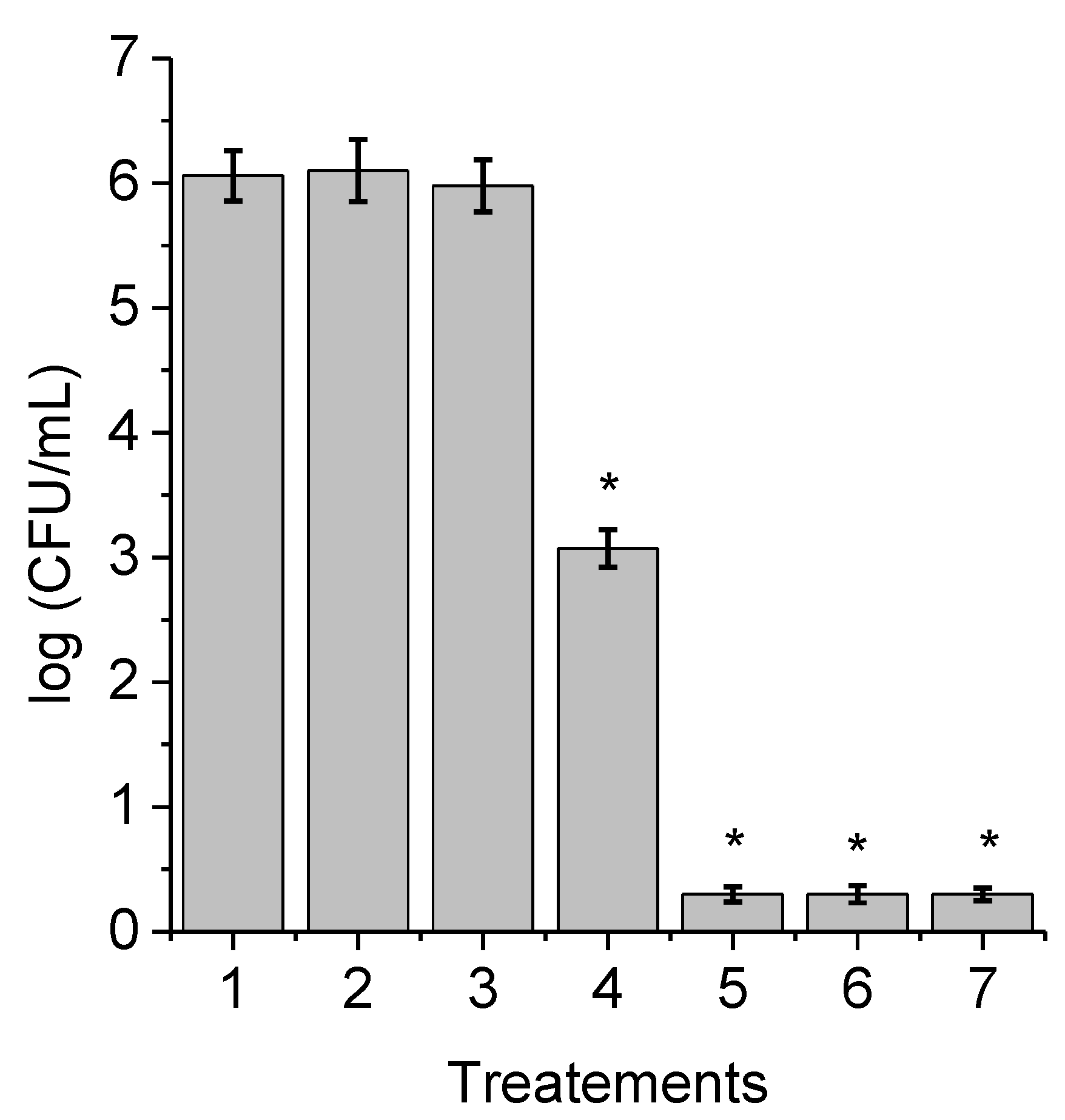
3.1. PDI of C. albicans Planktonic Cells and Photodynamic Action Mechanism

Photosensitized inactivation of C. albicans was investigated after incubation with 2.5 µM TAPC and irradiated with white light for different periods. This concentration was chosen to not produce a complete eradication of C. albicans cells at short irradiation times and thus be able to observe the effect produced by the ROS scavengers. Yeast cell survivals are shown in Figure 2. No toxicity was found for the cells treated with TAPC for 30 min in the dark (Figure 2, line 3). Moreover, the viability of C. albicans cultures without chlorin was not affected by irradiation (Figure 2, line 2). Photoinactivation of C. albicans in presence of TAPC was dependent on irradiation times. After 2 min irradiation, PDT treatment produced a reduction of 3 log in the cell survival. A fast decrease in C. albicans survival (>5 log) was detected after 5 min irradiation and no colony formation was detected under these conditions. An increase in the irradiation to 30 min was not accompanied by an enhancement in the PDI efficiency, indicating a complete eradication of yeast cells.
In order to establish the photodynamic action mechanism sensitized by TAPC that predominantly occurs in the damage of yeast cells, PDI of C. albicans cell suspensions in PBS were compared with those obtained in the presence of ROS scavengers, such as sodium azide, D-mannitol and DMSO (Figure 3, Figure 4 and Figure 5). Cells incubated with these ROS scavengers in the dark or exposed to light in the absence of TAPC do not show a reduction in their viability (Figure 3, Figure 4 and Figure 5, lines 1 and 2). Additionally, no toxicity was found for cells incubated with ROS scavengers and TAPC in the dark (Figure 3, Figure 4 and Figure 5, line 3). To estimate the involvement of O2(1Δg) in the photoinactivation of yeast cells, sodium azide was used as a quencher of O2(1Δg) [22,23]. When cultures were treated with 50 mM sodium azide, a reduction in the photoinactivation was observed for PDI of C. albicans. The presence of azide ions caused a photoprotection in the inactivation of yeast cells, reducing cell death to less than 1 log (Figure 3, lines 4–7). Only a decrease in cell viability of 1 log was observed after 30 min irradiation. This indicates that azide ions produced a significant decrease in TAPC-sensitized photodynamic action by quenching O2(1Δg). On the other hand, D-mannitol was used as an inhibitor of type I reactions because this compound can act as a radical scavenger, such as superoxide anion radical (O2) and hydroxyl radical (HO) [24,25]. In the presence of 100 mM D-mannitol, the photocytotoxic effect induced by TAPC was reduced mainly at shorter irradiation times (Figure 4, lines 4–6). After 2 min irradiation, protection of 1.5 log (Figure 4, line 4) was found in comparison with PDT without D-mannitol (Figure 2, line 4). However, no difference in cell death was observed after 30 min irradiation. Furthermore, PDI experiments were performed in presence of DMSO, a powerful scavenger of HO [26]. The addition of 100 mM DMSO produced a slight photoprotection of 1 log in cell survival of C. albicans after 2 min irradiation (Figure 5, line 4). After 15 min of irradiation, no protection of yeast cells was observed (Figure 5, line 6) in relation to the experiments without DMSO (Figure 2, line 6). Therefore, under similar conditions, the protective effect of DMSO was less than that found for D-mannitol. The photoprotective action produced by D-mannitol and DMSO in C. albicans indicates some contribution of a type I mechanism.
On the other hand, the photostability of a PS is an important requirement for antimicrobial PDI in order to achieve practical applications. Therefore, the photobleaching of TAPC was tested under irradiation conditions used with the cultures. A solution of chlorin in DMF was exposed to white light at different times (Figure S4). The photodegradation was studied by observing the decrease in the absorption of the Soret band. Moreover, the formation of new bands was not detected in the visible region. Photobleaching of TAPC exhibited a slight decrease with the irradiation times, reaching 8.5% decomposition after 30 min.

3.2. Photosensitized Effect of TAPC on the Growth of C. albicans Cells

The photocytotoxic activity mediated by TAPC was investigated on the growth of C. albicans cultures in a Sabouraud broth. These tests were carried out to determine that the PDI of C. albicans is possible when the cells are not starved or in the stationary phase suspended in PBS [20]. Thus, 5 μM TAPC was added to fresh cultures of C. albicans and the cells in the growth medium were continuously irradiated at 37 °C. Figure 6 shows the photocytotoxic effect sensitized by the chlorin on yeast cell growth. As can be observed, C. albicans cells treated with TAPC in the dark or not treated with the PS and irradiated showed similar behavior as the controls. In contrast, cell growth was delayed when C. albicans cultures containing TAPC were irradiated with white light. After 12 h irradiation in the presence of 5 μM chlorin, the cells did not appear to be growing as measured by turbidity at 650 nm.

3.3. Photoinactivation of C. albicans Pseudohyphae

A striking feature of C. albicans is its ability to grow either as a unicellular budding yeast or in filamentous pseudohyphae and hyphae forms [27]. The morphological plasticity of C. albicans is a virulence factor, as the pseudohyphae form has key roles in the infection process [28]. Therefore, the susceptibility of C. albicans pseudohyphae to the photodynamic effect sensitized by TAPC was determined in PBS and in HS. The induction of dimorphic state of C. albicans was obtained when cells were resuspended in HS for 4 h at 37 °C. The formation of pseudohyphae was corroborated by optical microscopy (Figure S2). Photoinactivation of C. albicans pseudohyphae cell suspensions (~106 CFU/mL) incubated with 5 µM TAPC was studied in both media, incubating the cultures for 30 min in the dark at 37 °C. These conditions were selected to obtain a mode optimal for PDI in cell suspensions, according to previous results in planktonic cells [15]. Survivals of pseudohyphae after different periods of irradiations (2, 5, 15 and 30 min) with white light are shown in Figure 7. The viability of the pseudohyphae was unaffected by irradiation without incubation with TAPC. Moreover, no toxicity was found in both media for cells incubated with TAPC in the dark for 30 min (Figure S5). Therefore, the pseudohyphae inactivation originated after irradiation of the cultures treated with the TAPC was due to the photosensitization effect of the chlorin induced by white light. As can be observed in Figure 7, TAPC was effective in photoinactivate pseudohyphae suspended in PBS, producing a decrease in cell viability of 2.7 log after 2 min irradiation. Furthermore, the PDI reached a reduction of 5 log in cell survival when the cultures were irradiated for 30 min. However, TAPC-induced photoinactivation was considerably lower in pseudohyphae suspended in HS at shorter irradiation times. A statistically significant reduction in cell survival of 2 log was observed after 30 min irradiation.

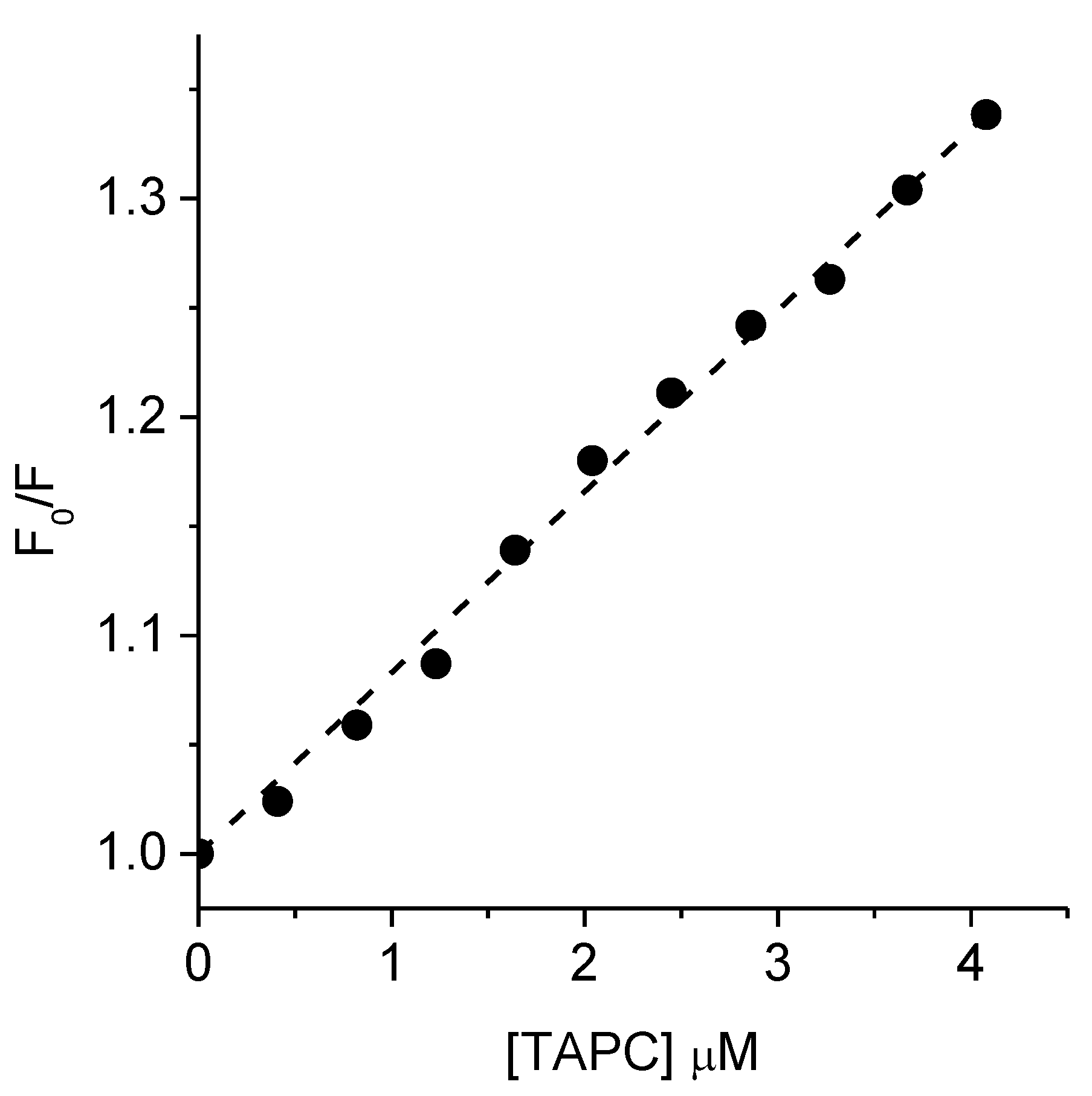
3.4. Binding of TAPC to C. albicans Pseudohyphae

In order to explain the differences observed in the ability of TAPC to inhibit C. albicans pseudohyphae in presence of HS, the binding of the PS to the cells and the amount remaining in the supernatant were determined. The C. albicans pseudohyphae suspensions in PBS or HS (~106 CFU/mL) were incubated with 5 μM TAPC for 30 min at 37 °C in the dark. The amount of chlorin recovered from the pellets and supernatants is shown in Table 1. In PBS, the quantity of TAPC in the supernatant was 1.33 times higher than in the pellet, while this ratio was 11.35 times in HS. These results indicate that in HS the TAPC mainly remained in the supernatant. Additionally, the amount of chlorin recovered in pellets from pseudohyphae treated in HS was ~10 times lower than the PS measured in the supernatant, which only represents 8% chlorin bonded to cells. In contrast, a similar amount of TAPC in pellets compared to supernatant was found in the pseudohyphae treatments in PBS. These results attempt to explain the difference in PDI between the two media.

3.5. Interaction of TAPC with BSA

Albumin is the main protein present in serum and one of the most abundant in humans. This protein is significantly involved in physiological roles and participates in effective drug delivery functions [29]. It has acidic characteristics con a negative net charge at the physiological pH, which produces electrostatic interaction with cationic drugs. Therefore, in order to explain why TAPC binding to cells was reduced in the presence of serum, the interaction of TAPC with albumin was quantitatively evaluated. First, the binding constant (K) of the TAPC-BSA complex was determined in an aqueous medium from the fluorescence intensity changes of TAPC in presence of a different concentration of BSA by Equation (1) [30,31].
1 Δ F = 1 Δ F max + 1 K [ BSA ] ( 1 Δ F max )
where ΔF = F − F0 and ΔFmax = F − F0, in which F0, F and F are the fluorescence intensities of TAPC at 650 nm (λexc = 420 nm) in the absence of BSA, in presence of different concentrations of BSA, and TACP saturated with BSA, respectively. Figure 8 shows the plot 1/ΔF vs. 1/[BSA] with 1/ΔFmax = 0.99 ± 0.02 a.u.−1 and 1/(K ΔFmax) = (1.46 ± 0.02) × 10−5 a.u−1 M−1. Thus, a binding constant K = (6.8 ± 0.2) × 104 M−1 was obtained from the ratio between interest and slope.
Furthermore, an aqueous solution of BSA was titrated with varying concentrations of TAPC. Diminution of BSA emission at 340 nm (λexc = 295 nm) was observed with an increase in TAPC concentrations. These results were used to determine the binding constant K and the number of binding sites n by Equation (2) [30,32].
log ( F 0 F ) F = log K + n   log   [ TAPC ]
where F0 and F fluorescence intensities in the absence or presence of TAPC were represented, respectively. Figure 9 shows the dependence of log [(F0 − F)/F] on the value of log [TAPC], which was fitted by Equation (2). This plot was used to obtain the values of n = 0.99 from the slope and log K = 4.87 from the interest, which gives a value of K = (7.4 ± 0.46) × 104 M−1.
In addition, the intrinsic fluorescence of BSA due to the tryptophan residue (Trp214) was used to evaluate the quenching effect generated by the addition of TAPC [33]. The addition of TAPC produced a noticeable decrease in BSA fluorescence intensity. The results allowed us to estimate what kind of quenching was happening, that is, if it occurs by a static or dynamic process. To analyze the data from the quenching experiments, the Stern–Volmer plot was performed according to Equation (3) [30].
F 0 F = 1 + K SV [ TAPC ] = 1 + k q τ 0 [ TAPC ]  
where F and F0 are the fluorescence intensities in the presence and absence of TAPC, respectively. KSV represents the Stern–Volmer quenching constant, kq is the bimolecular quenching rate constant, and τ0 is the fluorescence lifetime of BSA in absence of TAPC. The Stern–Volmer plot is shown in Figure 10. From the above Equation (3), the values of KSV = (8.29 ± 0.06) × 104 M−1 and kq = (8.29 ± 0.08) × 1012 s−1 M−1 were calculated, considering a τ0 = 10 ns [33,34].

3.6. Inactivation of Planktonic Cells in Presence of BSA

In view of the results of the previous experiments that include the interaction between TAPC with BSA, the PDI of C. albicans planktonic cells were evaluated in the presence of BSA in the media. Thus, C. albicans cell suspensions (~106 CFU/mL) were treated with 5 μM TAPC in PBS containing different amounts of BSA (1 and 4.5% w/v) for 30 min at 37 °C in the dark. The survival curves of yeast cells are shown in Figure 11. These conditions were not toxic neither for the irradiated cells without TAPC nor for the cultures treated with the PS kept in the dark (Figure S6). In absence of BSA, the PDI sensitized by TAPC revealed a marked decrease in C. albicans cell viability of more than 5 log at short irradiation times (<5 min). The viability of cells was dependent on the amount of BSA in the medium. An increase in the BSA concentration produced a decrease in the PDI efficiency. In contrast with those results in PBS, when the cells were supplemented with only 1% BSA a decrease of 2.5 log in the CFU count was observed after 30 min irradiation. At short times, the presence of 4.5% BSA in the PBS significantly reduced the ability of TAPC to cause cell death, being negligible from cell controls. However, when a longer irradiation time (30 min) was used, a statistically significant 2 log decrease was found in cell survival.

4. Discussion

Recent studies indicate that C. albicans is the most prevalent species found in patients with candidemia throughout the world [35]. It was demonstrated an increment in the presence of this yeast in fungal infections both mucocutaneous and invasive, especially in patients immunocompromised or with underlay diseases [36]. Furthermore, clinical failure was continuously observed in treatments involving the use of conventional antifungals. This has become a growing problem for medical systems because conventional drugs for clinical use invariably lead to more widespread resistance from pathogenic yeasts. Therefore, the development of more effective antifungal therapies is imperative [37,38]. In this sense, for some decades PDI was proposed as a new strategy to selectively kill prokaryotic or eukaryotic microorganisms [10,12].
In the search for efficient phototherapeutic agents, in this work, we investigate the capacity of TAPC to eradicate C. albicans in different media and conditions. This PS is characterized by a chlorine unit substituted in the periphery by four terminal amino groups, which are separated from the macrocycle by an aliphatic chain (Figure 1). This spacer gives the amino groups greater mobility to interact with the cell envelope and keeps the properties of the PS unchanged. TAPC does not have intrinsic positive charges in its structure but the basic aliphatic amino groups (3-(N,N-dimethylamine)propanol, pKa = 9.51) in the periphery of the macrocycle can be protonated in aqueous media [39]. The formation of cationic groups on the PS structure allows better interaction with yeast cells [19]. Moreover, TAPC absorbs in the visible region, showing the characteristic Soret band at 421 nm and the four Q-bands between 515 and 650 nm (Figure S7) [15]. In particular, the chlorin exhibited an intense absorption in the phototherapeutic window. Additionally, this compound emits red fluorescence (Figure S7) with quantum yields of 0.15 and it was able to photosensitize O2(1Δg) with quantum yields of 0.54. In addition, TAPC can sensitize the formation of O2 in the presence of NADH through a type I pathway. TAPC showed an appropriate photostability under the irradiation conditions used to inactivate yeast cells. If the sensitizer degrades very rapidly during PDT treatments, the inactivation of microbial cells may be incomplete.
PDI studies using C. albicans planktonic cells in PBS indicated that cultures treated with 2.5 μM TAPC produced an inactivation of 99.9% after 2 min irradiation. Under these conditions, no colony formation units were detected when the C. albicans cells were exposed for 5 min to white light, which means a cell inactivation greater than 99.9996%. In order to find insights into the predominant mechanism of action induced by TAPC, the PDI of C. albicans was investigated in the presence of different ROS scavengers. Sodium azide can avoid the photocytotoxic effect produced by O2(1Δg) in yeast cells [18,23]. A high photoprotection of C. albicans cells sensitized by TAPC was found in presence of azide ions even after 30 min irradiation. This decrease in PDI indicates the involvement of O2(1Δg) in the cell death of C. albicans. On the other hand, D-mannitol was added as a free radical scavenger to evaluate the contribution of the type I mechanism [25,40]. Photoinactivation of C. albicans sensitized by TAPC was reduced in presence of D-mannitol, mainly to the shorter irradiation times used in the PDI. Unlike azide ions, the protective effect of mannitol was not significant at 30 min of irradiation. Furthermore, DMSO was added to determine the formation of HO [26]. Similar to that found with D-mannitol, a reduction in the PDI of C. albicans was observed mainly to short periods of exposure. However, in this case, no protection was found in the photokilling when the cells were irradiated for 15 or 30 min. Therefore, the contribution of the type I pathway for the photokilling of C. albicans induced by TAPC was less important than the mediation of O2(1Δg). Similar results were previously found for the PDI of C. albicans sensitized by cationic porphyrins [23]. Moreover, it was previously found that O2(1Δg) was the main reactive species involved in cell damage induced by a porphyrin containing basic amino substituents [18,41]. Therefore, this family of compounds appears to act primarily through a type II pathway to inactivate C. albicans under aerobic conditions.
The photodynamic activity mediated by TAPC was examined to inactivate C. albicans cells in growth conditions of cultures. The growth was delayed when yeast cells were treated with 5 μM TAPC and continuously irradiated with white light. A smaller delay on the growth of C. albicans cultures was observed with 5,10,15,20-tetra(4-N,N,N-trimethylammoniumphenyl)porphyrin (TMAP4+) [19]. Moreover, 5,10,15,20-tetra(4-N-methylpyridyl)porphyrin (TMPyP4+) showed a lower photodynamic activity on the growth delay [42]. In addition, TAPC was more effective than its tetrapyrrolic analogous 5,10,15,20-tetrakis[4-(3-N,N-dimethylaminopropoxy)phenyl]porphyrin (TAPP) to produce a reduction in the growth of C. albicans [20].
One of the most extraordinary characteristics of C. albicans is its ability to change into different morphological forms ranging from unicellular yeast to true hyphae [27]. Several growth states are observed between those forms, and these are collectively referred to as pseudohyphae [28]. This biological feature is induced strongly in presence of serum and it is commonly considered a virulence factor due to the filamentous forms, which are needed for growth within the host [43]. For this reason, we attempt to evaluate the photoinactivation of C. albicans pseudohyphae incubated with 5 µM TAPC in the presence or absence of HS after different illumination times. TAPC was able to kill pseudohyphae suspensions without HS, producing over 99.999% of photoinactivation after 30 min irradiation. Therefore, in planktonic cells with only 2 min of irradiation, cell survival decreases 3 log, then with 5 min of irradiation it decreases more than 5.5 log. This means that colony formation is not detected according to the quantification method used. The same result occurred at 15 min and 30 min and colony formation was no longer detected. In the case of pseudohyphae, PDI was more progressive, reaching a 5 log decrease after 30 min of irradiation. This indicates that for TAPC, it was more difficult to photoinactivate pseudohyphae than planktonic cells. On the other hand, photokilling of pseudohyphae was reduced to 99% in presence of HS. These results may be due to an interaction between chlorine and serum proteins, which can affect the binding of TAPC to pseudohyphae. Several studies report interactions between PSs and serum albumin [44,45]. Therefore, the uptake of TAPC by pseudohyphae was evaluated using suspensions in PBS and in HS. Furthermore, the amount of PS remaining in the supernatants was determined in both media. The results clearly showed that TAPC was bound to the pseudohyphae. However, the uptake was over five times more concentrated in cells suspended in PBS than those suspended in HS. Moreover, the remaining concentration of TAPC in HS suggests that this chlorin interacts with HS more than fungal cells. This can explain the lower photodynamic activity observed on pseudohyphae suspended in HS. In this sense; when germ tubes (one kind of elongated form) of C. albicans were treated with Photofrin; the uptake of PS was poor in C. albicans blastoconidia grown in nutrient broth. The conversion of blastoconidia to filamentous forms produced a considerable Photofrin uptake. Under this condition, irradiation of the cultures induced considerable cell damage [46]. However, a gradual loss of photosensitivity was observed by increasing serum concentrations, due to the gradual leaching of Photofrin to the external medium [47]. In the present study, we attempted to understand the relationship between TAPC and HS for a plausible cause of minor photodynamic effect in the treatment of pseudohyphae suspended in serum. In this sense, albumin is a 66 kDa water-soluble, a monomeric protein present in blood plasma and serum in high concentrations (about 40–50 mg/mL). Its structure is stabilized with a disulfide bond, and its function is to be a carrier of several solutes including cations, fatty acids, and therapeutic drugs among others [48]. Therefore, in the present investigation, two approaches were used to analyze the interaction of TAPC with BSA in aqueous media. First, the fluorescence emission of TAPC increased as BSA was added as a titrant. On the other hand, the intrinsic fluorescence of BSA, due to the tryptophan residue present in its structure, decreased with increasing TAPC concentration as it is titrated. From these results, a binding constant greater than 104 M−1 was obtained with the number of binding sites of one, suggesting an effective interaction between TAPC and BPS. Furthermore, from the Stem–Volmer constant a value of kq greater than 1012 s−1 M−1 was calculated. This result indicates that kq is more than two orders higher than the limiting diffusion constant of the biomolecule, which proved evidence that there was specific interaction between BSA and TAPC. Therefore, quenching between TAPC and albumin occurred through a static process. Similar behavior was previously found for the interaction of cationic PSs with albumin [33,49].
In order to evaluate the effect of albumin in vitro, the PDI of C. albicans cell suspensions in PBS sensitized by TAPC was investigated in presence of different concentrations of albumin (0, 1 and 4.5% w/v). These concentrations of BSA were selected because the total protein concentration of human blood plasma is about 7.3% of which 4.5% is albumin [50]. When the inactivation was carried out with the planktonic cells suspended in PBS using 2.5 μM TAPC, complete eradication of C. albicans occurred after 5 min irradiation. In presence of 5 μM TAPC, this result was obtained with only 2 min irradiation. However, in the presence of BSA (1% and 4.5%) the photoinactivation decreases due to the interaction of TAPC with albumin. A considerable reduction of the photocytotoxic activity was observed with 1%, even the inactivation of C. albicans was insignificant in the presence of 4.5% of BSA in short periods of irradiation. Therefore, albumin plays an important role in the protection of C. albicans when human serum or plasma is in contact with yeast, reducing the effectiveness of the PSs. It was previously reported that changing the suspending medium from PBS to human blood plasma reduced the PDI of microorganisms sensitized by a tricationic porphyrin [50]. In particular, C. albicans was the most resistant microorganism in human blood plasma in a dose-dependent manner. At pH 7.4, albumin has a normal heart-like structure (N isoform) and has an expected total charge of nine negative charges [48]. This means that it interacts preferentially with positively charged molecules, such as TAPC that can be protonated at physiological pH [41]. These results are consistent with those previously found by fluorescence spectral analysis and suggest that in the presence of BSA there is a competition between the binding affinity of TAPC with albumin and C. albicans cells. This effect produced a reduction of TAPC bound to yeast cells and consequently a decrease in the photoinactivation capacity of the PS.

5. Conclusions

Eradication of C. albicans planktonic cells was possible at low TAPC concentrations and short irradiation times. Furthermore, this chlorin was able to suspend the growth of yeasts in the culture medium by means of continuous light irradiation. The most interesting result was the ability of TAPC to eradicate pseudohyphae, one of the major virulence factors in PBS. However, the presence of HS in the media reduced the photokilling capacity sensitized by this chlorin. This protection was assigned to the interaction of TAPC with the proteins contained in the HS, which considerably reduced the binding of the PS to pseudohyphae. In order to check this statement, BSA was used as a protein model contained in the serum. The high K and kq values found for the TAPC-albumin complex indicated that the quenching process occurred through a static process. As a consequence of this interaction, the photoinactivation of C. albicans cells in PBS was considerably decreased in the presence of albumin, although photoinactivation is still significant at longer irritation times. Therefore, this investigation allows optimizing the best conditions to inactivate C. albicans under different culture conditions.

Supplementary Materials

The following are available online at https://www.mdpi.com/article/10.3390/photochem1030033/s1. Strains and cultures of C. albicans; model of irradiation systems for PDI and emission spectrum of the light source (Figure S1); microscopic observation of C. albicans pseudohyphae (Figure S2); calibration curve for TAPC (Figure S3); photobleaching of TAPC (Figure S4); survival curves of C. albicans pseudohyphae incubated with TAPC and containing HS in the dark (Figure S5); survival curves of C. albicans incubated with TAPC and containing BSA in the dark (Figure S6) and spectra of TAPC (Figure S7).

Author Contributions

Conceptualization, P.V.C., M.G.A., M.E.P., D.D.F. and E.N.D.; methodology, P.V.C., D.D.F., M.E.P. and M.G.A.; validation, P.V.C., D.D.F., M.E.P. and M.G.A.; formal analysis, P.V.C., D.D.F., M.E.P. and M.G.A.; investigation, P.V.C., D.D.F., M.E.P. and M.G.A.; data curation, E.N.D.; writing—original draft preparation, P.V.C. and M.G.A.; writing—review and editing, E.N.D.; visualization, P.V.C., D.D.F., M.G.A. and E.N.D.; supervision, E.N.D.; project administration, E.N.D. All authors have read and agreed to the published version of the manuscript.

Funding

This research was funded by ANPCYT (PICT 0667/16) and UNRC-SECYT (PPI-2020 Res. 083/20).

Institutional Review Board Statement

Not applicable.

Informed Consent Statement

Not applicable.

Data Availability Statement

The data presented in this study are available in the article.

Acknowledgments

This work was supported by UNRC-SECYT (PPI-2020 Res. 083/20) and ANPCYT (PICT 2019- 02391). P.V.C., M.G.A. and E.N.D. are Scientific Members of CONICET. M.E.P. thanks CONICET for the research fellowship.

Conflicts of Interest

The authors declare no conflict of interest.

References

  1. Janbon, G.; Quintin, J.; Lanternier, F.; d’Enfert, C. Studying fungal pathogens of humans and fungal infections: Fungal diversity and diversity of approaches. Microbes Infect. 2019, 21, 237–245. [Google Scholar] [CrossRef] [PubMed]
  2. Pathakumari, B.; Liang, G.; Liu, W. Immune defence to invasive fungal infections: A comprehensive review. Biomed. Pharmacother. 2020, 130, 110550. [Google Scholar] [CrossRef]
  3. Perfect, J.R.; Ghannoum, M. Emerging issues in antifungal resistance. Infect. Dis. Clin. N. Am. 2020, 34, 921–943. [Google Scholar] [CrossRef]
  4. Williams, T.J.; Harvey, S.; Armstrong-James, D. Immunotherapeutic approaches for fungal infections. Curr. Opin. Microbiol. 2020, 58, 130–137. [Google Scholar] [CrossRef]
  5. Limon, J.J.; Skalski, J.H.; Underhill, D.M. Commensal fungi in health and disease. Cell Host Microbe 2017, 22, 156–165. [Google Scholar] [CrossRef]
  6. Antinori, S.; Milazzo, L.; Sollima, S.; Galli, M.; Corbellino, M. Candidemia and invasive candidiasis in adults: A narrative review. Eur. J. Intern. Med. 2016, 34, 21–28. [Google Scholar] [CrossRef]
  7. Epelbaum, O.; Chasan, R. Candidemia in the intensive care unit. Clin. Chest Med. 2017, 38, 493–509. [Google Scholar] [CrossRef] [PubMed]
  8. Sardi, J.C.O.; Scorzoni, L.; Bernardi, T.; Fusco-Almeida, A.M.; Mendes Giannini, M.J.S. Candida species: Current epidemiology, pathogenicity, biofilm formation, natural antifungal products and new therapeutic options. J. Med. Microbiol. 2013, 62, 10–24. [Google Scholar] [CrossRef]
  9. Capoor, M.R.; Subudhi, C.P.; Collier, A.; Bal, A.M. Antifungal stewardship with an emphasis on candidaemia. J. Glob. Antimicrob. Resist. 2019, 19, 262–268. [Google Scholar] [CrossRef]
  10. Calzavara-Pinton, P.; Rossi, M.T.; Sala, R.; Venturini, M. Photodynamic antifungal chemotherapy. Photochem. Photobiol. 2012, 88, 512–522. [Google Scholar] [CrossRef] [PubMed]
  11. Wang, Y.-Y.; Liu, Y.-C.; Sun, H.; Guo, D.-S. Type I photodynamic therapy by organic-inorganic hybrid materials: From strategies to applications. Coord. Chem. Rev. 2019, 395, 46–62. [Google Scholar] [CrossRef]
  12. Durantini, A.M.; Heredia, D.A.; Durantini, J.E.; Durantini, E.N. BODIPYs to the rescue: Potential applications in photodynamic inactivation. Eur. J. Med. Chem. 2018, 144, 651–661. [Google Scholar] [CrossRef]
  13. Alves, E.; Faustino, M.A.F.; Neves, M.G.P.M.S.; Cunha, Â.; Nadais, H.; Almeida, A. Potential applications of porphyrins in photodynamic inactivation beyond the medical scope. J. Photochem. Photobiol. C Photochem. Rev. 2015, 22, 34–57. [Google Scholar] [CrossRef] [Green Version]
  14. Martinez De Pinillos Bayona, A.; Mroz, P.; Thunshelle, C.; Hamblin, M.R. Design features for optimization of tetrapyrrole macrocycles as antimicrobial and anticancer photosensitizers. Chem. Biol. Drug. Des. 2017, 89, 192–206. [Google Scholar] [CrossRef] [Green Version]
  15. Ferreyra, D.D.; Reynoso, E.; Cordero, P.; Spesia, M.B.; Alvarez, M.G.; Milanesio, M.E.; Durantini, E.N. Synthesis and properties of 5,10,15,20-tetrakis[4-(3-N,N-dimethylaminopropoxy)phenyl] chlorin as potential broad-spectrum antimicrobial photosensitizers. J. Photochem. Photobiol. B Biol. 2016, 158, 243–251. [Google Scholar] [CrossRef] [PubMed]
  16. Ballatore, M.B.; Milanesio, M.E.; Fujita, H.; Lindsey, J.S.; Durantini, E.N. Bacteriochlorin-bis(spermine) conjugate affords an effective photodynamic action to eradicate microorganisms. J. Biophotonics 2020, 13, e201960061. [Google Scholar] [CrossRef]
  17. Pérez, M.E.; Durantini, J.E.; Reynoso, E.; Alvarez, M.G.; Milanesio, M.E.; Durantini, E.N. Porphyrin-Schiff base conjugates bearing basic amino groups as antimicrobial phototherapeutic agents. Molecules 2021, 26, 5877. [Google Scholar] [CrossRef] [PubMed]
  18. Quiroga, E.D.; Cordero, P.; Mora, S.J.; Alvarez, M.G.; Durantini, E.N. Mechanistic aspects in the photodynamic inactivation of Candida albicans sensitized by a dimethylaminopropoxy porphyrin and its equivalent with cationic intrinsic charges. Photodiagn. Photodyn. Ther. 2020, 31, 101877. [Google Scholar] [CrossRef] [PubMed]
  19. Cormick, M.P.; Alvarez, M.G.; Rovera, M.; Durantini, E.N. Photodynamic inactivation of Candida albicans sensitized by tri- and tetra-cationic porphyrin derivatives. Eur. J. Med. Chem. 2009, 44, 1592–1599. [Google Scholar] [CrossRef] [PubMed]
  20. Quiroga, E.D.; Mora, S.J.; Alvarez, M.G.; Durantini, E.N. Photodynamic inactivation of Candida albicans by a tetracationictentacle porphyrin and its analogue without intrinsic charges inpresence of fluconazole. Photodiagn. Photodyn. Ther. 2016, 13, 334–340. [Google Scholar] [CrossRef]
  21. Samaranayake, Y.H.; Cheung, B.P.K.; Yau, J.Y.Y.; Yeung, S.K.W.; Samaranayake, L.P. Human serum promotes candida albicans biofilm growth and virulence gene expression on silicone bio-material. PLoS ONE 2013, 8, e62902. [Google Scholar] [CrossRef] [Green Version]
  22. Da Silva, E.F.F.; Pedersen, B.W.; Breitenbach, T.; Toftegaard, R.; Kuimova, M.K.; Arnaut, L.G.; Ogilby, P.R. Irradiation- and sensitizer-dependent changes in the lifetime of intracellular singlet oxygen produced in a photosensitized process. J. Phys. Chem. B 2012, 116, 445–461. [Google Scholar] [CrossRef]
  23. Cormick, M.P.; Quiroga, E.D.; Bertolotti, S.G.; Alvarez, M.G.; Durantini, E.N. Mechanistic insight of the photodynamic effect induced by tri- and tetra-cationic porphyrins on Candida albicans cells. Photochem. Photobiol. Sci. 2011, 10, 1556–1561. [Google Scholar] [CrossRef] [PubMed]
  24. Maisch, T.; Bosl, C.; Szeimies, R.M.; Lehn, N.; Abels, C. Photodynamic effects of novel XF porphyrin derivatives on prokaryotic and eukaryotic cells. Antimicrob. Agents Chemother. 2005, 49, 1542–1552. [Google Scholar] [CrossRef] [PubMed] [Green Version]
  25. Gsponer, N.S.; Agazzi, M.L.; Spesia, M.B.; Durantini, E.N. Approaches to unravel pathways of reactive oxygen species in the photoinactivation of bacteria induced by a dicationic fulleropyrrolidini-um derivative. Methods 2016, 109, 167–174. [Google Scholar] [CrossRef] [PubMed]
  26. Dalle, A.A.; Domergue, L.; Fourcade, F.; Assadi, A.A.; Djelal, H.; Lendormi, T.; Soutrel, I.; Taha, S.; Amrane, A. Efficiency of DMSO as hydroxyl radical probe in an electrochemical advanced oxidation process—Reactive oxygen species monitoring and impact of the current density. Electrochim. Acta 2017, 246, 1–8. [Google Scholar] [CrossRef]
  27. Sudbery, P.E. Growth of Candida albicans hyphae. Nat. Rev. Microbiol. 2011, 9, 737–748. [Google Scholar] [CrossRef]
  28. Noble, S.M.; Gianetti, B.A.; Witchley, J.N. Candida albicans cell-type switching and functional plasticity in the mammalian host. Nat. Rev. Microbiol. 2017, 15, 96–108. [Google Scholar] [CrossRef] [Green Version]
  29. Bteich, M. An overview of albumin and alpha-1-acid glycoprotein main characteristics: Highlighting the roles of amino acids in binding kinetics and molecular interactions. Heliyon 2019, 5, e02879. [Google Scholar] [CrossRef] [Green Version]
  30. An, W.; Jiao, Y.; Dong, C.; Yang, C.; Inoue, Y.; Shua, S. Spectroscopic and molecular modeling of the binding of meso-tetrakis(4-hydroxyphenyl)porphyrin to human serum albumin. Dyes Pigm. 2009, 81, 1–9. [Google Scholar] [CrossRef]
  31. Bhattacharya, B.; Nakka, S.; Guruprasad, L.; Samanta, A. Interaction of bovine serum albumin with dipolar molecules: Fluorescence and molecular docking studies. J. Phys. Chem. B 2009, 113, 2143–2150. [Google Scholar] [CrossRef]
  32. Bose, B.; Dube, A. Interaction of chlorin p6 with bovine serum albumin and photodynamic oxidation of protein. J. Photochem. Photobiol. B Biol. 2006, 86, 49–55. [Google Scholar] [CrossRef] [PubMed]
  33. Durmus, M.; Yaman, H.; Göl, C.; Ahsen, V.; Nyokong, T. Water-soluble quaternized mercaptopyridine-substituted zinc-phthalocyanines: Synthesis, photophysical, photochemical and bovine serum albumin binding properties. Dyes Pigm. 2011, 91, 153–163. [Google Scholar] [CrossRef]
  34. Zhang, Y.-Z.; Zhou, B.; Liu, Y.-X.; Zhou, C.-X.; Ding, X.-L.; Liu, Y. Fluorescence study on the interaction of bovine serum albumin with p-aminoazobenzene. J. Fluoresc. 2008, 18, 109–118. [Google Scholar] [CrossRef]
  35. McCarty, T.P.; White, C.M.; Pappas, P.G. Candidemia and invasive candidiasis. Infect. Dis. Clin. N. Am. 2021, 35, 389–413. [Google Scholar] [CrossRef] [PubMed]
  36. Austermeier, S.; Kasper, L.; Westman, J.; Gresnigt, M.S. I want to break free-macrophage strategies to recognize and kill Candida albicans, and fungal counter-strategies to escape. Curr. Opin. Microbiol. 2020, 58, 15–23. [Google Scholar] [CrossRef]
  37. Ben-Ami, R.; Kontoyiannis, D.P. Resistance to antifungal drugs. Infect. Dis. Clin. N. Am. 2021, 35, 279–311. [Google Scholar] [CrossRef]
  38. Garcia-Rubio, R.; Monteiro, M.C.; Mellado, E. Azole antifungal drugs: Mode of action and resistance. Encyclop. Mycol. 2021, 1, 427–437. [Google Scholar] [CrossRef]
  39. Rayer, A.V.; Sumon, K.Z.; Jaffari, L.; Henni, A. Dissociation constants (pKa) of tertiary and cyclic amines: Structural and temperature dependences. J. Chem. Eng. Data 2014, 59, 3805–3813. [Google Scholar] [CrossRef]
  40. Ballatore, M.B.; Spesia, M.B.; Milanesio, M.E.; Durantini, E.N. Mechanistic insight into the photodynamic effect mediated by porphyrin-fullerene C60 dyads in solution and in Staphylococcus aureus cells. RSC Adv. 2018, 8, 22876–22886. [Google Scholar] [CrossRef] [Green Version]
  41. Scanone, A.C.; Gsponer, N.S.; Alvarez, M.G.; Durantini, E.N. Porphyrins containing basic aliphatic amino groups as potential broad spectrum antimicrobial agents. Photodiagn. Photodyn. Ther. 2018, 24, 220–227. [Google Scholar] [CrossRef] [PubMed]
  42. Quiroga, E.D.; Alvarez, M.G.; Durantini, E.N. Susceptibility of Candida albicans to photodynamic action of 5,10,15,20-tetra(4-N-methylpyridyl)porphyrin in different media. FEMS Immunol. Med. Microbiol. 2010, 60, 123–131. [Google Scholar] [CrossRef] [PubMed] [Green Version]
  43. Wu, Y.; Wu, M.; Wang, Y.; Chen, Y.; Gao, J.; Ying, C. ERG11 couples oxidative stress adaptation, hyphal elongation and virulence in Candida albicans. FEMS Yeast Res. 2018, 18, 1–7. [Google Scholar] [CrossRef] [PubMed]
  44. Lebedeva, N.S.; Gubarev, Y.A.; Yurina, E.S.; Syrbu, S.A. Features of interaction of tetraiodide meso-tetra(N-methyl-3-pyridyl) porphyrin with bovine serum albumin. J. Mol. Liq. 2018, 265, 664–667. [Google Scholar] [CrossRef]
  45. Kollara, J.; Machacek, M.; Jancarova, A.; Kubat, P.; Kucera, R.; Miletina, M.; Novakova, V.; Zimcika, P. Effect of bovine serum albumin on the photodynamic activity of sulfonated tetrapyrazino-porphyrazine. Dyes Pigm. 2019, 162, 358–366. [Google Scholar] [CrossRef]
  46. Bliss, J.M.; Bigelow, C.E.; Foster, T.H.; Haidaris, C.G. Susceptibility of Candida species to photodynamic effects of photofrin. Antimicrob. Agents Chemother. 2004, 48, 2000–2006. [Google Scholar] [CrossRef] [Green Version]
  47. Chabrier-Roselló, Y.; Foster, T.H.; Pérez-Nazario, N.; Mitra, S.; Haidaris, C.G. Sensitivity of Candida albicans germ tubes and biofilms to photofrin-mediated phototoxicity. Antimicrob. Agents Chemother. 2005, 49, 4288–4295. [Google Scholar] [CrossRef] [PubMed] [Green Version]
  48. Baler, K.; Martin, O.A.; Carignano, M.A.; Ameer, G.A.; Vila, J.A.; Szleifer, I. Electrostatic unfolding and interactions of albumin driven by pH changes: A molecular dynamics study. J. Phys. Chem. B 2014, 118, 921–930. [Google Scholar] [CrossRef]
  49. Zhou, B.; Zhang, Z.; Zhang, Y.; Li, R.; Xiao, Q.; Liu, Y.; Li, Z. Binding of cationic porphyrin to human serum albumin studied using comprehensive spectroscopic methods. J. Pharm. Sci. 2009, 98, 105–113. [Google Scholar] [CrossRef]
  50. Lambrechts, S.A.G.; Aalders, M.C.G.; Verbraak, F.D.; Lagerberg, J.W.M.; Dankert, J.B.; Schuitmaker, J.J. Effect of albumin on the photodynamic inactivation of microorganisms by a cationic porphyrin. J. Photochem. Photobiol. B Biol. 2005, 79, 51–57. [Google Scholar] [CrossRef]
Figure 1. Molecular structure of TAPC.
Photochem 01 00033 g001
Figure 2. Survival of C. albicans (~106 UFC/mL) incubated with 2.5 μM TAPC in dark for 30 min to 37 °C and irradiated with white light (90 mW/cm2) for different times; (1) dark control; (2) irradiated control; (3) cells treated with TAPC in dark; (4) cells treated with TAPC and 2 min irradiation; (5) cells treated with TAPC and 5 min irradiation; (6) cells treated with TAPC and 15 min irradiation; 7) cells treated with TAPC and 30 min irradiation (* p < 0.05, compared with control).
Photochem 01 00033 g002
Figure 3. Survival of C. albicans (~106 UFC/mL) incubated with 50 mM NaN3 and 2.5 μM TAPC in dark for 30 min to 37 °C and irradiated with white light (90 mW/cm2) for different times; (1) dark control treated with NaN3; (2) irradiated control treated with NaN3; (3) cells treated with NaN3 and TAPC in dark; (4) cells treated with NaN3 and TAPC and 2 min irradiation; (5) cells treated with NaN3 and TAPC and 5 min irradiation; (6) cells treated with NaN3 and TAPC and 15 min irradiation; (7) cells treated with NaN3 and TAPC and 30 min irradiation (* p < 0.05, compared with control).
Photochem 01 00033 g003
Figure 4. Survival of C. albicans (~106 UFC/mL) incubated with 100 mM D-mannitol and 2.5 μM TAPC in dark for 30 min to 37 °C and irradiated with white light (90 mW/cm2) for different times; (1) dark control treated with D-mannitol; (2) irradiated control treated with D-mannitol; (3) cells treated with D-mannitol and TAPC in dark; (4) cells treated with D-mannitol and TAPC and 2 min irradiation; (5) cells treated with D-mannitol and TAPC and 5 min irradiation; (6) cells treated with D-mannitol and TAPC and 15 min irradiation; (7) cells treated with D-mannitol and TAPC and 30 min irradiation (* p < 0.05, compared with control).
Photochem 01 00033 g004
Figure 5. Survival of C. albicans (~106 UFC/mL) incubated with 100 mM DMSO and 2.5 μM TAPC in dark for 30 min to 37 °C and irradiated with white light (90 mW/cm2) for different times; (1) dark control treated with DMSO; (2) irradiated control treated with DMSO; (3) cells treated with DMSO and TAPC in dark; (4) cells treated with DMSO and TAPC and 2 min irradiation; (5) cells treated with DMSO and TAPC and 5 min irradiation; (6) cells treated with DMSO and TAPC and 15 min irradiation; (7) cells treated with DMSO and TAPC and 30 min irradiation (* p < 0.05, compared with control).
Photochem 01 00033 g005
Figure 6. Growth curves of C. albicans treated with 5 µM TAPC (▲) and exposed to white light in SB at 37 °C. Controls: untreated cells in dark (○), untreated cells irradiated (●), cells treated with 5 µM TAPC in dark (△).
Photochem 01 00033 g006
Figure 7. Survival curves of pseudohyphae of C. albicans (~106 CFU/mL) incubated with 5 μM TAPC in PBS (▲) and in HS (▼) for 30 min at 37 °C in dark and exposed to visible light for different irradiation times. Control pseudohyphae of C. albicans untreated with TAPC and irradiated (●), (* p < 0.05, compared with control).
Photochem 01 00033 g007
Figure 8. Titration of TAPC (0.5 × 10⁻⁶ M) with BSA (λexc = 420 nm, λem = 650 nm) in water. Lineal fit by Equation (1) (dashed line).
Photochem 01 00033 g008
Figure 9. Titration of BSA (3 × 10⁻⁵ M) with TAPC (λexc = 295 nm, λem = 340 nm) in water. Lineal fit by Equation (2) (dashed line).
Photochem 01 00033 g009
Figure 10. Stern–Volmer plot of TAPC quenching of BSA (3 × 10⁻⁵ M) in water (λexc = 295 nm, λem = 340 nm).
Photochem 01 00033 g010
Figure 11. Survival curves of C. albicans (~106 CFU/mL) incubated with 5 μM TAPC in PBS (▲) and containing 1% (■) and 4.5% BSA (▼) for 30 min at 37 °C in dark and exposed to white light for different irradiation times. Control of C. albicans untreated with TAPC and irradiated (●), C. albicans treated with 5 µM TAPC in PBS in dark (△) (* p < 0.05, compared with control).
Photochem 01 00033 g011
Table 1. Binding of TAPC to C. albicans pseudohyphae cell suspensions (~106 CFU/mL) in PBS and HS incubated with 5 μM PS for 30 min in dark at 37 °C.
MediumTAPC (nmol/106 Cells)TAPC (%)
Cells-PBS a2.23 ± 0.2643
Supernatant-PBS b2.96 ± 0.4157
Cells-HS c0.45 ± 0.268
Supernatant-HS d5.11 ± 0.7492
a Pellet of cells in PBS, b supernatant in PBS, c Pellet of cells in HS, d supernatant in HS.
Publisher’s Note: MDPI stays neutral with regard to jurisdictional claims in published maps and institutional affiliations.

