Article

Clinical Validation of Estimated Muscle Activations during Phases of Elderly Gait

by
Athanasios Gkrekidis
1,
Georgios Giarmatzis
1,
Dimitrios Menychtas
1,
Evangelos Karakasis
1,
Vassilios Gourgoulis
1,
Maria Michalopoulou
1,
Ilias Smilios
1,
Helen T. Douda
1,
Georgios Ch. Sirakoulis
2 and
Nikolaos Aggelousis
1,*
1
School of Physical Education and Sport Science, Democritus University of Thrace, 69100 Komotini, Greece
2
Department of Electrical and Computer Engineering, Democritus University of Thrace, 67100 Xanthi, Greece
*
Author to whom correspondence should be addressed.
Biomechanics 2023, 3(4), 552-560; https://doi.org/10.3390/biomechanics3040044
Submission received: 30 August 2023 / Revised: 26 October 2023 / Accepted: 14 November 2023 / Published: 16 November 2023

Abstract

:
This study validated muscle activation estimations generated by OpenSim during the gait of elderly fallers. Ten healthy elderly participants walked on an instrumented treadmill, monitored by motion capture, force platforms, and 12 surface EMG sensors. Static optimization was used to calculate muscle activations, evaluated through cosine similarity, comparing them with EMG signals from 12 muscles of the right leg. Findings revealed varied similarity levels across muscles and gait phases. During stance phase, tibialis anterior (TIBA), peroneus longus (PERL), soleus (SOL), gastrocnemius lateralis (GASL), semitendinosus (SEMI), tensor fasciae latae (TFL), and rectus femoris (RECF) demonstrated poor similarity (cosim < 0.6), while gluteus medius (GMED), biceps femoris long head (BFLH), and vastus lateralis (VL) exhibited moderate similarity (0.6 ≤ cosim ≤ 0.8), and gluteus maximus (GMAX) and vastus medialis (VASM) displayed high similarity (cosim > 0.8). During the swing phase, only SOL demonstrated inadequate similarity, while GASL, GMAX, GMED, BFLH, SEMI, TFL, RECF, and VASL exhibited moderate similarity, and TIBA, PERL, and VASM showed high similarity. Comparing the different 10% intervals of the gait cycle generally produced more favorable similarity results. For most of the muscles and intervals, good agreement was found. Moderate agreement was estimated in the cases of TIBA (0–10%), PERL (60–70%), GASL (60–70%), TFL (10–20%), RECF (0–10%, 80–100%), and GMED (50–60%). Bad agreement was found in the cases of SOL (60–70%), GASL (0–10%), and TFL (0–10%). In conclusion, the study’s validation outcomes were acceptable in most cases, underlining the potential for user-friendly musculoskeletal modeling routines to study muscle output during elderly gait.

1. Introduction

After the age of 65, the frequency of falls and the severity of the resulting injuries increases in community-dwelling adults [1,2]. Typical related injuries vary between bruising, lacerations, and fractures to the upper and lower extremities, while some may even prove to be fatal [1,3,4,5]. Furthermore, long hospitalizations result in financial over-burdening of national health systems [6,7,8], with costs rising to €474.4 million in countries in the EU like Holland [9]. Their psychological impact is also worth noting since the fear of falling may impede the individual during everyday living and make him/her feel nonautonomous [1,10].
Studying the mechanics of falls and devising strategies for prevention is of utmost importance. This involves identifying factors that elevate the risk of falling and implementing effective interventions, such as exercise programs aimed at improving balance. Postural balance depends on various systems such as vestibular, visual, proprioceptive, and exteroceptive systems [11]. Falls often result from a person’s loss of stability, typically occurring when the body’s center of mass goes beyond the limits of its support base [12]. Utilizing motion capture analysis systems can aid in determining crucial biomechanical measurements, which can be employed to systematically investigate the underlying causes of balance and other gait deficits [5,13,14]. These systems capture kinematic data, which, when combined with ground reaction forces from force plates, allows for the calculation of joint angles and moments during various human movements using inverse kinematics and inverse dynamics algorithms.
Nevertheless, these methods are limited in their ability to estimate the muscular forces exerted by the body to execute the observed motions. This information is vital for gaining deeper insights into the functioning of the human body. To tackle this challenge, musculoskeletal modeling emerges as the sole non-invasive approach to acquire such valuable insights. OpenSim, an open-source software, serves as a valuable tool for musculoskeletal modeling and dynamic simulations of movement. It facilitates the estimation of muscle and joint forces, along with muscle activation patterns, thereby bridging the gap in our understanding [15]. The static optimization algorithm of OpenSim can be used to estimate muscle activations and forces based on kinetic and kinematic data from 3D motion analysis systems [16]. Since in vivo data of muscle forces do not exist, it is necessary to validate such outputs against indirect signals of muscle function, such as electromyography (EMG). Several studies have validated OpenSim estimations of muscle activation in young individuals [16,17,18,19,20], yielding diverse outcomes. While some studies have demonstrated strong similarity between recorded and calculated muscle activations [16,17,20], others have shown less favorable results [18,19]. However, it is important to exercise caution when extending these conclusions to the elderly population. This is because the way elderly individuals walk differs significantly, marked by shorter stride lengths, longer periods of double-support stance, reduced push-off power, and increased gait variability compared to younger individuals [21], among other distinctive characteristics.
To our knowledge there are only a few studies that checked the validity of the estimated muscle activations of OpenSim or other musculoskeletal modeling software during elderly gait. In the study of [22], significant correlations were found between the recorded muscle activity patterns and those calculated using the Anybody software for the vastus lateralis, gastrocnemius medialis, and tibialis anterior muscles in elderly individuals while pedaling. Low-to-moderate agreement between OpenSim results and EMG data during different gait subphases was reported by Karimi et al. [23] for only four elderly lower limb muscles. Two studies reported good agreement between simulated and experimental data for several lower limb elderly muscles for gait [24,25] or single step in reactive balance tests [26]; however, no explicit quantitative measures for each muscle are reported to draw safe conclusions on the validity of their outcomes. From the above, there is a substantial knowledge gap on the validity of calculated muscle forces in a variety of lower limb muscles during elderly gait.
Hence, this study aims to report on the validity of the estimated muscle activations from OpenSim during elderly gait in a variety of muscles along and in different parts of the gait cycle. We expect varying results amongst the muscles, as derived from the literature. The results will enhance usage of MSK modeling to address specific research questions regarding elderly locomotion.

2. Materials and Methods

2.1. Participants

A cohort of ten healthy senior citizens, consisting of one male and nine females, took part in a treadmill walking session. Their average age was 71.7 years (±8 years), with a mean weight of 69.1 kg (±7.78 kg) and an average height of 1.55 m (±0.06 m). These participants were selected between December 2022 and February 2023, provided their informed consent, and the study was approved by the Research Ethics Committee of Democritus University of Thrace, Greece. To be eligible for participation, individuals had to be 65 years or older, capable of walking without assistance, and free from any medical conditions affecting mobility, such as lower limb pain.

2.2. Gait Analysis

The gait analysis laboratory was equipped with a 10-camera motion capture system (Vicon Motion Systems), operating at a sampling rate of 100 Hz, a split-belt instrumented treadmill (Bertec , Columbus, OH, USA) with a sampling rate of 2000 Hz, and 12 wireless EMG sensors (Ultium Noraxon, Scottsdale, AZ, USA) with a sampling rate of 2000 Hz. The system was used to record and digitize synchronous 3D marker trajectories, surface EMG, and ground reaction forces data during gait on the instrumented treadmill. Fifty-seven retroreflective markers were affixed to specific anatomical locations on the participants’ bodies, based on full body protocol according to CGM2.4 guidelines [27].
The surface EMG sensors were accurately positioned on each participant, adhering to the guidelines established by SENIAM [28]. These sensors were firmly attached to specific muscles, including the lateral gastrocnemius (GASL), soleus (SOL), tibialis anterior (TIBA), peroneus longus (PERL), vastus lateralis (VASL), vastus medialis (VASM), rectus femoris (RECF), gluteus medius (GMED), gluteus maximus (GMAX), long head of biceps femoris (BFLH), semitendinosus (SEMI), and tensor fasciae latae (TFL) of the right leg. Subsequently, the recorded EMG data underwent a series of processing steps. This included applying a bandpass filter within the frequency range of 30–300 Hz, followed by rectification of the signals. To further refine the data, a low-pass filter at 6 Hz was applied. Lastly, the gait cycle data for each individual were normalized relative to their respective local peak EMG values.
The participants were first asked to walk at their normal pace on the ground to determine their natural walking speed. Then, each participant was granted a period of three minutes to familiarize themselves with walking on the split-belt treadmill, maintaining their natural speed without supporting themselves and walking as they normally would in their everyday life. The recording session extended for approximately one minute, and the last ten consecutive gait cycles were selected for further analysis to ensure appropriate levels of familiarization with the procedure.

2.3. Musculoskeletal Modeling

A generic full body musculoskeletal model [29] was scaled to each participant based on a static trial and individual mass. Joint angles for the right side were calculated using the inverse kinematics tool of OpenSim (version 4.3). Then, OpenSim static optimization (SO) analysis was performed to determine the muscle activations of the right limb by minimizing the sum of squared muscle activations for each frame.

2.4. Statistics

The agreement between the muscle activation estimations obtained from OpenSim and the recorded EMG data was evaluated using cosine similarity (cosim). This assessment was conducted during both the stance and swing phase of the gait cycle and at every 10% of the gait cycle via in-lab python scripts. Data from EMG and MSK modeling were selected based on specific gait cycle (heel strike to ipsilateral heel strike) events, that is the stance phase defined as the time frame between the heel strike and toe off and the swing time as the time frame between the toe off and subsequent heel strike. The cosine similarity method assesses similarity based solely on the angle between two vectors/curves, ignoring their magnitude.
For clarity of interpretation, the researchers classified cosim values as follows: values exceeding 0.8 were considered indicative of good agreement, values falling between 0.6 and 0.8 were regarded as moderate agreement, and values below 0.6 were considered indicative of poor agreement. The cosim values between the estimated and recorded data for the 12 muscles were computed for each trial on an individual subject basis. Subsequently, the mean cosim value for each muscle was computed across all trials for each subject. These individual muscle mean cosim values were then averaged across subjects.

3. Results

The average cosine similarity (cosim) values for each muscle throughout stance, swing, and every 10% interval of the gait cycle are reported in Table 1 and Table 2.
For enhanced clarity in interpreting the outcomes and facilitating inter-muscle comparisons, these results have been visually represented using multiple radar charts (Figure 1a–d). These radar charts offer a comprehensive perspective on the degree of agreement between the projected and observed muscle activations for each individual muscle.
Moreover, ensemble curves illustrating experimental and modeled muscle activations, along with their corresponding standard deviations, have been included as supplementary material (Figure S1). These curves are presented after normalizing EMG values by peak soleus (SO) activation, providing further insight into the agreement between experimental and modeled muscle activation profiles.
In the stance phase, substantial agreement was observed between the estimated and experimental muscle activations for GMAX and VASM. The agreement was of a moderate nature for GMED, BFLH, and VASL, and poor for TIBA, PERL, SOL, GASL, SEMI, TFL, and RECF (Figure 1a). During the swing phase, there was notable agreement between the estimated and experimental muscle activations for TIBA, PERL, and VASM. Moderately consistent agreement was evident for GASL, GMAX, GMED, BFLH, SEMI, TFL, RECF, and VASL, with SOL being the only muscle showing poor agreement (Figure 1a).
In relation to the 10% partition, for most of the muscles and intervals, good agreement was found. Moderate agreement was estimated in the cases of TIBA (0–10%), PERL (60–70%), GASL (60–70%), TFL (10–20%), RECF (0–10%, 80–100%), and GMED (50–60%). Bad agreement was found in the cases of SOL (60–70%), GASL (0–10%), and TFL (0–10%) (Table 2, Figure 1b–d).

4. Discussion

The current study has explored the validity of muscle force estimations during different sub-phases of elderly gait. Acceptable levels of agreement between experimental and calculated muscle activations for most muscles suggest that the combination of the least personalized MSK model with static optimization could be used to model elderly gait and implemented in clinical practice. Nevertheless, similarity values for single muscles varied depending on the different partitioning of the gait cycle.
Our findings for stance and swing phase demonstrated a mixed agreement with the existing literature. In the study by Karimi et al. [23], comparable low-to-moderate correlations were found for gastrocnemius medialis, TIBA, and BFLH of the elderly in the stance phase. In this study, agreement was evaluated using Pearson correlation on peak values, while computed muscle control was employed to estimate muscle forces. However, the agreement noted for vastus medialis (VASM) contradicts the strong concordance identified in our investigation.
Modeling elderly gait was also part of the study by Schloemer et al. [24], who reported good correlations (root-mean-squared error less than 0.18) using SO for BFLH, SEMI, RECF, TIBA, and VL, while identifying bad correlations for SOL, across the entire gait cycle. This contradicts our findings during the stance phase, but it corresponds to results during the swing phase.
Similar to our study, Roelker et al. [16] employed SO for muscle force estimation and cosine similarity (cosim) as the similarity measure, evaluating eight lower limb muscles in young individuals throughout the complete gait cycle. They reported unfavorable correlations for RECF, SOL, and TIBA. Conversely, the good agreement observed for GMAX and the moderate agreement found for BFLH, VASL, and GMED noted in our study do not align with the adverse findings of Roelker et al. [16].
It is evident that making direct comparisons between studies necessitates careful consideration of numerous methodological disparities, including distinct musculoskeletal (MSK) models and muscle force estimation algorithms, variations in motion and EMG recording parameters, and dissimilar similarity measures. As a result, conclusive validation of the estimated individual muscle activation profile cannot be definitively established.
Across the spectrum of muscles, the degree of similarity displayed notable variations between the stance and swing phases, with a tendency to favor the latter. For instance, suboptimal outcomes during the swing phase were exclusively observed for the soleus (SOL), whereas the majority of muscles fell within this category during the stance phase. Conversely, the majority of muscles demonstrated only moderate agreement during the swing phase. One possible rationale for this divergence may lie in the greater dynamic complexities inherent in the stance phase compared to the swing phase, leading to genuine muscle activations differing from the modeled ones, largely due to inherent assumptions and simplifications present in the musculoskeletal (MSK) model and algorithms. Additionally, the phenomenon of earlier activation observed in EMG during the loading phase, as opposed to SO, could further elucidate the reduced levels of agreement during the stance phase.
The difference in offsets between EMG and SO seems to alleviate its contribution to similarity measures when segmenting the gait cycle into ten intervals. As indicated in Table 2, most muscles exhibit substantial agreement when the stance phase is subdivided into six intervals, in contrast to the suboptimal agreement observed when considering the stance phase as a whole. This phenomenon primarily stems from the formulation of the cosine similarity equation, which yields values closer to one when the curves are partitioned into smaller segments with comparable orientation.
Nevertheless, TIBA with RECF showed moderate similarity, whereas GASL and TFL showed bad similarity during the first 0–10%, indicating that modeling those muscles’ function during early stance phase remains a challenge. One possible explanation for these results could be the shock generated during the weight acceptance phase of gait on an instrumented treadmill. When the limb makes contact with the treadmill belt, the resulting impact may lead to center of pressure (COP) calculations errors that can cause non-stable joint moments calculations, which could contribute to the observed discrepancies [30]. Hence, forming judgments about the accuracy of OpenSim estimations based solely on findings derived from dividing the gait cycle into the stance and swing phases could be misleading due to the conflicting outcomes.
Comparisons between EMG and estimated muscle activations but also between studies cannot be taken lightly. The contours of EMG envelopes are particularly susceptible to motion artifacts, the quality of equipment employed, and filtering preferences. This same sensitivity extends to joint angles and ground reaction forces, which serve as inputs for muscle force estimation. Consequently, when contrasting studies and subsequently assessing the accuracy of muscle force computations, it is crucial to acknowledge these limitations.
The existing literature has underscored that the validity of OpenSim-estimated muscle activations and forces is influenced by the methodology used for activation and force estimation [18,19], the chosen simulation model [16], and the specific muscles under scrutiny. Furthermore, distinct methods of segmenting the gait cycle for comparing muscle activations with EMG yield divergent similarity outcomes. Our findings elucidate that OpenSim exhibits varying performance levels in estimating muscle activations across different muscles and phases of the gait cycle. Nonetheless, OpenSim remains an invaluable tool that warrants validation for elderly subjects across a range of movements and motion capture settings.

5. Conclusions

In conclusion, the validity of the static optimization combined with the Rajagopal model, used to model healthy elderly gait, is sensitive to the selection of muscles and gait phases. In this study, the comparison of EMG data with estimated activations primarily focused on discrepancies in activation timing. This resulted in moderate to good similarities, depending on how the gait cycles were segmented. While further research is undoubtedly required, the existing findings suggest that OpenSim holds promise in effectively estimating muscle activations in elderly individuals during gait on an instrumented treadmill.

6. Limitations

In the interpretation of the study outcomes, it is essential to consider the limitations inherent in the research design. Firstly, the relatively small sample size employed in this study may impact the generalizability of the findings. However, the authors believe that, during walking, the validity of MSK modeling calculations depends mainly on the method and model used, rather than the characteristics of the participants. As previously shown, variations in anthropometric characteristics can impact force magnitudes but not their profile. Still, future research should employ more optimization algorithms and MSK models to model healthy elderly gait and also test their validity in different movements. The accuracy of EMG acquisition methods is crucial, yet potential limitations arise due to motion artifacts from equipment and the risk of crosstalk in specific muscles, potentially affecting the reliability of the obtained data. Another notable limitation lies in the musculoskeletal modeling approach, which did not incorporate subject-specific properties related to bone geometry and muscle force generation. Instead, the study relied on generic properties of the model, which might not fully capture the intricacies of individual anatomical variations. Additionally, the muscle force calculation algorithm utilized in the analysis has inherent limitations, which should be acknowledged when interpreting the results. Lastly, it is noteworthy that the study focused solely on the right leg, potentially limiting the comprehensive understanding of bilateral differences. These limitations highlight the need for cautious interpretation and suggest avenues for future research to address these constraints and enhance the robustness of the study’s conclusions. Future research should embrace a diverse array of models and optimization techniques to simulate the gait of elderly subjects, while simultaneously introducing streamlined and efficient approaches for estimating muscle activations and forces.

Supplementary Materials

The following supporting information can be downloaded at: https://www.mdpi.com/article/10.3390/biomechanics3040044/s1, Figure S1. Experimental and modeled muscle activations with their corresponding standard deviations. The black solid line represents the mean recorded EMG of each subject’s muscle and the shaded area around the solid black line represents the standard deviation of the EMG values during the 10 gait cycles. The dotted line represents the mean estimated activation values of every subject’s muscle and the shaded area around dotted line represents the standard deviation of the estimated activation values for the 10 gait cycles.

Author Contributions

Conceptualization, A.G., G.G. and N.A.; data curation, A.G., G.G. and D.M.; formal analysis, A.G., E.K. and V.G.; funding acquisition, G.C.S. and N.A.; investigation, A.G. and D.M.; methodology, A.G., G.G., V.G., I.S. and N.A.; project administration, M.M., G.C.S. and N.A.; resources, I.S. and H.T.D.; software, A.G., G.G. and E.K.; supervision, M.M. and N.A.; visualization, A.G. and H.T.D.; writing—original draft, A.G. and G.G.; writing—review and editing, D.M., E.K., V.G., M.M., I.S., H.T.D., G.C.S. and N.A. All authors have read and agreed to the published version of the manuscript.

Funding

This work was funded by the project “Study, Design, Development and Implementation of a Holistic System for Upgrading the Quality of Life and Activity of the Elderly” (MIS 5047294), which is implemented under the Action “Support for Regional Excellence”, funded by the Operational Program “Competitiveness, Entrepreneurship and Innovation” (NSRF 2014–2020) and co-financed by Greece and the European Union (European Regional Development Fund).

Institutional Review Board Statement

The study received ethical approval from the Research Ethics Committee of the Democritus University of Thrace (DUTH/EHDE/28061/165) and was in accordance with international ethical rules.

Informed Consent Statement

Informed consent was obtained from all subjects involved in the study.

Data Availability Statement

The data presented in this study are available on request from the corresponding author. The data are not publicly available due to privacy reasons.

Conflicts of Interest

The authors declare no conflict of interest.

References

  1. Fawzan, S.; Kozou, H.; Baki, F.; Asal, S. Fall risk assessment and effect of vestibular rehabilitation in the elderly population. Egypt. J. Otolaryngol. 2022, 38, 88. [Google Scholar] [CrossRef]
  2. Ganz, D.A.; Latham, N.K. Prevention of Falls in Community-Dwelling Older Adults. N. Engl. J. Med. 2020, 382, 734–743. [Google Scholar] [CrossRef] [PubMed]
  3. Kim, N.; Park, J.; Shin, H.; Bae, Y. Gastrocnemius Medial Head Stiffness Is Associated with Potential Fall Risk in Community-Dwelling Older Adults. Healthcare 2022, 10, 785. [Google Scholar] [CrossRef] [PubMed]
  4. Cattagni, T.; Harnie, J.; Jubeau, M.; Hucteau, E.; Couturier, C. Neural and muscular factors both contribute to plantar- fl exor muscle weakness in older fallers. Exp. Gerontol. 2018, 112, 127–134. [Google Scholar] [CrossRef] [PubMed]
  5. Karamanidis, K.; Arampatzis, A.; Mademli, L. Age-related deficit in dynamic stability control after forward falls is affected by muscle strength and tendon stiffness. J. Electromyogr. Kinesiol. 2008, 18, 980–989. [Google Scholar] [CrossRef]
  6. Lima, J.d.S.; de Quadros, D.V.; da Silva, S.L.C.; Tavares, J.P.; Pai, D.D. Custos das autorizações de internação hospitalar por quedas de idosos no Sistema Único de Saúde, Brasil, 2000–2020: Um estudo descritivo. Epidemiol. E Serviços Saúde 2022, 31, e2021603. [Google Scholar] [CrossRef]
  7. Rodrigues, F.; Domingos, C.; Monteiro, D.; Morouço, P. A Review on Aging, Sarcopenia, Falls, and Resistance Training in Community-Dwelling Older Adults. Int. J. Environ. Res. Public Health 2022, 19, 874. [Google Scholar] [CrossRef]
  8. Florence, C.S.; Bergen, G.; Atherly, A.; Burns, E.; Stevens, J.; Drake, C. Medical Costs of Fatal and Nonfatal Falls in Older Adults. J. Am. Geriatr. Soc. 2018, 66, 693–698. [Google Scholar] [CrossRef]
  9. Hartholt, K.A.; van Beeck, E.F.; Polinder, S.; van der Velde, N.; van Lieshout, E.M.M.; Panneman, M.J.M.; van der Cammen, T.J.M.; Patka, P. Societal Consequences of Falls in the Older Population: Injuries, Healthcare Costs, and Long-Term Reduced Quality of Life. J. Trauma Inj. Infect. Crit. Care 2011, 71, 748–753. [Google Scholar] [CrossRef]
  10. Liu, M.; Hou, T.; Li, Y.; Sun, X.; Szanton, S.L.; Clemson, L.; Davidson, P.M. Fear of falling is as important as multiple previous falls in terms of limiting daily activities: A longitudinal study. BMC Geriatr. 2021, 21, 350. [Google Scholar] [CrossRef]
  11. Era, P.; Schroll, M.; Ytting, H.; Gause-Nilsson, I.; Heikkinen, E.; Steen, B. Postural Balance and Its Sensory-Motor Correlates in 75-Year-Old Men and Women: A Cross-National Comparative Study. J. Gerontol. Ser. A Biol. Sci. Med. Sci. 1996, 51, M53–M63. [Google Scholar] [CrossRef]
  12. Hof, A.L. The equations of motion for a standing human reveal three mechanisms for balance. J. Biomech. 2007, 40, 451–457. [Google Scholar] [CrossRef] [PubMed]
  13. Roh, C.G. Physical exercise goals of the elderly through the analysis of kinetic and kinematic variables of quick walking—Results of the koreans elderly using a motion analysis system. Appl. Sci. 2021, 11, 225. [Google Scholar] [CrossRef]
  14. Yamaguchi, T.; Masani, K. Effects of age-related changes in step length and step width on the required coefficient of friction during straight walking. Gait Posture 2019, 69, 195–201. [Google Scholar] [CrossRef]
  15. Delp, S.L.; Anderson, F.C.; Arnold, A.S.; Loan, P.; Habib, A.; John, C.T.; Guendelman, E.; Thelen, D.G. OpenSim: Open-source software to create and analyze dynamic simulations of movement. IEEE Trans. Biomed. Eng. 2007, 54, 1940–1950. [Google Scholar] [CrossRef]
  16. Roelker, S.A.; Caruthers, E.J.; Hall, R.K.; Pelz, N.C.; Chaudhari, A.M.W.; Siston, R.A. Effects of optimization technique on simulated muscle activations and forces. J. Appl. Biomech. 2020, 36, 259–278. [Google Scholar] [CrossRef] [PubMed]
  17. Hamner, S.R.; Delp, S.L. Muscle contributions to fore-aft and vertical body mass center accelerations over a range of running speeds. J. Biomech. 2013, 46, 780–787. [Google Scholar] [CrossRef] [PubMed]
  18. Żuk, M.; Syczewska, M.; Pezowicz, C. Use of the surface electromyography for a quantitative trend validation of estimated muscle forces. Biocybern. Biomed. Eng. 2018, 38, 243–250. [Google Scholar] [CrossRef]
  19. Trinler, U.; Leboeuf, F.; Hollands, K.; Jones, R.; Baker, R. Estimation of muscle activation during different walking speeds with two mathematical approaches compared to surface EMG. Gait Posture 2018, 64, 266–273. [Google Scholar] [CrossRef]
  20. Michaud, F.; Lamas, M.; Lugrís, U.; Cuadrado, J. A fair and EMG—Validated comparison of recruitment criteri, musculotendon models and muscle coordination strategie, for the inverse—Dynamics based optimization of muscle forces during gait. J. Neuroeng. Rehabil. 2021, 18, 17. [Google Scholar] [CrossRef]
  21. Osoba, M.Y.; Rao, A.K.; Agrawal, S.K.; Lalwani, A.K. Balance And Gait In The Elderly: A Contemporary Review. Laryngoscope Investig. Otolaryngol. 2019, 4, 143–153. [Google Scholar] [CrossRef] [PubMed]
  22. Seo, J.; Choi, J.; Kang, D.; Yang, S.; Kim, D.; Tack, G. Forward and inverse dynamic study during pedaling: Comparison between the young and the elderly. Technol. Health Care 2016, 24 (Suppl. S2), S659–S664. [Google Scholar] [CrossRef] [PubMed]
  23. Karimi, M.T.; Hemmati, F.; Mardani, M.A.; Sharifmoradi, K.; Hosseini, S.I.; Fadayevatan, R.; Esrafilian, A. Determination of the correlation between muscle forces obtained from OpenSim and muscle activities obtained from electromyography in the elderly. Phys. Eng. Sci. Med. 2021, 44, 243–251. [Google Scholar] [CrossRef] [PubMed]
  24. Schloemer, S.A.; Thompson, J.A.; Silder, A.; Thelen, D.G.; Siston, R.A. Age-Related Differences in Gait Kinematics, Kinetics, and Muscle Function: A Principal Component Analysis. Ann. Biomed. Eng. 2017, 45, 695–710. [Google Scholar] [CrossRef]
  25. Lin, Y.C.; Walter, J.P.; Pandy, M.G. Predictive Simulations of Neuromuscular Coordination and Joint-Contact Loading in Human Gait. Ann. Biomed. Eng. 2018, 46, 1216–1227. [Google Scholar] [CrossRef]
  26. Graham, D.F.; Modenese, L.; Trewartha, G.; Carty, C.P.; Constantinou, M.; Lloyd, D.G.; Barrett, R.S. Hip joint contact loads in older adults during recovery from forward loss of balance by stepping. J. Biomech. 2016, 49, 2619–2624. [Google Scholar] [CrossRef]
  27. Leboeuf, F.; Baker, R.; Barré, A.; Reay, J.; Jones, R.; Sangeux, M. The conventional gait model, an open-source implementation that reproduces the past but prepares for the future. Gait Posture 2019, 69, 235–241. [Google Scholar] [CrossRef]
  28. Hermens, H.J.; Freriks, B.; Disselhorst-Klug, C.; Rau, G. Development of recommendations for SEMG sensors and sensor placement procedures. J. Electromyogr. Kinesiol. 2000, 10, 361–374. [Google Scholar] [CrossRef]
  29. Rajagopal, A.; Dembia, C.L.; DeMers, M.S.; Delp, D.D.; Hicks, J.L.; Delp, S.L. Full-Body Musculoskeletal Model for Muscle-Driven Simulation of Human Gait. IEEE Trans. Biomed. Eng. 2016, 63, 2068–2079. [Google Scholar] [CrossRef]
  30. Kim, H.-Y.; Sakurai, S.; Ahn, J.-H. Errors in the Measurement of Center of Pressure (CoP) Computed with Force Plate Affect on 3D Lower Limb Joint Moment During Gait. Int. J. Sport Health Sci. 2007, 5, 71–82. [Google Scholar] [CrossRef]
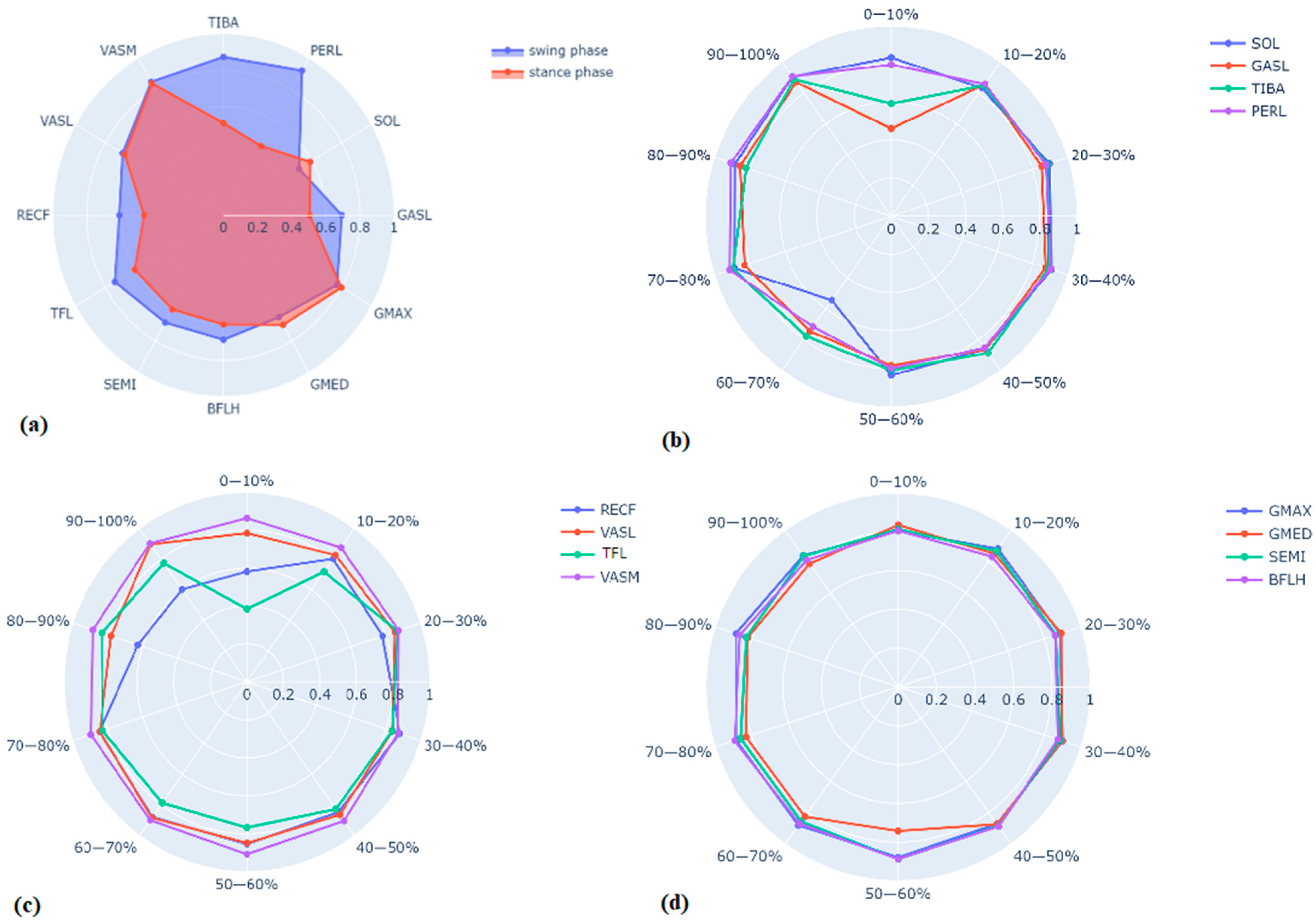
Figure 1. (a) Radar chart of mean cosine similarity values between recorded EMG data and estimated muscle activations (divided into swing and stance phase) of all subjects for the right leg. (b) Radar chart of mean cosine similarity values between recorded EMG data and estimated activations for every 10% of the gait cycle of all subjects for the right leg muscles SOL (soleus), GASL (lateral gastrocnemius), TIBA (tibialis anterior), PERL (peroneus longus). (c) Radar chart of mean cosine similarity values between recorded EMG data and estimated activations for every 10% of the gait cycle of all subjects for the right leg muscles RECF (rectus femoris), VASL (vastus lateralis), TFL (tensor fasciae latae), VASM (vastus medialis). (d) Radar chart of mean cosine similarity values between recorded EMG data and estimated activations for every 10% of the gait cycle of all subjects for the right leg muscles GMAX (gluteus maximus), GMED (gluteus medius), SEMI (semitendinosus), BFLH (long head of biceps femoris).
Biomechanics 03 00044 g001
Table 1. Mean (standard deviation) cosine similarity values for each muscle across 10 gait cycles of all subjects in stance and swing phases.
MuscleStance PhaseSwing Phase
Tibialis Anterior0.51 (0.12)0.87 (0.06)
Peroneus Longus0.44 (0.15)0.92 (0.02)
Soleus0.57 (0.12)0.51 (0.14)
Gastrocnemius Lateralis0.41 (0.21)0.69 (0.09)
Gluteus Maximus0.82 (0.1)0.77 (0.13)
Gluteus Medius0.69 (0.1)0.65 (0.13)
Biceps Femoris Long Head0.60 (0.13)0.69 (0.09)
Semitendinosus0.58 (0.12)0.68 (0.09)
Tensor Fasciae Latae0.58 (0.12)0.74 (0.1)
Rectus Femoris0.46 (0.14)0.61 (0.12)
Vastus Lateralis0.64 (0.1)0.68 (0.09)
Vastus Medialis0.85 (0.07)0.85 (0.08)
Table 2. Mean (and standard deviation) cosine similarity of 12 muscles of the right leg between recorded EMG data and OpenSim static optimization estimated activations for 10 gait cycles averaged for all subjects for every 10% of gait cycle.
PercentageTibialis AnteriorPeroneus LongusSoleusGastrocnemius LateralisTensor Fasciae LataeRectus Femoris
0–10%0.65 (0.1)0.86 (0.08)0.9 (0.03)0.5 (0.08)0.42 (0.1)0.63 (0.15)
10–20%0.92 (0.06)0.93 (0.04)0.89 (0.07)0.93 (0.04)0.79 (0.11)0.85 (0.13)
20–30%0.96 (0.05)0.94 (0.1)0.96 (0.04)0.91 (0.11)0.93 (0.04)0.83 (0.14)
30–40%0.96 (0.04)0.97 (0.01)0.98 (0.02)0.93 (0.09)0.89 (0.11)0.95 (0.04
40–50%0.95 (0.05)0.92 (0.03)0.93 (0.05)0.93 (0.05)0.89 (0.09)0.9 (0.14)
50–60%0.87 (0.09)0.86 (0.07)0.91 (0.06)0.84 (0.09)0.82 (0.09)0.91 (0.08)
60–70%0.83 (0.13)0.76 (0.16)0.56 (0.15)0.78 (0.12)0.83 (0.15)0.95 (0.03)
70–80%0.97 (0.03)0.99 (0.01)0.96 (0.03)0.91 (0.1)0.89 (0.06)0.91 (0.09)
80–90%0.87 (0.11)0.98 (0.02)0.96 (0.04)0.93 (0.07)0.9 (0.05)0.66 (0.14)
90–100%0.96 (0.03)0.98 (0.01)0.98 (0.01)0.93 (0.06)0.83 (0.09)0.65 (0.11)
PercentageVastus LateralisVastus MedialisGluteus MaximusGluteus MediusBiceps Femoris Long HeadSemitendinosus
0–10%0.85 (0.03)0.93 (0.03)0.86 (0.07)0.9 (0.04)0.87 (0.04)0.87 (0.05)
10–20%0.89 (0.05)0.95 (0.03)0.95 (0.04)0.91 (0.06)0.89 (0.07)0.93 (0.05)
20–30%0.91 (0.07)0.93 (0.08)0.96 (0.03)0.96 (0.06)0.93 (0.06)0.93 (0.07)
30–40%0.9 (0.12)0.94 (0.03)0.97 (0.03)0.96 (0.08)0.94 (0.06)0.95 (0.02)
40–50%0.93(0.05)0.98 (0.01)0.95 (0.07)0.94 (0.06)0.96 (0.05)0.96 (0.04)
50–60%0.91 (0.1)0.98 (0.02)0.94 (0.08)0.77 (0.26)0.95 (0.05)0.96 (0.02)
60–70%0.95 (0.04)0.97 (0.03)0.96 (0.04)0.88 (0.1)0.93 (0.09)0.92 (0.06)
70–80%0.9 (0.12)0.97 (0.03)0.95 (0.05)0.89 (0.090.96 (0.04)0.92 (0.08)
80–90%0.84 (0.15)0.95 (0.05)0.95 (0.04)0.88 (0.08)0.93 (0.04)0.9 (0.09)
90–100%0.97 (0.03)0.98 (0.02)0.9 (0.09)0.86 (0.07)0.88 (0.04)0.9 (0.02)
Disclaimer/Publisher’s Note: The statements, opinions and data contained in all publications are solely those of the individual author(s) and contributor(s) and not of MDPI and/or the editor(s). MDPI and/or the editor(s) disclaim responsibility for any injury to people or property resulting from any ideas, methods, instructions or products referred to in the content.

