Article

Can Slight Variations to Lateral Wedge Insoles Induce Significant Biomechanical Changes in Patients with Knee Osteoarthritis?

1
School of Health Sciences, University of Aveiro (ESSUA), 3810-193 Aveiro, Portugal
2
CIF2D, LABIOMEP, Faculdade de Desporto da Universidade do Porto, 4200-450 Porto, Portugal
3
Department of Orthopaedics, Centro Hospitalar Universitário do Porto, 4099-001 Porto, Portugal
4
Institute of Biomedical Sciences Abel Salazar (ICBAS), University of Porto, 4050-313 Porto, Portugal
5
Research Center in Sports Sciences, Health Sciences and Human Development, CIDESD, LABIOMEP (Porto Biomechanics Laboratory), 447-690 Maia, Portugal
*
Author to whom correspondence should be addressed.
Biomechanics 2022, 2(3), 342-351; https://doi.org/10.3390/biomechanics2030027
Submission received: 13 June 2022 / Revised: 12 July 2022 / Accepted: 13 July 2022 / Published: 16 July 2022
(This article belongs to the Topic Human Movement Analysis)

Abstract

:
Lateral wedge insoles are recommended in order to minimize the impacts of osteoarthritis of the knee. The amount of wedging required to induce a biomechanical response with clinical significance is still controversial. This study aimed to investigate the immediate biomechanical effects of different amounts of wedging in symptomatic medial knee OA. A 3D motion capture system and five force platforms were used to acquire walking kinematic and kinetic data along a 10 m walkway. Each participant was tested for six different lateral wedge insoles (0, 2, 4, 6, 8, and 10°) in a randomized order. Thirty-eight patients with medial osteoarthritis of the knee were recruited. The application of insoles resulted in an incremental reduction of the first peak of the external knee adduction moment under all experimental conditions in comparison with the control condition (0° insole). A significant increase (p < 0.05) was observed in peak ankle eversion and in ankle eversion at the first peak of the external knee adduction moment with insoles higher than 8° and 6°, respectively. Slight variations to lateral wedge insoles, greater than 2°, appear to induce significant biomechanical changes in patients with knee osteoarthritis.

1. Introduction

Osteoarthritis (OA) is a major disease that occurs throughout the world; it is associated with significant morbidity and is a cause of chronic pain and limited activity, such as walking, and can even affect the quality of sleeping [1,2]. Although relief from pain is a major and immediate aim, it is also important to minimize the risk of disease progression over time.
The mechanical factor of excessive load on the knee seems to explain the substantial number of cases of knee OA [3]. Biomechanical interventions are among the recommended treatments for managing the disease over time [4]. Lateral wedge insoles are orthotic devices that are placed within the shoes in order to significantly reduce the risk factors associated with knee OA [5]. The biomechanical result of the use of lateral wedge insoles seems to be a decrease in the external knee adduction moment (EKAM) [6,7]. EKAM is a parameter that quantifies the contribution of the ground reaction force (GRF) to the rotation of the lower leg around the knee. Increasing the values of EKAM may be caused by, and also contribute to, a more pronounced knee varus alignment, intensifying the load on the medial compartment of the knee [8]. The first peak of EKAM during the stance phase of the gait, which is often the higher one, has a strong correlation with the severity of the radiographic disease and has been associated with more intense and frequent pain [9].
The amount of wedging that is needed to induce a biomechanical response with clinical significance is an essential research question, given that the effects on the clinical condition are still controversial [5,10,11,12]. Different amounts of lateral wedging have been suggested, such as 4, 5, 6, 10, or 11° [13]. Previous studies have failed to demonstrate effectiveness in patients with osteoarthritis of the knee, despite the acute effects on the biomechanics of these types of insoles [14]. To the best of our knowledge, only one study has tried to understand the biomechanical effects of several wedging angles [15]. However, this previous study, regardless of some interesting findings, was carried out on young, healthy individuals. Another aspect that should be considered is the adaptation of patients to the use of insoles. Insoles with very high angulations are usually uncomfortable, especially angulations greater than 8° [13]. On the other hand, very low angulations may not have any biomechanical effects.
The fact that there are some patients who do not respond biomechanically and clinically to the application of different lateral wedge insoles [16,17,18] seems to provide justification for customized interventions. Therefore, understanding whether slight variations to lateral wedge insoles can induce significant biomechanical changes in patients with knee osteoarthritis could be an important contribution to justify their customization and it could improve the clinical condition, in particular for patients with similar characteristics, such as the knee morphology. Thus, the aim of the present study was to assess the acute effects that small variations in lateral wedge insoles have on the ankle and knee biomechanics in patients with varum malalignment and medial knee OA.

2. Materials and Methods

2.1. Participants

Thirty-eight participants (15 males and 23 females, mean age of 61.6 ± 8.4 years, mean mass of 75.8 ± 12.8 kg, mean height of 1.61 ± 0.09 m, and mean body mass index (BMI) of 29.3 ± 5.1 kg/m2), all of which had symptomatic medial knee OA, were recruited from local hospitals (Table 1). Of the 38 participants, 16 patients were grade 2 (42.1%) and 22 (7.9%) were grade 3 on the scale of Kellgren and Lawrence.
The clinical and radiological inclusion criteria followed those established by the American College of Rheumatology [19]. Other specific inclusion criteria were ages from 45 to 80 years old, grades 2 and 3 on the Kellgren and Lawrence grading scale [20], and a mechanical axis angle lower than 181° in females and 183° in males, indicating varus malalignment in the painful knee (scored by an orthopaedic doctor) on a full-length anteroposterior radiograph. Participants were excluded if they presented the following joint prostheses, symptomatic evidence of lateral knee OA, patellofemoral OA, knee surgeries within the previous six months, systemic arthritic conditions, previous use of insoles, BMI’s higher than 35, and any other condition that could impair assessments of gait or balance.
The protocol was approved by the hospital and university ethics committees and was performed in accordance with the World Medical Association Declaration of Helsinki-Ethical Principles for Medical Research involving Human Subjects. Written informed consent was received from all participants.

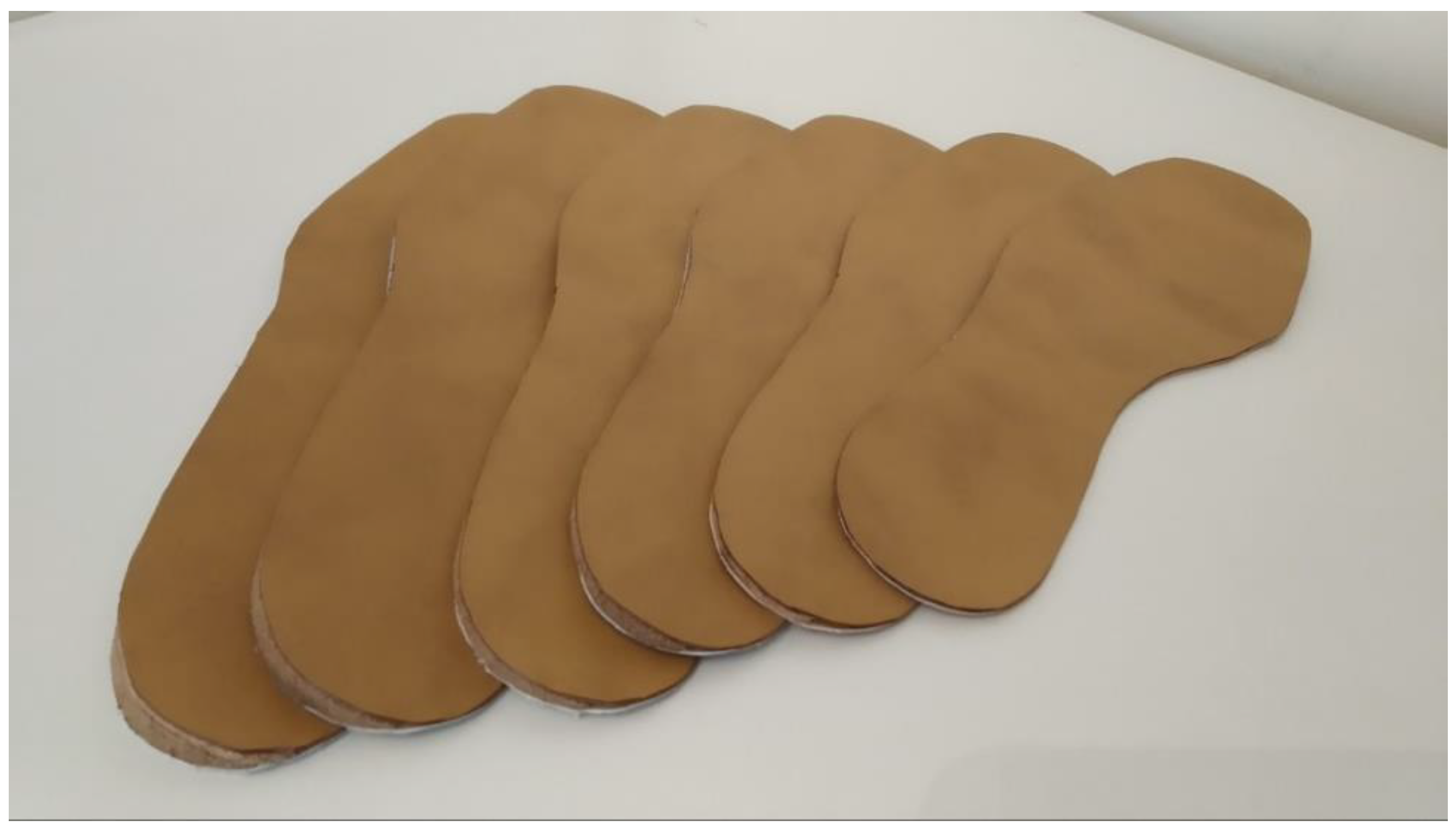
2.2. Equipment and Data Acquisition

A 3D motion capture system (Qualisys, Gothenburg, Sweden), which included 12 optical cameras (Oqus) sampling at 200 Hz and six force platforms (Bertec, Columbus, OH, USA) sampling at 1000 Hz, was used to acquire walking kinematic and kinetic data along a 10 m walkway.
The calibrated anatomical system technique (CAST) was used [21]. Forty-two reflective markers were placed on each subject’s bony landmarks, bilaterally, to reconstruct the position and orientation of the lower limbs and pelvis as described in previous studies [22,23]. The markers located at the calcaneus, as well as the first, second, and fifth metatarsal heads, were attached to the shoes, and the feet were assumed as rigid bodies. Four rigid clusters were also placed bilaterally, on the anterior−lateral aspect of the thigh and of the lower-leg to better follow the segments [24]. A static calibration of the markers was performed before walking trials for all of the conditions.
Participants were not given any instructions other than to walk at their self-selected paces until they had performed at least six valid trials on each of the experimental conditions. A valid trial was set when the subject had walked naturally, and the entire foot had contacted the force plate surface without going beyond its contours. Six experimental conditions were tested in a randomized order: a control condition (shoes with 0° insoles) and lateral wedge insoles with 2, 4, 6, 8, and 10°.
The insoles were full length, custom made with pronating wedges along the lateral border of the foot to the head of the fifth metatarsal (Capron Podologie, France; Ref.: 8004F) (Figure 1). Participants used their own shoes (flat shoes without heels or wedges) to decrease the relevant biomechanical changes with the use of unfamiliar shoes [25]. The insoles were adapted to the size of the feet and were worn bilaterally, but only the data for the most painful knees are presented.

2.3. Data Analysis

Data were previously treated using the Qualisys Track Manager software (Qualisys, Gothenburg, Sweden) to identify, edit, and process the marker trajectories. Post-processing calculations of the kinematic and kinetic time series data in the three planes were conducted using Visual3D software (C-Motion, Rockville, MD, USA) [26]. All of the data were filtered using a Butterworth fourth order filter (6 Hz). The ankle and knee joint centres were calculated as the midpoints between the malleoli and femoral epicondyles, respectively.
The main biomechanical parameters studied were range of motion, joint moments, and joint power. The lower limb joint angles (using an X-Y-Z Euler angle rotation sequence equivalent to the joint coordinate system) and joint moments (determined through inverse dynamics) were computed and expressed relative to the proximal segment. The peak values for the lower limb joint angles and joint moments of force, as well as time−distance parameters, were identified by the software for each set and for each participant. The first and second peaks were additionally identified from each of the EKAM waveforms. The kinematic joint data were expressed in degrees. The kinetic data were normalized to body mass (BM) (Nm/kg). The knee adduction angular impulse (KAAI) was calculated by integrating the EKAM signal for the stance phase (in % BM × time). The kinematic and kinetic data were normalized in time to 100% for the stance phase. Time to the first peak of the EKAM was calculated from the initial contact of the foot on the stance phase. Power (W/kg) was computed in the segment coordinate system, and a positive power output suggests concentric muscle action (power generation), while a negative power output indicates eccentric muscle activity (power absorption).

2.4. Statistical Analyses

The mean values of the kinematics and kinetics data were exported to SPSS 25.0 (IBM, Chicago, IL, USA) and were processed using a single factor: repeated measures analysis of variance. To adjust for multiple testing, Bonferroni corrections were made with an α level of 0.05 or less (two-sided) for determining statistical significance. Post hoc pairwise comparisons were used to understand the differences between the experimental conditions and the control condition (0° insole). The effect size was computed based on the differences between the means (Cohen’s d), and was interpreted as follows: small = 0.2, medium = 0.5, and large = 0.8 [27].

3. Results

The health status of the participants was assessed by the Knee Injury and Osteoarthritis Outcome Score (KOOS). The male gender presented the following in the KOOS subscales: 49.0 in pain, 60.4 in symptoms, 56.3 in activities of daily living, 34.0 in sports, and 35.0 in quality of life. The female gender presented the following in the KOOS subscales: 51.6 in pain, 59.2 in symptoms, 55.1 in activities of daily living, 37.8 in sports, and 43.7 in quality of life.

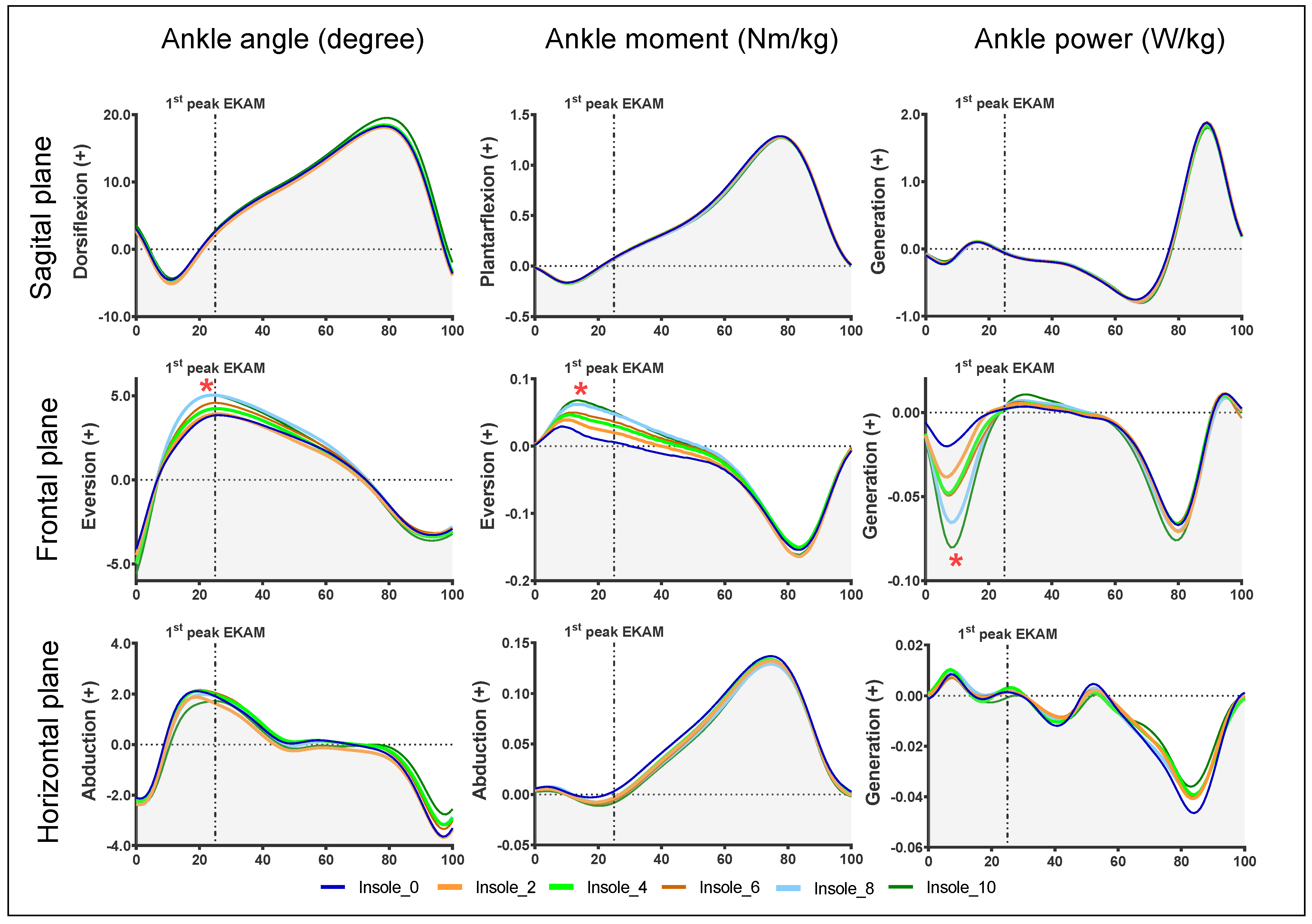
3.1. Ankle Biomechanical Parameters

Significant increases in peak ankle eversion and ankle eversion at the first peak of the EKAM were observed for insoles equal to or higher than 8° (Table 2).
There was a difference with statistical significance at the peak ankle eversion moment for insoles that were equal to or higher than 6°, with a medium effect size for the 6 and 8° insoles, and a large effect size for the 10° insole.
The ankle eversion moment at the first peak of EKAM increased, with statistical significance, for insoles equal to or higher than 4°, with a medium effect size for the 8 and 10° insoles. For insoles higher than 8°, there was a significant increase in peak ankle power absorption in the frontal plane, with a medium effect size for the 10° insole. This absolute peak occurred before the first peak of EKAM, immediately following the initial contact of the stance phase (Figure 2). In the first peak of the EKAM, there was a balance between power generation and absorption (in the frontal plane), with non-incremental effects due to the insoles.

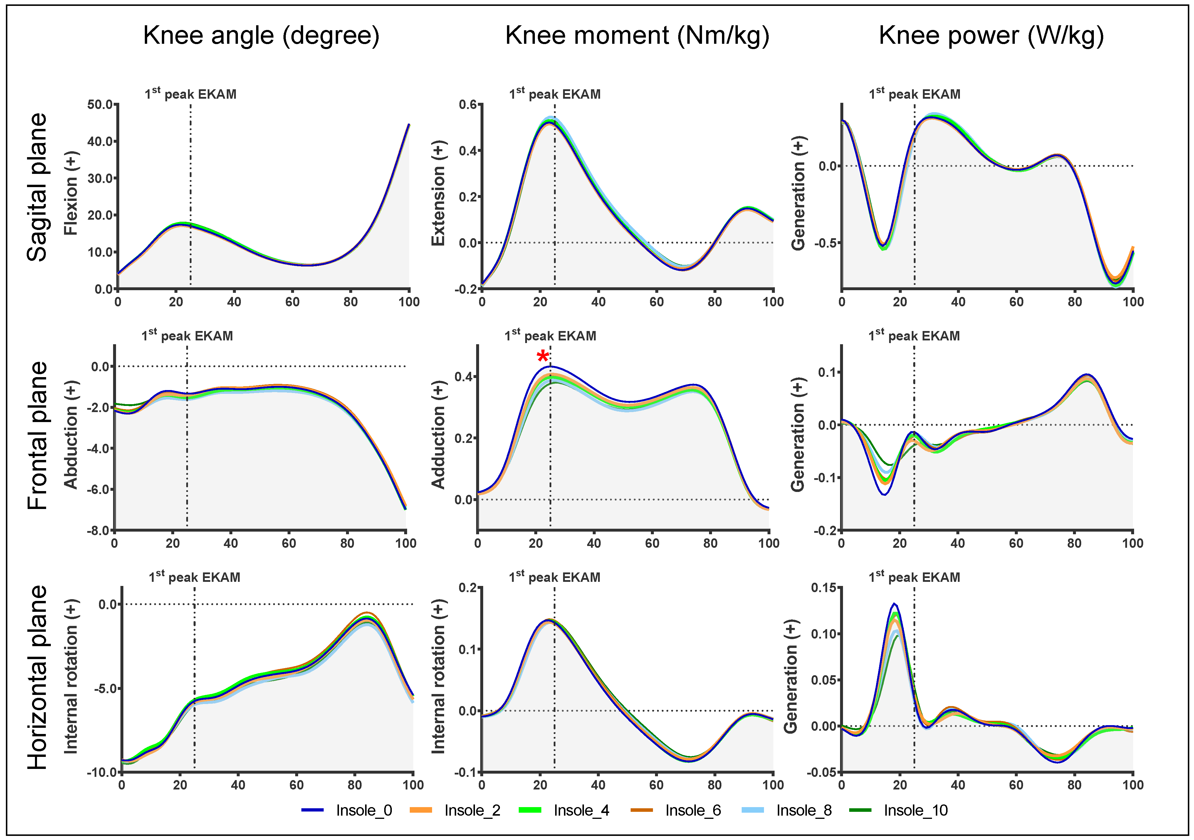
3.2. Knee Biomechanical Parameters

The data for the application of insoles showed an incremental reduction in the first peak of EKAM under all of the experimental conditions (Figure 3). With the 2° insole, there was a reduction of 0.024 Nm/kg, and with the 10° insole, there was a reduction of 0.050 Nm/kg (Table 2). There were no changes in the second peak of EKAM. However, there was a significant decrease in KAAI with insoles higher than 8°. In general, there was a small effect size in the main biomechanical parameters of EKAM. No differences were observed in knee power, except in the transverse plane, with a significant decrease with insoles higher than 8°. There were no changes in gait speed with the different insoles (m/s): 0° insole (1.145 ± 0.169), 2° insole (1.136 ± 0.147), 4° insole (1.138 ± 0.150), 6° insole (1.142 ± 0.155), 8° insole (1.147 ± 0.166), and 10° insole (1.136 ± 0.128).

4. Discussion

The aim of the current study was to assess the acute effects that small variations in lateral wedge insoles have on ankle and knee biomechanics in patients with varum malalignment and medial knee OA.
Generally, the participants presented significant changes in the ankle, mainly at the frontal plane of movement, and less biomechanical changes in the knee. The ankle eversion movement increased with the wedging angle, and this was significant for the 8° and 10° insoles. The angle of the ankle during the first peak of EKAM also increased, and this was significant with the 8 and 10° insoles. These adjustments were even more clear at the eversion moments, which is consistent with the literature [28,29]. No significant changes were observed at the sagittal and horizontal planes for the ankle and knee joints.
There was a significant increase in the peak ankle eversion moments, during the first peak of the EKAM for insoles equal to or greater than 4°. In the same way, ankle power absorption was observed before the first peak as a biomechanical response to wearing lateral wedge insoles. This absorption had statistical significance for insoles equal or higher than 8°. At the same time, in the knee, there was also a mechanism of power absorption, although without statistical significance and of a lesser magnitude (Figure 3). These adjustments can be explained in part by a change in neuromuscular control during movement with the presence of lateral wedge insoles. In the frontal plane, the presence of insoles at the initial contact phase will force the foot on the ground into greater eversion (Figure 2), generating a greater moment of eversion. Therefore, the foot attack to the ground in a slight inversion, with the presence of a lateral wedge insole, will improve the absorption of power. A possible explanation is that these movements that are under the control, in particular, of the tibialis anterior [30], as well as the presence of a lateral wedge insole, may reinforce the eccentric muscular action of this muscle [31,32].
In attempting to understand the effects that took place in the knee, it is interesting to note that the kinematics did not change with the different insole conditions. Nevertheless, a significant reduction in the first peak of EKAM for the experimental insoles was observed in comparison with the neutral insoles. The findings from the present study reinforce the evidence that lateral wedge insoles significantly reduce the first peak of EKAM [5,33]. However, in our study, there were changes even with the smallest angle applied (2° insoles). This result is similar to a previous study, suggesting that any slight change under the foot induces adaptations in the upper segments [34]. Therefore, the use of slight angulations could be a curious field of investigation, as high angulations under the foot usually cause greater discomfort during use [13]. Furthermore, the use of small changes in the material used in the insoles apparently also has an influence on the biomechanical parameters [35].
The time taken to reach the first peak of EKAM was extended with higher insoles, and this value was significant with insoles equal to and higher than 6°. This change was 8.2% higher than with the control insole (Table 2), and could be explained by the increased ankle eversion movement caused by the incremental increase in the insoles, which caused a slight delay to the peak of EKAM [36].
The changes in EKAM in our study are similar to those observed in previous studies [37,38], and it seems that this reduction is achieved essentially through the mechanical adaptation of the ankle during the initial contact of the stance phase. This adaptation, in turn, is responsible for orienting the GRF vector to a more medial position, thus reducing the moment arm of the knee. In addition, the results and observations shown in the charts (Figure 2) of our study indicate that these mechanical adaptations will trigger the capacity of the adjacent muscles to increase the absorption of forces generated by the support of the foot on the ground.
To the best of our knowledge, this is the first study that assesses the acute effects of small increments in lateral wedge insoles on biomechanics in patients with knee OA. Previous studies that attempted to examine the effects of lateral wedge insoles focused only on the knee or used only one or two insoles. The present study used six lateral wedge insoles with a 2° incremental difference between them, which allowed for a more extensive observation of the biomechanical effects of lateral wedge insoles. Understanding how these small changes in the angulation of the insoles influence the biomechanical behaviour may, in the future, allow for an improvement in patient symptoms. The prescription of an insole with the correct angulation, which potentiates the reduction of the mechanical load in fragile areas of the knee, may promote the quality of life of these patients.
The present study has some limitations. Initially, it did not consider the morphology of the feet. A previous study [39] suggested that foot alignment has different effects in kinematics when patients are wearing lateral wedge insoles. Analysing the effects of different angulations, taking into account the morphologies of the foot, can add useful information to this subject. On the other hand, our biomechanical model considers the foot as a rigid body. The use of other models, such as the Oxford model, may bring additional information. On the other hand, a differentiated analysis was not performed at different levels of the K/L scale. The increased severity of knee OA could impact the response to insoles. Finally, this study investigated only the immediate effects on biomechanics. Future studies should seek to understand the implications of small changes in the foot for the clinical condition in patients with knee OA, but in the long term.
In brief, our findings showed that lateral wedge insoles clearly reduce medial load on the knee, particularly at the first peak. This reduction in the mechanical medial load on the knee may have long-term clinical implications, such as delaying the progression of OA or even improving the clinical condition. Further research is needed over a longer time frame to conclusively determine the effects of lateral wedge insoles on joint structure.

5. Conclusions

Slight variations in the lateral wedge insoles induced adaptations in incremental ways only in the frontal plane, suggesting that these are the adaptations that trigger the changes in the EKAM. Increased ankle eversion in the foot during the stance phase seems to be the primary mechanism of the action of lateral wedge insoles. It appears that even small changes under the foot induce significant biomechanical changes in the knee. The use of this simple and low-cost orthosis with minor angulations may change the biomechanics of patients with knee OA.

Author Contributions

V.F., L.M. and P.R. conceptualized and designed the study. V.F., A.V. and F.X.-L. recruited and interacted with all of the participants. V.F. collated the data. L.M. and P.R. provided the supervision of study. V.F., L.M. and P.R. wrote the first draft. All authors have read and agreed to the published version of the manuscript.

Funding

This research received no external funding.

Institutional Review Board Statement

The study was conducted in accordance with the Declaration of Helsinki and was approved by the hospital (088-DEFI/086-CES) and university Ethics Committees (CEFADE 10.2016).

Informed Consent Statement

Informed consent was obtained from all of the subjects involved in the study.

Data Availability Statement

Data can be made available for non-commercial purposes upon request to the authors.

Conflicts of Interest

The authors declare no conflict of interest.

References

  1. Felson, D.T.; Zhang, Y.; Hannan, M.T.; Naimark, A.; Weissman, B.N.; Aliabadi, P.; Levy, D. The incidence and natural history of knee osteoarthritis in the elderly. The Framingham Osteoarthritis Study. Arthritis Rheum. 1995, 38, 1500–1505. [Google Scholar] [CrossRef]
  2. Buckley, J.G.; Scally, A.J.; Bhattacharjee, C. Living with Knee Osteoarthritis: The Positive Impact of Reducing the Knee Torque Induced When Sleeping Supine; A Randomised Clinical Trial. Biomechanics 2022, 2, 95–106. [Google Scholar] [CrossRef]
  3. Felson, D.T. Osteoarthritis as a disease of mechanics. Osteoarthr. Cartil./OARS Osteoarthr. Res. Soc. 2013, 21, 10–15. [Google Scholar] [CrossRef] [PubMed] [Green Version]
  4. McAlindon, T.E.; Bannuru, R.R.; Sullivan, M.C.; Arden, N.K.; Berenbaum, F.; Bierma-Zeinstra, S.M.; Hawker, G.A.; Henrotin, Y.; Hunter, D.J.; Kawaguchi, H.; et al. OARSI guidelines for the non-surgical management of knee osteoarthritis. Osteoarthr. Cartil./OARS Osteoarthr. Res. Soc. 2014, 22, 363–388. [Google Scholar] [CrossRef] [PubMed] [Green Version]
  5. Arnold, J.B.; Wong, D.X.; Jones, R.K.; Hill, C.L.; Thewlis, D. Lateral Wedge Insoles for Reducing Biomechanical Risk Factors for Medial Knee Osteoarthritis Progression: A Systematic Review and Meta-Analysis. Arthritis Care Res. 2016, 68, 936–951. [Google Scholar] [CrossRef] [Green Version]
  6. Hinman, R.S.; Bowles, K.A.; Metcalf, B.B.; Wrigley, T.V.; Bennell, K.L. Lateral wedge insoles for medial knee osteoarthritis: Effects on lower limb frontal plane biomechanics. Clin. Biomech. 2012, 27, 27–33. [Google Scholar] [CrossRef]
  7. Khosravi, M.; Babaee, T.; Daryabor, A.; Jalali, M. Effect of knee braces and insoles on clinical outcomes of individuals with medial knee osteoarthritis: A systematic review and meta-analysis. Assist. Technol. Off. J. RESNA 2021, 11, 1–17. [Google Scholar] [CrossRef]
  8. Telfer, S.; Lange, M.J.; Sudduth, A.S.M. Factors influencing knee adduction moment measurement: A systematic review and meta-regression analysis. Gait Posture 2017, 58, 333–339. [Google Scholar] [CrossRef]
  9. Kito, N.; Shinkoda, K.; Yamasaki, T.; Kanemura, N.; Anan, M.; Okanishi, N.; Ozawa, J.; Moriyama, H. Contribution of knee adduction moment impulse to pain and disability in Japanese women with medial knee osteoarthritis. Clin. Biomech. 2010, 25, 914–919. [Google Scholar] [CrossRef] [Green Version]
  10. Khosravi, M.; Arazpour, M.; Sharafat Vaziri, A. An evaluation of the use of a lateral wedged insole and a valgus knee brace in combination in subjects with medial compartment knee osteoarthritis (OA). Assist. Technol. Off. J. RESNA 2021, 33, 87–94. [Google Scholar] [CrossRef]
  11. Zhang, J.; Wang, Q.; Zhang, C. Ineffectiveness of lateral-wedge insoles on the improvement of pain and function for medial knee osteoarthritis: A meta-analysis of controlled randomized trials. Arch. Orthop. Trauma Surg. 2018, 138, 1453–1462. [Google Scholar] [CrossRef] [PubMed] [Green Version]
  12. Ferreira, V.; Machado, L.; Vilaca, A.; Xara-Leite, F.; Roriz, P. Effects of tailored lateral wedge insoles on medial knee osteoarthritis based on biomechanical analysis: 12-week randomized controlled trial. Clin. Rehabil. 2021, 35, 1235–1246. [Google Scholar] [CrossRef]
  13. Ferreira, V.; Simoes, R.; Goncalves, R.S.; Machado, L.; Roriz, P. The optimal degree of lateral wedge insoles for reducing knee joint load: A systematic review and meta-analysis. Arch. Physiother. 2019, 9, 18. [Google Scholar] [CrossRef] [PubMed]
  14. Jones, R.K.; Chapman, G.J.; Parkes, M.J.; Forsythe, L.; Felson, D.T. The effect of different types of insoles or shoe modifications on medial loading of the knee in persons with medial knee osteoarthritis: A randomised trial. J. Orthop. Res. 2015, 33, 1646–1654. [Google Scholar] [CrossRef] [PubMed] [Green Version]
  15. Tipnis, R.A.; Anloague, P.A.; Laubach, L.L.; Barrios, J.A. The dose-response relationship between lateral foot wedging and the reduction of knee adduction moment. Clin. Biomech. 2014, 29, 984–989. [Google Scholar] [CrossRef]
  16. Felson, D.T.; Parkes, M.; Carter, S.; Liu, A.; Callaghan, M.J.; Hodgson, R.; Bowes, M.; Jones, R.K. The Efficacy of a Lateral Wedge Insole for Painful Medial Knee Osteoarthritis After Prescreening: A Randomized Clinical Trial. Arthritis Rheumatol. 2019, 71, 908–915. [Google Scholar] [CrossRef]
  17. Jones, R.K.; Chapman, G.J.; Forsythe, L.; Parkes, M.J.; Felson, D.T. The relationship between reductions in knee loading and immediate pain response whilst wearing lateral wedged insoles in knee osteoarthritis. J. Orthop. Res. 2014, 32, 1147–1154. [Google Scholar] [CrossRef] [Green Version]
  18. Hunt, M.A.; Takacs, J.; Krowchuk, N.M.; Hatfield, G.L.; Hinman, R.S.; Chang, R. Lateral wedges with and without custom arch support for people with medial knee osteoarthritis and pronated feet: An exploratory randomized crossover study. J. Foot Ankle Res. 2017, 10, 20. [Google Scholar] [CrossRef] [Green Version]
  19. Altman, R.; Asch, E.; Bloch, D.; Bole, G.; Borenstein, D.; Brandt, K.; Christy, W.; Cooke, T.D.; Greenwald, R.; Hochberg, M.; et al. Development of criteria for the classification and reporting of osteoarthritis. Classification of osteoarthritis of the knee. Diagnostic and Therapeutic Criteria Committee of the American Rheumatism Association. Arthritis Rheum. 1986, 29, 1039–1049. [Google Scholar] [CrossRef]
  20. Kellgren, J.H.; Lawrence, J.S. Radiological assessment of osteo-arthrosis. Ann. Rheum. Dis 1957, 16, 494–502. [Google Scholar] [CrossRef] [Green Version]
  21. Cappozzo, A.; Catani, F.; Croce, U.D.; Leardini, A. Position and orientation in space of bones during movement: Anatomical frame definition and determination. Clin. Biomech. 1995, 10, 171–178. [Google Scholar] [CrossRef]
  22. Ferreira, V.; Machado, L.; Vilaça, A.; Leite, F.X.; Roriz, P. Biomechanics performance in 30-s chair stand test in patients with medial knee osteoarthritis. Int. J. Hum. Factors Ergon. 2019, 6, 319–330. [Google Scholar] [CrossRef]
  23. Ferreira, V.; Machado, L.; Roriz, P. The effects on lower limbs kinematics of different lateral wedge insoles. J. Mech. Eng. Biomech. 2020, 4, 77–82. [Google Scholar] [CrossRef]
  24. Wen, C.; Cates, H.E.; Weinhandl, J.T.; Crouter, S.E.; Zhang, S. Knee biomechanics of patients with total knee replacement during downhill walking on different slopes. J. Sport Health Sci. 2022, 11, 50–57. [Google Scholar] [CrossRef] [PubMed]
  25. Lewinson, R.T.; Worobets, J.T.; Stefanyshyn, D.J. Control conditions for footwear insole and orthotic research. Gait Posture 2016, 48, 99–105. [Google Scholar] [CrossRef]
  26. Documentation, C.-M.W. Visual3D Documentation. Available online: https://www.c-motion.com/v3dwiki/index.php?title=Visual3D_Documentation (accessed on 9 July 2022).
  27. Sawilowsky, S. New Effect Size Rules of Thumb. J. Mod. Appl. Stat. Methods 2009, 8, 597–599. [Google Scholar] [CrossRef]
  28. Chapman, G.J.; Parkes, M.J.; Forsythe, L.; Felson, D.T.; Jones, R.K. Ankle motion influences the external knee adduction moment and may predict who will respond to lateral wedge insoles?: An ancillary analysis from the SILK trial. Osteoarthr. Cartil./OARS Osteoarthr. Res. Soc. 2015, 23, 1316–1322. [Google Scholar] [CrossRef] [Green Version]
  29. Tse, C.T.F.; Ryan, M.B.; Hunt, M.A. Influence of foot posture on immediate biomechanical responses during walking to variable-stiffness supported lateral wedge insole designs. Gait Posture 2020, 81, 21–26. [Google Scholar] [CrossRef]
  30. Santilli, V.; Frascarelli, M.A.; Paoloni, M.; Frascarelli, F.; Camerota, F.; De Natale, L.; De Santis, F. Peroneus longus muscle activation pattern during gait cycle in athletes affected by functional ankle instability: A surface electromyographic study. Am. J. Sports Med. 2005, 33, 1183–1187. [Google Scholar] [CrossRef]
  31. Reeves, J.; Jones, R.; Liu, A.; Bent, L.; Plater, E.; Nester, C. A systematic review of the effect of footwear, foot orthoses and taping on lower limb muscle activity during walking and running. Prosthet. Orthot. Int. 2019, 43, 576–596. [Google Scholar] [CrossRef]
  32. Ludwig, O.; Kelm, J.; Frohlich, M. The influence of insoles with a peroneal pressure point on the electromyographic activity of tibialis anterior and peroneus longus during gait. J. Foot Ankle Res. 2016, 9, 33. [Google Scholar] [CrossRef] [PubMed] [Green Version]
  33. Hatfield, G.L.; Cochrane, C.K.; Takacs, J.; Krowchuk, N.M.; Chang, R.; Hinman, R.S.; Hunt, M.A. Knee and ankle biomechanics with lateral wedges with and without a custom arch support in those with medial knee osteoarthritis and flat feet. J. Orthop. Res. 2016, 34, 1597–1605. [Google Scholar] [CrossRef] [PubMed] [Green Version]
  34. Ma, C.C.; Lee, Y.J.; Chen, B.; Aruin, A.S. Immediate and short-term effects of wearing a single textured insole on symmetry of stance and gait in healthy adults. Gait Posture 2016, 49, 190–195. [Google Scholar] [CrossRef]
  35. Jin, L. The Influence of Different Footwear Insole Stiffness on Center of Pressure and Ankle Kinematics during Walking: A Case Report. Biomechanics 2022, 2, 205–212. [Google Scholar] [CrossRef]
  36. Shaw, K.E.; Charlton, J.M.; Perry, C.K.L.; de Vries, C.M.; Redekopp, M.J.; White, J.A.; Hunt, M.A. The effects of shoe-worn insoles on gait biomechanics in people with knee osteoarthritis: A systematic review and meta-analysis. Br. J. Sports Med. 2018, 52, 238–253. [Google Scholar] [CrossRef] [Green Version]
  37. Kluge, F.; Krinner, S.; Lochmann, M.; Eskofier, B.M. Speed dependent effects of laterally wedged insoles on gait biomechanics in healthy subjects. Gait Posture 2017, 55, 145–149. [Google Scholar] [CrossRef] [PubMed]
  38. Maly, M.R.; Culham, E.G.; Costigan, P.A. Static and dynamic biomechanics of foot orthoses in people with medial compartment knee osteoarthritis. Clin. Biomech. 2002, 17, 603–610. [Google Scholar] [CrossRef]
  39. Sawada, T.; Tanimoto, K.; Tokuda, K.; Iwamoto, Y.; Ogata, Y.; Anan, M.; Takahashi, M.; Kito, N.; Shinkoda, K. Rear foot kinematics when wearing lateral wedge insoles and foot alignment influence the effect of knee adduction moment for medial knee osteoarthritis. Gait Posture 2017, 57, 177–181. [Google Scholar] [CrossRef] [Green Version]
Figure 1. The six experimental insoles used in the study.
Biomechanics 02 00027 g001
Figure 2. Ankle angles, moments, and power waveforms during the stance phase (horizontal line) in the sagittal, frontal, and transverse plane. Each colour line represents the mean for different wedge insoles. The vertical dashed lined represents the timing of first peak of EKAM for the neutral condition (0° insole). Note the different vertical scales used for clarity. *: indicates statistical significance.
Biomechanics 02 00027 g002
Figure 3. Knee angles, moments, and power waveforms during the stance phase (horizontal line) in the sagittal, frontal, and transverse plane. Each colour line represents the mean of different wedge insoles. The vertical dashed lined represents the timing of the first peak of EKAM for the neutral condition (0° insole). Note the different vertical scales used for clarity. *: indicates statistical significance.
Biomechanics 02 00027 g003
Table 1. Patient demographics.
MinimumMaximumMeanStandard Deviation
Age (years)45.07861.68.4
Height1.381.771.60.01
Body mass index20.835.529.35.1
Table 2. Biomechanical parameters at the knee and ankle (bold indicates significance).
(n = 38)0° Insole2° Insole4° Insole6° Insole8° Insole10° Insole
Peak ankle eversion [°] 4.4 (±3.1)4.5 (±3.1)4.8 (±3.0)5.1 (±2.9)5.5 (±3.0)5.5 (±3.1)
p-values 1.0001.0000.1050.0010.009
Cohen’s d 0.0520.1360.2320.3740.364
Ankle eversion at 1st peak EKAM [°]3.8 (±3.1)3.9 (±3.0)4.3 (±3.0)4.6 (±3.0)5.0 (±3.1)4.9 (±3.3)
p-values 1.0001.0000.0870.0000.018
Cohen’s d 0.0430.1580.2530.4000.357
Peak ankle eversion moment [Nm/kg]0.052 (±0.039)0.061 (±0.038)0.066 (±0.040)0.074 (±0.044)0.080 (±0.039)0.087 (±0.044)
p-values 0.1970.6960.0430.0000.000
Cohen’s d 0.2340.3540.529 *0.718 *0.842 **
Ankle eversion moment at 1st peak EKAM [Nm/kg]0.020 (±0.065)0.032 (±0.062)0.041 (±0.058)0.050 (±0.062)0.058 (±0.056)0.060 (±0.067)
p-values 0.1040.0000.0000.0000.000
Cohen’s d 0.1890.3410.4720.626 *0.606 *
Peak ankle power absorption (frontal plane) [W/kg]−0.097 (±0.055)−0.113 (±0.064)−0.109 (±0.062)−0.114 (±0.069)−0.122 (±0.064)−0.138 (±0.072)
p-values 0.1730.3570.2220.0040.000
Cohen’s d 0.2680.2050.2720.4190.640 *
1st Peak EKAM [Nm/kg]0.452 (±0.183)0.428 (±0.181)0.421 (±0.182)0.424 (±0.189)0.410 (±0.183)0.402 (±0.182)
p-values 0.0050.0000.0000.0000.000
Cohen’s d 0.1310.1700.1510.2300.274
KAAI [Nm/kgxs]0.196 (±0.104)0.188 (±0.107)0.183 (±0.097)0.185 (±0.100)0.177 (±0.097)0.177 (±0.100)
p-values 0.5310.1090.0770.0010.000
Cohen’s d 0.0760.1290.1080.1890.186
Time to 1st peak EKAM [s]0.184 (±0.058)0.194 (±0.061)0.195 (±0.063)0.199 (±0.065)0.197 (±0.062)0.202 (±0.064)
p-values 0.0600.1150.0370.0010.000
Cohen’s d 0.1680.1820.2440.2170.295
Values reported as mean (±SD); p value: difference from the 0° insole; Cohen’s d: comparation with a 0° insole; * effect size: medium; ** effect size: large.
Publisher’s Note: MDPI stays neutral with regard to jurisdictional claims in published maps and institutional affiliations.

