Article

Cyclograms Reveal Alteration of Inter-Joint Coordination during Gait in People with Multiple Sclerosis Minimally Disabled

1
Department of Mechanical, Chemical and Materials Engineering, University of Cagliari, Piazza d’Armi, 09123 Cagliari, Italy
2
Multiple Sclerosis Regional Center of Sardinia, Binaghi Hospital, Via Is Guadazzonis, 2, 09126 Cagliari, Italy
3
Department of Medical Sciences and Public Health, University of Cagliari, Cittadella Universitaria di Monserrato-Blocco I, SS 554 bivio Sestu, 09042 Monserrato, Italy
*
Author to whom correspondence should be addressed.
Biomechanics 2022, 2(3), 331-341; https://doi.org/10.3390/biomechanics2030026
Submission received: 2 June 2022 / Revised: 29 June 2022 / Accepted: 11 July 2022 / Published: 13 July 2022
(This article belongs to the Topic Human Movement Analysis)

Abstract

:
Subtle alterations of gait patterns in people with Multiple Sclerosis (pwMS) with minimal or no disability often coexist with normal spatio-temporal parameters. Here, we retrospectively investigate the existence of possible anomalies in lower limb inter-joint coordination (i.e., the functional relationship between joint pairs) in pwMS with apparently physiologic gait features. Twenty-seven pwMS with Expanded Disability Status Scale scores ≤ 2, and 27 unaffected age-and-sex-matched individuals, were tested using 3D computerized gait analysis. Raw data were processed to extract the main spatio-temporal parameters and the kinematics in the sagittal plane at the hip, knee, and ankle joints. Angle-angle diagrams (cyclograms) were obtained by coupling the flexion-extension values for the hip-knee and knee-ankle joint pairs at each point of the gait cycle. Cyclogram area, perimeter, and dimensionless ratio were employed to quantify inter-joint coordination. The results demonstrate that cyclograms of pwMS are characterized by significantly reduced perimeters for both investigated joint pairs and reduced area at the hip–knee joint pair. In the latter pair, the differences between groups involved the entire swing phase. For the knee-ankle pair, the average cyclogram of pwMS departed from normality from the late stance until the mid-swing phase. Such findings suggest that inter-joint coordination is impaired even in minimally disabled pwMS who exhibit a normal gait pattern in terms of spatio-temporal parameters. The quantitative and qualitative study of cyclogram features may provide information that is useful for better understanding the underlying mechanisms of walking dysfunctions in MS.

1. Introduction

Alterations in locomotor efficiency, which represents one of the most distinctive features of Multiple Sclerosis (MS), play a major role in defining the disability of the MS-affected individual [1]. Indeed, population-based studies reported that a large percentage of people with MS (pwMS) complain about impaired ambulation [2,3,4,5] and consider gait one of the most valuable bodily functions threatened by the disease [6].
The precise identification of the period in which the gait of pwMS becomes anomalous is a crucial, yet mostly unsolved issue. While it has been recently hypothesized that early interventions in terms of lifestyle adaptations and physical training would be beneficial for delaying the worsening of motor dysfunctions (including gait [7]), there is no consensus about what parameters (and threshold values) could be considered the most effective for detecting meaningful and clinically relevant ambulation impairments.
In this context, using quantitative techniques to assess gait performance—currently a well-established practice in research laboratories and gradually becoming more widespread even in clinical settings—might certainly be a powerful method, due to the large and detailed dataset such techniques provide. In particular, optical motion capture systems, electronic walkways, and wearable inertial sensors have so far been successfully employed to characterize several aspects of gait in pwMS, such as spatio-temporal parameters (i.e., speed, stride/step length, cadence, step width, and double support phase duration), lower limb kinematics (i.e., angles at hip, knee, and ankle joints during the gait cycle), variability, symmetry, smoothness, etc. In this regard, spatio-temporal parameters are the most frequently used [8,9], as they can generally be considered sensitive enough to distinguish pwMS from unaffected individuals, especially when the Expanded Disability Status Scale (EDSS) score is 3 or higher.
However, in pwMS with minimal or no disability (e.g., those with EDSS ≤ 2), such capability appears markedly reduced. As a result, the literature on this subject comprises either studies that failed to detect any significant differences with respect to healthy controls [10,11,12,13,14] or others that detected them only for one or a few parameters [15,16,17,18,19,20,21]. While such mixed findings can be partly attributed to inhomogeneities in terms of sample size and selection, female to male ratio, data acquisition and processing techniques, and so on, it seems reasonable to state that more refined and sophisticated approaches are needed to detect subtle gait alterations.
To achieve this purpose, several studies have proposed the use of specific metrics such as variability of spatio-temporal parameters (using linear and non-linear approaches [17,22,23,24]) and smoothness (expressed by harmonic ratio, a parameter derived by trunk accelerations [13]). In some cases, they have investigated in further detail the kinematics of lower limb joints in the sagittal plane by detecting several alterations, especially at ankle level [11,20,25,26]. Despite the lower simplicity in terms of data interpretation, lower limb kinematics has been recognized as a valuable tool for analyzing the underlying mechanisms of walking dysfunctions in MS [27,28]; thus, it is reasonable to hypothesize that the information it provides can be also important for the early identification of gait anomalies in pwMS.
In this context, useful insights might be extracted by studying inter-joint coordination, the functional relationship between joint pairs during the whole gait cycle. This can be studied by means of angle-angle diagrams (also known as cyclograms). Cyclograms, first proposed by Grieve [29], are closed bidimensional curves that can provide data on inter-joint coordination by either relating the motion of the same joint for the left and right limb (in this case, they are meant to represent inter-limb symmetry) or, as in the case of the present study, expressing the relative motion of a certain joint couple (e.g., hip-knee, knee-ankle, etc.) during a whole cycle. The inter-joint coordination can be quantified by a range of techniques that provide differing complexity and accuracy of results [30]. However, a basic approach suitable for clinical use is based on the calculation of geometric properties such as area and perimeter. Several studies of individuals affected by orthopedic [31,32] and neurologic conditions [33,34,35] have demonstrated the capability of cyclograms to effectively detect the existence of alterations in coordination originated by the presence of MS. In recent years, this approach has been proposed even in a few studies on pwMS [36,37], which reported the existence of significant alterations in movement coordination during the stance phase in pwMS with respect to unaffected individuals and between pwMS with and without fall history. More recently, a retrospective study on 104 pwMS [38] proposed a detailed characterization of inter-joint coordination alterations using geometric properties of cyclograms and investigated the extent of their association with the disability level.
On the basis of the above-mentioned considerations, we propose a study aimed at verifying the actual capability of cyclograms to detect and characterize alterations of inter-joint coordination possibly exhibited during walking in pwMS with minimal or no disability. In particular, the goal of the research is to understand whether this approach is effective in distinguishing the inter-joint coordination features at hip-knee and knee-ankle joint pairs of pwMS and unaffected individuals.

2. Methods

2.1. Participants

This is a retrospective study that analyzed data originated from computerized three-dimensional gait analysis carried out on 270 pwMS (followed at the Regional Multiple Sclerosis Center of Sardinia, Cagliari, Italy), in the period May 2014–February 2022, at the Laboratory of Biomechanics and Industrial Ergonomics of the University of Cagliari (Cagliari, Italy). Diagnosis of MS was carried out by a neurologist expert in MS (EC, GC, JF) according to the 2010 revised criteria [39,40]. Participants were enrolled in a series of studies either aimed to characterize the kinematic alterations associated with MS or to quantitatively assess the effect of pharmacologic and rehabilitative treatments [38,41,42,43,44].
For the present study, we considered only pwMS who were scored with an EDSS ≤ 2 at the time of the gait analysis and free from any other neurologic or orthopedic condition potentially able to severely affect gait or balance. Such selection resulted in a group composed of 27 participants (17 women, 10 men, mean age 40.5 years). Twenty-seven unaffected individuals age-and-sex-matched, recruited among the staff of the MS Center and the University of Cagliari, composed the control group. The participants’ main anthropometric and clinical features are reported in Table 1.
The study was conducted according to the World Medical Association Declaration of Helsinki principles. All participants signed an informed consent form agreeing to participate.

2.2. Data Acquisition and Processing

We employed an eight-camera optical motion-capture system (Smart-D, BTS Bioengineering, Milan, Italy) set at 120 Hz frequency to acquire the trajectories of 22 spherical retro-reflective passive markers (14 mm diameter) placed on the skin of participants’ lower limbs and trunk, according to the protocol described by Davis et al. [45]. Preliminarily, the following anthropometric data were collected: height, body mass, anterior superior iliac spine distance, pelvis thickness, knee and ankle width, and leg length. Once the markers’ placement was complete, the participants were requested to walk at a self-selected speed on a 10 m walkway in the most natural manner at least six times. At the end of the trials, the marker’s trajectories were processed with the dedicated software, Smart Analyzer (BTS Bioengineering, Milan, Italy), to obtain:
  • The main spatio-temporal parameters of gait: speed, stride length, cadence, step width, stance, swing, and duration of double support phases.
  • The flexion-extension angle for hip and knee joints and the dorsi-plantarflexion angle for the ankle joint for each of the 100 points in which the gait cycle was divided. Such data were employed to quantify the inter-joint coordination (as described in detail later) and to calculate the dynamic range of motion (dynamic ROM) as the difference between the maximum and minimum values assumed by each angle within the gait cycle.
In all the subsequent analyses, for all the above-mentioned parameters, the mean value was calculated across the six trials.

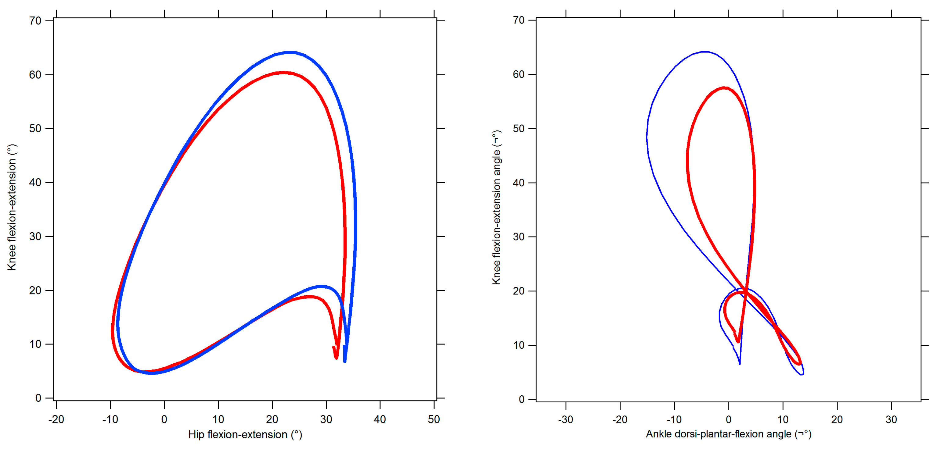
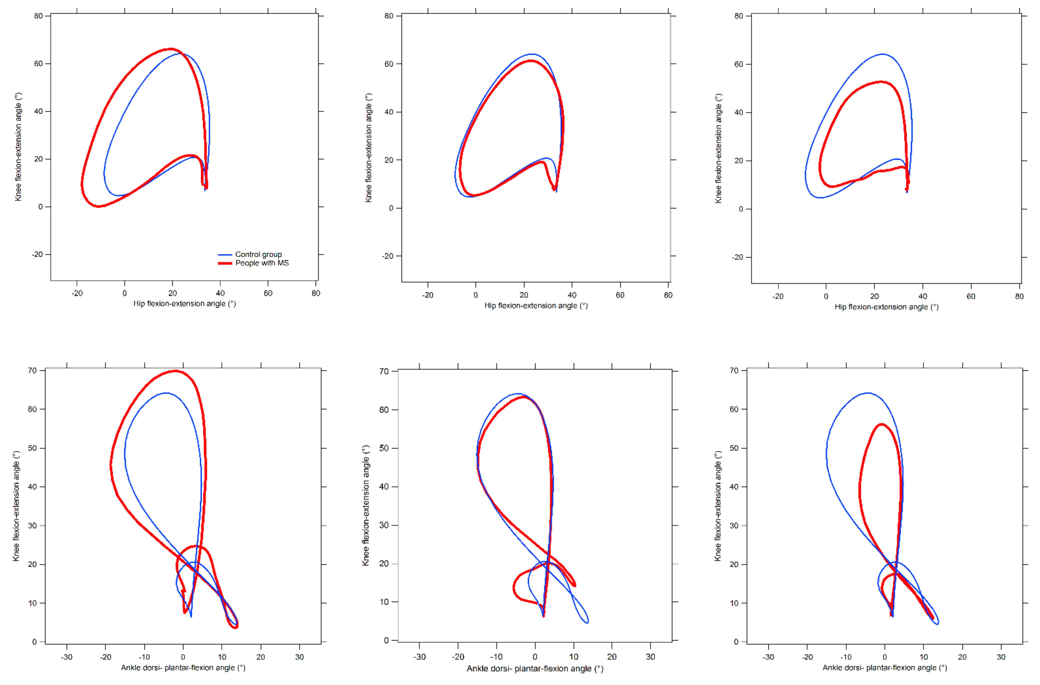
2.3. Quantification of Inter-Joint Coordination by Means of Cyclograms

The curves which refer to the angle variations at hip, knee, and ankle joints in the sagittal plane, were exported as text files to be processed by a custom routine developed under the Matlab® environment, which provides the hip-knee and knee-ankle cyclograms separately for the right and left limb as well as the value of their following geometrical properties:
  • Cyclogram area (degrees2): the area of the closed trajectory described by the simultaneous angular variation that occurs at the two joints of interest during the gait cycle [46]. The interpretation of this parameter is quite straightforward, as larger areas are usually representative of higher conjoint range of angular movements experienced at a certain joint pair within a complete gait cycle [46,47].
  • Cyclogram perimeter (degrees): the length of the trajectory previously described, which is typically expected to increase as the area increases. Thus, its interpretation is similar to that of the area. However, there are cases in which repeated abrupted angular variations (due to lack of coordination) originate relevant increases of the perimeter even without correspondent area changes [46].
  • Cyclogram dimensionless ratio: this parameter, obtained by the ratio of the perimeter and the square root of the area, represents the shape of the diagram. Lower values indicate cyclograms of regular shape (i.e., not particularly elongated towards a specific direction).

2.4. Statistical Analysis

Preliminarily, a t-test was carried out on the variables separately calculated for left and right limb to verify whether there were differences between the two sides. Given that the analysis found no significant differences, both were considered for the subsequent analysis. Thus, the data included in the statistical analysis refer to 108 limbs.
The existence of possible differences in the spatio-temporal parameters of gait and in inter-joint coordination (i.e., cyclograms) parameters introduced by the presence of MS was assessed by means of a one-way multivariate analysis of variance (MANOVA). The participant’s status (pwMS or unaffected individual) was set as an independent variable, while the dependent variables were the seven spatio-temporal parameters previously described; the three dynamic ROM at the hip, knee, and ankle joints; or the three cyclogram parameters (i.e., area, perimeter, and dimensionless ratio). The significance level was set at p = 0.05, and the effect sizes were assessed using the eta-squared (η2) coefficient. A univariate ANOVA was carried out as a post-hoc test by reducing the significance level to p = 0.007 (0.05/7) for spatio-temporal parameters of gait and p = 0.017 (0.05/3) for dynamic ROM and cyclogram parameters after a Bonferroni correction for multiple comparisons.

3. Results

Table 2, Table 3 and Table 4 summarize the analysis results of the spatio-temporal parameters of gait, dynamic ROM, and inter-joint coordination parameters, respectively.
The MANOVA did not detect a main effect of the individual’s status [F (7,46) = 1.23, p = 0.307, Wilks λ = 0.84, η2 = 0.16] regarding the spatio-temporal parameters of gait. However, a significant group effect was found for the dynamic ROM [F (3,104) = 4.94, p = 0.003, Wilks λ = 0.87, η2 = 0.13]. In particular, the follow-up analysis revealed that pwMS exhibit a smaller ROM with respect to unaffected individuals at the knee (p = 0.001) and ankle (p = 0.006) joints, but not at the hip.
The statistical analysis also detected a significant effect of the individual’s status on inter-joint coordination parameters for both hip-knee pair [F (3,104) = 4.49, p = 0.005, Wilks λ = 0.88, η2 = 0.12] and knee-ankle pair [F (3,104) = 6.34, p = 0.001, Wilks λ = 0.84, η2 = 0.16]. In case of the hip-knee joint couple, the post-hoc analysis revealed that pwMS were characterized by cyclograms with a significantly smaller area and perimeter with respect to unaffected individuals, while at the knee-ankle joint couple this was true only for the perimeter. No significant differences were found between groups regarding the dimensionless ratio.
Figure 1 shows the comparison between the average cyclograms calculated for pwMS and unaffected individuals. Figure 2 presents several examples of cyclograms for some of the tested pwMS compared with the average cyclogram of the control group. Using the cyclogram area value to perform a rough classification of their typology, we observed that the majority of pwMS exhibit either a cyclogram with a value of its area within ±10% with respect to normality (n = 12) in the hip–knee pair, or a reduced area (n = 12). In few cases, the diagram was larger or completely different in shape. As regards the knee–ankle joint pair, the classification is not as straightforward as in the previous case. In fact, the group of pwMS is practically split in two—those who exhibit a larger (n = 12) or smaller (n = 12) area than normality. Above all, the shape of their cyclograms tend to be markedly less regular with respect to unaffected individuals.

4. Discussion

4.1. General Considerations

The present study aims to assess the feasibility of using cyclograms as a tool to detect the existence of subtle inter-joint coordination alterations in a sample of pwMS with minimal or no disability. The integration of this information with that obtained through other variables associated with gait (i.e., spatio-temporal parameters, variability, smoothness etc.) would be helpful for better defining the complex and multifaceted nature of the walking dysfunction in MS. This is because it has been hypothesized that coordination, together with sensory organization and biomechanical factors, is one of the subcomponents of the underlying control of postural stability and, as such, it contributes to maintain stability in different situations such as: in response to external perturbations, in anticipation of voluntary limb movements and during voluntary motions of the body such as active sway and locomotion [48].
At first, the gait pattern of pwMS tested here did not significantly differ from that of unaffected individuals in any of the main spatio-temporal parameters, suggesting that, as a group, their walking abilities appear intact. While this result is consistent with several previous studies that tested pwMS with minimal or no disability, it should be considered that the literature reported quite controversial results. For instance, if we consider self-selected gait speed (the most commonly used outcome for studies targeted on gait assessment and the only spatio-temporal measure associated with disease severity, [49]) the majority of studies failed to detect significant differences with controls [10,11,12,13,14,18,19,20,50], even though pwMS were found to be slower in a few cases [15,16,17,21]. Generally speaking, differences between pwMS and unaffected individuals that involved all parameters [15,17] were seldom observed while, more frequently, the analysis revealed substantial modifications only in some. It is not trivial to understand the reasons of such discrepancies, even though factors such as sample size, used equipment, different MS phenotype and female-to-male ratios, and more compromised functional systems certainly play a relevant role.
The main hypothesis of the study, namely the existence of alterations in inter-joint coordination during gait in pwMS with minimal or no disability, was substantially confirmed by the results, as they exhibited significantly reduced cyclogram perimeters for both investigated joint pairs and a significantly smaller hip-knee cyclogram area when compared to unaffected individuals. In particular (as visible from Figure 1) in the case of the hip-knee joint pair, the differences between groups involve the entire swing phase, while in the knee-ankle pair, the average pwMS cyclogram departed from normality in the period from the late stance until the mid-swing phase. Overall, such results suggest that inter-joint coordination may be impaired even in pwMS who exhibit a normal gait pattern in terms of spatio-temporal parameters. What are the possible causes of this phenomenon? Previous studies suggested that pwMS exhibit coactivation of lower limb muscles even since the early stages of the disease [27,51], which is interpreted as a compensatory mechanism to cope with impairments of balance and gait. It is noteworthy that co-contractions of tibialis anterior and triceps surae have been hypothesized as responsible for the reduced ankle ROM [51], a fact also observed in the pwMS tested here. Another factor possibly involved is muscular weakness. In fact, several studies reported that MS originates reductions of skeletal muscle fiber cross-sectional area, muscle strength, and muscle mass of the lower limbs of mildly affected MS patients, particularly in the knee flexors and extensors [52,53,54]. Weakness of ankle plantar-flexor muscles, which negatively affects push-off power in the ankle joint [55,56], might also play a role in impairing coordination at a distal level (i.e., knee-ankle joint pair). However, the literature did not report specific studies on pwMS with minimal disability, so this aspect remains to be clarified. Finally, kinematic alterations of gait, especially in terms of lack of mid-stance knee extension, reduced knee flexion during the swing phase, and reduced hip extension at terminal stance/initial swing (all consistent with the deviations of the pwMS cyclograms from normality observed here) were previously reported for pwMS with low disability [26,57]. Of course, it is likely that some of these alterations are associated with the neural damage consequent of MS. In this regard, recent studies on minimally disabled pwMS that combine imaging techniques with instrumental gait analysis [58] found that subtle gait alterations are correlated with axonal loss in major sensorimotor pathways of the brain.
At last, it should be mentioned that, as shown in Figure 2, our analysis revealed a certain variability in terms of differences of pwMS’ cyclograms with respect to unaffected individuals, as deviations from normality can be more or less marked and involve different phases of the gait cycle. It is likely that this phenomenon is associated with specific individual’s features such as muscular strength, fatigue, presence of spasticity, etc. Thus, future studies should try to identify what factors among those mentioned, and to what extent, are implicated in significant alterations of inter-joint coordination.

4.2. Limitations and Strengths of the Study

As previously mentioned, the assessment of inter-joint coordination by cyclograms in MS is relatively unexplored, and the few existing data were obtained by testing either small samples of pwMS with moderate-severe disability [36] or under less ecologic conditions (i.e., participants walked on a treadmill, [37]). By focusing on pwMS with low disability who exhibit a regular gait pattern from the perspective of their spatio-temporal parameters, the present study attempted to clarify whether cyclograms may be a feasible approach for highlighting subtle inter-joint coordination alterations that possibly precede other kinds of walking dysfunctions. However, some limitations of the study should be acknowledged. Firstly, our sample of low EDSS was quite limited in number, and represents a percentage of the cohort tested in our lab probably smaller than those typical of a real-world distribution of disability (previous studies estimated that pwMS with EDSS ≤ 2 account for approximately 40%, [59], while here they account for 10% of those tested with 3D gait analysis in our lab). The limited sample also prevented us from stratifying the participants based on their sex, MS phenotype, or functional system more affected, which are all factors known to have some influence on gait alterations. Moreover, the qualitative analysis of the cyclograms, especially regarding the hip–knee joint pair, seemed to suggest that coordination may be altered according to different patterns (or even not altered at all). Further studies on larger cohorts are, thus, needed to understand what feature of MS can be associated with specific cyclogram shapes.

5. Conclusions

Using simple geometric features of hip-knee and knee–ankle cyclograms, we demonstrated that minimally disabled pwMS are characterized by inter-joint coordination alterations, which coexist with a gait pattern classifiable as “normal” from the perspective of the spatio-temporal parameters instrumentally assessed. In particular, when examined as a group, the cyclograms of the two investigated joint pairs of pwMS share relevant differences with unaffected individuals in the first half of the swing phase, while coordination appeared altered even in the late stance (knee-ankle pair) and for the whole swing phase (hip-knee pair).
Of course, the analysis of simultaneous joint movement is susceptible to further refinements, for instance accounting for the intrinsic dynamic nature of the process. In this context, inter-joint coordination can be investigated from a temporal point of view (using, for example, the Continuous Relative Phase method [60]). This approach has been found effective in detecting gait anomalies in Parkinson’s disease, cerebral palsy, and stroke. However, we think that even the simple quantification of cyclograms based on their area and perimeter, as well as their qualitative comparison with reference templates obtained from a population of unaffected individuals, may be quite intuitive for clinical use, especially in terms of early characterization of gait disturbances in newly diagnosed pwMS or in those with minimal disability who exhibit normal spatio-temporal parameters, thus allowing us to plan timely interventions to slow down the mobility deterioration possibly occurring during the disease.

Author Contributions

Conceptualization, M.P. (Massimiliano Pau); methodology, M.P. (Massimiliano Pau), B.L.; software, B.L.; validation, B.L. and M.P. (Massimiliano Pau); formal analysis, M.P. (Massimiliano Pau) and M.P. (Micaela Porta); investigation, E.C., J.F., G.C. and M.P. (Massimiliano Pau); resources, E.C.; data curation, M.P. (Massimiliano Pau) and M.P. (Micaela Porta); writing—original draft preparation, M.P. (Massimiliano Pau); writing—review and editing, E.C.; supervision, E.C.; project administration, E.C.; funding acquisition, E.C. All authors have read and agreed to the published version of the manuscript.

Funding

This research was funded by the Autonomous Region of Sardinia (grant RASSR42584 L.R. 7/2007).

Institutional Review Board Statement

Data used for this retrospective study were collected during previous studies conducted according to the guidelines of the Declaration of Helsinki and approved by the Local Ethics Committee (authorization numbers 180/2014, 102/2018 and 198/2019).

Informed Consent Statement

Informed consent was obtained from all subjects involved in the study.

Data Availability Statement

The data that support the findings of this study are available from the corresponding author (M.P.) upon request.

Acknowledgments

The authors are grateful to Federico Arippa, Federica Corona, and Giuseppina Pilloni for their valuable support during the data acquisition process.

Conflicts of Interest

The authors declare no conflict of interest.

References

  1. Motl, R.W. Ambulation and multiple sclerosis. Phys. Med. Rehabil Clin. N. Am. 2013, 24, 325–336. [Google Scholar] [CrossRef] [PubMed]
  2. Weinshenker, B.G.; Bass, B.; Rice, G.P.A.; Noseworthy, J.; Carriere, W.; Baskerville, J.; Ebers, G.C. The natural history of multiple sclerosis: A geographically based study: II predictive value of the early clinical course. Brain 1989, 112, 1419–1428. [Google Scholar] [CrossRef] [PubMed]
  3. Wu, N.; Minden, S.L.; Hoaglin, D.C.; Hadden, L.; Frankel, D. Quality of life in people with multiple sclerosis: Data from the Sonya Slifka longitudinal multiple sclerosis study. J. Health Hum. Serv. Adm. 2007, 30, 233–267. [Google Scholar] [PubMed]
  4. LaRocca, N.G. Impact of walking impairment in multiple sclerosis: Perspectives of patients and care partners. Patient 2011, 4, 189–201. [Google Scholar] [CrossRef]
  5. Gustavsen, S.; Olsson, A.; Søndergaard, H.B.; Andresen, S.R.; Sørensen, P.S.; Sellebjerg, F.; Oturai, A. The association of selected multiple sclerosis symptoms with disability and quality of life: A large Danish self-report survey. BMC Neurol. 2021, 21, 317. [Google Scholar] [CrossRef]
  6. Heesen, C.; Haase, R.; Melzig, S.; Poettgen, J.; Berghoff, M.; Paul, F.; Zettl, U.; Marziniak, M.; Angstwurm, K.; Kern, R.; et al. Perceptions on the value of bodily functions in multiple sclerosis. Acta Neurol. Scand. 2018, 137, 356–362. [Google Scholar] [CrossRef]
  7. Riemenschneider, M.; Hvid, L.G.; Stenager, E.; Dalgas, U. Is there an overlooked “window of opportunity” in MS exercise therapy? Perspectives for early MS rehabilitation. Mult. Scler. J. 2018, 24, 886–894. [Google Scholar] [CrossRef]
  8. Comber, L.; Galvin, R.; Coote, S. Gait deficits in people with multiple sclerosis: A systematic review and meta-analysis. Gait Posture 2017, 51, 25–35. [Google Scholar] [CrossRef]
  9. Coca-Tapia, M.; Cuesta-Gomez, A.; Molina-Rueda, F.; Carratalà-Tejada, M. Gait pattern in people with multiple sclerosis: A systematic review. Diagnostics 2021, 11, 584. [Google Scholar] [CrossRef]
  10. Spain, R.I.; St. George, R.J.; Salarian, A.; Mancini, M.; Wagner, J.M.; Horak, F.B.; Borduette, D. Body-worn motion sensors detect balance and gait deficits in people with multiple sclerosis who have normal walking speed. Gait Posture 2012, 35, 573–578. [Google Scholar] [CrossRef] [Green Version]
  11. Nogueira, L.A.C.; Teixeira, L.; Sabino, P.; Filho, H.A.; Alvarenga, R.M.P.; Thuler, L.C. Gait characteristics of multiple sclerosis patients in the absence of clinical disability. Disabil. Rehabil. 2013, 35, 1472–1478. [Google Scholar] [CrossRef] [PubMed]
  12. Pau, M.; Caggiari, S.; Mura, A.; Corona, F.; Leban, B.; Coghe, G.; Lorefice, L.; Marrosu, M.G.; Cocco, E. Clinical assessment of gait in individuals with multiple sclerosis using wearable inertial sensors: Comparison with patient-based measure. Mult. Scler. Relat. Disord. 2016, 10, 187–191. [Google Scholar] [CrossRef] [PubMed]
  13. Pau, M.; Mandaresu, S.; Pilloni, G.; Porta, M.; Coghe, G.; Marrosu, M.G.; Cocco, E. Smoothness of gait detects early alterations of walking in persons with multiple sclerosis without disability. Gait Posture 2017, 58, 307–309. [Google Scholar] [CrossRef]
  14. Morel, E.; Allali, G.; Laidet, M.; Assal, F.; Lalive, P.H.; Armand, S. Gait Profile Score in multiple sclerosis patients with low disability. Gait Posture 2017, 51, 169–173. [Google Scholar] [CrossRef] [PubMed]
  15. Benedetti, M.G.; Piperno, R.; Simoncini, L.; Bonato, P.; Tonini, A.; Giannini, S. Gait abnormalities in minimally impaired multiple sclerosis patients. Mult. Scler. 1999, 5, 363–368. [Google Scholar] [CrossRef] [PubMed]
  16. Martin, C.L.; Phillips, B.A.; Kilpatrick, T.J.; Butzkueven, H.; Tubridy, N.; McDonald, F.; Galea, M.P. Gait and balance impairment in early multiple sclerosis in the absence of clinical disability. Mult. Scler. 2006, 12, 620–628. [Google Scholar] [CrossRef]
  17. Sosnoff, J.J.; Sandroff, B.M.; Motl, R.W. Quantifying gait abnormalities in persons with multiple sclerosis with minimal disability. Gait Posture 2012, 36, 154–156. [Google Scholar] [CrossRef]
  18. Kalron, A.; Dvir, Z.; Givon, U.; Baransi, H.; Achiron, A. Gait and jogging parameters in people with minimally impaired multiple sclerosis. Gait Posture 2014, 39, 297–302. [Google Scholar] [CrossRef]
  19. Novotna, K.; Sobisek, L.; Horakova, D.; Havrdova, E.; Lizrova Preiningerova, J. Quantification of Gait Abnormalities in Healthy-Looking Multiple Sclerosis Patients (with Expanded Disability Status Scale 0-1.5). Eur. Neurol. 2016, 76, 99–104. [Google Scholar] [CrossRef]
  20. Liparoti, M.; Della Corte, M.; Rucco, R.; Sorrentino, P.; Sparaco, M.; Capuano, R.; Minino, R.; Lavorgna, L.; Agosti, V.; Sorrentino, G.; et al. Gait abnormalities in minimally disabled people with Multiple Sclerosis: A 3D-motion analysis study. Mult. Scler. Relat. Disord. 2019, 29, 100–107. [Google Scholar] [CrossRef]
  21. Müller, R.; Hamacher, D.; Hansen, S.; Oschman, P.; Keune, P.M. Wearable inertial sensors are highly sensitive in the detection of gait disturbances and fatigue at early stages of multiple sclerosis. BMC Neurol. 2021, 21, 337. [Google Scholar] [CrossRef] [PubMed]
  22. Kaipust, J.P.; Huisinga, J.M.; Filipi, M.; Stergiou, N. Gait variability measures reveal differences between multiple sclerosis patients and healthy controls. Mot. Control 2012, 16, 229–244. [Google Scholar] [CrossRef] [PubMed] [Green Version]
  23. Socie, M.J.; Motl, R.W.; Pula, J.H.; Sandroff, B.M.; Sosnoff, J.J. Gait variability and disability in multiple sclerosis. Gait Posture 2013, 38, 51–55. [Google Scholar] [CrossRef]
  24. Kalron, A. Gait variability across the disability spectrum in people with multiple sclerosis. J. Neurol. Sci. 2016, 361, 1–6. [Google Scholar] [CrossRef] [PubMed]
  25. Huisinga, J.M.; Schmid, K.K.; Filipi, M.L.; Stergiou, N. Gait mechanics are different between healthy controls and patients with multiple sclerosis. J. Appl. Biomech. 2013, 29, 303–311. [Google Scholar] [CrossRef] [PubMed]
  26. Severini, G.; Manca, M.; Ferraresi, G.; Caniatti, L.M.; Cosma, M.; Baldasso, F.; Basaglia, N. Evaluation of Clinical Gait Analysis parameters in patients affected by Multiple Sclerosis: Analysis of kinematics. Clin. Biomech. 2017, 45, 1–8. [Google Scholar] [CrossRef] [PubMed]
  27. Cofré Lizama, L.E.; Khan, F.; Lee, P.S.V.; Galea, M.P. The use of laboratory gait analysis for understanding gait deterioration in people with multiple sclerosis. Mult. Scler. 2016, 22, 1768–1776. [Google Scholar] [CrossRef]
  28. Filli, F.; Sutter, T.; Easthope, C.S.; Killeen, T.; Meyer, C.; Reuter, K.; Lörincz, L.; Bolliger, M.; Weller, M.; Curt, A.; et al. Profiling walking dysfunction in multiple sclerosis: Characterisation, classification and progression over time. Sci. Rep. 2018, 8, 4984. [Google Scholar] [CrossRef]
  29. Grieve, D.W. Gait patterns and the speed of walking. Bio-Med. Eng. 1968, 3, 119–122. [Google Scholar]
  30. Krasovsky, T.; Levin, M.F. Review: Toward a better understanding of coordination in healthy and poststroke gait. Neurorehabil. Neural. Repair 2010, 24, 213–224. [Google Scholar] [CrossRef]
  31. Longworth, L.A.; Chlosta, S.; Foucher, K.C. Inter-joint coordination of kinematics and kinetics before and after total hip arthroplasty compared to asymptomatic subjects. J. Biomech. 2018, 72, 180–186. [Google Scholar] [CrossRef] [PubMed]
  32. Park, J.H.; Lee, H.H.; Cho, J.; Kim, I.; Lee, J.; Jang, S.H. Effects of knee osteoarthritis severity on inter-joint coordination and gait variability as measured by hip-knee cyclograms. Sci. Rep. 2021, 11, 1789. [Google Scholar] [CrossRef] [PubMed]
  33. Field-Fote, E.C.; Tepavac, D. Improved intralimb coordination in people with incomplete spinal cord injury following training with body weight support and electrical stimulation. Phys. Ther. 2002, 82, 707–715. [Google Scholar] [CrossRef]
  34. Awai, L.; Curt, A. Intralimb coordination as a sensitive indicator of motor-control impairment after spinal cord injury. Front. Hum. Neurosci. 2014, 8, 148. [Google Scholar] [CrossRef]
  35. Lee, H.S.; Ryu, H.; Lee, S.; Cho, J.; You, S.; Park, J.H.; Jang, S. Analysis of gait characteristics using hip-knee cyclograms in patients with hemiplegic stroke. Sensors 2021, 21, 7685. [Google Scholar] [CrossRef] [PubMed]
  36. Shafizadeh, M.; Watson, P.J.; Mohammadi, B. Intra-limb coordination in gait pattern in healthy people and multiple sclerosis patients. Clin. Kinesiol. 2013, 67, 32–38. [Google Scholar]
  37. Salehi, R.; Mofateh, R.; Mehravar, M.; Negahban, H.; Tajali, S.; Monjezi, S. Comparison of the lower limb inter-segmental coordination during walking between healthy controls and people with multiple sclerosis with and without fall history. Mult. Scler. Relat. Disord. 2020, 41, 102053. [Google Scholar] [CrossRef]
  38. Pau, M.; Leban, B.; Massa, D.; Porta, M.; Frau, J.; Coghe, G.; Cocco, E. Inter-joint coordination during gait in people with multiple sclerosis: A focus on the effect of disability. Mult. Scler. Relat. Disord. 2022, 60, 103741. [Google Scholar] [CrossRef]
  39. Polman, C.H.; Reingold, S.C.; Edan, G.; Filippi, M.; Hartung, L.; Kappos, L.; Lublin, F.D.; Metz, L.H.; McFarland, H.F.; O’Connor, P.W.; et al. Diagnostic criteria for multiple sclerosis: 2005 revisions to the McDonald criteria. Ann. Neurol. 2005, 58, 840–846. [Google Scholar] [CrossRef] [Green Version]
  40. Polman, C.H.; Reingold, S.C.; Banwell, B.; Clanet, M.; Cohen, J.A.; Filippi, M.; Fujihara, K.; Havrdova, E.; Hutchinson, M.; Kappos, L.; et al. Diagnostic criteria for multiple sclerosis: 2010 revisions to the McDonald criteria. Ann. Neurol. 2011, 69, 292–302. [Google Scholar] [CrossRef] [Green Version]
  41. Pau, M.; Coghe, G.; Atzeni, C.; Corona, F.; Pilloni, G.; Marrosu, M.G.; Cocco, E. Novel characterization of gait impairments in people with multiple sclerosis by means of the gait profile score. J. Neurol. Sci. 2014, 345, 159–163. [Google Scholar] [CrossRef] [PubMed]
  42. Pau, M.; Coghe, G.; Corona, F.; Marrosu, M.G.; Cocco, E. Effect of spasticity on kinematics of gait and muscular activation in people with multiple sclerosis. J. Neurol. Sci. 2015, 358, 339–344. [Google Scholar] [CrossRef] [PubMed]
  43. Pau, M.; Coghe, G.; Corona, F.; Marrosu, M.G.; Cocco, E. Quantitative assessment of the effects of 6 months of adapted physical activity on gait in people with multiple sclerosis: A randomized controlled trial. Disabil. Rehabil. 2018, 40, 144–151. [Google Scholar] [CrossRef]
  44. Pau, M.; Leban, B.; Deidda, M.; Putzolu, F.; Porta, M.; Coghe, G.; Cocco, E. Kinematic analysis of lower limb joint asymmetry during gait in people with multiple sclerosis. Symmetry 2021, 13, 598. [Google Scholar] [CrossRef]
  45. Davis, R.B.; Õunpuu, S.; Tyburski, D. A gait analysis data collection and reduction technique. Hum. Mov. Sci. 1991, 10, 575–587. [Google Scholar] [CrossRef]
  46. Hershler, C.; Milner, M. Angle-angle diagrams in the assessment of locomotion. Am. J. Phys. Med. 1980, 59, 109–125. [Google Scholar] [PubMed]
  47. Goswami, A. A new gait parameterization technique by means of cyclogram moments: Application to human slope walking. Gait Posture 1998, 8, 15–36. [Google Scholar] [CrossRef]
  48. Horak, F.B. Clinical assessment of balance disorders. Gait Posture 1997, 6, 76–84. [Google Scholar] [CrossRef]
  49. Preiningerova, J.L.; Novotna, K.; Rusz, J.; Sucha, L.; Ruzicka, E.; Havrdova, E. Spatial and temporal characteristics of Gait as outcome measures in multiple sclerosis (EDSS 0 to 6.5). J. NeuroEng. Rehabil. 2015, 12, 14. [Google Scholar] [CrossRef] [Green Version]
  50. Flegel, M.; Knox, K.; Nickel, D. Step-length variability in minimally disabled women with multiple sclerosis or clinically isolated syndrome. Int. J. MS Care 2012, 14, 26–30. [Google Scholar] [CrossRef] [Green Version]
  51. Massot, C.; Guyot, M.; Donze, C.; Simoneau, E.; Gillet, C.; Leteneur, S. Ankle dysfunction in multiple sclerosis and the effects on walking. Disabil. Rehabil. 2021, 43, 2454–2463. [Google Scholar] [CrossRef] [PubMed]
  52. Checchia, G.A.; Giannone, F.; Miccoli, B.; Cantafora, N.; Gazzi, A. Isokinetic testing of muscular function and fatigue in patients with multiple sclerosis. Isokinet. Exerc. Sci. 1993, 3, 101–110. [Google Scholar] [CrossRef]
  53. Wens, I.; Dalgas, U.; Vandenabeele, F.; Krekels, M.; Grevendonk, L.; Eijnde, B.O. Multiple sclerosis affects skeletal muscle characteristics. PLoS ONE 2014, 9, e108158. [Google Scholar] [CrossRef] [PubMed]
  54. Sieljacks, P.S.; Søberg, C.A.; Michelsen, A.; Dalgas, U.; Hvid, L.G. Lower extremity muscle strength across the adult lifespan in multiple sclerosis: Implications for walking and stair climbing capacity. Exp. Gerontol. 2020, 139, 111025. [Google Scholar] [CrossRef] [PubMed]
  55. Bregman, D.J.J.; Van Der Krogt, V.; De Groot, V.; Harlaar, J.; Wisse, M.M.; Collins, S.H. The effect of ankle foot orthosis stiffness on the energy cost of walking: A simulation study. Clin. Biomech. 2011, 26, 955–961. [Google Scholar] [CrossRef] [Green Version]
  56. Kempen, J.C.E.; Doorenbosch, C.A.M.; Knol, D.L.; de Groot, V.; Beckerman, H. Newly identified gait patterns in patients with multiple sclerosis may be related to push-off quality. Phys. Ther. 2016, 96, 1744–1752. [Google Scholar] [CrossRef] [Green Version]
  57. Lôbo Corrêa, P.L.; Sisterolli Diniz, D.S.; Diniz Carneiro, M.A. Biomechanical analysis of gait disturbances in just diagnosed multiple sclerosis:A case series. Mult. Scler. 2016, 22, 84. [Google Scholar] [CrossRef]
  58. Cofré Lizama, L.E.; Bastani, A.; van der Walt, A.; Kilpatrick, T.; Khan, F.; Galea, M.P. Increased ankle muscle coactivation in the early stages of multiple sclerosis. Mult. Scler. J. Exp. Transl. Clin. 2020, 6, 2055217320905870. [Google Scholar] [CrossRef] [Green Version]
  59. Ohle, L.; Ellenberger, D.; Flachenecker, P.; Friede, T.; Haas, J.; Hellwig, K. Chances and challenges of a long-term data repository in multiple sclerosis: 20th birthday of the German MS registry. Sci. Rep. 2021, 11, 13340. [Google Scholar] [CrossRef]
  60. Lukšys, D.; Jatužis, D.; Jonaitis, G.; Griškevičius, J. Application of continuous relative phase analysis for differentiation of gait in neurodegenerative disease. Biomed. Signal Process. Control 2021, 67, 102558. [Google Scholar] [CrossRef]
Figure 1. Comparison between the average cyclograms of unaffected individuals (blue curve) and people with Multiple Sclerosis with minimal disability (red curve). Left: hip-knee joint couple; right: knee-ankle joint couple.
Biomechanics 02 00026 g001
Figure 2. Comparison between cyclograms of unaffected individual and people with Multiple Sclerosis. Top: hip-knee couple; bottom: knee-ankle couple. From left to right, examples of cyclograms larger, similar and smaller with respect to the normality. The curves have been made coincident at the initial contact to facilitate visual comparison.
Biomechanics 02 00026 g002
Table 1. Anthropometric and clinical features of participants. Values are expressed as mean (SD).
Healthy ControlsMultiple Sclerosis
Participants (M, F)27 (17 F, 10 M)27 (17 F, 10 M)
Age (years)41.6 (10.9)40.5 (7.0)
Body mass (kg)63.3 (12.0)66.2 (11.8)
Height (cm)166.4 (8.9)167.3 (8.7)
Type of MS-27 RR
Time since diagnosis (years)-6.8 (6.1)
EDSS-1.4 (0.5)
EDSS: Expanded Disability Status Scale; RR: Relapsing-Remitting.
Table 2. Comparison between spatio-temporal parameters of gait of pwMS and unaffected individuals. Values are expressed as mean (SD).
Healthy ControlsMultiple Sclerosis
Gait speed (m s−1)1.17 (0.15)1.17 (0.16)
Stride length (m)1.25 (0.08)1.20 (0.13)
Cadence (steps min−1)111.7 (10.1)114.2 (7.2)
Step width (m)0.19 (0.03)0.19 (0.03)
Stance phase (% of the gait cycle)59.19 (2.29)59.60 (2.51)
Swing phase (% of the gait cycle)39.78 (1.85)40.10 (2.81)
Double support (% of the gait cycle)20.85 (3.89)20.48 (3.49)
Table 3. Comparison between dynamic ROM during gait of pwMS and unaffected individuals. Values are expressed as mean (SD).
Healthy ControlsMultiple Sclerosis
Hip ROM (degrees)45.2 (3.7)44.0 (4.9)
Knee ROM (degrees)60.7 (3.4)57.9 (5.1) a
Ankle ROM (degrees)31.1 (5.0)27.8 (6.9) a
The symbol a indicates significant difference vs. Healthy Controls after Bonferroni correction (p = 0.017).
Table 4. Comparison between inter-joint coordination parameters of pwMS and unaffected individuals. Values are expressed as mean (SD).
Joint CoupleParameterHealthy ControlsMultiple Sclerosis
Hip–KneeCyclogram Area1746.94 (246.21)1568.20 (293.73) a
Cyclogram Perimeter192.85 (12.16)185.42 (17.18) a
Dimensionless Ratio4.64 (0.38)4.72 (0.43)
Knee–AnkleCyclogram Area789.29 (260.09)647.95 (260.23)
Cyclogram Perimeter186.76 (14.58)170.46 (19.22) a
Dimensionless Ratio6.83 (0.80)7.97 (5.17)
The symbol a indicates significant difference vs. Healthy Controls after Bonferroni correction (p = 0.017).
Publisher’s Note: MDPI stays neutral with regard to jurisdictional claims in published maps and institutional affiliations.

