

Heart Energy Harvesting and Cardiac Bioelectronics: Technologies and Perspectives

1
Laboratory for Processing of Advanced Composites (LPAC), École Polytechnique Fédérale de Lausanne (EPFL), 1015 Lausanne, Switzerland
2
Laboratory for Soft Bioelectronic Interfaces (LSBI), École Polytechnique Fédérale de Lausanne (EPFL), 1202 Geneva, Switzerland
Nanoenergy Adv. 2022, 2(4), 344-385; https://doi.org/10.3390/nanoenergyadv2040018
Submission received: 23 October 2022
/
Revised: 25 November 2022
/
Accepted: 30 November 2022
/
Published: 6 December 2022
Abstract
:Nanogenerators are a recently emerging technology which is able to cost-effectively harvest energy from renewable and clean energy sources at the micro/nano-scale. Their applications in the field of self-powered sensing systems and portable power supplying devices have been increasing in recent years. Wearable and implantable electromechanical/electrochemical transducers for energy harvesting represent a novel alternative to chemical batteries for low-power devices and to exploit the energy conveyed by human biomechanics. The human heart, in particular, is a compelling in vivo source of continuous biomechanical energy and is a natural battery which can power implantable or wearable medical devices. This review describes the recent advances in cardiac wearable/implantable soft and flexible devices and nanogenerators for energy harvesting (piezoelectric nanogenerators, triboelectric nanogenerators, biofuel cells, solar cells, etc.), as well as cardiovascular implantable electronic devices in a more general sense, as components of more complex self-sustainable bioelectronic systems for controlling irregular heartbeats or for interventional therapy for cardiac diseases. The main types of soft heart energy harvesters (HEHs) and heart bioelectronic systems (HBSs) are covered and classified, with a detailed presentation of state-of-the-art devices, and the advances in terms of materials choice, chemical functionalization, and design engineering are highlighted. In vivo bioelectronic cardiac interfaces are outlined as well as soft devices for in vitro cardiac models (patch and organoids). Cutting-edge 3D/4D bioprinting techniques of cardiac tissue are also mentioned. The technical challenges for the practical application and commercialization of soft HBSs are discussed at the end of this paper.
1. Introduction
The human heart is a compelling in vivo source of biomechanical energy: its rhythmic cycle of contraction and expansion serves as a pump for blood circulation and it provides a periodic excitation for devices that can transduce this energy into electrical energy. This concept of heart energy harvesting has been recently considered as one of the possible alternatives which can power implantable biomedical devices (IBDs), such as pacemakers, with clean energy sources [1,2,3,4,5]. The most common IBD is the heart pacemaker, which is currently used to maintain hearts beating regularly and for the treatment and monitoring of bradyarrhythmia. Standard pacemakers are based on chemical batteries which need periodic (every 5–12 years) surgical replacements due to their restricted lifetime, besides not being environmentally friendly and unsuitable for positioning in hostile/unusual places, such as the human skin or the in-body environment [6,7,8,9,10]. Difficulty in charging, insufficient battery life, risk of restart, costs of the surgical interventions, and risks of complications and infections are the main problems related to the use of pacemakers. Flexible transducers and nanogenerators have been proposed in recent years as a novel useful technology for scavenging the heart’s natural energy and providing power to self-sustained sensor systems or other miniaturized implantable devices, such as batteryless pacemakers [11,12]. These micro-systems are flexible, lightweight, and can convert energy from the available sources (mechanical, thermal, or biochemical) into electrical energy at the micro/nano-scale, based on several types of working mechanisms (piezoelectric, triboelectric, photovoltaic, biochemical, etc.) [2,4,13,14,15]. They find application in several fields not only as energy transducers but also as wearable/implantable smart sensors due to their dual property: they have been used for the sensing of human motion in sports [16,17,18], athletics [19,20,21], robotics [22,23,24], and for biomedical prosthetics [25,26], and also as biomedical devices for monitoring physiological parameters [16,17,18,19,20,21,27,28,29,30,31,32,33,34]. The advantageous features that make nanogenerators so attractive are: cost-effectiveness, downscaling capability, softness, conformality, biocompatibility, durability, and adaptable designs/architectures [35,36,37]. The most widely fabricated and adopted nanogenerators are the piezoelectric and triboelectric ones [15,35,38,39,40], which exploit either periodic mechanical deformations or friction-induced contact electrification. Their success over electromagnetic or electrostatic harvesters is due to their structural simplicity, light mass, and intrinsic capability of generating electrical charges. Hybrid nanogenerators that couple the aforementioned effects have emerged recently as promising technologies with better energy-generating and high-sensitivity-sensing performances rather than the single piezoelectric or triboelectric components [41,42,43]. Other energy harvesters worth being mentioned are solar/photovoltaic devices or biofuel cells. In this case, the devices scavenge, respectively, light energy or biochemical energy differently from the biomechanical energy deriving from the cardiac contraction. Very recently, efficient wearable/implantable examples of these micro-systems have been presented [44,45,46,47].
The recent trend is to integrate nanogenerators or energy harvesters as local energy suppliers into more complex self-powered sensing/actuating systems, in the more general context of the Internet of Bodies (IoB) [48], and this is also the case for cardiac technologies. The increasing demand for multifunctionalities, and biocompatibility and efficiency, has boosted the creation of self-sustained closed-loop systems applied inside and outside the human body that automatically maintain a desired physiological state or set point through feedback sensors and without human intervention, in order to extract specific information, e.g., life parameters, or to perform specific functions, e.g., neuromodulation, electrical/optical stimulation, regulation of the concentration of a biomolecule, etc. A generic closed-loop system consists of a network of devices, i.e., implantable microsystems to deliver a specific function and wearable devices that put them in communication with smartphones.
This review describes the recent advances in cardiac wearable/implantable soft and flexible devices and nanogenerators for energy harvesting. Section 2 provides a general description of the heart anatomy and the physiology of heart energy, as well as a summary of the standard cardiac technologies, such as pacemakers or implantable cardiac defibrillators. In Section 3, the main types of heart energy harvesters (HEHs) are outlined with a general overview of the working principles, the materials used, and state-of-the-art examples from the previous literature. In particular, the following examples are highlighted: piezoelectric nanogenerators (PNGs), triboelectric nanogenerators (TNGs), solar cells (SCs), and biofuel cells (BFCs). Section 4 describes, more generally, cardiovascular implantable bioelectronic devices that can exploit the harvesters which were previously presented into more complex self-sustainable systems which may serve to control irregular heartbeats for interventional therapy for cardiac diseases. These heart bioelectronic systems (HBSs) are classified and the main characteristics are presented concerning materials choices and design engineering. Recent examples of closed-loop systems are outlined as well as other soft devices and cardiac interfaces for in vitro models (cardiac patch and organoids). Section 5 casts some light on the field of 3D bioprinting of cardiac tissues which, in the future, can be coupled with advanced additive manufacturing techniques to integrate bioelectronics directly within the bio-printed inks (see Figure 1a). Finally, the applicative perspectives and future challenges for the advancement and commercialization of the HEHs and HBSs are discussed in the conclusion.
2. Heart Anatomy and Heart Energy
Situated between the lungs, the heart is one of the most important organs in the body and pumps blood into and from the rest of the body through the network of arteries, arterioles, veins, venules, and capillaries, which form the cardiovascular system. Blood is carried from the heart to the periphery through the arteries to deliver oxygen and nutrients to cells and tissues, whereas it is returned to the heart through the veins to remove carbon dioxide and waste products. Figure 1 shows the anatomy of the heart: it is made of two upper chambers, i.e., left and right atriums, and two lower chambers, i.e., left and right ventricles (Figure 1b). These are connected through four valves. The right atrium receives non-oxygenated blood from the body (through the superior and inferior vena cava) and pumps it through the tricuspid valve into the right ventricle. From here, the blood is pumped through the pulmonary valve towards the lungs where it becomes oxygenated and enters the left atrium, which pumps it through the mitral valve into the left ventricle. Finally, the oxygen-rich blood is pumped by the left ventricle through the aortic valve to the aorta and is delivered to the rest of the body (Figure 1c). The surface of the heart, represented by a membrane called pericardium, is irrorated by oxygen-rich blood flowing through the coronary arteries, whereas the contraction/relaxation motions of the heart are governed by electrical signals delivered by nerves.
The heart is a periodic pump that acts as a cyclic source of biomechanical energy. As reported in [49], the heart energy system is characterized by two phases: the internal load, provided by the mechanical behaviour of the ventricles, and the external load, given by the hydraulic energy and vascular resistance of the blood flow through the arterial network [50]. The heart generates a pulse that establishes the necessary pressure for driving blood circulation, and this is accompanied by another complementary driving mechanism attributed to the vessels (see Figure 1d) [51,52,53,54,55]. On one hand, the diastole determines the degree of filling of the ventricles and the strength of the upcoming systole, and the latter generates a pulse which is energetic enough to overcome the friction forces opposing ejection. The external load is characterized by a steady component given by the energy lost due to vascular resistance, and a pulsatile component represented by the energy lost in arterial pulsations due to aortic hydraulic impedance. Moreover, the heart is an aerobic pump and needs myocardial oxygen to be activated; thus, it is important to consider the cardiac mechanical efficiency, i.e., the useful stroke work compared to the oxygen utilized, which is normally ~25% and decreases in the case of diseases [49]. Cardiometabolic disorders are generally due to failure in energy transduction by the myocardium and the occurring of glycolysis and ketone oxidation. Besides the pharmacological administration of drugs, the cardiac metabolism can also be activated or enhanced by endocardial electrical stimulation [56].
The cardiac muscle has an average lifetime of 70 years, and it lasts for more than 1.8 billion cycles at a heart rate of 70 beats per minute [57]. The human heartbeat induces the myocardium deformation with a frequency of 1–3 Hz and a resulting strain of 15–23% in the radial, and 9–12% in the circumferential, direction [58]. The blood pressure gradient, in contrast, is in the range of 20–100 mmHg in the right and left ventricles and 40 mmHg in the arterial system. The blood vessels undergo remarkable diameter distensions (e.g., carotid artery 10%, and brachial artery 3.7%) which provide useable sources of biomechanical energy.
3. Standard Cardiac Technologies
In this section, the standard technologies (pacemakers, cardiac defibrillators) used in contact with the heart are described, with a focus on the materials and device fabrication.
Electrotherapy consists of using an external source of electricity to stimulate human tissues in order to achieve a beneficial therapeutic effect. The first attempt to treat bradycardia through electrotherapy consisted of external pulse generators connected to the heart with resulting mobility limitations and discomfort [59]. The devices were powered by series-wired mercury-zinc batteries which released hydrogen and, thus, were not compatible with sealant and encapsulations for implantable systems. The first fully-implantable pacemaker was introduced in 1958 in Sweden, and it was based on epicardial electrodes connected directly to the heart and guaranteed a functional operation for 3 h [60]. It was equipped with a rechargeable nickel-cadmium battery bearing a cell voltage of 1.25 VV and a capacity of 190 mAh. These first devices were bulky and had a limited lifetime; however, over the years, technological advancements improved the lead design, reduced the size, and increased the battery longevity, with a resulting increased functionality and reliability. To overcome the issue of restricted lifespan, nuclear-power (or isotope-based) pacemakers were introduced in 1970 [61], with a longer lifetime (>30 years) and synchronous pacing capabilities, although with safety and toxicity issues due to the use of isotopes (such as 238Pu). These pacemakers were then replaced in 1975 by lithium-iodine batteries which can provide power to the devices with a resulting stable pacing for 10 years or more, with the aid of software algorithms [62,63]. These batteries usually include a single central lithium anode surrounded by a cathode material made of 96% iodine and thermally fixed with a polymer [64]. The modern pacemakers have the capability of sensing the intrinsic rhythm in the atrium and ventricle and pace each chamber on demand Figure 2a. The essential key requirements for cardiac IBDs include: voltage, discharge current, intensity and duration of current pulses, power density, a long lifetime, and adaptability to different operating conditions, as well as a biocompatible, corrosion-resistant, hermetically encapsulated, and lightweight battery which can satisfy a power demand of 100–200 µW [58]. Lithium-solid-cathode primary batteries are usually preferred over lithium-liquid-cathode systems for implantable devices [58], and the typical materials used in these batteries are copper oxide (CuO), manganese dioxide (MnO2), vanadium oxide (V2O5), and carbon monofluoride (CF)n.
Standard pacemakers and cardiac electronic interfaces require external power supplies: they need ~15 μJ, resulting in an annual power consumption of 10–100 μW (0.5–2 Ah over 5–10 years); implantable cardiac defibrillators, and especially cardiac resynchronization therapy defibrillators, need even more power (~40 µJ) to generate the defibrillation shock [58]. Thus, they are associated with complications, such as defects in the batteries, housekeeping current drain, lead impedance, electromagnetic interferences with cellular devices, security systems, energy generators, and wireless power suppliers [65]. Electromagnetic fields can in fact hinder or inhibit the pacing provided by pacemakers. Another issue is related to the leads or generator insertion or malfunction, as well as risk of infections or thrombosis [66,67,68]. More recently, leadless pacemakers were developed (Figure 2b), in which the pulse generator, the battery, and the pacing electrodes are contained in a small capsule that is inserted into the ventricle via a steerable sheath through the femoral vein [69,70,71]. Thus, they do not have leads and their implantation does not require an open surgery. However, these devices require a large-bore venous delivery system and provide only single-chamber pacing, even though dual leadless pacemakers can be developed with wireless technologies for pacing in both chambers [72] (Figure 2c). Additionally, these pacemakers present the same drawbacks of normal pacemakers; in fact, their battery life lasts six–seven years and, after the battery is depleted, the pacemakers need to be extracted and replaced by a new device [73]. Other types of pacemakers, with a subcutaneous generator connected to endovascular pacing leads, can provide single-chamber, dual-chamber, biventricular, and other pacing modalities. Cingolani et al. [74] give a comprehensive overview of the standard electronic pacemakers and describe, also, the next-generation pacemakers which combine different sensing systems (e.g., accelerometer, impedance, blood pressure, etc.) and software algorithms.
Figure 2.
(a) Illustration of a standard pacemaker with indication of the space occupied by the battery and the electronic circuitry. (b) Evolution of electronic pacemakers: the first implantable devices were big with epicardial leads for asynchronous pacing, modern devices are smaller, endowed with catheter-like endocardial leads which can be implanted with minimally invasive techniques and capable of providing synchronous pacing. More recently, leadless pacemakers have been introduced which can wirelessly provide pacing and can be implanted percutaneously through the femoral vein. (c) Example of leadless pacemaker, with the indication of the electronic interface used to provide pacing. Adapted with permission from reference [72], 2018 Copyright Elsevier.

4. Micro-Devices for Heart Energy Harvesting
One of the main issues of standard cardiac technologies and pacemakers is the need for frequent replacement of the chemical batteries. Usually, they are replaced every ~10 years; however, it also depends on the specific utilization of the device. The surgeries required for these interventions represent a cumulative risk for the patient’s health, which could be avoided if the devices were self-powered without the need for recharging. This is the reason why, in recent years, many efforts have been made towards self-sustaining energy-scavenging technologies based on clean and affordable energy sources. Common oscillation generators (OGs) for harvesting the heart’s kinetic energy are based on quartz clocks. Goto et al. [75] presented an OG device implanted on the right ventricular wall of a dog, which was capable of storing 13 pJ per heartbeat. Zurbuchen et al. [57], instead, described a mass-imbalance electromagnetic OG implanted on a sheep heart, consisting of clockwork from a commercially available automatic wrist watch and which was able to generate 11 µJ per heartbeat. The OGs, however, are bulky, heavy, and not reliable in the long-term. On the other hand, advanced heart energy harvesting systems (HEHs) are usually microfabricated devices which can be implemented on or inside the human body and can rely on different energy transducing mechanisms. They have been explored in plenty of preclinical studies in different animal models although additional tests would be required in the future to evaluate their reliability and robustness, in perspective, for clinical purposes. In this section, piezoelectric and triboelectric nanogenerators (PNGs and TNGs), solar-powered devices, and biofuel cells (BFCs) for cardiac applications are described, and the main examples are listed in Table 1 and Table 2.
4.1. Piezoelectric Nanogenerators (PNGs)
Mechanical energy is ubiquitous and abundant, and the biomechanical energy conveyed by the heart is continuous, periodic, and endless. The rhythmic cycle of contraction/expansion provides the optimal conditions for exploiting the piezoelectric effect: when a PNG is subjected to a mechanical force, the piezoelectric active material intrinsically establishes an internal electrical charge and potential [76,77,78]. In order to exhibit piezoelectricity, a material needs to have a non-centrosymmetric crystalline structure [79]: under a mechanical strain, a polarization occurs in the microstructure due to a displacement of the positive and negative charges (anions and cations), and the resulting potential distribution triggers the electrons flow through an external circuit, generating current and power [80]. Piezoelectric materials are commonly used and synthesized as thin films [81,82,83,84,85] or as nanostructured layers [86,87,88], and they belong, basically, to two big families, i.e., piezo-ceramics and piezo-polymers [80]. Flexible PNGs are made either monolithically by piezoelectric polymers or by piezo-ceramics deposited onto flexible substrates [89]. Another possibility is to embed piezoelectric ceramic micro/nanoparticles into a polymeric soft matrix. Lead zirconate titanate (PbZr1−xTixO3, PZT) and its derivatives are the most highly-performing piezoelectric materials; however, their lead content makes them not-environmentally-friendly and even toxic. Thus, many efforts have been made over the years to find lead-free alternatives, such as zinc oxide (ZnO) [81,90], barium titanate (BaTiO3) [91], lithium niobite (LiNbO3) [92,93], potassium sodium niobite (KNN) [94], and aluminium nitride (AlN) [95,96]. AlN [14,15,97,98,99,100], for instance, can be deposited by room-temperature reactive sputtering in the form of transparent µm-thick films onto flexible substrates [43,95,101,102,103], and it exhibits great properties such as biocompatibility [82,89], high-temperature and humidity resistance [104], and mechanical and chemical stability [105,106].
Poly(vinylidene fluoride) (PVDF) and its co-polymers are widely used for flexible PNGs [107,108] due to their outstanding properties, i.e., light mass, softness, and compatibility with wearable/implantable patches [108,109,110], even though they exhibit high dissipation factors, high dielectric constants, poor heat resistance, and lower performance than piezo-ceramics [111].
Li et al. [112] demonstrated the first PNG able to scavenge energy from respiratory and cardiac movements (Figure 3a): the authors performed the implanting of the devices in a live rat, showing the potential of applying nanogenerators for scavenging low-frequency dynamic muscle energy created by small-scale physical motion. The PNG was based on a ZnO nanowire with a diameter of 100–800 nm and length of 100–500 mm, with the two ends tightly fixed to the surface of a flexible polyimide substrate. The in vivo performances provided voltage and current outputs of around 3 mV and 30 pA: the presence of two kinds of peaks in the detected signal was explained by the nature of the heartbeat, which is not simply a stretching-release cycle.
Lu et al. [113] used an ultra-flexible PNG to scavenge energy from a pig’s heart, placing the device from the left ventricular apex to the right ventricle (Figure 3b). Their PNG were based on PZT films on soft substrates and was fabricated via transfer printing technology. In vivo experimental studies were performed in swine, when the animal’s chest was being opened and closed and also when the swine resumed consciousness after anesthesia, revealing a peak-to-peak voltage of 3 V.
Kim et al. [114] demonstrated the implementation of a high-performance single-crystalline flexible PNG for energy harvesting from heartbeats in a large animal model (pig) (Figure 3c); in particular, the device based on PMN-PZT was sutured onto the epicardium after a median sternotomy and was able to generate an output voltage of 17.8 V and current of 1.75 µA. The detected signal of the output current was well-synchronized with the porcine ECG, which makes the device suitable as a heart monitoring sensor.
Zhang et al. [115] presented a first attempt to harvest the energy from the pulsation of the ascending aorta, which has the largest amplitude of deformation in all blood vessels. They used a PVDF-based PNG, wrapping it around the ascending aorta through in vivo study, without direct contact with the heart or with the blood, thus with no risks of blocking blood flow or thrombosis and stroke. The energy harvester generated an output voltage and current of 1.5 V and 300 nA, respectively, under a heart rate of 120 bpm and blood pressure of 160/105 mmHg, with a resulting instantaneous generated power of 30 nW for a long-lasting duration of 700 ms.
Li et al. [116] reported an integration strategy of a double-PMN-PZT-based piezoelectric nanogenerator to directly power a modern full-function cardiac pacemaker, also for myocardial stimulation and cardioversion therapy. The device implanted on a porcine heart generated a maximal output voltage of 20 V and current of 8 µA in series mode, and 12 V and 15 µA, respectively, in parallel mode.
Natta et al. [117] described an example of a PNG used in sensor mode and applied around a vessel. They proposed an ultrathin and flexible smart patch based on an AlN PNG, integrated on the extraluminal surface of a vascular graft: the system was tested in vitro using a pulsatile recirculating flow system mimicking the blood flow, and it was able to detect real-time variations in the hemodynamics parameters, exhibiting a sensitivity of 0.012 V Pa−1 m−2.
Dong et al. [118] presented a unique design based on existing pacemaker leads tailored for compact energy harvesting through PNG. The device was made of flexible porous poly(vinylidene fluoride-trifluoroethylene) (PVDF-TrFE) placed within a dual-cantilever structure wrapping around the pacemaker lead. The maximum electrical voltage and current generated by the device were, respectively, 0.5 V and 43 nA at a frequency of 1 Hz.
Azimi et al. [119] proposed a biocompatible PNG based on the synergistic effects of ZnO, reduced graphene oxide (rGO), and PVDF nanofibers in enhancing the piezoelectric response, for harvesting energy from the left ventricle and powering, in vivo, a pacemaker (Figure 3d). The composite with 0.1% wt of ZnO:rGO (90:10) exhibited a 10-fold increase in the output voltage compared to the pristine PVDF nanofiber mat, with a resulting power density of 138 µW/cm3. The PNG was implanted in a dog model, generating a maximum energy of 0.487 µJ from the heartbeat.
Xu et al. [120] reported a Kirigami-inspired energy harvester which was seamlessly integrated into a pacemaker lead and based on a piezoelectric composite film. This composite was made of a first pure PVDF-TrFE layer, and a second one with embedded ZnO nanoparticles and multi-wall carbon nanotubes (MWCNTs). A Kirigami pattern was hand-crafted on the composite by blade shaping, conferring a great shape-adaptive property, and the final device was encapsulated in two PDMS layers. In vivo implantation on a porcine heart showed a generated voltage up to 0.7 V.
Figure 3.
Examples of PNGs for scavenging heart energy. (a) PNG attached to a live rat’s diaphragm and heart, with the I-V characteristics (i) and the in vivo testing for energy harvesting from the rat’s heartbeat (short-circuit current and open-circuit voltage signals in forward and reverse connection (ii)). Adapted with permission from ref. [112], 2010 Copyright Wiley. (b) PNG based on PZT capacitor stack and PI encapsulation (i), for energy harvesting from the systole-diastole cardiac cycle (ii). Adapted from reference [113]. Creative Commons CC BY license. (c) In vivo self-powered system made of a flexible PMN-PZT-based PNG implanted on a porcine heart (i), with outputs signals compared to the ECG (ii). Reprinted with permission from reference [114], 2017 Copyright Wiley. (d) PNG based on PVDF nanofibers with embedded hybrid nanofillers (i), for energy harvesting in an adult female dog and powering a pacemaker (ii). Adapted with permission from reference [119], 2021 Copyright Elsevier.

4.2. Triboelectric Nanogenerators (TNGs)
In a triboelectric nanogenerator, the periodic relative contact between two dissimilar materials induces a redistribution of charges at the surface of the materials due to the electrostatic induction: these charges can then be collected with an external circuit, resulting in an alternate current flow [121,122,123,124,125,126]. Each material has a different capability of attaining electrical charges, which depends on its surface triboelectric polarizability. A list of the main triboelectric materials ordered according to their electron affinity and work function can be found in the triboelectric series [121,127,128]. Different methods exist to enhance contact electrification and favour the charge transfer: in particular, the material surface can be nanostructured or patterned in order to increase the surface roughness or induce surface porosities and increase the friction area [129,130,131,132,133,134,135,136,137,138]. Traditional TNGs rely on the vertical contact-separation mode, where two triboelectric layers are brought into contact vertically and separated cyclically. However, other operation modes can be exploited, such as the sliding mode or the free-standing mode [14].
Compared to electromagnetic generators, TNGs present attractive properties, e.g., low density, light mass, higher scalability, no need of bulky magnets, cost-effectiveness, and wide choice of materials, especially for the fabrication of flexible devices [16,36,126,139,140,141,142,143,144]. The main polymers used in TNGs are: polytetrafluoroethylene (PTFE), polyethylenetereftalate (PET), Polyimide (PI), polydimethyl siloxane (PDMS), and Parylene C (poly(para-xylylene)). The latter has emerged recently for several outstanding properties, among which are conformality, chemical inertness, biocompatibility, and extreme flexibility and tunability of surface properties through chemical or physical treatments [14,102,145,146,147,148,149,150,151,152,153,154].
Compared to PNGs, TNGs exhibit higher power output; however, they need to be protected from humidity and liquids, and they are less resistant to long-term operation [155].
Zheng et al. [156] demonstrated in vivo biomechanical energy harvesting from respiratory movements using a TNG for the first time and implanted the device in a living rat under the left thoracic skin (Figure 4a). The device was based on 100 µm-thick PDMS films with patterned pyramid arrays, a Kapton substrate, a 400 µm-thick PET spacer, and an Al foil with nano-surface modification to serve as both the contact layer and electrode. The energy scavenged from breathing was directly used to power a prototype pacemaker, with an open circuit voltage of 3.75 V and a short circuit current of 0.14 µA.
Ma et al. [157] implanted a multifunctional TNG in the space between the pericardium and epicardium of a pig animal model to provide real-time and continuous monitoring functions, such as the rates of heartbeats and respiration (Figure 4b). The device was based on a 50 µm-thick nanostructured PTFE film as one triboelectric layer, fixed on a Kapton substrate. Au was deposited on the back side of the Kapton film as one of the electrodes, whereas a 100 µm-thick Al foil was used both as a second triboelectric layer and electrode. A core-shell encapsulating multilayer strategy based on PDMS and parylene C was adopted to guarantee the in vivo durability, hermeticity, and bidirectional biocompatibility of the device. The TNG provided an open circuit voltage of 10 V and a short circuit current of 4 µA.
Ouyang et al. [158] reported on a fully-implanted symbiotic pacemaker based on energy-harvesting TNG which was able to successfully correct sinus arrhythmia and prevent deterioration (Figure 4c). The system was also based on a core-shell structure with two triboelectric layers: a support and the shell, made of two encapsulation layers. One tribo-layer was nanostructured PTFE, a 3D elastic EVA sponge was the spacer, and a memory alloy ribbon was the kneel; a Teflon film and PDMS were used to package the full device. The device generated 0.495 µJ for each cardiac motion cycle, which is enough to power the pacemaker since it is higher than the endocardial pacing threshold energy (0.377 µJ). In order to meet requirements for a minimally invasive implantation procedure and for better comfort in the long term, the implantable TNG would need a smaller size, higher energy density, efficient fixation with the biological tissues, and more effective power management units [159].
Ryu et al. [160] presented an inertia-driven in vivo TNG with the size of a commercial coin battery, made of amine-functionalised poly(vinil alcohol) (PVA) and perfluoroalkoxy (PFA) as triboelectric layers. Through a Bluetooth low-energy (BLE) information-transmitting system, the implantable TNG could be used effectively to recharge a cardiac pacemaker. Five-stacked TNGs generated 136 V peak and 2 µApeak/cm3, with a resulting maximum power density of 4.9 µWRMS/cm3. The device charged energy storage components (capacitors) by scavenging energy from small movements while an adult mongrel was asleep.
Jiang et al. [161] reported various fully-bioabsorbable natural-materials-based TNGs made of cellulose, silk fibroin, chitin, rice paper, and egg white, and they used these devices to harvest energy and provide power for regulating the beating rates of dysfunctional cardiomyocytes in rats. The bioabsorbable capability makes these devices advantageous because, after biodegration, no additional surgery interventions are required and no side effects will occur after the first implantation.
Besides being placed on the surface of the heart or close by, TNGs have also been employed transcutaneously in order to receive energy from ultrasounds and provide power to implantable devices, i.e., pacemakers. For instance, Hinchet et al. [162] investigated the use of a high-frequency vibrating and implantable TNG for harvesting ultrasounds in vivo underneath the skin. The TNG was based on a large membrane of PFA suspended on a thin Cu electrode and integrated with a rectifier, transformer, voltage regulator, and battery. The working principle was that ultrasound can induce micrometer-scale displacements of the polymer membrane and generate energy through contact electrification: the generated outputs were 2.4 V and 156 µA under porcine tissue, high enough for small medical implants.
4.3. Hybrid Piezo/Triboelectric Nanogenerators (HNGs)
The hybridization of piezoelectricity and triboelectricity has emerged recently as a promising strategy to increase the overall energy-generation performances of the single components. As demonstrated in [43], coupling a PNG and a single-electrode TNG results in a non-algebraic summative effect in the final hybrid signal generated by the full device. Additionally, the hybridization allows for a better adaptability of the overall device to different operating conditions. Mariello [42] provided a comprehensive review of the hybrid nanogenerators (HNGs) employed in several fields of applications. Examples of HNGs for implantable applications and, in particular, for cardiac bioelectronics or heart energy harvesting can be found in the literature [163,164], even though the deployment of these devices is still not fully explored because of the slightly more complex architectures compared to the single PNG or TNG components. Shi et al. [165], for example, presented a self-powered system made of a HNG based on BaTiO3 nanoparticle-embedded PDMS, and used it to power an electronic thermometer for detecting the subcutaneous temperature of a live rat.
4.4. Solar Cells (SCs)
Solar is the most abundant form of natural energy and, in recent years, remarkable efforts have been made for the research on solar/photovoltaic cells [166]. Differently from PNGs, TNGs, or their derivatives, which convert biomechanical energy and are activated by mechanical deformations or motions, solar-powered implantable devices utilize a subcutaneous solar module to convert transcutaneous light into electrical energy which can then be provided to cardiac pacemakers [74]. A typical solar cell consists of nonlinear semiconductor devices that generate electricity while exposed to light; in particular, it presents an anode and a cathode in a molecular dye. Solar energy harvesting for IBDs is still challenging due to problems related to the placement of the cell on the human body and its connection with the IBD, as well as their efficiency and durability; however, solar energy is an attractive source for wearable/implantable devices. Subdermal solar cells, in fact, can power bioelectronic implants as described in previous works related to in vitro tests under porcine skin [167,168], in pigs [44,169], and in mice [170,171]. Bereuter et al. [172] reported the first real-life validation data of energy harvesting by subcutaneous solar cells worn by volunteers in different seasons. They demonstrated that a 3.6-cm2 subdermal solar cell would generate enough energy (19 µW cm−2) to power a cardiac pacemaker.
Tholl et al. [173] conducted Monte Carlo simulations of light distribution in human skin to estimate the power output of subdermal solar cells, which is affected by several factors, i.e., implantation depth, optical skin properties, geographical latitude, and solar panel size (Figure 5a). They concluded that, for the darkest skin type, a 2 cm2 solar cell implanted subdermally at a depth of 3 mm would generate enough power under exposure to 11 min of midday for a modern standard cardiac pacemaker.
Haeberlin et al. [169] presented a device with a 3.24 cm2 solar module for subcutaneous implantation into a pig at a depth of 2.8–3.84 mm. The output power was higher than 3500 µW/cm2, enough to successfully perform a battery-free VVI pacing. The same authors [44] proposed another 4.6-cm2 solar module implanted in a pig, which was capable of empowering a pacemaker, even indoors.
Song et al. [171] developed a subdermally-implanted flexible ultrathin photovoltaic device which delivered 647 µW to DC power a pacemaker in hairless mouse models (Figure 5b). The device was based on dual-junction solar microcells (0–76 mm × 0.76 mm × 5.7 µm) of GaInP/GaAs, transfer-printed on a flexible polyimide film, and interconnected through sputterd Ti/Au metals; finally, the device was encapsulated with multiple layers of biocompatible, transparent polymers (SU-8, NOA, PDMS, etc.).
Prominski et al. [174] reported on a fast method to obtain pure-silicon porosity-based heterojunctions to transduce light pulses into electrical stimuli for optically-induced biomodulation and for overdriving heart pacing. The device was, in fact, interfaced conformally (thanks to capillary forces and without adhesives) with the left ventricle of an isolated cardiac tissue in a Langendorff apparatus. Under light pulses, the heart immediately synchronized to the light pulsing frequency, owing to the injection of photoelectrochemical currents and the depolarization of the myocardium. An amount of 4 mW mm−2 of a 532 nm laser light was enough to stimulate the heart. The proposed device could be used to scavenge energy from light and power a pacemaker or for direct heart stimulation.
Among the materials used for solar/photovoltaic cells, amorphous silicon is flexible, cost-effective, and highly sensitive to natural light, allowing for the fabrication of flexible photovoltaic devices [175], even though it has a relative low efficiency [176]. Dieffenderfer et al. [177] presented a solar-powered, wireless, wrist-worm device to produce photoplethysmogram signals from the radial artery: the system includes two monocrystalline solar cells to charge a 20 mAh lithium polymer battery and a supercapacitor. The solar modules were placed under pig skin flaps and generated output powers of 1963 µW/mm2, 206 µW/mm2, and 4 µW/mm2, respectively, for full sunlight outdoors, shade outdoors, and indoors.
4.5. Biofuel Cells (BFCs)
Implantable glucose biofuel cells are a subset of conventional biofuel cells (BFCs) that use hydrogen or alcohols as energy sources, and rely on glucose in the body to convert the biochemical energy of its bonds into electrical energy useable for supplying power to IBDs, e.g., cardiac pacemakers. These devices are generally based on two catalytic electrodes: the anode oxidizes glucose while the cathode reduces oxygen via electrocatalytic processes. Glucose BFCs, miniaturized and implanted inside the heart, can scavenge the energy of the glucose present in the blood stream to generate energy continuously, given the abundancy of glucose in the body. Glucose is, in fact, one of the most important sources of energy in living organisms; it is provided by the splitting of carbohydrates and it is oxidized to CO2 and H2O through aerobic metabolic processes, releasing energy (up to 16 kWs/g). Cosnier et al. [178] gave a comprehensive review of implantable glucose biofuel cells, classifying them into abiotic and enzyme-catalyst BFCs. Abiotic glucose BFCs are based on noble metals, alloys, or activated carbon as a catalyst for oxygen reduction and glucose oxidation. Drake et al. [179] presented a tissue-implantable glucose BFC using a dialysis membrane for partial isolation of the cathode by phase separation of O2 and CO2 gases. The device was implanted in the flank of a dog and generated an open circuit voltage of 0.5 V and a stable power density of 2.2 µW cm−2 for 30 days of continuous operation, whereas short-term testing showed possible power densities in the range of 6.4 µW cm−2. Simons et al. [180] presented a ceramic glucose fuel cell based on a crack-free, self-supported freestanding sub-400-nm-thick membrane consisting of a Pt anode, the proton-conducting electrolyte ceria (CeO2), and a Pt cathode. The authors demonstrated the fabrication of 150 individual glucose BFCs on a Si chip, exhibiting a power density of 43 µW cm−2: these devices represent the smallest potentially implantable energy harvesters to date.
Holade et al. [46] demonstrated an abiotic BFC composed of a catalytic electrode modified with inorganic nanoparticles of various compositions (AuxPty) deposited on carbon black (CB) (Figure 6a). The electrode materials selected for the study were buckypaper composed of carbon nanotubes and carbon paper with carbon fibers: Au/CB on buckypaper was selected for catalyzing glucose oxidation and Au60Pt40/CB on carbon paper was chosen to catalyze oxygen reduction. The output open-circuit voltage, short-circuit current density, and power produced were, respectively, 0.35 V, 0.65 mAcm−2, and 104 µW, in human serum at 5.4 mM glucose. An energy harvesting power-management circuit was used to amplify the generated voltage and to power a pacemaker.
However, metal-based catalysts generally exhibit low electrocatalytic activity at neutral pH; thus, abiotic glucose BFCs cannot produce enough power for the supply of electronic devices. This is the reason why research has moved towards biocatalysts and enzyme-catalyst-based glucose BFCs [181,182], since enzymes are highly efficient at performing electrocatalysis when in contact with the corresponding electrode: the main enzymes for bioanodes are glucose oxidase (GOx) and glucose dehydrogenase (GDH), whereas those for biocathodes are copper oxidases (e.g., laccase or bilirubin oxidase (BOD)). The reactions in an enzyme-catalysts glucose BFC occur through two possible pathways: (i) mediated electron transfer (MET), where active molecules or polymers are used with their redox potential close to that of the enzyme [183,184], or direct electron transfer (DET), where electrons are directly exchanged by the catalytic site of the enzyme [181]. In order to achieve DET, different methods can be adopted, such as lowering the overpotentials of glucose oxidation and oxygen reduction or developing nanostructured 3D electrodes to increase the number of wired enzymes per surface or volume.
Cinquin et al. [185] presented the first functional glucose BFC implanted in the retroperitoneal space of freely moving Wistar rats (Figure 6b). The BFC was characterized by a mechanical confinement of various enzymes and redox mediators without bonding them covalently to the electrodes surface. In particular, a composite graphite pellet was used which contained glucose oxidase and ubiquinone (anode), and polyphenol oxidase and quinone (cathode). The generated power was 24.4 µWmL−1, which was higher than the requirements for pacemakers. Carbon nanotubes (CNTs) have also been used to favour the DET due to their high specific surface and hydrophobicity, which allow for large enzyme densities, as well as their high porosity and electrical conductivity [186,187]. Halamkova et al. [187], for instance, used a PQQ-dependent glucose dehydrogenase and laccase immobilized on a CNTs-based buckypaper, and adopted a cross-linking agent to connect the enzymes to the CNT covalently. The device provided an open circuit voltage of 530 mV and a power density of 30 µWcm−2. Zebda et al. [188] designed a BFC based on unconventional electrodes made of compressed, multi-walled CNTs and implanted it in a living rat, obtaining a voltage of 0.95 V and power density of 1.3 mWcm−2 (Figure 6c). As an alternative approach for BFCs implanted in blood vessels, Castorena-Gonzalez et al. [189] prepared buckypaper electrodes with multi-walled CNTs coated with the enzyme and directly deposited the bioelectrodes of the BFC on the cremaster tissue of a rat during the surgical operation (Figure 6d).
Sales et al. [190] demonstrated an intravenous implantable glucose hybrid enzyme-Pt BFC in which the bioanode was made of a flexible carbon fiber microelectrode modified with neutral red redox mediator and glucose oxidase, whereas the biocathode was composed of another carbon fiber electrode modified with Pt nanoparticles stabilized on PAMAM-G4 dendrimer. Under physiological conditions, the implanted BFC captured glucose from rat blood and dissolved molecular oxygen acted as an oxidizing agent: in the jugular vein of the rat, the BFC supplied a power density of 95 µW cm−2. Southcott et al. [47] assembled a flow BFC filled with serum solution, mimicking the human blood stream, based on biocatalytic electrodes composed of buckypaper modified with PQQ-dependent glucose dehydrogenase on the anode and with laccase on the cathode. The outputs of the BFC were 470 mV and 5 mA, which were enough to sustainably power a pacemaker through a charge pump and a DC/DC converter. MacVittie et al. [191] presented an implanted BFC operating in vivo in lobsters and in a model closely mimicking human physiology. A commercial pacemaker with an estimated power consumption of 90 mW and a required voltage of 2.8 V was cut open, and the internal battery was removed and replaced with a circuit of five biofuel cells connected in electrical series. The activation of the pacemaker was successfully demonstrated.
Therefore, implantable BFCs can provide the required energy for IBDs in many living organism; however, some challenges need to be addressed in the future, among which the selectivity of metal catalysts for abiotic BFCs, the biological stability of the employed enzymes (up to date it is approximately one month in vitro), the toxicity of some mediators used in BFCs, and mass transfer issues related to the available amount of “fuel” [192].
Table 1 summarizes the advantages and disadvantages of the aforementioned micro-devices for heart energy harvesting.
Figure 6.
BFCs for heart energy harvesting. (a) Flow abiotic BFC coupled with an energy harvesting circuit (i) to power a pacemaker and make it apply electrical pulses (ii). Adapted with permission from reference [46], 2014 Copyright Wiley. (b) Glucose BFC based on graphite modified with ubiquinone, glucose oxidase, and catalase (anode), and with quinhydrone and polyphenol oxidase (cathode) (i), implanted in rats for energy harvesting (ii). Adapted from reference [185]. Creative Commons Attribution License. (c) BFC based on compressed carbon nanotube-enzyme electrodes (i). At the anode (ii), glucose is oxidized to gluconolactone, where the electrons are transferred from the GOX to CNT. Catalase decomposes hydrogen peroxide into oxygen and water. At the cathode, electrons are transferred from CNT to laccase where dioxygen is reduced to water. Adapted from reference [188]. Creative Commons Attribution-NonCommercial-ShareAlike 3.0. (d) BFC based on buckypaper and MWCNTs (i), with the biocatalytic electrodes in contact with the cremaster tissue of a rat (ii). Reprinted with permission from reference [189], 2013 Copyright, Wiley.

5. Bioelectronic Systems with Cardiac Interfaces
In the previous section, several principles for heart energy harvesting were presented, and the devices described all consisted of a generator connected to the cardiac pacemaker. These devices represent only a building block for more complex bioelectronic devices used for the interrogation and intervention of cardiac systems (i.e., sensing or stimulation). Rigid cardiac interfaces are constrained by structural and mechanical mismatches with the host soft tissues; thus, more recently, great interest has been raised in soft (flexible or stretchable) bioelectronics which can be symbiotically applied to treatment and rehabilitation. In this section, different classes of bioelectronic systems for cardiac interfaces are presented: in vivo epicardial systems, in vivo endocardial systems and minimally invasive cardiac catheters, in vitro systems for cardiac patch, and in vitro systems for cardiac organoids. A more comprehensive review of these bioelectronic devices is provided by Tang et al. [193], who classified them in terms of flexible or stretchable bioelectronics. They explain that, in order to accommodate the real mechanical motions of the heart and guarantee a long-term functionality, the modern bioelectronic interfaces must be designed with stretchable structures, where the sensing and computing components are patterned in rigid islands and the interconnections are generally made wavy, wrinkled, or in different strategic shapes (serpentines, star-shapes, etc.).
5.1. In Vivo Epicardial Systems
Flexible bioelectronics commonly rely on thin-film active inorganic layers deposited/transferred onto or encapsulated into flexible polymeric substrates. Multilayer structures and design choices are generally adopted to minimize the stresses applied onto these active electronic components [194,195]. Epicardial cardiac bioelectronic interfaces which exploit the same concepts are based on soft polymers with functional thin films, attached conformally onto the surface of the heart and protected by ultra-high barrier encapsulations [196]. Viventi et al. [197] presented a high-performance, conformal bioelectronic system which was intimately integrated with dynamic, living biological tissues and used to measure cardiac electrophysiological activity in a porcine animal model with high temporal and spatial resolution (~500 µm), and with high durability (>10,000 cardiac contraction cycles) (Figure 7a). The device included an 18 × 16 array of amplified and multiplexed electrodes spaced 800 µm apart, and it was based on doped single crystal silicon nanoribbons deposited on a Si wafer and transfer-printed onto a thin polyimide sheet. Then, functional layers and multilayer encapsulations completed the structure, resulting in 2016 n-type metal-oxide-semiconductor field-effect transistors. Another example of an ultrathin cardiac bioelectronic interface being applied on the epicardium was given by Fang et al. [198]. They used a perfused rabbit heart and showed that an ultrathin, leakage-free, and biocompatible dielectric layer (SiO2) can hermetically seal an underlying array of 396 multiplexed capacitive sensors based on silicon nanomembrane transistors. The electrophysiological measurements showed that it is possible to collect a signal even without direct metal contact with the tissue, thus extending the overall lifetime.
Choi et al. [199] reported a revolutionary, fully implantable and bioresorbable, leadless, battery-free, and externally programmable cardiac pacemaker based on a poly(lactide-go-glycolide) (PLGA) thin-film dielectric interlayer, W/Mg biodegradable electrodes, and a Si nanomembrane diode, used for the treatment of atrioventricular (AV) nodal heart block. The authors showed that the devices could be tuned with tailored designs, be powered wirelessly via resonant inductive coupling, and provide effective pacing of hearts with stable signals throughout the desired period of use in several animal models (mice, rats, rabbits, canine, and human models). This type of soft bioresorbable bioelectronics paves the way for the next generation of cardiac technologies that will not require percutaneous hardware, thus reducing the risk of device-associated infections and dislodgement.
The same authors [200] presented a more complex, transient, and closed-loop system that embodied several time-synchronized components (Figure 7b), i.e., a temporary, bioresorbable, and stretchable epicardial pacemaker; a bioresorbable steroid-eluting interface to reduce inflammation; a subcutaneous bioresorbable energy harvesting module; an array of soft, skin-interfaced sensors to detect ECGs, heart rate, respiratory parameters, and cerebral hemodynamics; a wireless RF module for power transfer; a soft, skin-interfaced haptic actuator; a handheld device for real-time visualization of data. This system is an example of integration of different devices with cardiac interfaces and paves the way for closed-loop temporary electrotherapy through wirelessly connected, body-integrated bioelectronics.
Kim et al. [201] proposed a class of stretchable sensors and actuators based on epicardial webs that can conformally wrap around the heart surface and allow the interconnections to move jointly with the cardiac tissue, resulting in a stable ECG recording and pacing under normal rhythms, as well as for abnormal tachycardia rhythms. The epicardial web was fabricated through microelectronic processes and a silk sacrificial layer was used to transfer the pattern onto the surface of the heart: this layer dissolved after exposure to moisture and left the web as a laminate on the heart. In vivo studies on a porcine heart demonstrated that the adopted design provides ultralow effective stiffness and high degrees of deformability, a robust and reliable adhesion to the epicardium via capillary forces and without any additional adhesives, wide coverage of the tissue, and allowance for direct visual and optical interrogation.
Xu et al. [202] demonstrated a 1-mm2-area 3D multifunctional integumentary thin elastic membrane integrating 68 Au electrodes and custom-formed to completely and conformally envelope the heart, in the same way as the natural pericardium, guaranteeing robust and non-invasive contacts throughout dynamic cardiac cycles. The fabrication of this membrane included the 3D printing of a solid model of the heart to serve as a substrate for mounting ultrathin electronic or optoelectronic sensor systems (such as inorganic InGaN-based LEDs, Si nanomembranes for strain gauges, Au electrodes for electrical stimulation, IrOx pads for pH sensors), separately prefabricated through planar technologies and with the top face attached to the heart; then, a thin layer of silicone was cast on the heart model and, after curing, it was removed, resulting in a thin elastomeric envelope with integrated device components. An ex vivo test on perfused rabbit hearts showed the possibility to create a high-density epicardial platform for cardiac mapping/stimulation.
Xu et al. [203] exploited fractal geometry to design and fabricate compliant, large-area, low-impedance electrodes for electrical stimulation (Figure 7c), based on platinum-iridium (Pt-Ir) alloys and poly(3,4-ethylenedioxythiophene):poly(styrene sulfonate) (PEDOT:PSS). The authors presented these very stretchable constructs integrated with high-performance sensors for cardiac electrotherapy ex vivo on Langendorff-perfused rabbit hearts, with high spatiotemporal resolution.
Lee et al. [204] proposed an ultrathin, stretchable grid-patterned active organic electrochemical transistor (OECT) matrix directly attached to the surface of a rat’s beating heart. The 4 × 4 OECT array was used to map an ECG and exhibited a signal-to-noise ratio of 52 dB, a total thickness of 2.6 µm and durability and signal stability over long periods (1 h). In terms of microfabrication and materials structure, the system was based on a 1.2-µm-thick parylene honeycomb grid substrate which conferred mechanical stability and stretchability, and was dip-coated with 100 nm-tick poly(3-methoxypropyl acrylate) (PMC3A) in order to achieve antithrombotic properties.
Park et al. [205] introduced an epicardial mesh for electromechanical cardioplasty and electrotherapy based on a blend of sliver metal nanowire network (AgNWs) and styrene-butadiene-styrene (SBS) rubber as stretchable interconnects and electrodes that resembled the elastic and electrical properties of cardiac tissue. The blend was prepared using the ligand exchange of AgNWs with polyvinylpyrrolidone (PVP) partially exchanged to hexylamine (Ham) through NOBF4, which allowed for a phase transfer of the water-dispersed AgNW to the organic phase (SBS). The epicardial mesh was integrated in postinfarct rats and was able to deliver synchronized electrical impulses to restore cardiac contractile functionality, and to terminate ventricular tachyarrhythmia serving as an epicardial defibrillator.
Choi et al. [206] reported on Ag-Au nanocomposites made of ultralong Au-coated Ag nanowires embedded in an elastomeric block-copolymer matrix which exhibited a high percolation connectivity and an optimized stretchability. The Au coating prevented the Ag surface oxidation and ion leakage. The authors demonstrated the fabrication of 2D fan-shaped implantable devices with 42 multichannel electrodes which was based on these nanocomposites and was applied on swine heart for continuous electrophysiological recording (surface ECG and intracardiac ECG).
Gutruf et al. [207] provided an example of integration of miniaturized wireless energy-harvesting and digital communication electronics to fabricate pacing devices which were implanted subdermally in rats without performance losses and with optimal biocompatibility (Figure 7d). The stimulation electrodes were made of Pt-coated copper, while parylene C was used as an encapsulation layer. The stretchable devices were capable of delivering pacing stimuli in rodents electrically, and they also included microscale inorganic LEDs for optogenetic applications, i.e., to stimulate ChR2-expressing hearts.
Sim et al. [208] described an epicardial bioelectronic patch entirely made of stretchable rubbery materials with cardiac tissue-like mechanical softness, used for spatiotemporal mapping of electrophysiological activity as well as strain/temperature sensing and electrical pacing on a beating porcine heart. The materials employed were: PDMS, conductive robbery paste, AgNWs/PDMS rubbery conducting layer, PDMS mixed with poly(3-hexylthiophene-2,5-diyl) nanofibrils (P3HT-NFs) acting as a stretchable semiconductor, and an ionic gel as rubbery dielectric.
Liu et al. [209] developed a fully elastic electrode with tissue-like softness (~10 kPa Young’s modulus) made of intrinsically stretchable materials to improve biocompatibility, reduce immune responses, and avoid tissue damage. In particular, they fabricated a device (called elastrode) with 64 electrodes packed in a 0.25 cm2 area and made of PEDOT:PSS hydrogel and fluorinated elastomers: it allowed for robust and intimate contact with the cardiac tissue, and, at the same time, demonstrated optimal electrical properties during cardiac cycles on porcine hearts. The same authors, Liu et al. [210], showed, for the first time, a fully stretchable organic light-emitting electrochemical cell array driven by a solution-processed organic thin-film transistor active-matrix; even though it was developed for wearable applications, the processes are compatible for implantable devices and, especially, for cardiac bioelectronic interfaces.
Figure 7.
Epicardial bioelectronic systems. (a) Conformal electronics for cardiac electrophysiology based on doped silicon nanomembranes (i), implanted on a porcine heart (ii). Adapted with permission from reference [197], 2010 Copyright, The American Association for the Advancement of Science. (b) Transient closed-loop system for temporary cardiac pacing based on a wireless bioresorbable pacemaker (i), for treatment of temporary bradycardia on a Langendorff-perfused human whole-heart model (ii). Reprinted with permission from reference [200], 2022 Copyright, The American Association for the Advancement of Science. (c) 3D multifunctional integumentary membrane on a Langendorff-perfused rabbit heart (i), with indication of the maximum strain during deformation (ii). Reprinted with permission from reference [203], 2015 Copyright Wiley. (d) Wireless, battery-free multimodal and multisite electrical and optical pacemaker for providing successful pacing in rats and mice. Adapted from reference [207], 2019 Creative Commons CC-BY License.

5.2. In Vivo Endocardial Systems
Endocardial bioelectronic interfaces consist of devices integrated into basket and balloon catheters, and they are generally delivered non-invasively through blood vessels [211,212,213]. Traditional catheters are rigid, unsuitable for being coupled with soft tissues without any damage, require precise and guided placement for effective diagnosis and therapy, and have limited space for the integration of sensing or stimulation elements. Standard balloons, in particular, are attractive because they enable minimally invasive insertion into lumens and other organs, and their size and shape can be tailored to have controlled inflation and specific non-destructive interaction with the tissue. However, they are limited in their therapeutic potential because they rely only on mechanical forces applied to the walls of blood vessels, and they lack electronically active materials. Soft bioelectronics can replace or be integrated into these systems and implanted for high-resolution endocardial recording/mapping and stimulation [214].
Klinker et al. [215] developed a novel smart balloon catheter system containing stretchable electrodes and thermal-based blood flow sensors for monitoring hemodynamic parameters, electrical stimulation, and ablation therapy (Figure 8a).
Kim et al. [201,216] demonstrated an array of integrated stretchable radio-frequency electrodes and multimodal (temperature, flow, and tactile) sensors on a balloon catheter for controlled local cardiac ablation therapy. The tactile sensors were used to detect circumferential contact between the balloon surface and the endocardium, avoiding the use of X-ray and radioactive contrast agents during the ablation procedure. Thin-film Au serpentine tracks were designed to achieve optimal stretchability (up to 200% tensile strains) and were used for recordings of the anterior right ventricle and the left atrium surfaces.
Han et al. [217] reported on endocardial balloon catheter-integrated soft multilayer electronic arrays for multiplexed sensing and actuation during minimally-invasive forms of cardiac surgery. They used plastic heart models and Langendorff animal and human hearts to validate the devices and demonstrate its high-density spatiotemporal mapping of temperature, pressure and electrophysiological parameters, programmable electrical stimulation, and other multifunctional diagnostic and therapeutic functions.
Liu et al. [218] presented a flexible, self-powered endocardial pressure sensor for real-time electrophysiology monitoring and the detection of ventricular fibrillation or ventricular premature contraction (Figure 8b).
Figure 8.
Endocardial cardiac bioelectronic systems. (a) Balloon catheter integrated electronics with a stretchable interconnected array of electrodes and flow sensors (i), exhibiting high deformability during deflation-inflation cycles (ii). Adapted with permission from reference [215], 2015 Copyright Elsevier. (b) Self-powered ultrasensitive endocardial pressure sensor (i), and in vivo testing in an adult Yorkshire swine’s heart (ii). Adapted with permission from reference [218], 2018 Copyright, Wiley.

5.3. In Vitro Systems for Cardiac Patch
Cardiac patch is a functional engineered tissue grown by the primary culture of cardiomyocytes in 3D tissue scaffolds, and it represents a realistic in vitro cardiac model which mimics the structure and function of the heart [193,219,220,221,222,223]. The utility of a soft bioelectronics-integrated cardiac patch is to overcome the issues related to the conventional scaffold materials and conventional cardiac electronics, for which their mechanical and structural disparities, in fact, prevent the long-term 3D interrogation of cardiac tissue with high spatiotemporal resolution. Liu et al. [224] proposed a new strategy to prepare ordered 3D interconnected microporous nanoelectronics networks from ordered 2D nanowire nanoelectronics precursors in order to achieve the seamless and minimally invasive integration of 3D bioelectronics within host tissues. The microporous network was mixed with organic gels and polymers to form hybrid materials containing nanowire devices (nanoelectronics scaffold) with ~14-nm resolution, without damaging or altering the tissue.
Tian et al. [225] developed a microporous, flexible and free-standing Si nanowire nanoelectronics scaffold (nanoES) to mimic the structure of natural tissue scaffolds. The device was based on field-effect transistors on SU8 polymer backbones, and then the nanoES was integrated with natural or synthetic biomaterials, i.e., electrospun nanofibers or alginate hydrogels. A cardiac patch was grown on the nanoES from primary rat cardiomyocytes and, inside this hybrid scaffold, the nanoES could monitor the extracellular electrical activity (local field potentials). Dai et al. [226] presented a tissue-scaffold-mimicking 3D nanoelectronics array with 64 devices to enable real-time sub-millisecond recording of extracellular action potential in developing rat cardiac tissues and in a transient arrhythmia disease model.
Wang et al. [227] reported another example of a 3D porous, flexible electronic scaffold formed through the compressive buckling of 2D precursor structures (Figure 9a), with microelectrodes integrated within thermoresponsive extracellular matrix-based hydrogels acting as structural support for engineered cardiac tissues, with the final purpose of sensing, stimulation, drug release, and regulation of tissue function. Feiner et al. [228] integrated a cardiac patch, with flexible, free-standing electronics (32 spatially distributed Au electrodes) and a 3D nanocomposite scaffold, in order to record cellular electrical activities, stimulate cardiac cells to obtain synchronized contraction, regulate cardiac functions, and provide programmable drug release through the use of electroactive polymers deposited onto the metal electrodes. The same authors [229] introduced elastic, biodegradable, and electronic scaffolds composed of electrospun albumin fibers as substrate and as encapsulation for Au electrodes (Figure 9b). The scaffolds were integrated with cardiac patches grown by cardiomyocytes and, after one–three weeks of implantation into adult rats, they bio-degraded, leading to the dissociation of the composing inorganic materials.
Lind et al. [230] proposed a method based on multimaterial 3D printing for fabricating novel instrumented cardiac microphysiological devices (heart-on-chip electronics): they prepared six functional inks, made of biocompatible, piezo-resistive soft materials, such as thermoplastic polyurethane, PDMS, and carbon black nanoparticles, and they embedded, by 3D printing, mechanical sensors within the self-assembled laminar cardiac tissues to provide non-invasive, electronic readouts of tissue contractile stresses in vitro.
5.4. In Vitro Systems for Cardiac Organoids
Cardiac organoids are biological constructs derived from the self-assembly of embryonic or pluripotent stem cells and represent in vitro cardiac models that mimic the structure and functions of the heart, and which are useful for studies in regenerative medicine, drug delivery, and cell therapy [231,232,233,234,235,236]. The development and maturation of cardiac organoids can be characterized and monitored through different multimodal methods, such as fluorescence imaging, single-cell RNA sequencing, and bioelectronic organs-on-chip, to assess the environment pH, O2, temperature, protein biomarker, or morphology [237]. Soft bioelectronics are preferable for integration with cardiac (as well as brain) organoids because they can adapt to their rapid variation in volume, and they need to detect 3D electrophysiology with cellular-scale spatial resolution and millisecond temporal resolution in order to capture the change in the cell’s structure and connectivity [238].
Kalmykov et al. [239] introduced a flexible organ-on-electronic-chip device (Figure 10a): it consisted of 3D self-rolled biosensor arrays composed of field-effect transistors or passive graphene-based microelectrodes applied onto human cardiac spheroids in order to enable electrophysiological investigation of the dynamic 3D cellular assemblies and provide continuous and stable multiplexed (12-channel) recordings of field potentials.
Li et al. [240] reported on the creation of cyborg organoids, i.e., the 3D assembly of stretchable sub-µm-thick mesh nanoelectronics (SMN) across the organoids through organogenesis and cell–cell attractive forces (Figure 10b). They fabricated the SMN by using a releasable layer, and demonstrated that the SMN can grow and transform from an initial 2D cell-seeded layer into a 3D organoid-like structure, maintaining an intimate and seamless contact with the cardiac tissue and providing a stable, high-resolution electrical recording.
Kim et al. [241] proposed an unconventional method for multimodal mechanophysiological characterization of 3D human-induced pluripotent stem-cell-derived cardiac organoids using a single device composed of an active-matrix array of pressure-sensitive transistors and 3D liquid metal-based electrodes (Figure 10c). This intra-organoid electronic interface was used for electrophysiological recording and stimulation.
6. Bioprinting of Cardiac Tissue
Bioprinting of cardiac tissue is an emerging field that attempts to address a crucial requirement of studies on the functions of the heart. Cardiac tissue is in fact dynamic and made of complex cellular and extracellular structures; thus, there is an increasing interest in the creation of biomimetic and dynamic scaffolds for tissue organization and regeneration for applications in drug screening, transplants, disease models, electrophysiological investigations, etc. 3D bioprinting is generally based on a layer-by-layer additive approach and is advantageous over conventional scaffold fabrication technologies thanks to better control of compositions and structural complexities. 3D bioprinting supported by imaging technologies (3D scanners, computer tomography, magnetic resonance imaging, and computer-aided design) can be useful for the creation of in vitro constructs that mimic the complex architectures of cardiac tissues and for a better understanding of cardiac abnormalities. Jafari et al. [242] summarized the main 3D bioprinting techniques and the biomaterials available for the realization of cardiac constructs. Kato et al. [243] reviewed recent approaches of 3D cardiac tissue construction based on biomaterials, e.g., alginate, gelatin, collagen, and decellularized extracellular matrix, coupled with bioprinting methods (i.e., extrusion, inkjet, laser-assisted, etc.).
Jia et al. [244] explored the use of an extrusion-based bioprinting technique to deposit perfusable vascular structures with highly ordered arrangements (Figure 11a). They developed a cell-responsive bioink consisting of gelatin methacryloyl (GelMA), sodium alginate, and 4-arm poly(-ethylene glycol)-tetra-acrylate (PEGTA), and this was used in combination with a multilayered coaxial extrusion system. The stable bioprinted constructs were created through the ionic- and photo-crosslinking of GelMA and PEGTA, and they demonstrated the capability of supporting the spreading and proliferation of encapsulated endothelial and stem cells.
Sanjuan-Alberte et al. [245] developed a 3D printed bioink composed of MWCNTs and decellularized extracellular matrix. Electrical stimulation of 3D printed structures with this bioink, containing human pluripotent stem cell-derived cardiomyocytes, was performed: in the absence of stimulation, the conductive properties of MWCNTs improved the contractile behavior of the cardiac cells, and this was enhanced in the case of stimulation.
Current scaffolds are mainly static and, although they can be integrated with nano-bioelectronic meshes or stretchable ultrasensitive devices, they are unable to adapt to changes in the in vitro/in vivo cardiac environment [229,246,247,248,249]. Bioprinting of 4D smart biomaterials for dynamic scaffolds represents an affordable strategy in this context. These adaptive materials can change their form, structure, shape, or properties in response to biological, magnetic, or electrical stimuli, as well as light, temperature, ultrasound, pH, UV light, or osmotic pressure. Miao et al. [250] provided an extensive summary of the state-of-the-art technological advances in the field of transformation-preprogrammed 4D printing and 4D printing of shape memory polymers for tissue and organ regeneration. In particular, bisphenol A diglycidyl ether, poly(propylene glycol) bis(2-aminopropyl) ether, and decylamine can be used to create dynamic myocardial tissue-like scaffolds.
Wang et al. [251] developed a digital light processing-based printing technique to fabricate 4D near-infrared (NIR) light-sensitive cardiac constructs which can change their curvature and shape on-demand to mimic the curved topology of the myocardial tissue (Figure 11b). The NIR light-sensitive 4D material was based on a shape memory polymer and graphene and was tested with human-induced pluripotent stem cell-derived cardiomyocytes, and mesenchymal stem cells and endothelial cells.
Ma et al. [252] presented 4D printed shape memory composites based on a polylactic acid (PLA) matrix and semi-crystalline linear polymer polycaprolactone (PCL): the material was temperature-sensitive and was used to create a controllable drug release system. Pedron et al. [253] used a diacrylated triblock copolymer composed of poly(ethylene glycol) and poly(lactic acid) (PLA-PEG-PLA), and poly(N-isopropyl acrylamide) (PIPAAm), for the creation of biocompatible constructs for cardiac microtissue transplantation.
The combination of bioprinted scaffolds of 4D printed materials with bioelectronics and biosensors is also a promising method to create dynamic constructs for the study of the cardiovascular system. The bioelectronic devices can integrate additional functions to favour or accelerate tissue regeneration and biosensing of the complex cell signalling networks which contribute to the dynamic cardiac environment and evolution [254,255,256].
Figure 11.
Examples of 3D/4D bioprinted cardiac tissues. (a) Crosslinking processes of bioinks based on alginate, GelMA, and PEGTA, ionically and covalently crosslinked upon exposure to CaCl2 solution and UV light (i) for 3D bioprinting of perfusable tubular constructs (ii). Adapted with permission from reference [244], 2016 Copyright, Elsevier. (b) 4D printed cardiac patch with aligned myofibers and adjustable curvature for myocardial regeneration (i). Continuous imaging of 4D printed structures at subsequent time steps (I, II, III, IV) (ii). Reprinted with permission from reference [251], 2021 Copyright, American Chemical Society.

Table 2 lists the main examples from previous literature for all the aforementioned devices for cardiac bioelectronic interfaces.

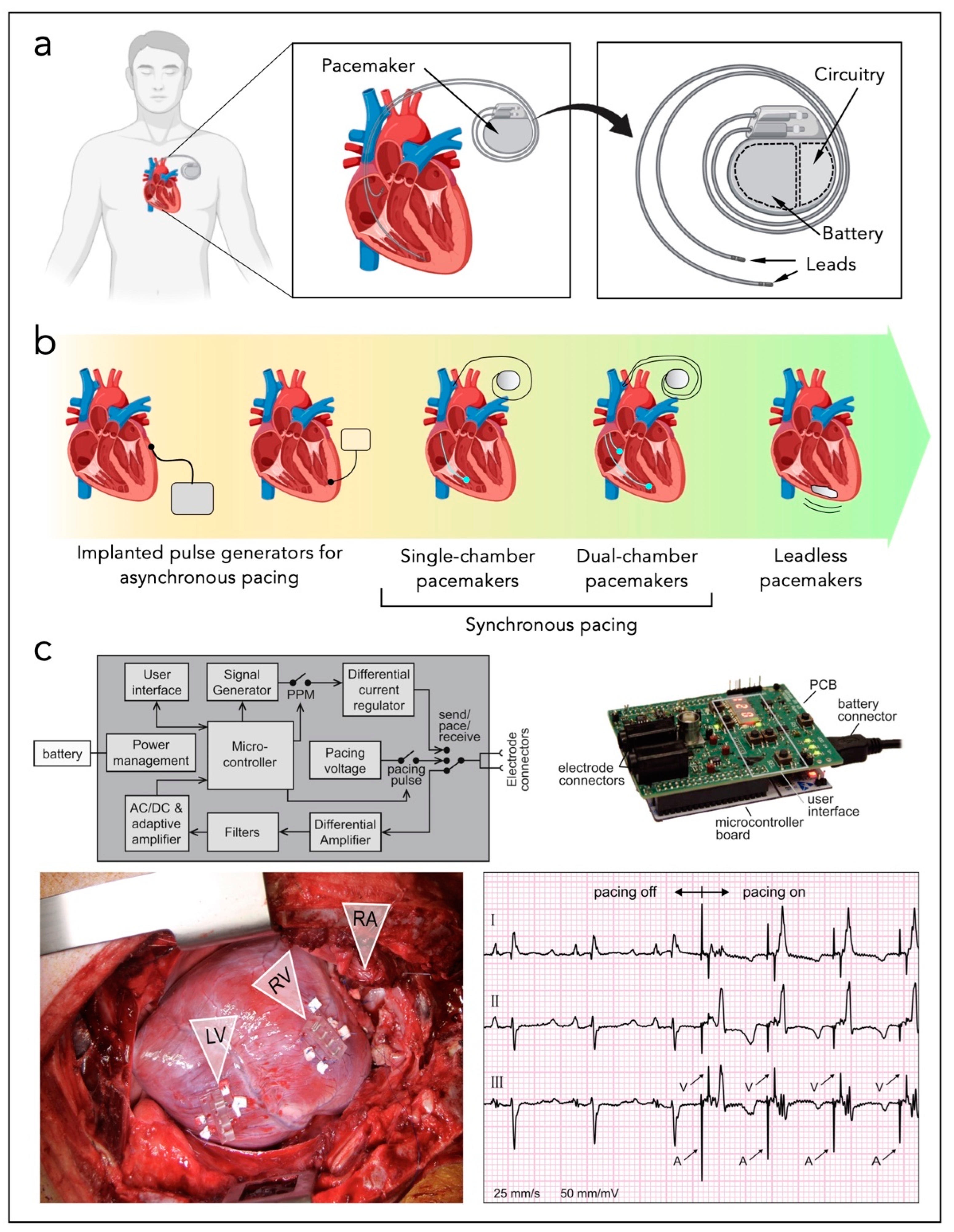
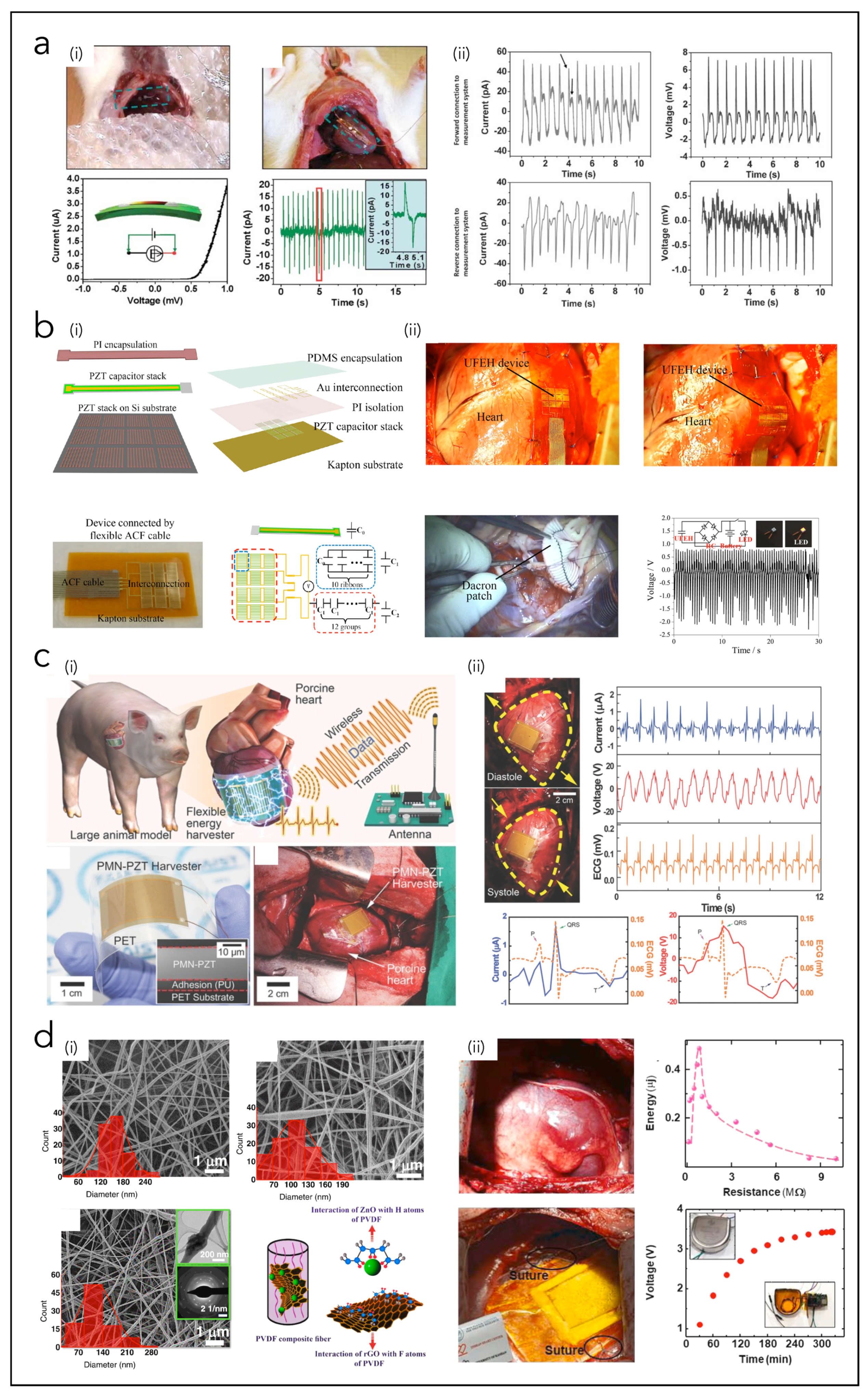
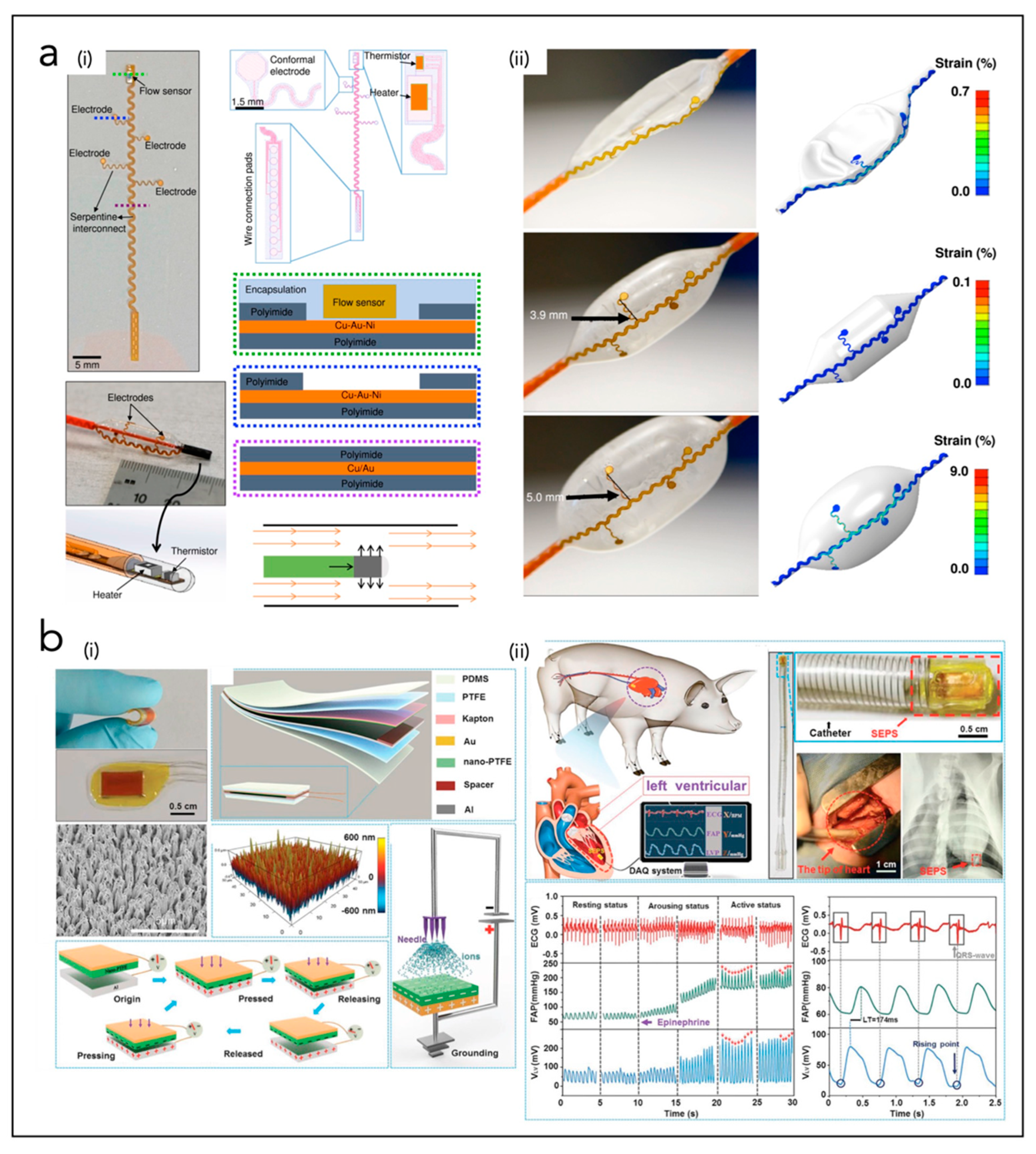
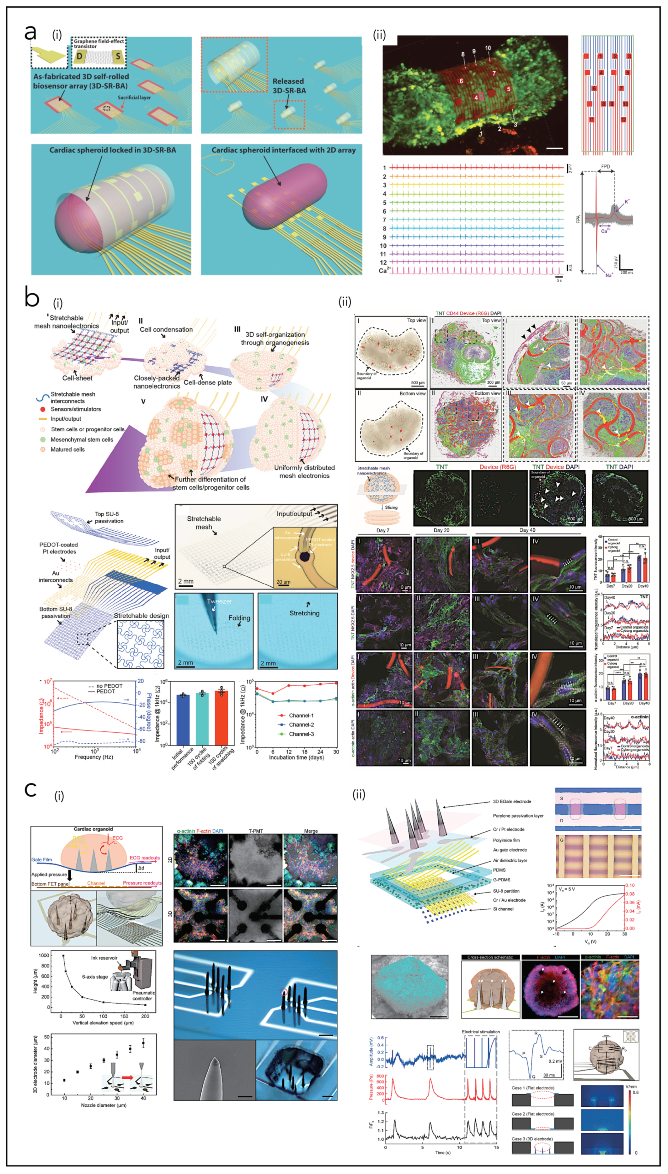
Table 2.
Cardiac bioelectronic devices, classified in terms of materials, performances, and applicative context. The blue, pink, grey, green, and yellow colors are, respectively, for: piezoelectric nanogenerator (PNG), triboelectric nanogenerator (TNG), solar cells (SCs), electrochemical generators and biofuel cells (BFCs), and cardiac bioelectronic systems.
| Ref. | Type of Device | Materials | Output Performances/Working Principle | Application Field |
|---|---|---|---|---|
| Dagdeviren et al. [257] | PNG | PZT/Pt/Ti/SiO2 Ti/Pt, Cr/Au electrodes | VOC = 1 mV; ISC = 1 pA E = 0.001 pJ | EH from heart motion in rabbits |
| Xie et al. [258] | PNG | PMN-Pt | VOC = 3.2 V; ISC = 54 nA | EH from heart motion in rats |
| Hwang et al. [259] | PNG | PZT, Au, PET | VOC = 8.2 mV; ISC = 0.223 mA E = 1.8286 µJ | EH from heart motion in rats |
| Lu et al. [113] | PNG | PZT, Ti/Pt, SiO2, Si, polyimide | VOC = 3 V | EH from heart motion in pigs |
| Kim et al. [114] | PNG | PZT, PU, PET | VOC = 17.8 V; ISC = 1.74 µA E = 30.972 µJ | EH from heart motion in pigs |
| Li et al. [116] | PNG | PZT, Cr/Au, Be, PDMS | VOC = 20 V; ISC = 15 µA E = 300 µJ | EH from heart motion in pigs |
| Zhang et al. [115] | PNG | PVDF, Al | VOC = 1.5 mV; ISC = 0.3 mA E = 0.45 µJ | EH from vases in pigs |
| Li et al. [260] | PNG | ZnO, polyimide | VOC = 3 mV; ISC = 30 pA E = 0.06 pJ | EH from heart motion in rats |
| Dong et al. [118] | PNG | Cu, Au, Kapton, PVDF, porous PVDF | VOC = 0.5 V; ISC = 43 nA | EH from pacemaker leads |
| Azimi et al. [119] | PNG | PVDF-TrFE NFs, ZnO, rGO | E = 0.487 µJ | EH from heartbeat in dogs |
| Hyun Kim et al. [114] | PNG | PMN-PZT | VOC = 17.8 V; ISC = 1.74 µA | EH from heartbeat in pigs |
| Zheng et al. [33] | TNG | PTFE, Kapton, Au, Al | VOC = 14 V; ISC = 5 µA E = 70 µJ | EH from heart motion in pigs |
| Liu et al. [218] | TNG | PTFE, PDMS, Kapton, Au, Al | VOC = 0.008 V | EH from heart motion in pigs |
| Ouyang et al. [158] | TNG | PDMS, Ti, Kapton, Au, PTFE, Al | VOC = 65.2 V; ISC = 0.5 µA E = 32.6 µJ | EH from heart motion in pigs |
| Zheng et al. [156] | TNG | PDMS, Kapton, Au | VOC = 3.75 V; ISC = 0.14 µA E = 0.525 µJ | EH from breath in rats |
| Ryu et al. [160] | TNG | PVA-NH2, PFA | VOC = 3.75 V p = 4.9 µW/cm3 | EH from body motion in mongrels |
| Ma et al. [157] | TNG | PTFE, Kapton, Au | VOC = 10 V; ISC = 4 µA E = 40 µJ | EH from heat motion in pigs |
| Haeberlin et al. [44] | SC | Commercial modules | p = 1963 µW/cm2 (direct outdoor sunlight) p = 206 µW/cm2 (shade outdoors) p = 4 µW/cm2 (indoors) | EH from solar light to power pacemaker in pigs |
| Prominski et al. [174] | SC | Si NMs | - | Harvesting solar light for cardiac modulation in rats |
| Dan Tchin-Iou et al. [45] | SC | - | VOC = 4.5 V; ISC = 23 mA p = 80.6 mW | EH from solar light to charge artificial heart |
| Song et al. [171] | SC | PDMS, Polyimide, SU8, NOA, GaInP/GaAs | p = 647 µW | EH from solar light to power pacemaker in mice |
| Holade et al. [46] | BFC | AuxPty NPs, carbon black (CB), buckypaper | VOC = 0.35 V; ISC = 0.65 mAcm−2 p = 104 µW | EH from glucose oxidation to activate a pacemaker |
| Southcott et al. [47] | BFC | Buckypaper, CNTs, laccase PQQ-dependent glucose dehydrogenase | VOC = 0.47 V; ISC = 0.83 mAcm−2 | EH from glucose oxidation to activate a pacemaker |
| MacVittie et al. [191] | BFC | Buckypaper, CNTs, laccase PQQ-dependent glucose dehydrogenase | VOC = 3 V | EH from lobsters to power a pacemaker |
| Choi et al. [200] | BP/CLS | Si NM, Mo, b-DCPU, PLGA, elastomer | Transient closed-loop system for cardiac electrotherapy | |
| Viventi et al. [197] | Epi-CS | Si NM, PI | High-performance, conformal bioelectronic system used to measure cardiac electrophysiological activity | |
| Fang et al. [198] | Epi-CS | Si NM; SiO2 | Hermetically sealed array of 396 multiplexed capacitive sensors based on Si NM transistors | |
| Choi et al. [199] | Epi-CS | PLGA, W/Mg, SI NM diodes | Fully implantable and bioresorbable, leadless, battery-free and externally programmable cardiac pacemaker for the treatment of atrioventricular (AV) nodal heart block. | |
| Kim et al. [201] | Epi-CS | PI, PMMA, Cr/Au, silk, Ti/Pt | Stretchable sensors and actuators based on epicardial webs that can conformally wrap around the heart | |
| Xu et al. [202] | Epi-CS | Au, Si or INGaN, PI, PMMA, PET, PDMS | 3D multifunctional integumentary thin elastic membrane integrating 68 electrodes and custom-formed to envelope completely and conformally the heart, | |
| Xu et al. [203] | Epi-CS | Pt-Ir allow, PEDOT:PSS | Compliant, large-area, low-impedance electrodes for electrical stimulation and cardiac electrotherapy | |
| Lee et al. [204] | Epi-CS | Parylene, Au, PMC3A, PEDOT:PSS | Ultrathin, stretchable grid-patterned active OECT matrix directly attached to the surface of a rat’s beating heart. | |
| Park et al. [205] | Epi-CS | AgNWs, SBS rubber | Epicardial mesh for electromechanical cardioplasty and electrotherapy | |
| Choi et al. [206] | Epi-CS | Ag-Au, elastomers | 2D fan-shaped implantable devices with 42 multichannel electrodes and based on nanocomposites applied on the swine heart for continuous electrophysiological recording | |
| Gutruf et al. [207] | Epi-CS | Parylene, Ti/Pt, Cu, PI | Miniaturized wireless energy-harvesting and digital communication electronics implanted subdermally to provide pacing in rats | |
| Sim et al. [208] | Epi-CS | PDMS, AgNWs/PDMS, Ion gel, P3HT-NFs/PDMS | Epicardial bioelectronic stretchable patch with cardiac tissue-like softness, for spatiotemporal mapping of electrophysiological activity, strain/temperature sensing and electrical pacing on a beating porcine heart. | |
| Liu et al. [209] | Epi-CS | PEDOT:PSS, fluorinated elastomers | Fully elastic electrode with tissue-like softness with 64 electrodes (elastrode) | |
| Klinker et al. [215] | Endo-CS | PI, Cu/Au/Ni | Smart balloon catheter system containing stretchable electrodes and thermal-based blood flow sensors | |
| Kim et al. [201,216] | Endo-CS | PI, PMMA, Cr/Au, silk, Ti/Pt | Array of integrated stretchable RF electrodes and multimodal (temperature, flow, tactile) sensors on a balloon catheter | |
| Han et al. [217] | Endo-CS | Cr/Au, PI, SiO2, Ti/Cu | Balloon catheter-integrated soft multilayer electronic array for multiplexed sensing and actuation during cardiac surgery | |
| Liu et al. [218] | Endo-CS | PDMS, PTFE, Kapton, Au, Al | Flexible self-powered endocardial pressure sensor for real-time electrophysiology monitoring. | |
| Liu et al. [224] | CP | Si NWs, SU8, Cr/Pd | 3D interconnected microporous nanoelectronics networks assembled from 2D precursors, integrated with cardiac patch. | |
| Tian et al. [225] | CP | Si NWs, SU-8, alginate, electrospun NFs | Microporous, flexible and free-standing nanoelectronics scaffold based on field-effect transistors and integrated with natural or synthetic biomaterials. | |
| Dai et al. [226] | CP | Si NWs, SiO2/Si, Ni, SU8 meshes, PLGA NFs | Tissue-scaffold-mimicking 3D nanoelectronics array (64 electrodes) for real-time sub-millisecond recording of extracellular action potential in developing rat cardiac tissues | |
| Wang et al. [227] | CP | PI, Au, PVA, Ti, SiO2, Dragon Skin | 3D porous, flexible electronic scaffold formed through compressive buckling of 2D precursor structures, with microelectrodes integrated within thermoresponsive extracellular matrix-based hydrogels | |
| Feiner et al. [228] | CP | SU8 mesh, Au, TiN, PCL NFs | Flexible, free-standing electronics (32 electrodes) and a 3D nanocomposite scaffold, integrated with a cardiac patch | |
| Feiner et al. [229] | CP | Electrospun albumin fibers, Au | Elastic, biodegradable, electronic scaffolds | |
| Lind et al. [230] | CP | Dextran, TPU, CB, PDMS, Ag | Multimaterial 3D-printed heart-on-chip electronics | |
| Kalmykov et al. [239] | CO | SU8, Au, graphene | Flexible organ-on-electronic-chip device based on 3D self-rolled biosensor arrays | |
| Li et al. [240] | CO | SU8, PEDOT, Pt, Au | Cyborg human cardiac organoids based on the 3D assembly of stretchable sub-µm thick mesh nanoelectronics | |
| Kim et al. [241] | CO | EGaIn, Parylene, Cr/Pt, PI, Au, PDMS, SU8, Si | Multimodal mechanophysiological characterization of 3D human cardiac organoids | |
PNG: Piezoelectric nanogenerator; TNG: triboelectric nanogenerator; SC: solar cell; BFC: biofuel cell; EH: energy harvesting; BP: bioresorbable pacemaker; CLS: closed-loop system. Epi-CS: epicardial system; Endo-CS: endocardial system; CP: cardiac patch; CO: cardiac organoid. NFs: nanofibers; NM: nanomembrane; CNTs: carbon nanotubes; NWs: nanowires.
7. Conclusions and Future Challenges
This paper reviews different types of bioelectronic interfaces used for monitoring, pacing, or studying the heart and its in vitro models. The most recent technologies for heart energy harvesting are described in comparison with the standard ones, and the main types of micro-devices used for this purpose are presented. The necessity of novel cardiac energy generators is motivated by several factors, especially by the need for reliability and the powering of implantable biomedical electronic devices, in order to avoid the reliance on chemical batteries, characterized by restricted lifetime and requiring frequent replacements with risky surgical interventions. Battery-less pacemakers represent the first important advancement in this field, and they are based on micro-devices for harvesting the energy conveyed by the cardiac contraction/expansion cycle; in particular, the cardiac systole is a source of periodic pressure and endless clean energy. Implantable/wearable nanogenerators and heart energy harvesters are, thus, described with a focus on materials and state-of-the-art examples from previous works. Against other electromagnetic generators, piezoelectric and triboelectric nanogenerators, solar cells, and biofuel cells represent the most promising technologies for the realization of biocompatible, self-powered biomedical devices. In particular, PNGs and TNGs are the most widely used alternatives to chemical batteries or wireless inductive charging, because they offer attractive properties, e.g., light weight, simple architectures, biocompatibility, optimal performances, compatibility with most microfabrication processes, a wide variety of employable materials, easy processability, suitability to be conformally applied onto the heart or in endocardial systems, and sensitivity to quasi-static mechanical deformations. Thus, they could easily be applicatied as heart energy harvesters. However, they also present some disadvantages: in particular, their scarce resistance to humidity makes their deployment for implantable applications quite challenging, revealing the need for hermetic encapsulations for protecting them from the surrounding tissues. The research of new biocompatible high-performance materials also represents another future challenge for PNGs and TNGs as HEHs.
Moreover, more general soft bioelectronic cardiac interfaces are presented in the second part of the review, classified as epicardial and endocardial bioelectronics, and devices for applications with in vitro cardiac models (cardiac patches and cardiac organoids). Thanks to their flexibility, stretchability, and reliability, these systems can accommodate the mechanical deformations and movements of the heart, of blood vessels, and of the growing cells in cardiac models, and they can provide a continuous, robust, reliable, and high-density electrophysiological mapping with high spatiotemporal resolution. The most recent advances in the 3D and 4D bioprinting of cardiac tissues are, finally, highlighted with emphasis on the possible combination and hybrid integration of bioprinted scaffolds and dynamic biomaterials with morphing bioelectronics. The main applications of bioprinting of cardiac tissues reside in regenerative medicine and tissue engineering; however, the seamless integration of biomimetic bioelectronics allows for multiple additional functions, e.g., biosensing, electrical stimulation, and drug delivery.
Heart energy harvesters (HEHs) and heart bioelectronic systems (HBSs) are promising technologies for the next-generation implantable biomedical devices, and future research work is needed to assess their efficiency, adaptivity to the environment, biocompatibility, flexibility and conformality, and complexity of their electronic interfaces and circuits. Future developments in materials science and nanotechnology will improve the realization of more biocompatible and more efficient cardiac bioelectronic devices.
The main challenges related to these technologies are summarized and illustrated in Figure 12. Concerning HEHs and cardiac nanogenerators, the most crucial points to take into account for their future development and commercialization regard the output generation performances, their reliability, their electronic interfaces and power management circuitry, their biocompatibility, and their connection into more complex closed-loop systems. Increasing their performances is possible through the development of novel energy transducing materials, through nanostructuring techniques that enhance the charge generation [261,262], or even by acting on the design and architectures of the devices [262,263]. The use of hermetic packages and encapsulations is a key aspect to guarantee long-term operation and stable reliability of cardiac implantable devices [264]. The electronic circuitry serves to extract the signal from the HEH and collect the charges to generate power; therefore, it requires minimized losses and maximized signal-to-noise ratios and efficiencies. Additionally, the advances of soft bioelectronics and microfabrication techniques allow for the integration of the circuitry and power managements modules directly in the space of the real device, minimizing the hindrance and making the interconnections flexible or stretchable according to the needs. Connecting HEHs into more complex HBSs requires the processing of the signal of multiple devices working simultaneously, i.e., the energy harvester should be synchronized with the IBD under consideration, and, in the case of multiple IBDs, multiplexing instrumentation is needed on a miniaturized scale. The advent of artificial intelligence and deep learning approaches will allow for the creation of intelligent systems of sensors/transducers/devices [265].
Regarding, more generally, the cardiac bioelectronic interfaces, the main challenges for the future can be ascribed to the following points: (i) increasing the number of electrodes per unit area to achieve high-density electrical recording/stimulation; (ii) enhancing multifunctionality and multimodality in order to detect simultaneously different cardiac physiological activities (electrophysiology, temperature, metabolism, mechanical forces, etc.); (iii) developing wireless, biodegradable devices with online data processing algorithms; (iv) miniaturizing technologies up to the cellular level to perform in situ molecular phenotype characterization for a better cardiac diagnosis and therapy; (v) enabling large-scale production for a better cost-effectiveness, in order to enable commercialization and clinical applications; (vi) conducting more studies on animal-derived cells and animal models, clinical trials and biocompatibility analyses to assess the long-term safety, the immune reaction, and the devices’ stability over time, and establishing validation protocols.
Funding
This research received no external funding.
Conflicts of Interest
The author declares no conflict of interest.
References
- Lai, Y.-C.; Hsiao, Y.-C.; Wu, H.-M.; Wang, Z.L. Waterproof Fabric-Based Multifunctional Triboelectric Nanogenerator for Universally Harvesting Energy from Raindrops, Wind, and Human Motions and as Self-Powered Sensors. Adv. Sci. 2019, 6, 1801883. [Google Scholar] [CrossRef] [PubMed] [Green Version]
- Zhang, Z.; Li, X.; Yin, J.; Xu, Y.; Fei, W.; Xue, M.; Wang, Q.; Zhou, J.; Guo, W. Emerging hydrovoltaic technology. Nat. Nanotechnol. 2018, 13, 1109–1119. [Google Scholar] [CrossRef] [PubMed]
- Qin, Y.; Wang, X.; Wang, Z.L. Microfibre–nanowire hybrid structure for energy scavenging. Nature 2008, 451, 809–813. [Google Scholar] [CrossRef]
- Wang, Z.L.; Song, J. Piezoelectric Nanogenerators Based on Zinc Oxide Nanowire Arrays. Science 2006, 312, 242–246. [Google Scholar] [CrossRef]
- Wang, Z.L. Catch wave power in floating nets. Nature 2017, 542, 159–160. [Google Scholar] [CrossRef]
- Wang, Z.L. Self-Powered Nanosensors and Nanosystems. Adv. Mater. 2012, 24, 280–285. [Google Scholar] [CrossRef]
- Wu, Z.; Cheng, T.; Wang, Z.L. Self-Powered Sensors and Systems Based on Nanogenerators. Sensors 2020, 20, 2925. [Google Scholar] [CrossRef]
- Wang, Z.L. Energy harvesting for self-powered nanosystems. Nano Res. 2008, 1, 1–8. [Google Scholar] [CrossRef] [Green Version]
- Pu, X.; Li, L.; Liu, M.; Jiang, C.; Du, C.; Zhao, Z.; Hu, W.; Wang, Z.L. Wearable Self-Charging Power Textile Based on Flexible Yarn Supercapacitors and Fabric Nanogenerators. Adv. Mater. 2015, 28, 98–105. [Google Scholar] [CrossRef] [PubMed]
- Sim, H.J.; Choi, C.; Kim, S.H.; Kim, K.M.; Lee, C.J.; Kim, Y.T.; Lepró, X.; Baughman, R.H.; Kim, S.J. Stretchable Triboelectric Fiber for Self-powered Kinematic Sensing Textile. Sci. Rep. 2016, 6, 35153. [Google Scholar] [CrossRef]
- Nielsen, M. Batteryless Pacemaker Could Use Heart’s Energy for Power; AIP Publishing LLC: Melville, NY, USA, 2021. [Google Scholar]
- Sun, M.; Li, Z.; Yang, C.; Lv, Y.; Yuan, L.; Shang, C.; Liang, S.; Guo, B.; Liu, Y.; Li, Z.; et al. Nanogenerator-based devices for biomedical applications. Nano Energy 2021, 89, 106461. [Google Scholar] [CrossRef]
- Helseth, L.E.; Guo, X.D. Contact Electrification and Energy Harvesting Using Periodically Contacted and Squeezed Water Droplets. Langmuir 2015, 31, 3269–3276. [Google Scholar] [CrossRef] [PubMed]
- Mariello, M.; Guido, F.; Mastronardi, V.M.; Madaro, F.; Mehdipour, I.; Todaro, M.T.; Rizzi, F.; De Vittorio, M. Chapter 13—Micro- and Nanodevices for Wind Energy Harvesting. In Nano Tools and Devices for Enhanced Renewable Energy; Devasahayam, S., Hussain, C.M., Eds.; Micro and Nano Technologies; Elsevier: Newark, NJ, USA, 2021; pp. 291–374. ISBN 978-0-12-821709-2. [Google Scholar]
- Mariello, M.; Guido, F.; Mastronardi, V.; Todaro, M.; Desmaële, D.; De Vittorio, M. Nanogenerators for harvesting mechanical energy conveyed by liquids. Nano Energy 2018, 57, 141–156. [Google Scholar] [CrossRef]
- Zhang, Z.; Du, K.; Chen, X.; Xue, C.; Wang, K. An air-cushion triboelectric nanogenerator integrated with stretchable electrode for human-motion energy harvesting and monitoring. Nano Energy 2018, 53, 108–115. [Google Scholar] [CrossRef]
- Mitcheson, P.D.; Yeatman, E.M.; Rao, G.K.; Holmes, A.S.; Green, T.C. Energy Harvesting from Human and Machine Motion for Wireless Electronic Devices. Proc. IEEE 2008, 96, 1457–1486. [Google Scholar] [CrossRef] [Green Version]
- Guido, F.; Qualtieri, A.; Algieri, L.; Lemma, E.D.; De Vittorio, M.; Todaro, M.T. AlN-based flexible piezoelectric skin for energy harvesting from human motion. Microelectron. Eng. 2016, 159, 174–178. [Google Scholar] [CrossRef]
- Yang, Y.; Zhang, H.; Lin, Z.-H.; Zhou, Y.S.; Jing, Q.; Su, Y.; Yang, J.; Chen, J.; Hu, C.; Wang, Z.L. Human Skin Based Triboelectric Nanogenerators for Harvesting Biomechanical Energy and as Self-Powered Active Tactile Sensor System. ACS Nano 2013, 7, 9213–9222. [Google Scholar] [CrossRef] [Green Version]
- Shi, Q.; Wang, H.; Wu, H.; Lee, C. Self-powered triboelectric nanogenerator buoy ball for applications ranging from environment monitoring to water wave energy farm. Nano Energy 2017, 40, 203–213. [Google Scholar] [CrossRef]
- Olsen, M.; Zhang, R.; Örtegren, J.; Andersson, H.; Yang, Y.; Olin, H. Frequency and voltage response of a wind-driven fluttering triboelectric nanogenerator. Sci. Rep. 2019, 9, 5543. [Google Scholar] [CrossRef] [Green Version]
- Shi, Q.; Zhang, Z.; Chen, T.; Lee, C. Minimalist and multi-functional human machine interface (HMI) using a flexible wearable triboelectric patch. Nano Energy 2019, 62, 355–366. [Google Scholar] [CrossRef]
- O’Flynn, B.; Sanchez-Torres, J.; Tedesco, S.; Walsh, M. Challenges in the Development of Wearable Human Machine Interface Systems. In Proceedings of the 2019 IEEE International Electron Devices Meeting (IEDM), San Francisco, CA, USA, 7–11 December 2019; pp. 10.4.1–10.4.4. [Google Scholar]
- Sun, Z.; Zhu, M.; Lee, C. Progress in the Triboelectric Human–Machine Interfaces (HMIs)-Moving from Smart Gloves to AI/Haptic Enabled HMI in the 5G/IoT Era. Nanoenergy Adv. 2021, 1, 81–120. [Google Scholar] [CrossRef]
- Gerratt, A.P.; Michaud, H.O.; Lacour, S.P. Elastomeric Electronic Skin for Prosthetic Tactile Sensation. Adv. Funct. Mater. 2015, 25, 2287–2295. [Google Scholar] [CrossRef]
- Brunelli, D.; Tadesse, A.M.; Vodermayer, B.; Nowak, M.; Castellini, C. Low-Cost Wearable Multichannel Surface EMG Acquisition for Prosthetic Hand Control. In Proceedings of the 2015 6th International Workshop on Advances in Sensors and Interfaces (IWASI), Gallipoli, Italy, 18–19 June 2015; pp. 94–99. [Google Scholar]
- Akaydin, H.D.; Elvin, N.; Andreopoulos, Y. Energy Harvesting from Highly Unsteady Fluid Flows using Piezoelectric Materials. J. Intell. Mater. Syst. Struct. 2010, 21, 1263–1278. [Google Scholar] [CrossRef]
- Dagdeviren, C.; Joe, P.; Tuzman, O.L.; Park, K.-I.; Lee, K.J.; Shi, Y.; Huang, Y.; Rogers, J.A. Recent progress in flexible and stretchable piezoelectric devices for mechanical energy harvesting, sensing and actuation. Extreme Mech. Lett. 2016, 9, 269–281. [Google Scholar] [CrossRef] [Green Version]
- Choi, A.Y.; Lee, C.J.; Park, J.; Kim, D.; Kim, Y.T. Corrugated Textile based Triboelectric Generator for Wearable Energy Harvesting. Sci. Rep. 2017, 7, 45583. [Google Scholar] [CrossRef] [Green Version]
- Chen, X.; Shao, J.; An, N.; Li, X.; Tian, H.; Xu, C.; Ding, Y. Self-powered flexible pressure sensors with vertically well-aligned piezoelectric nanowire arrays for monitoring vital signs. J. Mater. Chem. C 2015, 3, 11806–11814. [Google Scholar] [CrossRef]
- Zhang, X.-S.; Han, M.-D.; Wang, R.-X.; Zhu, F.-Y.; Li, Z.-H.; Wang, W.; Zhang, H.-X. Frequency-Multiplication High-Output Triboelectric Nanogenerator for Sustainably Powering Biomedical Microsystems. Nano Lett. 2013, 13, 1168–1172. [Google Scholar] [CrossRef] [PubMed]
- Kim, B.-Y.; Lee, W.-H.; Hwang, H.-G.; Kim, D.-H.; Kim, J.-H.; Lee, S.-H.; Nahm, S. Resistive Switching Memory Integrated with Nanogenerator for Self-Powered Bioimplantable Devices. Adv. Funct. Mater. 2016, 26, 5211–5221. [Google Scholar] [CrossRef]
- Zheng, Q.; Zhang, H.; Shi, B.; Xue, X.; Liu, Z.; Jin, Y.; Ma, Y.; Zou, Y.; Wang, X.; An, Z.; et al. In Vivo Self-Powered Wireless Cardiac Monitoring via Implantable Triboelectric Nanogenerator. ACS Nano 2016, 10, 6510–6518. [Google Scholar] [CrossRef]
- Gu, Y.; Zhang, T.; Chen, H.; Wang, F.; Pu, Y.; Gao, C.; Li, S. Mini Review on Flexible and Wearable Electronics for Monitoring Human Health Information. Nanoscale Res. Lett. 2019, 14, 263. [Google Scholar] [CrossRef]
- Fan, F.R.; Tang, W.; Wang, Z.L. Flexible Nanogenerators for Energy Harvesting and Self-Powered Electronics. Adv. Mater. 2016, 28, 4283–4305. [Google Scholar] [CrossRef] [PubMed]
- Khan, U.; Kim, S.-W. Triboelectric Nanogenerators for Blue Energy Harvesting. ACS Nano 2016, 10, 6429–6432. [Google Scholar] [CrossRef] [PubMed]
- Lee, J.-H.; Lee, K.Y.; Kumar, B.; Tien, N.T.; Lee, N.-E.; Kim, S.-W. Highly sensitive stretchable transparent piezoelectric nanogenerators. Energy Environ. Sci. 2012, 6, 169–175. [Google Scholar] [CrossRef]
- Mariello, M.; Scarpa, E.; Algieri, L.; Guido, F.; Mastronardi, V.M.; Qualtieri, A.; De Vittorio, M. Mechanical energy harvesting through a novel flexible contact-separation mode triboelectric nanogenerator based on metallized porous PDM and Parylene-C. In Proceedings of the 19th International Conference on Micro and Nanotechnology for Power Generation and Energy Conversion Applications (PowerMEMS), Krakow, Poland, 2–6 December 2019. [Google Scholar] [CrossRef]
- Wang, A.; Wu, C.; Pisignano, D.; Wang, Z.L.; Persano, L. Polymer nanogenerators: Opportunities and challenges for large-scale applications. J. Appl. Polym. Sci. 2018, 135, 45674. [Google Scholar] [CrossRef]
- Todaro, M.T.; Guido, F.; Algieri, L.; Mastronardi, V.M.; Desmaele, D.; Epifani, G.; De Vittorio, M. Biocompatible, Flexible, and Compliant Energy Harvesters Based on Piezoelectric Thin Films. IEEE Trans. Nanotechnol. 2018, 17, 220–230. [Google Scholar] [CrossRef]
- Ryu, H.; Yoon, H.; Kim, S. Hybrid Energy Harvesters: Toward Sustainable Energy Harvesting. Adv. Mater. 2019, 31, e1802898. [Google Scholar] [CrossRef] [PubMed]
- Mariello, M. Recent Advances on Hybrid Piezo-Triboelectric Bio-Nanogenerators: Materials, Architectures and Circuitry. Nanoenergy Adv. 2022, 2, 64–109. [Google Scholar] [CrossRef]
- Mariello, M.; Fachechi, L.; Guido, F.; De Vittorio, M. Multifunctional sub-100 µm thickness flexible piezo/triboelectric hybrid water energy harvester based on biocompatible AlN and soft parylene C-PDMS-Ecoflex™. Nano Energy 2021, 83, 105811. [Google Scholar] [CrossRef]
- Haeberlin, A.; Zurbuchen, A.; Walpen, S.; Schaerer, J.; Niederhauser, T.; Huber, C.; Tanner, H.; Servatius, H.; Seiler, J.; Haeberlin, H.; et al. The first batteryless, solar-powered cardiac pacemaker. Heart Rhythm 2015, 12, 1317–1323. [Google Scholar] [CrossRef]
- Tchin-Iou, A.D.; Min, B. Design of the Solar Cell System for Recharging the External Battery of the Totally-Implantable Artificial Heart. Int. J. Artif. Organs 1999, 22, 823–826. [Google Scholar] [CrossRef]
- Holade, Y.; MacVittie, K.; Conlon, T.; Guz, N.; Servat, K.; Napporn, T.W.; Kokoh, K.B.; Katz, E. Pacemaker Activated by an Abiotic Biofuel Cell Operated in Human Serum Solution. Electroanalysis 2014, 26, 2445–2457. [Google Scholar] [CrossRef]
- Southcott, M.; MacVittie, K.; Halámek, J.; Halámková, L.; Jemison, W.D.; Lobel, R.; Katz, E. A pacemaker powered by an implantable biofuel cell operating under conditions mimicking the human blood circulatory system—Battery not included. Phys. Chem. Chem. Phys. 2013, 15, 6278–6283. [Google Scholar] [CrossRef] [PubMed]
- Lee, M.; Boudreaux, B.; Chaturvedi, R.; Romanosky, S.; Downing, B. The Internet of Bodies: Opportunities, Risks, and Governance; RAND Corporation: Santa Monica, CA, USA, 2020. [Google Scholar]
- Al-Suhaimi, E.A.; Aljafary, M.A.; Alfareed, T.M.; Alshuyeh, H.A.; Alhamid, G.M.; Sonbol, B.; Almofleh, A.; Alkulaifi, F.M.; Altwayan, R.K.; Alharbi, J.N.; et al. Nanogenerator-Based Sensors for Energy Harvesting from Cardiac Contraction. Front. Energy Res. 2022, 10, 900534. [Google Scholar] [CrossRef]
- Muñoz, H.R.; Sacco, C.M. Cardiac mechanical energy and effects on the arterial tree. J. Cardiothorac. Vasc. Anesth. 1997, 11, 289–298. [Google Scholar] [CrossRef]
- Papp, L. Isn’t the heart the source of energy for blood circulation? “The heart doesn’t know the basic laws of physics”. Orv. Hetil. 2008, 149, 1443–1447. [Google Scholar] [CrossRef]
- Thudichum, I.L.W. On the Cause of the Emptiness of the Arteries after Death. Assoc. Med. J. 1855, 3, 122–127. [Google Scholar] [CrossRef] [Green Version]
- Manteuffel-Szoege, L. Energy Sources of Blood Circulation and the Mechanical Action of the Heart. Thorax 1960, 15, 47–53. [Google Scholar] [CrossRef] [Green Version]
- Furst, B. Hemodynamics of the Early Embryo Circulation. In The Heart and Circulation: An Integrative Model; Furst, B., Ed.; Springer International Publishing: Cham, Switzerland, 2020; pp. 21–29. ISBN 978-3-030-25062-1. [Google Scholar]
- Li, Z.; Pollack, G.H. Surface-induced flow: A natural microscopic engine using infrared energy as fuel. Sci. Adv. 2020, 6, eaba0941. [Google Scholar] [CrossRef]
- Moreno, A.; Walton, R.; Constantin, M.; Bernus, O.; Vigmond, E.J.; Bayer, J.D. Wide-area low-energy surface stimulation of large mammalian ventricular tissue. Sci. Rep. 2019, 9, 15863. [Google Scholar] [CrossRef]
- Zurbuchen, A.; Pfenniger, A.; Stahel, A.; Stoeck, C.T.; Vandenberghe, S.; Koch, V.M.; Vogel, R. Energy Harvesting from the Beating Heart by a Mass Imbalance Oscillation Generator. Ann. Biomed. Eng. 2013, 41, 131–141. [Google Scholar] [CrossRef] [Green Version]
- Moerke, C.; Wolff, A.; Ince, H.; Ortak, J.; Öner, A. New strategies for energy supply of cardiac implantable devices. Herzschrittmachertherapie Elektrophysiologie 2022, 33, 224–231. [Google Scholar] [CrossRef]
- Aquilina, O. A brief history of cardiac pacing. Images Paediatr. Cardiol. 2006, 8, 17–81. [Google Scholar] [PubMed]
- Van Hemel, N.M.; Van Der Wall, E.E. 8 October 1958, D Day for the implantable pacemaker. Neth. Heart J. 2008, 16, S3–S4. [Google Scholar] [CrossRef] [PubMed] [Green Version]
- Parsonnet, V.; Driller, J.; Cook, D.; Rizvi, S.A. Thirty-One Years of Clinical Experience with “Nuclear-Powered” Pacemakers. Pacing Clin. Electrophysiol. 2006, 29, 195–200. [Google Scholar] [CrossRef] [PubMed]
- Mond, H.G.; Freitag, G. The Cardiac Implantable Electronic Device Power Source: Evolution and Revolution. Pacing Clin. Electrophysiol. 2014, 37, 1728–1745. [Google Scholar] [CrossRef] [PubMed]
- Biffi, M.; Bertini, M.; Saporito, D.; Ziacchi, M.; Martignani, C.; Diemberger, I.; Boriani, G. Actual Pacemaker Longevity: The Benefit of Stimulation by Automatic Capture Verification. Pacing Clin. Electrophysiol. 2010, 33, 873–881. [Google Scholar] [CrossRef]
- Untereker, D.F.; Schmidt, C.L.; Jain, G.; Tamirisa, P.A.; Hossick-Schott, J.; Viste, M. 8—Power Sources and Capacitors for Pacemakers and Implantable Cardioverter-Defibrillators. In Clinical Cardiac Pacing, Defibrillation and Resynchronization Therapy, 5th ed.; Ellenbogen, K.A., Wilkoff, B.L., Kay, G.N., Lau, C.-P., Auricchio, A., Eds.; Elsevier: Philadelphia, PA, USA, 2017; pp. 251–269. ISBN 978-0-323-37804-8. [Google Scholar]
- Hauser, R.G.; Casey, S.A.; Gitter, C.B.; Tang, C.Y.; Abdelhadi, R.H.; Gornick, C.C.; Stanberry, L.; Sengupta, J.D. Reliability and longevity of implantable defibrillators. J. Interv. Card. Electrophysiol. 2021, 62, 507–518. [Google Scholar] [CrossRef]
- Hauser, R.G.; Hayes, D.L.; Kallinen, L.M.; Cannom, D.S.; Epstein, A.E.; Almquist, A.K.; Song, S.L.; Tyers, G.F.O.; Vlay, S.C.; Irwin, M. Clinical experience with pacemaker pulse generators and transvenous leads: An 8-year prospective multicenter study. Heart Rhythm. 2007, 4, 154–160. [Google Scholar] [CrossRef]
- Baddour, L.M.; Epstein, A.E.; Erickson, C.C.; Knight, B.P.; Levison, M.E.; Lockhart, P.B.; Masoudi, F.A.; Okum, E.J.; Wilson, W.R.; Beerman, L.B.; et al. Update on Cardiovascular Implantable Electronic Device Infections and Their Management. Circulation 2010, 121, 458–477. [Google Scholar] [CrossRef] [Green Version]
- Basar, N.; Cagli, K.; Basar, O.; Sen, N.; Gurel, O.M.; Akpinar, I.; Ozlu, M.F.; Okten, S. Upper-extremity deep vein thrombosis and downhill esophageal varices caused by long-term pacemaker implantation. Tex. Heart Inst. J. 2010, 37, 714–716. [Google Scholar]
- Miller, M.A.; Neuzil, P.; Dukkipati, S.R.; Reddy, V.Y. Leadless Cardiac Pacemakers: Back to the Future. J. Am. Coll. Cardiol. 2015, 66, 1179–1189. [Google Scholar] [CrossRef] [PubMed] [Green Version]
- Reddy, V.Y.; Knops, R.E.; Sperzel, J.; Miller, M.A.; Petru, J.; Simon, J.; Sediva, L.; De Groot, J.R.; Tjong, F.V.; Jacobson, P.; et al. Permanent Leadless Cardiac Pacing: Results of the LEADLESS Trial. Circulation 2014, 129, 1466–1471. [Google Scholar] [CrossRef] [PubMed]
- Piccini, J.P.; Stromberg, K.; Jackson, K.P.; Laager, V.; Duray, G.Z.; El-Chami, M.; Ellis, C.R.; Hummel, J.; Jones, D.R.; Kowal, R.C.; et al. Long-term outcomes in leadless Micra transcatheter pacemakers with elevated thresholds at implantation: Results from the Micra Transcatheter Pacing System Global Clinical Trial. Heart Rhythm. 2017, 14, 685–691. [Google Scholar] [CrossRef] [PubMed] [Green Version]
- Bereuter, L.; Gysin, M.; Kueffer, T.; Kucera, M.; Niederhauser, T.; Fuhrer, J.; Heinisch, P.; Zurbuchen, A.; Obrist, D.; Tanner, H.; et al. Leadless Dual-Chamber Pacing: A Novel Communication Method for Wireless Pacemaker Synchronization. JACC Basic Transl. Sci. 2018, 3, 813–823. [Google Scholar] [CrossRef] [PubMed]
- Ansari, M.H.; Karami, M.A. Piezoelectric energy harvesting from heartbeat vibrations for leadless pacemakers. J. Phys. Conf. Ser. 2015, 660, 012121. [Google Scholar] [CrossRef] [Green Version]
- Cingolani, E.; Goldhaber, J.; Marbán, E. Next-generation pacemakers: From small devices to biological pacemakers. Nat. Rev. Cardiol. 2018, 15, 139–150. [Google Scholar] [CrossRef]
- Goto, H.; Sugiura, T.; Kazui, T. Feasibility of the Automatic Generating System (AGS) for Quartz Watches as a Leadless Pacemaker Power Source: A Preliminary Report. In Proceedings of the 20th Annual International Conference of the IEEE Engineering in Medicine and Biology Society. Vol.20 Biomedical Engineering Towards the Year 2000 and Beyond (Cat. No.98CH36286), Hong Kong, China, 1 November 1998; Volume 1, pp. 417–419. [Google Scholar]
- Kim, S.-G.; Priya, S.; Kanno, I. Piezoelectric MEMS for energy harvesting. MRS Bull. 2012, 37, 1039–1050. [Google Scholar] [CrossRef] [Green Version]
- Tian, W.; Ling, Z.; Yu, W.; Shi, J. A Review of MEMS Scale Piezoelectric Energy Harvester. Appl. Sci. 2018, 8, 645. [Google Scholar] [CrossRef] [Green Version]
- Li, P.; Ryu, J.; Hong, S. Piezoelectric/Triboelectric Nanogenerators for Biomedical Applications; IntechOpen: London, UK, 2019; ISBN 978-1-83881-060-3. [Google Scholar]
- Curie, J.; Curie, P. Phénomènes électriques des cristaux hémièdres à faces inclinées. J. Phys. Theor. Appl. 1882, 1, 245–251. [Google Scholar] [CrossRef] [Green Version]
- Kim, H.S.; Kim, J.-H.; Kim, J. A review of piezoelectric energy harvesting based on vibration. Int. J. Precis. Eng. Manuf. 2011, 12, 1129–1141. [Google Scholar] [CrossRef]
- Wang, P.; Du, H. ZnO thin film piezoelectric MEMS vibration energy harvesters with two piezoelectric elements for higher output performance. Rev. Sci. Instrum. 2015, 86, 075002. [Google Scholar] [CrossRef] [PubMed]
- Algieri, L.; Todaro, M.T.; Guido, F.; Mastronardi, V.; Desmaële, D.; Qualtieri, A.; Giannini, C.; Sibillano, T.; De Vittorio, M. Flexible Piezoelectric Energy-Harvesting Exploiting Biocompatible AlN Thin Films Grown onto Spin-Coated Polyimide Layers. ACS Appl. Energy Mater. 2018, 1, 5203–5210. [Google Scholar] [CrossRef]
- Eom, C.-B.; Trolier-McKinstry, S. Thin-film piezoelectric MEMS. MRS Bull. 2012, 37, 1007–1017. [Google Scholar] [CrossRef] [Green Version]
- Wasa, K.; Matsushima, T.; Adachi, H.; Kanno, I.; Kotera, H. Thin-Film Piezoelectric Materials For a Better Energy Harvesting MEMS. J. Microelectromech. Syst. 2012, 21, 451–457. [Google Scholar] [CrossRef]
- Won, S.S.; Sheldon, M.; Mostovych, N.; Kwak, J.; Chang, B.-S.; Ahn, C.W.; Kingon, A.I.; Kim, I.W.; Kim, S.-H. Piezoelectric poly(vinylidene fluoride trifluoroethylene) thin film-based power generators using paper substrates for wearable device applications. Appl. Phys. Lett. 2015, 107, 202901. [Google Scholar] [CrossRef]
- Fang, J.; Wang, X.; Lin, T. Electrical power generator from randomly oriented electrospun poly(vinylidene fluoride) nanofibre membranes. J. Mater. Chem. 2011, 21, 11088–11091. [Google Scholar] [CrossRef] [Green Version]
- Chen, X.; Xu, S.; Yao, N.; Shi, Y. 1.6 V Nanogenerator for Mechanical Energy Harvesting Using PZT Nanofibers. Nano Lett. 2010, 10, 2133–2137. [Google Scholar] [CrossRef]
- Bai, S.; Xu, Q.; Gu, L.; Ma, F.; Qin, Y.; Wang, Z.L. Single crystalline lead zirconate titanate (PZT) nano/micro-wire based self-powered UV sensor. Nano Energy 2012, 1, 789–795. [Google Scholar] [CrossRef]
- Todaro, M.T.; Guido, F.; Mastronardi, V.M.; Desmaele, D.; Epifani, G.; Algieri, L.; De Vittorio, M. Piezoelectric MEMS vibrational energy harvesters: Advances and outlook. Microelectron. Eng. 2017, 183–184, 23–26. [Google Scholar] [CrossRef]
- Singh, H.H.; Khare, N. Flexible ZnO-PVDF/PTFE based piezo-tribo hybrid nanogenerator. Nano Energy 2018, 51, 216–222. [Google Scholar] [CrossRef]
- Wang, Z.; Narita, F. Corona Poling Conditions for Barium Titanate/Epoxy Composites and their Unsteady Wind Energy Harvesting Potential. Adv. Eng. Mater. 2019, 21, 1900169. [Google Scholar] [CrossRef]
- Clementi, G.; Lombardi, G.; Margueron, S.; Suarez, M.A.; Lebrasseur, E.; Ballandras, S.; Imbaud, J.; Lardet-Vieudrin, F.; Gauthier-Manuel, L.; Dulmet, B.; et al. LiNbO3 films—A low-cost alternative lead-free piezoelectric material for vibrational energy harvesters. Mech. Syst. Signal Process. 2021, 149, 107171. [Google Scholar] [CrossRef]
- Bartasyte, A.; Margueron, S.; Baron, T.; Oliveri, S.; Boulet, P. Toward High-Quality Epitaxial LiNbO3and LiTaO3Thin Films for Acoustic and Optical Applications. Adv. Mater. Interfaces 2017, 4, 1600998. [Google Scholar] [CrossRef]
- Kosec, M.; Malič, B.; Benčan, A.; Rojac, T. KNN-Based Piezoelectric Ceramics. In Piezoelectric and Acoustic Materials for Transducer Applications; Safari, A., Akdoğan, E.K., Eds.; Springer: Boston, MA, USA, 2008; pp. 81–102. ISBN 978-0-387-76540-2. [Google Scholar]
- Qualtieri, A.; Rizzi, F.; Todaro, M.; Passaseo, A.; Cingolani, R.; De Vittorio, M. Stress-driven AlN cantilever-based flow sensor for fish lateral line system. Microelectron. Eng. 2011, 88, 2376–2378. [Google Scholar] [CrossRef]
- Caro, M.A.; Zhang, S.; Riekkinen, T.; Ylilammi, M.; Moram, M.A.; Lopez-Acevedo, O.; Molarius, J.; Laurila, T. Piezoelectric coefficients and spontaneous polarization of ScAlN. J. Phys. Condens. Matter 2015, 27, 245901. [Google Scholar] [CrossRef] [PubMed] [Green Version]
- Koblmueller, G.; Averbeck, R.; Geelhaar, L.; Riechert, H.; Hösler, W.; Pongratz, P. Growth diagram and morphologies of AlN thin films grown by molecular beam epitaxy. J. Appl. Phys. 2003, 93, 9591–9596. [Google Scholar] [CrossRef]
- Chen, D.; Colas, J.; Mercier, F.; Boichot, R.; Charpentier, L.; Escape, C.; Balat-Pichelin, M.; Pons, M. High temperature properties of AlN coatings deposited by chemical vapor deposition for solar central receivers. Surf. Coat. Technol. 2019, 377, 124872. [Google Scholar] [CrossRef]
- Mariello, M.; Blad, T.; Mastronardi, V.; Madaro, F.; Guido, F.; Staufer, U.; Tolou, N.; De Vittorio, M. Flexible piezoelectric AlN transducers buckled through package-induced preloading for mechanical energy harvesting. Nano Energy 2021, 85, 105986. [Google Scholar] [CrossRef]
- Mariello, M.; Guido, F.; Algieri, L.; Mastronardi, V.M.; Qualtieri, A.; Pisanello, F.; De Vittorio, M. Microstructure and Electrical Properties of Novel Piezo-Optrodes Based on Thin-Film Piezoelectric Aluminium Nitride for Sensing. IEEE Trans. Nanotechnol. 2021, 20, 10–19. [Google Scholar] [CrossRef]
- Akiyama, M.; Morofuji, Y.; Kamohara, T.; Nishikubo, K.; Tsubai, M.; Fukuda, O.; Ueno, N. Flexible piezoelectric pressure sensors using oriented aluminum nitride thin films prepared on polyethylene terephthalate films. J. Appl. Phys. 2006, 100, 114318. [Google Scholar] [CrossRef]
- Mariello, M.; Fachechi, L.; Guido, F.; De Vittorio, M. Conformal, Ultra-thin Skin-Contact-Actuated Hybrid Piezo/Triboelectric Wearable Sensor Based on AlN and Parylene-Encapsulated Elastomeric Blend. Adv. Funct. Mater. 2021, 31, 2101047. [Google Scholar] [CrossRef]
- Signore, M.A.; Rescio, G.; De Pascali, C.; Iacovacci, V.; Dario, P.; Leone, A.; Quaranta, F.; Taurino, A.; Siciliano, P.; Francioso, L. Fabrication and characterization of AlN-based flexible piezoelectric pressure sensor integrated into an implantable artificial pancreas. Sci. Rep. 2019, 9, 17130. [Google Scholar] [CrossRef] [PubMed] [Green Version]
- Jackson, N.; Keeney, L.; Mathewson, A. Flexible-CMOS and biocompatible piezoelectric AlN material for MEMS applications. Smart Mater. Struct. 2013, 22, 115033. [Google Scholar] [CrossRef]
- Hu, X.; Yang, F.; Guo, M.; Pei, J.; Zhao, H.; Wang, Y. Fabrication of polyimide microfluidic devices by laser ablation based additive manufacturing. Microsyst. Technol. 2020, 26, 1573–1583. [Google Scholar] [CrossRef]
- Sun, Y.; Lacour, S.P.; Brooks, R.A.; Rushton, N.; Fawcett, J.; Cameron, R.E. Assessment of the biocompatibility of photosensitive polyimide for implantable medical device use. J. Biomed. Mater. Res. Part A 2009, 90, 648–655. [Google Scholar] [CrossRef]
- Pi, Z.; Zhang, J.; Wen, C.; Zhang, Z.-B.; Wu, D. Flexible piezoelectric nanogenerator made of poly(vinylidenefluoride-co-trifluoroethylene) (PVDF-TrFE) thin film. Nano Energy 2014, 7, 33–41. [Google Scholar] [CrossRef]
- Mariello, M.; Qualtieri, A.; Mele, G.; De Vittorio, M. Metal-Free Multilayer Hybrid PENG Based on Soft Electrospun/-Sprayed Membranes with Cardanol Additive for Harvesting Energy from Surgical Face Masks. ACS Appl. Mater. Interfaces 2021, 13, 20606–20621. [Google Scholar] [CrossRef]
- Chiu, Y.-Y.; Lin, W.-Y.; Wang, H.-Y.; Huang, S.-B.; Wu, M.-H. Development of a piezoelectric polyvinylidene fluoride (PVDF) polymer-based sensor patch for simultaneous heartbeat and respiration monitoring. Sensors Actuators A Phys. 2013, 189, 328–334. [Google Scholar] [CrossRef]
- Bae, J.-H.; Chang, S.-H. PVDF-based ferroelectric polymers and dielectric elastomers for sensor and actuator applications: A review. Funct. Compos. Struct. 2019, 1, 012003. [Google Scholar] [CrossRef]
- Liu, J.; Yu, D.; Zheng, Z.; Huangfu, G.; Guo, Y. Lead-free BiFeO3 film on glass fiber fabric: Wearable hybrid piezoelectric-triboelectric nanogenerator. Ceram. Int. 2021, 47, 3573–3579. [Google Scholar] [CrossRef]
- Li, Z.; Zhu, G.; Yang, R.; Wang, A.; Wang, Z.L. Muscle-Driven In Vivo Nanogenerator. Adv. Mater. 2010, 22, 2534–2537. [Google Scholar] [CrossRef] [PubMed]
- Lu, B.; Chen, Y.; Ou, D.; Chen, H.; Diao, L.; Zhang, W.; Zheng, J.; Ma, W.; Sun, L.; Feng, X. Ultra-flexible Piezoelectric Devices Integrated with Heart to Harvest the Biomechanical Energy. Sci. Rep. 2015, 5, 16065. [Google Scholar] [CrossRef] [PubMed] [Green Version]
- Kim, D.H.; Shin, H.J.; Lee, H.; Jeong, C.K.; Park, H.; Hwang, G.-T.; Lee, H.-Y.; Joe, D.J.; Han, J.H.; Lee, S.H.; et al. In Vivo Self-Powered Wireless Transmission Using Biocompatible Flexible Energy Harvesters. Adv. Funct. Mater. 2017, 27, 1700341. [Google Scholar] [CrossRef]
- Zhang, H.; Zhang, X.-S.; Cheng, X.; Liu, Y.; Han, M.; Xue, X.; Wang, S.; Yang, F.; Smitha, A.S.; Zhang, H.; et al. A flexible and implantable piezoelectric generator harvesting energy from the pulsation of ascending aorta: In vitro and in vivo studies. Nano Energy 2015, 12, 296–304. [Google Scholar] [CrossRef]
- Li, N.; Yi, Z.; Ma, Y.; Xie, F.; Huang, Y.; Tian, Y.; Dong, X.; Liu, Y.; Shao, X.; Jin, L.; et al. Direct Powering a Real Cardiac Pacemaker by Natural Energy of a Heartbeat. ACS Nano 2019, 13, 2822–2830. [Google Scholar] [CrossRef] [PubMed]
- Natta, L.; Mastronardi, V.M.; Guido, F.; Algieri, L.; Puce, S.; Pisano, F.; Rizzi, F.; Pulli, R.; Qualtieri, A.; De Vittorio, M. Soft and flexible piezoelectric smart patch for vascular graft monitoring based on Aluminum Nitride thin film. Sci. Rep. 2019, 9, 8392. [Google Scholar] [CrossRef] [Green Version]
- Dong, L.; Han, X.; Xu, Z.; Closson, A.B.; Liu, Y.; Wen, C.; Liu, X.; Escobar, G.P.; Oglesby, M.; Feldman, M.; et al. Flexible Porous Piezoelectric Cantilever on a Pacemaker Lead for Compact Energy Harvesting. Adv. Mater. Technol. 2019, 4, 1800148. [Google Scholar] [CrossRef] [Green Version]
- Azimi, S.; Golabchi, A.; Nekookar, A.; Rabbani, S.; Amiri, M.H.; Asadi, K.; Abolhasani, M.M. Self-powered cardiac pacemaker by piezoelectric polymer nanogenerator implant. Nano Energy 2021, 83, 105781. [Google Scholar] [CrossRef]
- Xu, Z.; Jin, C.; Cabe, A.; Escobedo, D.; Gruslova, A.; Jenney, S.; Closson, A.B.; Dong, L.; Chen, Z.; Feldman, M.D.; et al. Implantable Cardiac Kirigami-Inspired Lead-Based Energy Harvester Fabricated by Enhanced Piezoelectric Composite Film. Adv. Healthc. Mater. 2021, 10, e2002100. [Google Scholar] [CrossRef]
- Niu, S.; Wang, Z.L. Theoretical systems of triboelectric nanogenerators. Nano Energy 2015, 14, 161–192. [Google Scholar] [CrossRef]
- Fan, F.-R.; Tian, Z.-Q.; Wang, Z.L. Flexible triboelectric generator. Nano Energy 2012, 1, 328–334. [Google Scholar] [CrossRef]
- Niu, S.; Liu, Y.; Wang, S.; Lin, L.; Zhou, Y.S.; Hu, Y.; Wang, Z.L. Theoretical Investigation and Structural Optimization of Single-Electrode Triboelectric Nanogenerators. Adv. Funct. Mater. 2014, 24, 3332–3340. [Google Scholar] [CrossRef]
- Henniker, J. Triboelectricity in Polymers. Nature 1962, 196, 474. [Google Scholar] [CrossRef]
- Luo, J.; Wang, Z.L. Recent progress of triboelectric nanogenerators: From fundamental theory to practical applications. EcoMat 2020, 2, e12059. [Google Scholar] [CrossRef]
- Wang, Z.; Lin, L.; Chen, J.; Niu, S.; Zi, Y. Triboelectric Nanogenerators; Green Energy and Technology; Springer International Publishing: Atlanta, GA, USA, 2016; ISBN 978-3-319-40038-9. [Google Scholar]
- Zou, H.; Zhang, Y.; Guo, L.; Wang, P.; He, X.; Dai, G.; Zheng, H.; Chen, C.; Wang, A.C.; Xu, C.; et al. Quantifying the triboelectric series. Nat. Commun. 2019, 10, 1427. [Google Scholar] [CrossRef] [Green Version]
- Ibrahim, M.; Jiang, J.; Wen, Z.; Sun, X. Surface Engineering for Enhanced Triboelectric Nanogenerator. Nanoenergy Adv. 2021, 1, 58–80. [Google Scholar] [CrossRef]
- Zhang, H.; Yao, L.; Quan, L.; Zheng, X. Theories for triboelectric nanogenerators: A comprehensive review. Nanotechnol. Rev. 2020, 9, 610–625. [Google Scholar] [CrossRef]
- Narasimulu, A.A.; Zhao, P.; Soin, N.; Prashanthi, K.; Ding, P.; Chen, J.; Dong, S.; Chen, L.; Zhou, E.; Montemagno, C.D.; et al. Significant triboelectric enhancement using interfacial piezoelectric ZnO nanosheet layer. Nano Energy 2017, 40, 471–480. [Google Scholar] [CrossRef] [Green Version]
- Ravichandran, A.N.; Ramuz, M.; Blayac, S. Increasing surface charge density by effective charge accumulation layer inclusion for high-performance triboelectric nanogenerators. MRS Commun. 2019, 9, 682–689. [Google Scholar] [CrossRef]
- Kim, H.-J.; Yim, E.-C.; Kim, J.-H.; Kim, S.-J.; Park, J.-Y.; Oh, I.-K. Bacterial Nano-Cellulose Triboelectric Nanogenerator. Nano Energy 2017, 33, 130–137. [Google Scholar] [CrossRef]
- Wen, R.; Guo, J.; Yu, A.; Zhang, K.; Kou, J.; Zhu, Y.; Zhang, Y.; Li, B.-W.; Zhai, J. Remarkably enhanced triboelectric nanogenerator based on flexible and transparent monolayer titania nanocomposite. Nano Energy 2018, 50, 140–147. [Google Scholar] [CrossRef]
- Kil Yun, B.; Kim, H.S.; Ko, Y.J.; Murillo, G.; Jung, J.H. Interdigital electrode based triboelectric nanogenerator for effective energy harvesting from water. Nano Energy 2017, 36, 233–240. [Google Scholar] [CrossRef]
- Mariello, M.; Scarpa, E.; Algieri, L.; Guido, F.; Mastronardi, V.M.; Qualtieri, A.; De Vittorio, M. Novel Flexible Triboelectric Nanogenerator based on Metallized Porous PDMS and Parylene C. Energies 2020, 13, 1625. [Google Scholar] [CrossRef] [Green Version]
- Qian, Y.; Lyu, Z.; Kim, D.-H.; Kang, D.J. Enhancing the output power density of polydimethylsiloxane-based flexible triboelectric nanogenerators with ultrathin nickel telluride nanobelts as a co-triboelectric layer. Nano Energy 2021, 90, 106536. [Google Scholar] [CrossRef]
- Qian, Y.; Kang, D.J. Large-Area High-Quality AB-Stacked Bilayer Graphene on h-BN/Pt Foil by Chemical Vapor Deposition. ACS Appl. Mater. Interfaces 2018, 10, 29069–29075. [Google Scholar] [CrossRef]
- Harnchana, V.; Van Ngoc, H.; He, W.; Rasheed, A.; Park, H.; Amornkitbamrung, V.; Kang, D.J. Enhanced Power Output of a Triboelectric Nanogenerator using Poly(dimethylsiloxane) Modified with Graphene Oxide and Sodium Dodecyl Sulfate. ACS Appl. Mater. Interfaces 2018, 10, 25263–25272. [Google Scholar] [CrossRef]
- Kim, D.Y.; Kim, H.S.; Kong, D.S.; Choi, M.; Kim, H.B.; Lee, J.-H.; Murillo, G.; Lee, M.; Kim, S.S.; Jung, J.H. Floating buoy-based triboelectric nanogenerator for an effective vibrational energy harvesting from irregular and random water waves in wild sea. Nano Energy 2018, 45, 247–254. [Google Scholar] [CrossRef]
- Chen, J.; Yang, J.; Li, Z.; Fan, X.; Zi, Y.; Jing, Q.; Guo, H.; Wen, Z.; Pradel, K.C.; Niu, S.; et al. Networks of Triboelectric Nanogenerators for Harvesting Water Wave Energy: A Potential Approach toward Blue Energy. ACS Nano 2015, 9, 3324–3331. [Google Scholar] [CrossRef]
- Wang, Z.L.; Jiang, T.; Xu, L. Toward the blue energy dream by triboelectric nanogenerator networks. Nano Energy 2017, 39, 9–23. [Google Scholar] [CrossRef]
- Xiong, J.; Lin, M.-F.; Wang, J.; Gaw, S.L.; Parida, K.; Lee, P.S. Wearable All-Fabric-Based Triboelectric Generator for Water Energy Harvesting. Adv. Energy Mater. 2017, 7, 1701243. [Google Scholar] [CrossRef]
- Luo, J.; Tang, W.; Fan, F.R.; Liu, C.; Pang, Y.; Cao, G.; Wang, Z.L. Transparent and Flexible Self-Charging Power Film and Its Application in a Sliding Unlock System in Touchpad Technology. ACS Nano 2016, 10, 8078–8086. [Google Scholar] [CrossRef] [PubMed]
- Luo, J.; Wang, Z.; Xu, L.; Wang, A.C.; Han, K.; Jiang, T.; Lai, Q.; Bai, Y.; Tang, W.; Fan, F.R.; et al. Flexible and durable wood-based triboelectric nanogenerators for self-powered sensing in athletic big data analytics. Nat. Commun. 2019, 10, 5147. [Google Scholar] [CrossRef] [PubMed] [Green Version]
- Gorham, W.F. A New, General Synthetic Method for the Preparation of Linear Poly-p-xylylenes. J. Polym. Sci. Part A-1 Polym. Chem. 1966, 4, 3027–3039. [Google Scholar] [CrossRef]
- Mariello, M.; Guido, F.; Mastronardi, V.M.; De Donato, F.; Salbini, M.; Brunetti, V.; Qualtieri, A.; Rizzi, F.; De Vittorio, M. Captive-air-bubble aerophobicity measurements of antibiofouling coatings for underwater MEMS devices. Nanomater. Nanotechnol. 2019, 9, 1847980419862075. [Google Scholar] [CrossRef] [Green Version]
- Mariello, M.; Guido, F.; Mastronardi, V.M.; Giannuzzi, R.; Algieri, L.; Qualteri, A.; Maffezzoli, A.; De Vittorio, M. Reliability of Protective Coatings for Flexible Piezoelectric Transducers in Aqueous Environments. Micromachines 2019, 10, 739. [Google Scholar] [CrossRef] [Green Version]
- Golda-Cepa, M.; Brzychczy-Wloch, M.; Engvall, K.; Aminlashgari, N.; Hakkarainen, M.; Kotarba, A. Microbiological investigations of oxygen plasma treated parylene C surfaces for metal implant coating. Mater. Sci. Eng. C 2015, 52, 273–281. [Google Scholar] [CrossRef]
- Chen, T.-N.; Wuu, D.-S.; Wu, C.-C.; Chiang, C.-C.; Chen, Y.-P.; Horng, R.-H. Improvements of Permeation Barrier Coatings Using Encapsulated Parylene Interlayers for Flexible Electronic Applications. Plasma Process. Polym. 2007, 4, 180–185. [Google Scholar] [CrossRef]
- Applerot, G.; Abu-Mukh, R.; Irzh, A.; Charmet, J.; Keppner, H.; Laux, E.; Guibert, G.; Gedanken, A. Decorating Parylene-Coated Glass with ZnO Nanoparticles for Antibacterial Applications: A Comparative Study of Sonochemical, Microwave, and Microwave-Plasma Coating Routes. ACS Appl. Mater. Interfaces 2010, 2, 1052–1059. [Google Scholar] [CrossRef]
- Cieślik, M.; Engvall, K.; Pan, J.; Kotarba, A. Silane–parylene coating for improving corrosion resistance of stainless steel 316L implant material. Corros. Sci. 2011, 53, 296–301. [Google Scholar] [CrossRef]
- Lo, H.-W.; Tai, Y.-C. Parylene-based electret power generators. J. Micromech. Microeng. 2008, 18, 104006. [Google Scholar] [CrossRef]
- Genter, S.; Langhof, T.; Paul, O. Electret-based Out-Of-Plane Micro Energy Harvester with Parylene-C Serving as the Electret and Spring Material. Procedia Eng. 2015, 120, 341–344. [Google Scholar] [CrossRef] [Green Version]
- Calcagnile, P.; Blasi, L.; Rizzi, F.; Qualtieri, A.; Athanassiou, A.; Gogolides, E.; De Vittorio, M. Parylene C Surface Functionalization and Patterning with pH-Responsive Microgels. ACS Appl. Mater. Interfaces 2014, 6, 15708–15715. [Google Scholar] [CrossRef] [PubMed]
- Li, Z.; Zheng, Q.; Wang, Z.L.; Li, Z. Nanogenerator-Based Self-Powered Sensors for Wearable and Implantable Electronics. Research 2020, 2020, 8710686. [Google Scholar] [CrossRef] [PubMed] [Green Version]
- Zheng, Q.; Shi, B.; Fan, F.; Wang, X.; Yan, L.; Yuan, W.; Wang, S.; Liu, H.; Li, Z.; Wang, Z.L. In Vivo Powering of Pacemaker by Breathing-Driven Implanted Triboelectric Nanogenerator. Adv. Mater. 2014, 26, 5851–5856. [Google Scholar] [CrossRef]
- Ma, Y.; Zheng, Q.; Liu, Y.; Shi, B.; Xue, X.; Ji, W.; Liu, Z.; Jin, Y.; Zou, Y.; An, Z.; et al. Self-Powered, One-Stop, and Multifunctional Implantable Triboelectric Active Sensor for Real-Time Biomedical Monitoring. Nano Lett. 2016, 16, 6042–6051. [Google Scholar] [CrossRef]
- Ouyang, H.; Liu, Z.; Li, N.; Shi, B.; Zou, Y.; Xie, F.; Ma, Y.; Li, Z.; Li, H.; Zheng, Q.; et al. Symbiotic cardiac pacemaker. Nat. Commun. 2019, 10, 1821. [Google Scholar] [CrossRef] [Green Version]
- Niu, S.; Wang, X.; Yi, F.; Zhou, Y.S.; Wang, Z.L. A universal self-charging system driven by random biomechanical energy for sustainable operation of mobile electronics. Nat. Commun. 2015, 6, 8975. [Google Scholar] [CrossRef] [Green Version]
- Ryu, H.; Park, H.-M.; Kim, M.-K.; Kim, B.; Myoung, H.S.; Kim, T.Y.; Yoon, H.-J.; Kwak, S.S.; Kim, J.; Hwang, T.H.; et al. Self-rechargeable cardiac pacemaker system with triboelectric nanogenerators. Nat. Commun. 2021, 12, 4374. [Google Scholar] [CrossRef]
- Jiang, W.; Li, H.; Liu, Z.; Li, Z.; Tian, J.; Shi, B.; Zou, Y.; Ouyang, H.; Zhao, C.; Zhao, L.; et al. Fully Bioabsorbable Natural-Materials-Based Triboelectric Nanogenerators. Adv. Mater. 2018, 30, e1801895. [Google Scholar] [CrossRef]
- Hinchet, R.; Yoon, H.-J.; Ryu, H.; Kim, M.-K.; Choi, E.-K.; Kim, D.-S.; Kim, S.-W. Transcutaneous ultrasound energy harvesting using capacitive triboelectric technology. Science 2019, 365, 491–494. [Google Scholar] [CrossRef]
- Shawon, S.M.A.Z.; Sun, A.X.; Vega, V.S.; Chowdhury, B.D.; Tran, P.; Carballo, Z.D.; Tolentino, J.A.; Li, J.; Rafaqut, M.S.; Danti, S.; et al. Piezo-tribo dual effect hybrid nanogenerators for health monitoring. Nano Energy 2021, 82, 105691. [Google Scholar] [CrossRef]
- Dong, L.; Jin, C.; Closson, A.B.; Trase, I.; Richards, H.R.; Chen, Z.; Zhang, J.X.J. Cardiac Cardiac energy harvesting and sensing based on piezoelectric and triboelectric designs. Nano Energy 2020, 76, 105076. [Google Scholar] [CrossRef]
- Shi, B.; Zheng, Q.; Jiang, W.; Yan, L.; Wang, X.; Liu, H.; Yao, Y.; Li, Z.; Wang, Z.L. A Packaged Self-Powered System with Universal Connectors Based on Hybridized Nanogenerators. Adv. Mater. 2016, 28, 846–852. [Google Scholar] [CrossRef]
- Ippili, S.; Jella, V.; Thomas, A.; Yoon, S.-G. The Recent Progress on Halide Perovskite-Based Self-Powered Sensors Enabled by Piezoelectric and Triboelectric Effects. Nanoenergy Adv. 2021, 1, 3–31. [Google Scholar] [CrossRef]
- Wu, T.; Redoute, J.-M.; Yuce, M.R. A Wireless Implantable Sensor Design With Subcutaneous Energy Harvesting for Long-Term IoT Healthcare Applications. IEEE Access 2018, 6, 35801–35808. [Google Scholar] [CrossRef]
- Chen, Z.; Law, M.-K.; Mak, P.-I.; Martins, R.P. A Single-Chip Solar Energy Harvesting IC Using Integrated Photodiodes for Biomedical Implant Applications. IEEE Trans. Biomed. Circuits Syst. 2017, 11, 44–53. [Google Scholar] [CrossRef] [PubMed]
- Haeberlin, A.; Zurbuchen, A.; Schaerer, J.; Wagner, J.; Walpen, S.; Huber, C.; Haeberlin, H.; Fuhrer, J.; Vogel, R. Successful pacing using a batteryless sunlight-powered pacemaker. EP Eur. 2014, 16, 1534–1539. [Google Scholar] [CrossRef]
- Burks, S.; Kolcun, J.P.; Wang, M.Y. Subcutaneous Solar Cells Harvest Energy for Pulse Generation. Neurosurgery 2017, 81, N24. [Google Scholar] [CrossRef] [Green Version]
- Song, K.; Han, J.H.; Lim, T.; Kim, N.; Shin, S.; Kim, J.; Choo, H.; Jeong, S.; Kim, Y.-C.; Wang, Z.L.; et al. Subdermal Flexible Solar Cell Arrays for Powering Medical Electronic Implants. Adv. Healthc. Mater. 2016, 5, 1572–1580. [Google Scholar] [CrossRef] [Green Version]
- Bereuter, L.; Williner, S.; Pianezzi, F.; Bissig, B.; Buecheler, S.; Burger, J.; Vogel, R.; Zurbuchen, A.; Haeberlin, A. Energy Harvesting by Subcutaneous Solar Cells: A Long-Term Study on Achievable Energy Output. Ann. Biomed. Eng. 2017, 45, 1172–1180. [Google Scholar] [CrossRef]
- Tholl, M.; Akarçay, H.; Tanner, H.; Niederhauser, T.; Zurbuchen, A.; Frenz, M.; Haeberlin, A. Subdermal solar energy harvesting—A new way to power autonomous electric implants. Appl. Energy 2020, 269, 114948. [Google Scholar] [CrossRef]
- Prominski, A.; Shi, J.; Li, P.; Yue, J.; Lin, Y.; Park, J.; Tian, B.; Rotenberg, M.Y. Porosity-based heterojunctions enable leadless optoelectronic modulation of tissues. Nat. Mater. 2022, 21, 647–655. [Google Scholar] [CrossRef] [PubMed]
- Wu, T.; Wu, F.; Redoute, J.-M.; Yuce, M.R. An Autonomous Wireless Body Area Network Implementation Towards IoT Connected Healthcare Applications. IEEE Access 2017, 5, 11413–11422. [Google Scholar] [CrossRef]
- Shuvo, M.H.; Titirsha, T.; Amin, N.; Islam, S.K. Energy Harvesting in Implantable and Wearable Medical Devices for Enduring Precision Healthcare. Energies 2022, 15, 7495. [Google Scholar] [CrossRef]
- Dieffenderfer, J.P.; Beppler, E.; Novak, T.; Whitmire, E.; Jayakumar, R.; Randall, C.; Qu, W.; Rajagopalan, R.; Bozkurt, A. Solar Powered Wrist Worn Acquisition System for Continuous Photoplethysmogram Monitoring. In Proceedings of the 2014 36th Annual International Conference of the IEEE Engineering in Medicine and Biology Society, Chicago, IL, USA, 26–30 August 2014; pp. 3142–3145. [Google Scholar]
- Cosnier, S.; Le Goff, A.; Holzinger, M. Towards glucose biofuel cells implanted in human body for powering artificial organs: Review. Electrochem. Commun. 2013, 38, 19–23. [Google Scholar] [CrossRef]
- Drake, R.F.; Kusserow, B.K.; Messinger, S.; Matsuda, S. A tissue implantable fuel cell power supply. Trans. Am. Soc. Artif. Intern. Organs 1970, 16, 199–205. [Google Scholar] [PubMed]
- Simons, P.; Schenk, S.A.; Gysel, M.A.; Olbrich, L.F.; Rupp, J.L.M. A Ceramic-Electrolyte Glucose Fuel Cell for Implantable Electronics. Adv. Mater. 2022, 34, 2109075. [Google Scholar] [CrossRef]
- Holzinger, M.; Le Goff, A.; Cosnier, S. Carbon nanotube/enzyme biofuel cells. Electrochim. Acta 2012, 82, 179–190. [Google Scholar] [CrossRef]
- Gellett, W.; Kesmez, M.; Schumacher, J.; Akers, N.; Minteer, S.D. Biofuel Cells for Portable Power. Electroanalysis 2010, 22, 727–731. [Google Scholar] [CrossRef]
- Heller, A. Miniature biofuel cells. Phys. Chem. Chem. Phys. 2004, 6, 209–216. [Google Scholar] [CrossRef]
- Mano, N.; Mao, F.; Heller, A. Characteristics of a Miniature Compartment-less Glucose−O2 Biofuel Cell and Its Operation in a Living Plant. J. Am. Chem. Soc. 2003, 125, 6588–6594. [Google Scholar] [CrossRef] [PubMed]
- Cinquin, P.; Gondran, C.; Giroud, F.; Mazabrard, S.; Pellissier, A.; Boucher, F.; Alcaraz, J.-P.; Gorgy, K.; Lenouvel, F.; Mathé, S.; et al. A Glucose BioFuel Cell Implanted in Rats. PLoS ONE 2010, 5, e10476. [Google Scholar] [CrossRef] [PubMed]
- Rasmussen, M.; Ritzmann, R.E.; Lee, I.; Pollack, A.J.; Scherson, D. An Implantable Biofuel Cell for a Live Insect. J. Am. Chem. Soc. 2012, 134, 1458–1460. [Google Scholar] [CrossRef] [PubMed]
- Halámková, L.; Halámek, J.; Bocharova, V.; Szczupak, A.; Alfonta, L.; Katz, E. Implanted Biofuel Cell Operating in a Living Snail. J. Am. Chem. Soc. 2012, 134, 5040–5043. [Google Scholar] [CrossRef]
- Zebda, A.; Gondran, C.; Le Goff, A.; Holzinger, M.; Cinquin, P.; Cosnier, S. Mediatorless high-power glucose biofuel cells based on compressed carbon nanotube-enzyme electrodes. Nat. Commun. 2011, 2, 370. [Google Scholar] [CrossRef] [Green Version]
- Castorena-Gonzalez, J.A.; Foote, C.; MacVittie, K.; Halámek, J.; Halámková, L.; Martinez-Lemus, L.A.; Katz, E. Biofuel Cell Operating in Vivo in Rat. Electroanalysis 2013, 25, 1579–1584. [Google Scholar] [CrossRef]
- Sales, F.C.P.F.; Iost, R.M.; Martins, M.V.A.; Almeida, M.C.; Crespilho, F.N. An intravenous implantable glucose/dioxygen biofuel cell with modified flexible carbon fiber electrodes. Lab Chip 2013, 13, 468–474. [Google Scholar] [CrossRef]
- MacVittie, K.; Halámek, J.; Halámková, L.; Southcott, M.; Jemison, W.D.; Lobel, R.; Katz, E. From “cyborg” lobsters to a pacemaker powered by implantable biofuel cells. Energy Environ. Sci. 2012, 6, 81–86. [Google Scholar] [CrossRef]
- Barton, S.C.; Gallaway, J.; Atanassov, P. Enzymatic Biofuel Cells for Implantable and Microscale Devices. Chem. Rev. 2004, 104, 4867–4886. [Google Scholar] [CrossRef]
- Tang, X.; He, Y.; Liu, J. Soft bioelectronics for cardiac interfaces. Biophys. Rev. 2022, 3, 011301. [Google Scholar] [CrossRef]
- Kim, D.-H.; Ahn, J.-H.; Choi, W.M.; Kim, H.-S.; Kim, T.-H.; Song, J.; Huang, Y.Y.; Liu, Z.; Lu, C.; Rogers, J.A. Stretchable and Foldable Silicon Integrated Circuits. Science 2008, 320, 507–511. [Google Scholar] [CrossRef] [PubMed] [Green Version]
- Rogers, J.A.; Lagally, M.G.; Nuzzo, R.G. Synthesis, assembly and applications of semiconductor nanomembranes. Nature 2011, 477, 45–53. [Google Scholar] [CrossRef] [PubMed]
- Phan, H.-P.; Zhong, Y.; Nguyen, T.-K.; Park, Y.; Dinh, T.K.; Song, E.; Vadivelu, R.K.; Masud, M.K.; Li, J.; Shiddiky, M.J.A.; et al. Long-Lived, Transferred Crystalline Silicon Carbide Nanomembranes for Implantable Flexible Electronics. ACS Nano 2019, 13, 11572–11581. [Google Scholar] [CrossRef]
- Viventi, J.; Kim, D.-H.; Moss, J.D.; Kim, Y.-S.; Blanco, J.A.; Annetta, N.; Hicks, A.; Xiao, J.; Huang, Y.; Callans, D.J.; et al. A Conformal, Bio-Interfaced Class of Silicon Electronics for Mapping Cardiac Electrophysiology. Sci. Transl. Med. 2010, 2, 24ra22. [Google Scholar] [CrossRef] [Green Version]
- Fang, H.; Yu, K.J.; Gloschat, C.; Yang, Z.; Song, E.; Chiang, C.-H.; Zhao, J.; Won, S.M.; Xu, S.; Trumpis, M.; et al. Capacitively coupled arrays of multiplexed flexible silicon transistors for long-term cardiac electrophysiology. Nat. Biomed. Eng. 2017, 1, 0038. [Google Scholar] [CrossRef] [PubMed] [Green Version]
- Choi, Y.S.; Yin, R.T.; Pfenniger, A.; Koo, J.; Avila, R.; Lee, K.B.; Chen, S.W.; Lee, G.; Li, G.; Qiao, Y.; et al. Fully implantable and bioresorbable cardiac pacemakers without leads or batteries. Nat. Biotechnol. 2021, 39, 1228–1238. [Google Scholar] [CrossRef]
- Choi, Y.S.; Jeong, H.; Yin, R.T.; Avila, R.; Pfenniger, A.; Yoo, J.; Lee, J.Y.; Tzavelis, A.; Lee, Y.J.; Chen, S.W.; et al. A transient, closed-loop network of wireless, body-integrated devices for autonomous electrotherapy. Science 2022, 376, 1006–1012. [Google Scholar] [CrossRef]
- Kim, D.-H.; Ghaffari, R.; Lu, N.; Wang, S.; Lee, S.P.; Keum, H.; D’Angelo, R.; Klinker, L.; Su, Y.; Lu, C.; et al. Electronic sensor and actuator webs for large-area complex geometry cardiac mapping and therapy. Proc. Natl. Acad. Sci. USA 2012, 109, 19910–19915. [Google Scholar] [CrossRef] [Green Version]
- Xu, L.; Gutbrod, S.R.; Bonifas, A.P.; Su, Y.; Sulkin, M.S.; Lu, N.; Chung, H.-J.; Jang, K.-I.; Liu, Z.; Ying, M.; et al. 3D multifunctional integumentary membranes for spatiotemporal cardiac measurements and stimulation across the entire epicardium. Nat. Commun. 2014, 5, 3329. [Google Scholar] [CrossRef] [Green Version]
- Xu, L.; Gutbrod, S.R.; Ma, Y.; Petrossians, A.; Liu, Y.; Webb, R.C.; Fan, J.A.; Yang, Z.; Xu, R.; Whalen, J.J., III; et al. Materials and Fractal Designs for 3D Multifunctional Integumentary Membranes with Capabilities in Cardiac Electrotherapy. Adv. Mater. 2015, 27, 1731–1737. [Google Scholar] [CrossRef]
- Lee, W.; Kobayashi, S.; Nagase, M.; Jimbo, Y.; Saito, I.; Inoue, Y.; Yambe, T.; Sekino, M.; Malliaras, G.G.; Yokota, T.; et al. Nonthrombogenic, stretchable, active multielectrode array for electroanatomical mapping. Sci. Adv. 2018, 4, eaau2426. [Google Scholar] [CrossRef] [PubMed] [Green Version]
- Park, J.; Choi, S.; Janardhan, A.H.; Lee, S.-Y.; Raut, S.; Soares, J.; Shin, K.; Yang, S.; Lee, C.; Kang, K.-W.; et al. Electromechanical cardioplasty using a wrapped elasto-conductive epicardial mesh. Sci. Transl. Med. 2016, 8, 344ra86. [Google Scholar] [CrossRef] [PubMed] [Green Version]
- Choi, S.; Han, S.I.; Jung, D.; Hwang, H.J.; Lim, C.; Bae, S.; Park, O.K.; Tschabrunn, C.M.; Lee, M.; Bae, S.Y.; et al. Highly conductive, stretchable and biocompatible Ag–Au core–sheath nanowire composite for wearable and implantable bioelectronics. Nat. Nanotechnol. 2018, 13, 1048–1056. [Google Scholar] [CrossRef] [PubMed]
- Gutruf, P.; Yin, R.T.; Lee, K.B.; Ausra, J.; Brennan, J.A.; Qiao, Y.; Xie, Z.; Peralta, R.; Talarico, O.; Murillo, A.; et al. Wireless, battery-free, fully implantable multimodal and multisite pacemakers for applications in small animal models. Nat. Commun. 2019, 10, 5742. [Google Scholar] [CrossRef] [Green Version]
- Sim, K.; Ershad, F.; Zhang, Y.; Yang, P.; Shim, H.; Rao, Z.; Lu, Y.; Thukral, A.; Elgalad, A.; Xi, Y.; et al. An epicardial bioelectronic patch made from soft rubbery materials and capable of spatiotemporal mapping of electrophysiological activity. Nat. Electron. 2020, 3, 775–784. [Google Scholar] [CrossRef]
- Liu, J.; Zhang, X.; Liu, Y.; Rodrigo, M.; Loftus, P.D.; Aparicio-Valenzuela, J.; Zheng, J.; Pong, T.; Cyr, K.J.; Babakhanian, M.; et al. Intrinsically stretchable electrode array enabled in vivo electrophysiological mapping of atrial fibrillation at cellular resolution. Proc. Natl. Acad. Sci. USA 2020, 117, 14769–14778. [Google Scholar] [CrossRef]
- Liu, J.; Wang, J.; Zhang, Z.; Molina-Lopez, F.; Wang, G.-J.N.; Schroeder, B.C.; Yan, X.; Zeng, Y.; Zhao, O.; Tran, H.; et al. Fully stretchable active-matrix organic light-emitting electrochemical cell array. Nat. Commun. 2020, 11, 3362. [Google Scholar] [CrossRef] [PubMed]
- Pappone, C.; Santinelli, V. Multielectrode basket catheter: A new tool for curing atrial fibrillation? Heart Rhythm 2006, 3, 385–386. [Google Scholar] [CrossRef]
- Kim, D.-H.; Ghaffari, R.; Lu, N.; Rogers, J.A. Flexible and Stretchable Electronics for Biointegrated Devices. Annu. Rev. Biomed. Eng. 2012, 14, 113–128. [Google Scholar] [CrossRef] [Green Version]
- Oesterlein, T.; Frisch, D.; Loewe, A.; Seemann, G.; Schmitt, C.; Dössel, O.; Luik, A. Basket-Type Catheters: Diagnostic Pitfalls Caused by Deformation and Limited Coverage. BioMed Res. Int. 2016, 2016, e5340574. [Google Scholar] [CrossRef]
- Su, Y.; Liu, Z.; Wang, S.; Ghaffari, R.; Kim, D.-H.; Hwang, K.-C.; Rogers, J.A.; Huang, Y. Mechanics of stretchable electronics on balloon catheter under extreme deformation. Int. J. Solids Struct. 2014, 51, 1555–1561. [Google Scholar] [CrossRef] [Green Version]
- Klinker, L.; Lee, S.; Work, J.; Wright, J.; Ma, Y.; Ptaszek, L.; Webb, R.C.; Liu, C.; Sheth, N.; Mansour, M.; et al. Balloon catheters with integrated stretchable electronics for electrical stimulation, ablation and blood flow monitoring. Extreme Mech. Lett. 2015, 3, 45–54. [Google Scholar] [CrossRef]
- Kim, D.H.; Lu, N.; Ghaffari, R.; Kim, Y.-S.; Lee, S.P.; Xu, L.; Wu, J.; Kim, R.-H.; Song, J.; Liu, Z.; et al. Materials for multifunctional balloon catheters with capabilities in cardiac electrophysiological mapping and ablation therapy. Nat. Mater. 2011, 10, 316–323. [Google Scholar] [CrossRef] [PubMed]
- Han, M.; Chen, L.; Aras, K.; Liang, C.; Chen, X.; Zhao, H.; Li, K.; Faye, N.R.; Sun, B.; Kim, J.-H.; et al. Catheter-integrated soft multilayer electronic arrays for multiplexed sensing and actuation during cardiac surgery. Nat. Biomed. Eng. 2020, 4, 997–1009. [Google Scholar] [CrossRef] [PubMed]
- Liu, Z.; Ma, Y.; Ouyang, H.; Shi, B.; Li, N.; Jiang, D.; Xie, F.; Qu, D.; Zou, Y.; Huang, Y.; et al. Transcatheter Self-Powered Ultrasensitive Endocardial Pressure Sensor. Adv. Funct. Mater. 2019, 29, 1807560. [Google Scholar] [CrossRef]
- Dvir, T.; Timko, B.; Brigham, M.; Naik, S.R.; Karajanagi, S.S.; Levy, O.; Jin, H.; Parker, K.K.; Langer, R.; Kohane, D.S. Nanowired three-dimensional cardiac patches. Nat. Nanotechnol. 2011, 6, 720–725. [Google Scholar] [CrossRef] [Green Version]
- Mei, X.; Cheng, K. Recent Development in Therapeutic Cardiac Patches. Front. Cardiovasc. Med. 2020, 7, 610364. [Google Scholar] [CrossRef]
- Chantre, C.O.; Gonzalez, G.M.; Ahn, S.; Cera, L.; Campbell, P.H.; Hoerstrup, S.P.; Parker, K.K. Porous Biomimetic Hyaluronic Acid and Extracellular Matrix Protein Nanofiber Scaffolds for Accelerated Cutaneous Tissue Repair. ACS Appl. Mater. Interfaces 2019, 11, 45498–45510. [Google Scholar] [CrossRef]
- Yang, Q.; Wei, T.; Yin, R.T.; Wu, M.; Xu, Y.; Koo, J.; Choi, Y.S.; Xie, Z.; Chen, S.W.; Kandela, I.; et al. Photocurable bioresorbable adhesives as functional interfaces between flexible bioelectronic devices and soft biological tissues. Nat. Mater. 2021, 20, 1559–1570. [Google Scholar] [CrossRef]
- Li, J.; Celiz, A.D.; Yang, J.; Yang, Q.; Wamala, I.; Whyte, W.; Seo, B.R.; Vasilyev, N.V.; Vlassak, J.J.; Suo, Z.; et al. Tough adhesives for diverse wet surfaces. Science 2017, 357, 378–381. [Google Scholar] [CrossRef]
- Liu, J.; Xie, C.; Dai, X.; Jin, L.; Zhou, W.; Lieber, C.M. Multifunctional three-dimensional macroporous nanoelectronic networks for smart materials. Proc. Natl. Acad. Sci. USA 2013, 110, 6694–6699. [Google Scholar] [CrossRef] [PubMed] [Green Version]
- Tian, B.; Liu, J.; Dvir, T.; Jin, L.; Tsui, J.H.; Qing, Q.; Suo, Z.; Langer, R.; Kohane, D.S.; Lieber, C.M. Macroporous nanowire nanoelectronic scaffolds for synthetic tissues. Nat. Mater. 2012, 11, 986–994. [Google Scholar] [CrossRef] [PubMed] [Green Version]
- Dai, X.; Zhou, W.; Gao, T.; Liu, J.; Lieber, C.M. Three-dimensional mapping and regulation of action potential propagation in nanoelectronics-innervated tissues. Nat. Nanotechnol. 2016, 11, 776–782. [Google Scholar] [CrossRef] [Green Version]
- Wang, X.; Feiner, R.; Luan, H.; Zhang, Q.; Zhao, S.; Zhang, Y.; Han, M.; Li, Y.; Sun, R.; Wang, H.; et al. Three-dimensional electronic scaffolds for monitoring and regulation of multifunctional hybrid tissues. Extreme Mech. Lett. 2020, 35, 100634. [Google Scholar] [CrossRef]
- Feiner, R.; Engel, L.; Fleischer, S.; Malki, M.; Gal, I.; Shapira, A.; Shacham-Diamand, Y.; Dvir, T. Engineered hybrid cardiac patches with multifunctional electronics for online monitoring and regulation of tissue function. Nat. Mater. 2016, 15, 679–685. [Google Scholar] [CrossRef] [Green Version]
- Feiner, R.; Fleischer, S.; Shapira, A.; Kalish, O.; Dvir, T. Multifunctional degradable electronic scaffolds for cardiac tissue engineering. J. Control. Release 2018, 281, 189–195. [Google Scholar] [CrossRef] [PubMed]
- Lind, J.U.; Busbee, T.A.; Valentine, A.D.; Pasqualini, F.S.; Yuan, H.; Yadid, M.; Park, S.-J.; Kotikian, A.; Nesmith, A.P.; Campbell, P.H.; et al. Instrumented cardiac microphysiological devices via multimaterial three-dimensional printing. Nat. Mater. 2017, 16, 303–308. [Google Scholar] [CrossRef]
- Drakhlis, L.; Biswanath, S.; Farr, C.-M.; Lupanow, V.; Teske, J.; Ritzenhoff, K.; Franke, A.; Manstein, F.; Bolesani, E.; Kempf, H.; et al. Human heart-forming organoids recapitulate early heart and foregut development. Nat. Biotechnol. 2021, 39, 737–746. [Google Scholar] [CrossRef] [PubMed]
- Hofer, M.; Lutolf, M.P. Engineering organoids. Nat. Rev. Mater. 2021, 6, 402–420. [Google Scholar] [CrossRef]
- Lewis-Israeli, Y.R.; Wasserman, A.H.; Gabalski, M.A.; Volmert, B.D.; Ming, Y.; Ball, K.A.; Yang, W.; Zou, J.; Ni, G.; Pajares, N.; et al. Self-assembling human heart organoids for the modeling of cardiac development and congenital heart disease. Nat. Commun. 2021, 12, 5142. [Google Scholar] [CrossRef]
- Richards, D.J.; Coyle, R.C.; Tan, Y.; Jia, J.; Wong, K.; Toomer, K.; Menick, D.R.; Mei, Y. Inspiration from heart development: Biomimetic development of functional human cardiac organoids. Biomaterials 2017, 142, 112–123. [Google Scholar] [CrossRef] [PubMed]
- Zhao, D.; Lei, W.; Hu, S. Cardiac organoid—A promising perspective of preclinical model. Stem Cell Res. Ther. 2021, 12, 272. [Google Scholar] [CrossRef] [PubMed]
- Mills, R.; Hudson, J. An in vitro model of myocardial infarction. Nat. Biomed. Eng. 2020, 4, 366–367. [Google Scholar] [CrossRef] [PubMed]
- Zhang, Y.S.; Aleman, J.; Shin, S.R.; Kilic, T.; Kim, D.; Shaegh, S.A.M.; Massa, S.; Riahi, R.; Chae, S.; Hu, N.; et al. Multisensor-integrated organs-on-chips platform for automated and continual in situ monitoring of organoid behaviors. Proc. Natl. Acad. Sci. USA 2017, 114, E2293–E2302. [Google Scholar] [CrossRef] [PubMed] [Green Version]
- Le Floch, P.; Li, Q.; Lin, Z.; Zhao, S.; Liu, R.; Tasnim, K.; Jiang, H.; Liu, J. Stretchable Mesh Nanoelectronics for 3D Single-Cell Chronic Electrophysiology from Developing Brain Organoids. Adv. Mater. 2022, 34, 2106829. [Google Scholar] [CrossRef]
- Kalmykov, A.; Huang, C.; Bliley, J.; Shiwarski, D.; Tashman, J.; Abdullah, A.; Rastogi, S.K.; Shukla, S.; Mataev, E.; Feinberg, A.W.; et al. Organ-on-e-chip: Three-dimensional self-rolled biosensor array for electrical interrogations of human electrogenic spheroids. Sci. Adv. 2019, 5, eaax0729. [Google Scholar] [CrossRef] [Green Version]
- Li, Q.; Nan, K.; Le Floch, P.; Lin, Z.; Sheng, H.; Blum, T.S.; Liu, J. Cyborg Organoids: Implantation of Nanoelectronics via Organogenesis for Tissue-Wide Electrophysiology. Nano Lett. 2019, 19, 5781–5789. [Google Scholar] [CrossRef]
- Kim, M.; Hwang, J.C.; Min, S.; Park, Y.-G.; Kim, S.; Kim, E.; Seo, H.; Chung, W.G.; Lee, J.; Cho, S.-W.; et al. Multimodal Characterization of Cardiac Organoids Using Integrations of Pressure-Sensitive Transistor Arrays with Three-Dimensional Liquid Metal Electrodes. Nano Lett. 2022, 22, 7892–7901. [Google Scholar] [CrossRef]
- Jafari, A.; Ajji, Z.; Mousavi, A.; Naghieh, S.; Bencherif, S.A.; Savoji, H. Latest Advances in 3D Bioprinting of Cardiac Tissues. Adv. Mater. Technol. 2022, 7, 2101636. [Google Scholar] [CrossRef]
- Kato, B.; Wisser, G.; Agrawal, D.K.; Wood, T.; Thankam, F.G. 3D bioprinting of cardiac tissue: Current challenges and perspectives. J. Mater. Sci. Mater. Med. 2021, 32, 54. [Google Scholar] [CrossRef]
- Jia, W.; Gungor-Ozkerim, P.S.; Zhang, Y.S.; Yue, K.; Zhu, K.; Liu, W.; Pi, Q.; Byambaa, B.; Dokmeci, M.R.; Shin, S.R.; et al. Direct 3D bioprinting of perfusable vascular constructs using a blend bioink. Biomaterials 2016, 106, 58–68. [Google Scholar] [CrossRef] [PubMed] [Green Version]
- Sanjuan-Alberte, P.; Whitehead, C.; Jones, J.N.; Silva, J.C.; Carter, N.; Kellaway, S.; Hague, R.J.; Cabral, J.M.; Ferreira, F.C.; White, L.J.; et al. Printing biohybrid materials for bioelectronic cardio-3D-cellular constructs. iScience 2022, 25, 104552. [Google Scholar] [CrossRef]
- Tamay, D.G.; Usal, T.D.; Alagoz, A.S.; Yucel, D.; Hasirci, N.; Hasirci, V. 3D and 4D Printing of Polymers for Tissue Engineering Applications. Front. Bioeng. Biotechnol. 2019, 7, 164. [Google Scholar] [CrossRef] [PubMed]
- Saska, S.; Pilatti, L.; Blay, A.; Shibli, J. Bioresorbable Polymers: Advanced Materials and 4D Printing for Tissue Engineering. Polymers 2021, 13, 563. [Google Scholar] [CrossRef]
- Cox-Pridmore, D.M.; Castro, F.A.; Silva, S.R.P.; Camelliti, P.; Zhao, Y. Emerging Bioelectronic Strategies for Cardiovascular Tissue Engineering and Implantation. Small 2022, 18, 2105281. [Google Scholar] [CrossRef] [PubMed]
- Yong, U.; Kang, B.; Jang, J. 3D bioprinted and integrated platforms for cardiac tissue modeling and drug testing. Essays Biochem. 2021, 65, 545–554. [Google Scholar] [CrossRef]
- Miao, S.; Castro, N.; Nowicki, M.; Xia, L.; Cui, H.; Zhou, X.; Zhu, W.; Lee, S.-J.; Sarkar, K.; Vozzi, G.; et al. 4D printing of polymeric materials for tissue and organ regeneration. Mater. Today 2017, 20, 577–591. [Google Scholar] [CrossRef]
- Wang, Y.; Cui, H.; Wang, Y.; Xu, C.; Esworthy, T.J.; Hann, S.Y.; Boehm, M.; Shen, Y.-L.; Mei, D.; Zhang, L.G. 4D Printed Cardiac Construct with Aligned Myofibers and Adjustable Curvature for Myocardial Regeneration. ACS Appl. Mater. Interfaces 2021, 13, 12746–12758. [Google Scholar] [CrossRef]
- Ma, S.; Jiang, Z.; Wang, M.; Zhang, L.; Liang, Y.; Zhang, Z.; Ren, L.; Ren, L. 4D printing of PLA/PCL shape memory composites with controllable sequential deformation. Bio-Design Manuf. 2021, 4, 867–878. [Google Scholar] [CrossRef]
- Pedron, S.; van Lierop, S.; Horstman, P.; Penterman, R.; Broer, D.J.; Peeters, E. Stimuli Responsive Delivery Vehicles for Cardiac Microtissue Transplantation. Adv. Funct. Mater. 2011, 21, 1624–1630. [Google Scholar] [CrossRef]
- Abraham, W.T.; Perl, L. Implantable Hemodynamic Monitoring for Heart Failure Patients. J. Am. Coll. Cardiol. 2017, 70, 389–398. [Google Scholar] [CrossRef] [PubMed]
- Xu, Y.; Xie, X.; Duan, Y.; Wang, L.; Cheng, Z.; Cheng, J. A review of impedance measurements of whole cells. Biosens. Bioelectron. 2015, 77, 824–836. [Google Scholar] [CrossRef]
- Vashistha, R.; Dangi, A.K.; Kumar, A.; Chhabra, D.; Shukla, P. Futuristic biosensors for cardiac health care: An artificial intelligence approach. 3 Biotech 2018, 8, 358. [Google Scholar] [CrossRef] [PubMed] [Green Version]
- Dagdeviren, C.; Yang, B.D.; Su, Y.; Tran, P.L.; Joe, P.; Anderson, E.; Xia, J.; Doraiswamy, V.; Dehdashti, B.; Feng, X.; et al. Conformal piezoelectric energy harvesting and storage from motions of the heart, lung, and diaphragm. Proc. Natl. Acad. Sci. USA 2014, 111, 1927–1932. [Google Scholar] [CrossRef] [PubMed] [Green Version]
- Xie, F.; Qian, X.; Li, N.; Cui, D.; Zhang, H.; Xu, Z. An experimental study on a piezoelectric vibration energy harvester for self-powered cardiac pacemakers. Ann. Transl. Med. 2021, 9, 880. [Google Scholar] [CrossRef] [PubMed]
- Hwang, G.-T.; Byun, M.; Jeong, C.K.; Lee, K.J. Flexible Piezoelectric Thin-Film Energy Harvesters and Nanosensors for Biomedical Applications. Adv. Healthc. Mater. 2015, 4, 646–658. [Google Scholar] [CrossRef]
- Sun, J.; Yang, A.; Zhao, C.; Liu, F.; Li, Z. Recent progress of nanogenerators acting as biomedical sensors in vivo. Science Bulletin. 2019, 64, 1336–1347. [Google Scholar] [CrossRef] [Green Version]
- Yu, Y.; Li, Z.; Wang, Y.; Gong, S.; Wang, X. Sequential Infiltration Synthesis of Doped Polymer Films with Tunable Electrical Properties for Efficient Triboelectric Nanogenerator Development. Adv. Mater. 2015, 27, 4938–4944. [Google Scholar] [CrossRef]
- Wang, H.; Xu, L.; Wang, Z. Advances of High-Performance Triboelectric Nanogenerators for Blue Energy Harvesting. Nanoenergy Adv. 2021, 1, 32–57. [Google Scholar] [CrossRef]
- Pongampai, S.; Charoonsuk, T.; Pinpru, N.; Pulphol, P.; Vittayakorn, W.; Pakawanit, P.; Vittayakorn, N. Triboelectric-piezoelectric hybrid nanogenerator based on BaTiO3-Nanorods/Chitosan enhanced output performance with self-charge-pumping system. Compos. Part B Eng. 2021, 208, 108602. [Google Scholar] [CrossRef]
- Mariello, M.; Kim, K.; Wu, K.; Lacour, S.P.; Leterrier, Y. Recent Advances in Encapsulation of Flexible Bioelectronic Implants: Materials, Technologies, and Characterization Methods. Adv. Mater. 2022, 34, e2201129. [Google Scholar] [CrossRef] [PubMed]
- Jiang, M.; Li, B.; Jia, W.; Zhu, Z. Predicting output performance of triboelectric nanogenerators using deep learning model. Nano Energy 2022, 93, 106830. [Google Scholar] [CrossRef]
Figure 1.
(a) Cardiac devices described in this review: pacemakers, heart energy harvesters and nanogenerators, epicardial and endocardial bioelectronics, cardiac patches and cardiac organoids, and 3D/4D bioprinting of cardiac tissue. (b) Simplified illustration of the heart anatomy. (c) Illustration of blood circulation. (d) Hear contraction/expansion cycle and electrocardiogram (ECG) signal.

Figure 4.
Examples of TNGs for heart energy harvesting. (a) Implantable TNG based on patterned PDMS (i) for powering a pacemaker (ii) to regulate the heartbeat in a rat (iii). Adapted with permission from reference [156], 2014 Copyright Wiley. (b) Implantable TNG based on nanostructured PTFE triboelectric layer (i) for energy harvesting from the heartbeat in different implanting sites: lateral wall (LLW), right lateral wall (RLW), and posterior wall (PW) of the heart (ii). Adapted with permission from reference [157], 2016 Copyright American Chemical Society. (c) Symbiotic cardiac pacemaker system based on implantable TNG made of nanostructured PTFE and 3D elastic sponge structure (i); (ii) illustration of the pacemaker turned on by wireless passive trigger in vivo in a pig model, with the ECG of successful pacing. Adapted from reference [158]. Creative Commons Attribution 4.0 International License.

Figure 5.
SCs for heart energy harvesting. (a) Illustration of a solar cardiac pacemaker implanted subdermally (i). Simulated fraction of light absorbed by a subdermal solar cell (ii). Reprinted with permission from reference [173], 2020 Copyright Elsevier. (b) Subdermal implantable photovoltaic device based on SU-8, PDMS, NOA, and solar microcells (i) for powering a subdermally-implanted flexible cardiac pacemaker in a live mouse (ii); the leads from the implanted self-powered pacemaker contact the right atrium (RA) and left ventricle (LV) of the heart. Adapted with permission from reference [171], 2016 Copyright Wiley.

Figure 9.
Bioelectronics integrated with cardiac patches as in vitro cardiac models. (a) 3D electronic scaffolds formed by mechanically-guided assembly (i) and integrated into an extracellular matrix hydrogel (ii) for monitoring and regulation of multifunctional hybrid tissues. Adapted with permission from reference [227], 2020 Copyright Elsevier. (b) Degradable electronic scaffolds based on electrospun albumin for cardiac tissue engineering. Adapted with permission from reference [229], 2018 Copyright Elsevier.

Figure 10.
Bioelectronics integrated with cardiac organoids as in vitro cardiac models. (a) 3D self-rolled biosensor array (organ-on-e-chip) (i) for electrical interrogation of human embryonic stem cell-derived cardiomyocytes spheroids (ii). Adapted with permission from reference [239], 2019 Copyright Creative Commons Attribution-Noncomercial License. (b) Cyborg organoids based on stretchable mesh nanoelectronics (i), consisting of SU8, Pt, PEDOT, Au, integrated into organoids through organogenesis, in particular into human cardiac organoids (ii). Stage I: transferring and laminating the mesh-like plane of nanoelectronics onto a 3D sheet of stem cells. Stage II: cell aggregation, proliferation and migration shrink the cell sheet into a cell-dense plate and compress the nanoelectronics into a closely packed architecture. Stage III: Contraction and curling of the interwoven cell-nanoelectronics structure. Stage IV: 3D spherical morphology. **, p < 0.01; ***, p < 0.001; ****, p < 0.0001. Reprinted with permission from reference [240], 2019 Copyright, American Chemical Society. (c) Multimodal sensing device based on directly printed 3D electrodes and pressure-sensitive transistor arrays (i), for simultaneous electrical stimulation of cardiac organoids with multimodal sensing (ii). Reprinted with permission from reference [241], 2022 Copyright, American Chemical Society, Creative Commons CC-BY-NC-ND License.

Figure 12.
Illustrations of challenges and issues to be solved for the future employment of heart energy harvesters (HEHs) and heart bioelectronic systems (HBSs). OGs: oscillator generators; PNGs: piezoelectric nanogenerators; TNGs: triboelectric nanogenerators; SCs: solar cells; BFCs: biofuel cells; Epi: epicardial bioelectronics; Endo: endocardial bioelectronics; CP: cardiac patch; CO: cardiac organoids.

Table 1.
Summary of the main working principles and advantages/disadvantages of the micro-devices for heart energy harvesting described in Section 4.
Table 1.
Summary of the main working principles and advantages/disadvantages of the micro-devices for heart energy harvesting described in Section 4.
| Heart Energy Harvesters (HEHs) | Working Principle | Advantages | Disadvantages |
|---|---|---|---|
| Piezoelectric nanogenerators (PNGs) | The energy generation is provided by the intrinsic properties of piezoelectric materials which convert even quasi-static mechanical deformation into electrical signals and power. |
|
|
| Triboelectric nanogenerators (TNGs) | The energy generation is provided by the contact or sliding between two dissimilar materials with different surface properties. |
|
|
| Hybrid nanogenerators (HNGs) | The energy generation is provided by the hybridization of piezoelectricity and triboelectricity in a single device. |
|
|
| Solar cells (SCs) | The energy generation is provided by the interaction of light with the materials of the SC. |
|
|
| Biofuel Cells (BFCs) | The energy generation is provided by electron transfer during electrochemical reactions. |
|
|
Publisher’s Note: MDPI stays neutral with regard to jurisdictional claims in published maps and institutional affiliations. |
© 2022 by the author. Licensee MDPI, Basel, Switzerland. This article is an open access article distributed under the terms and conditions of the Creative Commons Attribution (CC BY) license (https://creativecommons.org/licenses/by/4.0/).
Share and Cite
MDPI and ACS Style
Mariello, M. Heart Energy Harvesting and Cardiac Bioelectronics: Technologies and Perspectives. Nanoenergy Adv. 2022, 2, 344-385. https://doi.org/10.3390/nanoenergyadv2040018
AMA Style
Mariello M. Heart Energy Harvesting and Cardiac Bioelectronics: Technologies and Perspectives. Nanoenergy Advances. 2022; 2(4):344-385. https://doi.org/10.3390/nanoenergyadv2040018
Chicago/Turabian StyleMariello, Massimo. 2022. "Heart Energy Harvesting and Cardiac Bioelectronics: Technologies and Perspectives" Nanoenergy Advances 2, no. 4: 344-385. https://doi.org/10.3390/nanoenergyadv2040018