Review

Role of MOB4 in Cell Proliferation and Neurogenesis

by
Inês B. Santos
1,2,3,
Juan Garrido-Maraver
1,2,†,
Carolina Gonçalves
1,2,3,
Bruna I. Oliveira
1,3 and
Álvaro A. Tavares
1,2,3,*
1
Faculty of Medicine and Biomedical Sciences, University of Algarve, 8005-139 Faro, Portugal
2
Centre for Biomedical Research, University of Algarve, 8005-139 Faro, Portugal
3
Algarve Biomedical Center (ABC), University of Algarve, 8005-139 Faro, Portugal
*
Author to whom correspondence should be addressed.
Current address: Centro Andaluz de Biología de Desarollo, Universidad Pablo de Olavide, 41013 Sevilla, Spain.
BioChem 2023, 3(4), 182-196; https://doi.org/10.3390/biochem3040013
Submission received: 29 July 2023 / Revised: 30 September 2023 / Accepted: 27 November 2023 / Published: 6 December 2023
(This article belongs to the Special Issue Selected Papers from XXI SPB National Congress of Biochemistry 2021)

Abstract

:
Signaling pathways that integrate a large set of inputs (both extra- and intracellular) to control cell proliferation are essential during both development and adult stages to guarantee organism homeostasis. Mobs are small adaptor proteins that participate in several of these signaling pathways. Here, we review recent advances unravelling Mob4 cellular functions, a highly conserved non-catalytic protein, that plays a diversity of roles in cell proliferation, sperm cell differentiation and is simultaneously involved in synapse formation and neural development. In addition, the gene is often overexpressed in a large diversity of tumors and is linked to poor clinical outcomes. Nevertheless, Mob4 molecular functions remain poorly defined, although it integrates the core structure of STRIPAK, a kinase/phosphatase protein complex, that can act upstream of the Hippo pathway. In this review we focus on the recent findings of Mob4 functions, that have begun to clarify its critical role on cell proliferation and the development of tissues and individuals.

1. Introduction

Tissue homeostasis requires a fine balance between cellular proliferation, differentiation and cell death. Throughout evolution, several mechanisms have evolved to ensure the fine-tuning of cell proliferation in multicellular organisms. The Mps-one binder (Mob) family of genes encodes for a highly conserved group of proteins with a central role in regulating some of these mechanisms [1,2]. Mob1p, initially identified in S. cerevisiae, was the first Mob gene to be identified and described to be a part of the mitotic exit network (MEN) through its interaction with Dbf2-like kinases [3,4]. In the last twenty five years, Mob homologues have been identified in several model organisms. In eukaryotes, the Mob family is divided into four classes—Mob1, Mob2, Mob3 and Mob4—with non-overlapping functions. Seven Mob genes are encoded in the human genome, all with a high degree of sequence similarity [5,6]. Molecularly, Mob proteins act as adaptor proteins without catalytical activity, that can bind kinases and modulate their activity.
Mob1, the first Mob identified in metazoans, functions as a core component of the Hippo signaling pathway [7], which regulates cell proliferation and organ size [8,9]. Interestingly, the other members of the Mob family can also modulate the activation of the Hippo signaling pathway. For example, Mob2 displays an antagonist function of Mob1 by negatively restricting Hippo signaling [10,11]. On the other hand, in response to apoptotic stimuli and cell–cell contact, Mob3 protects against the induction of apoptosis, thereby sustaining cell proliferation and tumor growth [12]. Finally, Mob4, the focus of this review, can compete with Mob1, thereby restricting Hippo signaling.
The Hippo signaling pathway is a major regulator of cell proliferation, organ size, cellular homeostasis and regeneration. The pathway is evolutionarily conserved and is modulated by a variety of signals such as cell–cell contact, ligands of G-protein coupled receptors, cell polarity, mechanical cues and cellular energy status [13]. The core of the Hippo pathway is composed of a kinase cascade wherein MST1/2 kinases phosphorylates and activates the complex formed by LATS and Mob1, that in turn phosphorylates and inactivates the oncoprotein YAP/TAZ (yes-associated protein 1/tafazzin) that promotes the expression of cell proliferative and antiapoptotic genes [13].
The Hippo pathway is negatively regulated by a large protein complex named STRIPAK (Striatin-interacting phosphatase and kinase) complex. Core members of STRIPAK include the catalytical (PP2A-C), scaffolding (PP2A-A) and regulatory (Striatins or PP2A-B‴) subunits of the serine/threonine protein phosphatase 2A (PP2A), MST3/4 kinases (mammalian sterile 20-like kinases 3 and 4) and the adaptor proteins CCM3 (cerebral cavernous malformation 3), Mob4 and STRIP1/2 (Striatin-interacting proteins 1 and 2) [14,15,16]. The STRIPAK complex regulates several signaling pathways through the modulation of the phosphorylation levels of its interacting proteins [17,18]. The diversity of proteins associated with STRIPAK highlights its key roles across various biological systems [19,20]. Importantly, Mob4 as a core component of the STRIPAK complex, can restrict Hippo signaling through this second mechanism.

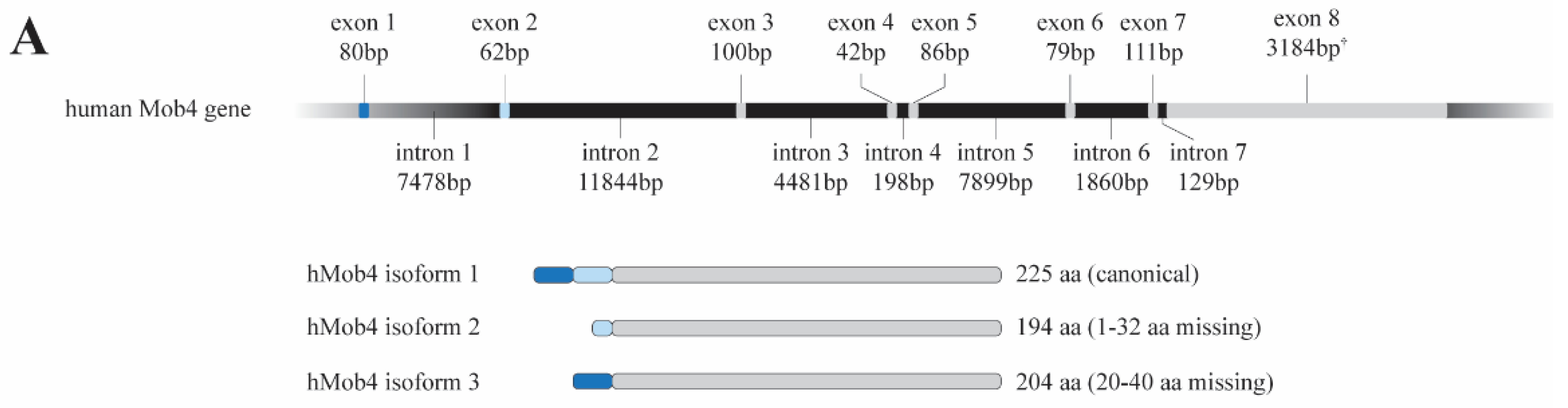
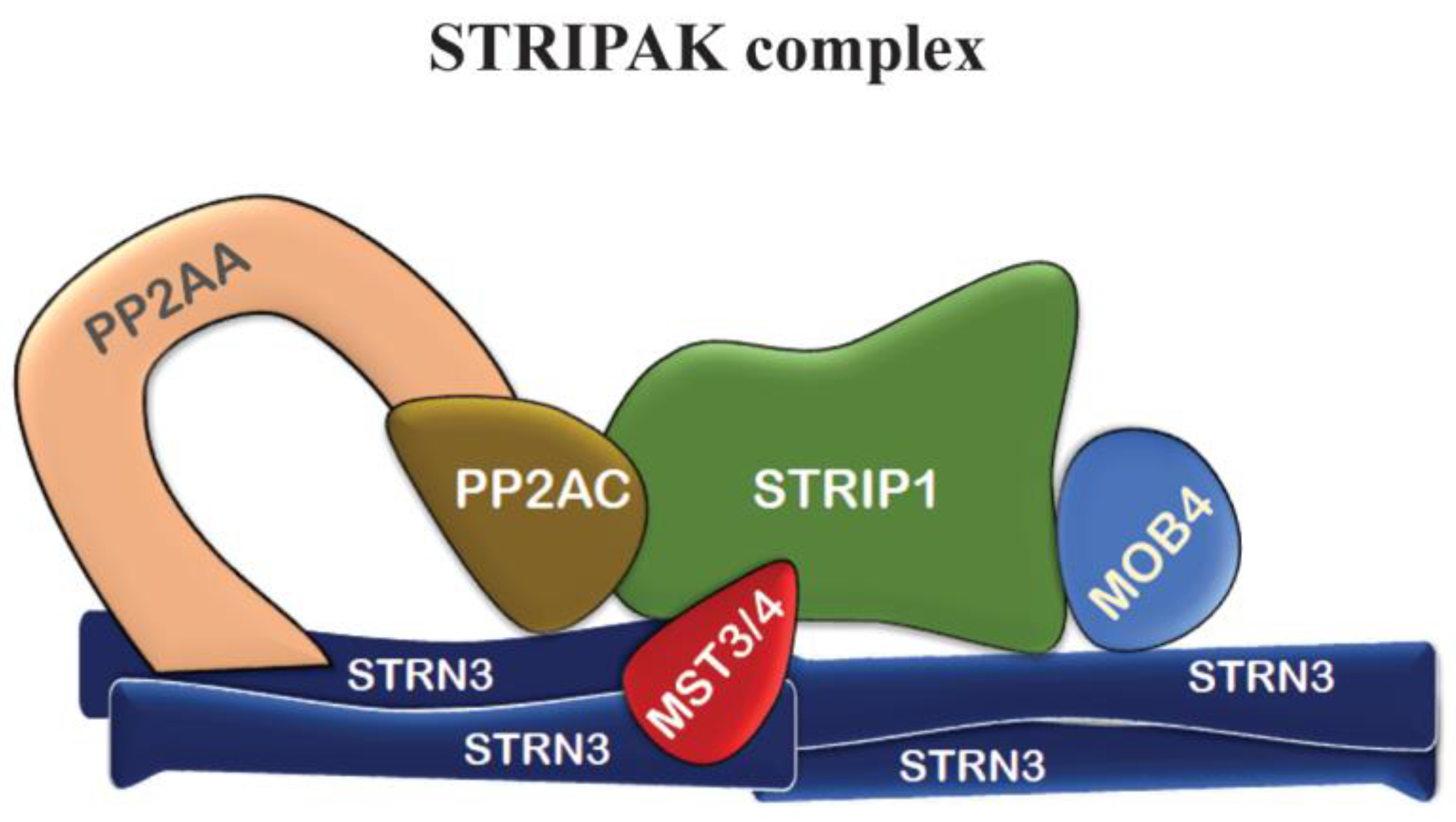
2. MOB4: From the Gene to the Function

Initially named as Phocein [21], it was soon renamed Mob4 because of the high homology it displays with other Mob proteins. The Mob4 gene has been referred in the literature as Mob1, 2C4D, CGI-95, class II mMOB1, Mob1 homolog 3, Mob3, Mps-one binder kinase activator-like 3 (MOBKL3) and preimplantation protein 3 (PREI3). In this work, we follow the HUGO Gene Nomenclature Committee (HGNC) and use Mob4 as the product of the gene ID 17261 (NCBI Entrez Gene: 25843).
Structurally, the canonical Mob fold consists of a four-helix bundle at its core, with three short α-helices at the N-terminal extension (amino acids (aa) 1−61) [22]. Of note, Mob4 share a high structural homology with Mob1, even at the N-terminal, a region where the various Mob proteins diverge.
The human Mob4 gene is predicted to generate alternatively spliced transcriptional variants, producing three predicted protein isoforms: a canonical isoform with 26 kDa (isoform 1) and two smaller variants (Figure 1). One variant results from an alternative exon, containing a different start codon and thus generating a smaller protein with a different N-terminal with 22.3 kDa (isoform 2), and a second variant lacking an in-frame exon generating an isoform with 23.5 kDa (isoform 3) but sharing the N- and C-termini with the canonical isoform. Like all Mob proteins, Mob4 is highly conserved across evolution; for example, there is 80% aa identity and 88% similarity between Drosophila melanogaster Mob4 (dMob4) and its human ortholog (hMob4).

3. Neuronal Functions of Mob4

Mob4 was initially identified on screenings in neural cell libraries as a potential interactor of Striatins, by the groups of Ariane Monneron and of David Pallas, independently [21,23]. These initial biochemical studies led to the characterization of Mob4 as a core component of the STRIPAK complex (Figure 2) and indicated that Mob4 had a putative role in neuronal function. In agreement with this idea, it was found that mammalian Mob4 is highly expressed in melanized dopamine neurons as well as in the central and peripheral human nervous system [21,24]. In addition, it is highly enriched in dendritic spines, the actin-rich protrusions emerging from dendrites [24,25,26].
Immunogold labeling detected Mob4 in close vicinity to endocytic-like membranes in the neuronal dendritic spines, suggesting vesicular trafficking functions [24,25]. Immunocytochemical studies also confirmed that Mob4 is strictly somato-dendritic, extending down to the neuronal spines [21]. Noticeably, Mob4 was detected in cell bodies and dendrites but not in axons [21], similarly to what has been described for striatin. In addition, immunoreactivity for Mob4 was only detected in neurons, not in glial cells. Recent studies in zebrafish confirm that Mob4 is highly expressed in the central nervous system; Mob4 was detected in the full extension of the neuroectoderm and in the brain in the initial stages of zebrafish development. Prior to hatching, Mob4 expression is almost limited to the brain, and its expression remains enriched in the brain after hatching [27]. A neural role for Mob4 was first demonstrated by genetic studies in Drosophila, where it was shown to be a key regulator of neuronal structure and development [26]. Importantly, these studies showed that Mob4 is an essential gene, as Drosophila individuals lacking Mob4 do not survive past the larval stages. The selective somatic downregulation of Drosophila Mob4 resulted in a disrupted neuronal morphology. Neurons that developed without Mob4 showed abnormal branching patterns and hyperbranching, and established incorrect connections to target cells. In addition, the lack of Mob4 resulted in severe defects in microtubule organization, synaptic development and axonal transport. Finally, mutant Drosophila individuals displayed a defective neuronal transport of cargo from the cell body to the synapses [26]. dMob4 mutants had neuromere clusters that were smaller in size, although with shorter neurites and a higher thickness of neurite bundles.
Synapses are highly specialized structures that process and transmit information in the brain. These cell–cell communication hubs are thought to be under constant modification during development and by experiences throughout life. Most excitatory synapses localizes to dendritic spines, small actin-rich protrusions on the surface of dendrites. The association of mammalian Mob4 with endocytic-like membranes and neuronal spines suggests a function on endocytosis and vesicular trafficking [24,25]. Intracellular traffic depends on cytoskeleton functioning as rails for the cargo. dMob4 mutants have a disorganized microtubule cytoskeleton [26], indicating a possible association of Mob4 function with cytoskeleton organization and stability.
Recently, the role of Mob4 in vertebrate neurodevelopment has been highlighted by studying the consequences of Mob4 depletion in zebrafish embryogenesis using morpholinos [27]. The authors found that the knockdown of zfMob4 using translation-blocking morpholinos in young embryos lead to severe neurologic defects. Zebrafish embryos at 24 h post fertilization lacked the midbrain–hindbrain boundary and showed reduced eye size. Mob4 morphants were also shown to be significantly smaller than their control morpholinos siblings. Notably, these differences are not found in the axial trunk but only in the head [27]. This suggests that Mob4 has important functions in the neurodevelopment of the brain but not the spinal cord. In addition, the authors have observed that, in these morphant embryos, not only is the hindbrain and eye regions smaller, but the rate of cell division is severely diminished in the hindbrain and eye regions, arguing that the reduced hindbrain size and eye size are a consequence of impaired cell divisions [27]. Altogether, these results strongly indicate a Mob4 involvement in the process of cell proliferation during neurodevelopment in vertebrates. In agreement with these observations for Mob4 and supporting such functions in neurons, it has been shown that the STRIPAK complex promotes the organization, development and maturation of striatal neurons. In fact, the dysfunction of STRIPAK has been linked to a range of clinical neurological conditions [28]. For example, knockdown of Striatin, that binds directly to Mob4, blocks dendrite formation [29], an observation that suggests that the knockdown of Mob4 affects neuronal morphology at least in part by affecting STRIPAK function.
Taking these observations altogether, Mob4 seems to be required for the regulation of neuronal functions at different levels. Firstly, by being required for the assembly of a normal microtubule cytoskeleton [26]. Second, biochemical studies indicated Mob4 to be involved in endocytosis and vesicular trafficking [24,25,26]. It seems therefore that Mob4 contributes both to postnatal synaptogenesis and to the dendritic-activity-dependent plasticity in the adult. It is noteworthy to recall that Mob4 is highly conserved throughout the animal kingdom. For this reason, it is tempting to speculate that a deficient Mob4 function may be related to mechanisms of neurological diseases in humans.

4. Mob4 and Cytoskeleton

Recent reports indicate that Mob4 has important roles in cytoskeletal regulation. Studies on Drosophila cells reported Mob4 to be involved in mitotic spindle microtubule focusing [30], while studies on the fly nervous system showed Mob4 to be important for the organization of microtubule networks within postmitotic neurons [26]. A recent report shows that in Zebrafish, Mob4 function is required for the incorporation α-actin into organized sarcomeres in skeletal muscle [31]. In this work, the authors found an interaction between Mob4 and the actin-folding chaperonin TRiC (TCP-1 ring complex, also called chaperonin containing TCP-1), suggesting that Mob4 affects TRiC to control actin biogenesis and thus myofibril growth. This report supports a previously described Mob4 molecular interaction with TRiC [14].
The proper folding of proteins is essential for cellular function, and protein misfolding is believed to be the primary cause of many neurodegenerative disorders. In eukaryotes, the folding of misfolded or unfolded proteins is mediated by molecular chaperones, one of which is the protein complex TRiC. Chaperonins are required for the proper folding, transport and degradation of proteins. TRiC chaperonin display limited substrate specificity and assists the folding of many key structural proteins, such as the cytoskeletal proteins actin and α- and β-tubulin [32,33,34]. The importance of TRiC complex in protein folding was first shown in Caenorhabditis elegans where individuals with reduced TRiC function display defective microtubule cytoskeletons [35]. Co-immunoprecipitations assays had shown that Mob4 and TRiC are part of a multiprotein complex and that a direct interaction between Mob4 protein and TRiC was demonstrated to occur in nematodes [14,36]. Recently, Berger et al. showed that Zebrafish Mob4 mutants also display deficient microtubule networks, which is in line with the fact that a main folding substrate of TRiC is tubulin. Therefore, it is possible that such defective microtubule networks are the cause of the observed compromised neuronal connectivity in zebrafish Mob4 mutants. In line with these observations, neuronal neurite formation is strongly affected in TRiC-deficient zebrafish [32,33].
Additionally, Berger et al. also found that in zebrafish, Mob4 protein, co-localized with the marker α-actinin at the sarcomere’s Z-discs. Importantly, whereas the loss of Mob4 function led to a smaller amount of myofibrils, increased Mob4 expression induced an increase in the amount of organized myofibrils. These findings indicate that Mob4 function might be required for the regulation of the number of organized myofibrils. Lack of Mob4 function results in reduced numbers of myofibrils and impaired movement due to skeletal muscle defects.
As Mob4 functions within the protein complex STRIPAK, Berger and colleagues looked for similar defects in strn3-deficient mutants. In fact, the authors found that strn3-deficient mutants featured both neuronal and muscle defects. These observations confirm not just Mob4 as a core component of STRIPAK in Zebrafish but also imply a role for the STRIPAK complex in sarcomerogenesis. Unexpectedly, zebrafish TRiC mutants still develop into almost normal larvae that, nonetheless, still show highly specific skeletal muscle defects, resulting from the defective folding of α-actin at Z-disks in the skeletal muscle, with the result of a reduced sarcomere assembly.
The work of Berger et al. highlights the fact that Mob4 function involves at least two different protein complexes, STRIPAK and TRiC. The former has a large diversity of cellular functions, and the latter is required for actin and tubulin biogenesis. Mob4 and strn3 mutants featured both neuronal and muscle defects, given that neuronal axons are formed by polar arrays of microtubules, while the major protein components of smooth-muscle thin filaments is actin. This supports the idea that STRIPAK and TRiC may interact through Mob4 to coordinate the growth of the myofibril and of the microtubule network during neural development.

5. Cell Proliferation Function of Mob4

From a physiological point of view, Mob4 plays important roles in the control of cell proliferation. As previously mentioned, the downregulation of zfMob4 results in a severe decrease in the number of mitotic cells in the brain and in the developing eye [27]. The control of stem cell number also seems to be one function of Mob4, as studies on planarian indicated that mob4 function limits body size through limiting stem cell numbers [37]. Planarians have been highly studied for their ability to regenerate body parts after injury and keeping body proportionality [38]. The inhibition of Mob4 in planaria dramatically increased posterior length after injury, affecting the polarity along the apical posterior axis [37].
This an important step forward in deciphering the molecular mechanisms that allow animals to reconstruct body parts while simultaneously integrating newly regenerated tissues into pre-existing old ones after injury. The process of regeneration in planarians is complex and involves the coordination of multiple cellular signaling pathways essential for normal physiological functions, such as organ size, cell proliferation cycle, asymmetric cell divisions, programmed cell death, and cell/tissue polarity determination [39]. One such signaling pathway is the evolutionarily conserved Wnt pathway, known to be involved in a myriad of processes namely cell proliferation, differentiation, and apoptosis, as well as in stem cell maintenance [40]. In planaria, the Wnt signaling pathway is the key regulator of the head–tail polarity, and it is at the core of the decision-making process to regenerate a head or a tail [41,42], as reviewed in [39]. The regionalization of the planarian Antero-Posterior axis is controlled by constitutive localized expression of Wnt ligands (expressed posteriorly) and of Wnt inhibitors (expressed anteriorly). The results obtained by Schad and Petersen support a model in which Mob4 regulates wnt pole cell numbers by limiting stem cell formation, and wnt pole cells in turn control tail proportionality along the antero-posterior axis [37]. These results therefore indicate that Mob4 is involved in the scaling of tail size with respect to body size via the regulation of wnt1. Considering that Mob4 is constitutively expressed, even in the absence of injury, the recovery of normal proportions through regeneration may involve a mechanism that establishes a balance of local signaling processes. These results suggest that the suppression of wnt signaling by Mob4 (through STRIPAK) is a critical pathway regulating scaling and whole-body proportionality in planarians. Interestingly, in planaria, the members of the Hippo pathway do not seem to be required for keeping antero-posterior tissue proportionality, suggesting that Mob4 and STRIPAK exert their action independently of the Hippo signaling pathway (Figure 3). On the other hand, a crosstalk between the Hippo pathway and the Wnt pathway has been previously described [43], and Hippo plays an important role in the regulation of the cell cycle, which is equally crucial for planarian regeneration.
Importantly, Mob proteins have been demonstrated to be involved in defining cell polarity both in humans and in Tetrahymena [44,45]. The importance of cell polarity in a number of physiological processes, including cell differentiation, cell migration, asymmetric cell division, cancer progression and immune response, has been extensively described and reviewed in [46]. In Tetrahymena, a unicellular organism, the single Mob protein encoded in the genome, is required for correct division-plane placement by establishing the anterior–posterior axis. The downregulation of Mob in Tetrahymena induces the misplacement of the division plane with consequent abscission failure; daughter cells fail to separate and form trails of interconnected abnormal cells [44]. Interestingly, the authors found that the Mob protein accumulates at the future site of cell division prior to constriction start, thereby defining the anterior and posterior ends of the future new daughter cells. This finding highlights the importance of Mob in cell polarity inception through a cell-intrinsic mechanism and how polarity is coupled to growth in Tetrahymena. Likewise, in human HeLa cultured cells, the downregulation of Mob1 also results in abscission failure and, importantly, cell polarity is affected in such a way that it allows cells to become motile [45].
Finally, a role for Mob4 in cell proliferation and tissue formation has also been described in the filamentous fungi Sodaria macrospora, where the downregulation of SmMob3 (the Mob4 homologue in Sodaria) results in impaired vegetative growth accompanied by a sexually sterile strain unable to undergo self-fusion and fusion [47]. On the other hand, in Caenorhabditis elegans, mob-4-deficient mutant individuals do not show an obvious abnormal phenotype under the normal growth conditions. Nevertheless, longevity and thermotolerance are affected in these individuals [48]. These observations, together with a previous report showing that C. elegans YAP-1 overexpression shortens these individuals’ life span (whereas yap-1 deficiency prolongs life span) [49] suggests that in contrast to humans and Drosophila, in which Mob4 activates YAP1 [50], in worms, Mob4 does not act upstream of YAP-1. Taken together, these observations led to suggestion that in C. elegans, the Hippo pathway is not conserved [49].
Mob4 may be involved in cell proliferation through its function within the STRIPAK complex or independently of it. STRIPAK negatively regulates the Hippo signaling pathway, thereby participating in the control of cell proliferation [28,51,52]. The deletion of the amino-terminal residues of Mob4 abolishes the assembly of STRIPAK [22] while the disruption of the sites responsible for Mob4 and STRN3 interaction causes aberrant Hippo signaling regulation. Thus, Mob4 can affect cell proliferation by participating in STRIPAK assembly and Hippo pathway activity.
Given the role of Mob4 in regulating cell proliferation it is not surprising that Mob4 may be involved in cancer initiation/progression. Cancer development, due to excessive cell proliferation, is highly associated with the activation of oncogenic pathways [53,54,55] or the deregulation of genes with tumor- suppressing functions [56,57,58,59]. In most human cancers, there is a moderate/high protein expression of Mob4, and a higher expression of Mob4 is associated with a poor prognosis for renal and liver cancers (https://www.proteinatlas.org, accessed on 1 December 2022). The Hippo pathway, per se, is an important regulator of cell proliferation and tissue growth, and mice mutant in Hippo components (Sav (Salvador), MST1/2, Lats (Large tumor suppressor kinase) and Mob1) are prone to develop malignant growths [60,61,62,63]. In addition, it has been described that the different Mob family members behave either as tumor suppressors or oncogenes. For example, the complete loss of Mob1, a component of the Hippo pathway, in mice promotes tumorigenesis and embryonic death [60]; Mob2 has recently been reported as a tumor suppressor in glioblastoma [64]; and in contrast, Mob3 has been found to be upregulated in glioblastoma multiforme, and it is proposed as an oncoprotein by suppressing MST1 activity [12].
Interestingly, Mob4 can also bind to the protein kinase MST4 forming a complex that antagonizes the complex formed by Mob1 and the kinases MST1/2 [50] (Figure 3). But while MST1-Mob1 acts as a tumor suppressor, MST4-Mob4 is oncogenic by activating YAP signaling and promoting cell proliferation. In fact, Mob4 can alternatively pair with either MST4 or MST1 due to the high structural similarities of both Mobs and of both MST kinases. Mob4 can therefore sequester MST1, consequently inhibiting the Hippo pathway and promoting cell proliferation [50], thus acting as an oncogene. It is worth remembering that simultaneously, on an alternative mechanism and as a component of STRIPAK complex, Mob4 negatively regulates the Hippo signaling pathway and therefore acts as a tumor suppressor gene.

6. Mob4 and Spermatogenesis

Spermatogenesis is the process by which germinal stem cells give rise to haploid spermatozoa. In Drosophila, spermatids morphogenesis generally occurs within a syncytium, with all spermatid nuclei remaining interconnected via an extensive network of cytoplasmic bridges. As spermiogenesis progresses, the syncytium (or cyst) is then resolved into individual cells in a process referred to as sperm individualization. Although with some differences in the control of hormonal regulation and in testicular structure, the different stages of spermatogenesis are highly conserved from fly to human. Importantly, many of the genes involved in Drosophila spermatogenesis were shown to be conserved in humans.
Santos et al. recently described a requirement for Mob4 during spermatogenesis in Drosophila: males without Mob4 function in the gonads are sterile, while females are fully fertile. In the lack of Mob4 function, cyst elongation still occurs, meaning that spermatids are capable of elongating an axoneme, but spermatid individualization was observed to fail. Consequently, the migration of sperm into the seminal vesicle does not occur, and consequently, males are sterile. In order to determine what defects in spermiogenesis cause spermatid individualization failure, the authors have examined the ultrastructure of the developing axoneme and found that the mitochondrial derivatives fail to form correctly. Importantly, defects in the axonemal structure were also found. These defects include the loss of microtubule doublets, and most interestingly, the stereotypical 9 + 2 microtubule doublets of the axoneme is affected, with the axoneme suffering a large radial expansion [65].
The use of a GFP:Mob4 transgene reveals that Mob4 has dynamic sub-cellular localization in different cell types throughout spermatogenesis. During meiosis, Mob4 accumulates in the reticulum surrounding the meiotic spindle structures. Post-meiotically, Mob4 transiently accumulates at the basal side of the nuclei in the vicinity of the basal body at the early canoe stage of spermatid differentiation. The dynamic behavior of Mob4 throughout different stages of spermatogenesis is suggestive of multiple roles in the parafusorial membranes and associated microtubules during meiosis, and at the basal body or transition zone in the initiation of axoneme elongation.
As mentioned, spermatogenesis is a highly conserved process, and the genes involved in spermatogenesis usually have their function conserved across species. Mob4 seem to follow the trend as the insertion of the human Mob4 paralog gene into Drosophila was capable of rescuing all the meiotic defects in mob4 mutant, including full fertility [65], suggesting that human Mob4 and Drosophila Mob4 are functional orthologs.
Considering that Mob4 is a component of STRIPAK, one may wonder if the defective spermatogenesis results from lack of STRIPAK function, or is an isolated function of Mob4. To answer this question Santos et al. looked for a function of Strip and Cka (two other components of STRIPAK) in Drosophila testes. They found that, like for Mob4, either Strip or Cka downregulation in testes results equally in male sterility; on the other hand, Strip or Cka downregulation did not seem to affect female ovaries and are not required for female oogenesis. In addition, the investigators also showed that similar failures in sperm individualization to Mob4 are observed after the downregulation of either Strip or Cka, suggesting that STRIPAK complex activity is required for spermatogenesis, and that Mob4 is probably acting through STRIPAK.

7. Other Mob4 Functions

A different set of studies indicated that Mob4 is also required for mitosis progression. In Drosophila S2 cultured cells, Mob4 was found to associate with centrosomes and kinetochores, and its downregulation resulted in the formation of monopolar spindles and defective mitosis [30]. Moreover, in human cells, Frost and collaborators showed that the downregulation of Mob4 causes an increase in DNA content, abnormal spindle formation and mitosis failure, triggering cell death [66]. In addition, the authors found that Mob4, together with the STRIPAK complex, bridges the centrosome with the cis-Golgi and the outer nuclear membrane [21,66] (Figure 4). The disruption of these bridges may be the cause of the defective mitotic progression.
Human Mob4 may also have a role in DNA damage signaling since its downregulation results in γH2AX phosphorylation in cells [67], which is an early marker for DNA damage [68]. Moreover, the Ser147 of human Mob4 is a possible target of ATM kinase suggesting a possible role for Mob4 within the DNA damage response [69]. Other MOB family members are also involved in the DNA damage signaling. For example, Mob2 can interact with RAD50 promoting the assembly of the MRN DNA damage sensor complex this way activating ATM kinase, a well-known protein orchestrating DNA damage response. In addition, Mob2 competes with Mob1 for NDR1/2 binding (NDR–Nuclear Dbf2-Related Kinase), and NDR-mediated phosphorylation is important in the G2/M DNA damage checkpoint by promoting the degradation of the CDC25A phosphatase [70].
Apoptosis is a process important for the maintenance of cell numbers, and its regulation has been previously associated with Mob4 and Mob1 due to their association with MST1 kinase. MST1 modulates oxidative-stress-induced neuronal death [71], and several studies have reported how the MST1 phosphorylation of FOXO proteins enhances their nuclear translocation, promoting the transcription of apoptosis-related genes [72,73]. A neuronal-specific isoform of YAP (the end target of the Hippo pathway), YAPdeltaC, acts as a neuronal apoptosis protector that decreases with progression for amyotrophic lateral sclerosis (ALS), whereas the full-length YAP remains constant during the late and severe stage of the disease [74].
Specific connections between Mob proteins and neurodegeneration can be established through their interaction with NDR/LATS kinases. It was found that all four Drosophila Mob genes can genetically interact with tricornered (Trc) (Drosophila homologue of NDR kinases) [75]. Trc is required for morphological changes, such as the outgrowth of epidermal hair and dendritic tiling in sensory neurons and Wts (Drosophila homologue of LATS) plays a role in dendritic maintenance in sensory neurons [76,77,78]. Mob2 has been directly associated with neuronal functions. In neuronal cell lines, Mob2 is required for sustaining neurite formation [79]. Mob2 expression is required to regulate the growth of Drosophila larval neuromuscular junction [80] and for normal neuronal distribution in mice developmental cortex [81]. In addition, human Mob2 has been identified as a specific protein of cerebral amyloid angiopathy (CAA), a condition in which amyloid plaques are deposited on the walls of cortical and blood vessels of the brain [82]. On the other hand, increasing evidence shows a potential role for Mob3 in neurodegenerative diseases. The human Mob3A gene was identified as a target for the nuclear respiratory factor 1 (NRF1) [83]. NRF1 is a master transcription factor that promotes the transcriptional activation of genes required for mitochondrial biogenesis and proteosome function. The deregulation of NRF1 target genes has been shown in neurodegenerative disease models where proteosome capacity is diminished [84]. On a second study, human Mob3 proteins were found to bind to the oligomeric Aβ42, a hallmark protein complex of Alzeimer’s disease, suggesting the potential involvement of Mob3 proteins with this condition [85]. Therefore, Mob4 is not the only Mob with a role in the control of cell proliferation and apoptosis, which is relevant for the clearance of excessive neural cells during normal development [86]. Finally, very recently, Guo et al. [87] suggested a new function for Mob4, through its role within STRIPAK, in the regulation of autophagy in Drosophila muscle tissue.
In summary, Mob family proteins have important functions in the control of cell proliferation. Mob4 displays a range of essential functions that spans from neuronal development to spermatogenesis and the control of cell proliferation, both in vertebrates and in Drosophila (Figure 5). These functions are likely exerted through different mechanisms. Mob4 can act as a scaffold on the assembly of the STRIPAK complex (Figure 2 and Figure 3), thus participating in axonal transport, dendritic development and synapse assembly. But Mob4 also appears to act independently of STRIPAK by interacting with the TRiC complex or directly with MST1. Most human cancers show a moderate/high protein expression of Mob4, and renal and liver cancer patients with a high expression of Mob4 show a reduced survival probability compared to those with low expression, suggesting that Mob4 is an unfavorable prognostic marker for renal and liver cancers. Therefore, elucidating the underlying mechanisms of Mob4 action and regulation may be of help to identify novel therapeutic targets and diagnostic markers for cancer and neurodegenerative diseases.

Author Contributions

Original draft preparation, writing—review and editing: I.B.S., J.G.-M., C.G., B.I.O. and Á.A.T.; funding acquisition: Á.A.T. All authors have read and agreed to the published version of the manuscript.

Funding

Work funded by the Algarve 2020 Program, grant number ALG-01-0145-FEDER-030014, and co-financed by FEDER Funds through the Operational Program for Competitiveness Factors—COMPETE 2020 and by National Funds through FCT—Foundation for Science and Technology under the Project PTDC/BIA-CEL/30014/2017. I.B.Santos was funded by FCT fellowship SFRH/BD/141734/2018.

Institutional Review Board Statement

Not applicable.

Informed Consent Statement

Not applicable.

Data Availability Statement

Data are contained within the article and references.

Conflicts of Interest

The authors declare no conflict of interest.

References

  1. Delgado, I.; Carmona, B.; Nolasco, S.; Santos, D.; Leitão, A.; Soares, H. MOB: Pivotal Conserved Proteins in Cytokinesis, Cell Architecture and Tissue Homeostasis. Biology 2020, 9, 413. [Google Scholar] [CrossRef] [PubMed]
  2. Duhart, J.C.; Raftery, L.A. Mob Family Proteins: Regulatory Partners in Hippo and Hippo-Like Intracellular Signaling Pathways. Front. Cell Dev. Biol. 2020, 8, 161. [Google Scholar] [CrossRef] [PubMed]
  3. Luca, F.C.; Winey, M. Regulation of Mob1p, an essential budding yeast protein required for completion of mitosis and spindle pole body duplication. Mol. Biol. Cell 1998, 9, 12A. [Google Scholar] [CrossRef] [PubMed]
  4. Luca, F.C.; Winey, M. MOB1, an essential yeast gene required for completion of mitosis and maintenance of ploidy. Mol. Biol. Cell 1998, 9, 29–46. [Google Scholar] [CrossRef]
  5. Ye, X.; Nikolaidis, N.; Nei, M.; Lai, Z. Evolution of the mob Gene Family. Open Cell Signal. J. 2009, 1, 1–11. [Google Scholar] [CrossRef]
  6. Vitulo, N.; Vezzi, A.; Galla, G.; Citterio, S.; Marino, G.; Ruperti, B.; Zermiani, M.; Albertini, E.; Valle, G.; Barcaccia, G. Characterization and evolution of the cell cycle-associated mob domain-containing proteins in eukaryotes. Evol. Bioinform. 2007, 3, 121–158. [Google Scholar] [CrossRef]
  7. Lai, Z.C.; Wei, X.; Shimizu, T.; Ramos, E.; Rohrbaugh, M.; Nikolaidis, N.; Ho, L.L.; Li, Y. Control of cell proliferation and apoptosis by mob as tumor suppressor, mats. Cell 2005, 120, 675–685. [Google Scholar] [CrossRef]
  8. Pan, D. Hippo signaling in organ size control. Genes Dev. 2007, 21, 886–897. [Google Scholar] [CrossRef]
  9. Halder, G.; Johnson, R.L. Hippo signaling: Growth control and beyond. Development 2011, 138, 9–22. [Google Scholar] [CrossRef]
  10. Weiss, E.L.; Kurischko, C.; Zhang, C.; Shokat, K.; Drubin, D.G.; Luca, F.C. The Saccharomyces cerevisiae Mob2p-Cbk1p kinase complex promotes polarized growth and acts with the mitotic exit network to facilitate daughter cell-specific localization of Ace2p transcription factor. J. Cell Biol. 2002, 158, 885–900. [Google Scholar] [CrossRef]
  11. Kohler, R.S.; Schmitz, D.; Cornils, H.; Hemmings, B.A.; Hergovich, A. Differential NDR/LATS interactions with the human MOB family reveal a negative role for human MOB2 in the regulation of human NDR kinases. Mol. Cell. Biol. 2010, 30, 4507–4520. [Google Scholar] [CrossRef]
  12. Tang, F.; Zhang, L.; Xue, G.; Hynx, D.; Wang, Y.; Cron, P.D.; Hundsrucker, C.; Hergovich, A.; Frank, S.; Hemmings, B.A.; et al. hMOB3 modulates MST1 apoptotic signaling and supports tumor growth in glioblastoma multiforme. Cancer Res 2014, 74, 3779–3789. [Google Scholar] [CrossRef]
  13. Ma, S.; Meng, Z.; Chen, R.; Guan, K.L. The Hippo Pathway: Biology and Pathophysiology. Annu. Rev. Biochem. 2019, 88, 577–604. [Google Scholar] [CrossRef] [PubMed]
  14. Goudreault, M.; D’Ambrosio, L.M.; Kean, M.J.; Mullin, M.J.; Larsen, B.G.; Sanchez, A.; Chaudhry, S.; Chen, G.I.; Sicheri, F.; Nesvizhskii, A.I.; et al. A PP2A phosphatase high density interaction network identifies a novel striatin-interacting phosphatase and kinase complex linked to the cerebral cavernous malformation 3 (CCM3) protein. Mol. Cell. Proteom. 2009, 8, 157–171. [Google Scholar] [CrossRef] [PubMed]
  15. Glatter, T.; Wepf, A.; Aebersold, R.; Gstaiger, M. An integrated workflow for charting the human interaction proteome: Insights into the PP2A system. Mol. Syst. Biol. 2009, 5, 237. [Google Scholar] [CrossRef] [PubMed]
  16. Ribeiro, P.S.; Josué, F.; Wepf, A.; Wehr, M.C.; Rinner, O.; Kelly, G.; Tapon, N.; Gstaiger, M. Combined functional genomic and proteomic approaches identify a PP2A complex as a negative regulator of Hippo signaling. Mol. Cell 2010, 39, 521–534. [Google Scholar] [CrossRef] [PubMed]
  17. Gordon, J.; Hwang, J.; Carrier, K.J.; Jones, C.A.; Kern, Q.L.; Moreno, C.S.; Karas, R.H.; Pallas, D.C. Protein phosphatase 2a (PP2A) binds within the oligomerization domain of striatin and regulates the phosphorylation and activation of the mammalian Ste20-Like kinase Mst3. BMC Biochem. 2011, 12, 54. [Google Scholar] [CrossRef]
  18. Hornbeck, P.V.; Zhang, B.; Murray, B.; Kornhauser, J.M.; Latham, V.; Skrzypek, E. PhosphoSitePlus, 2014: Mutations, PTMs and recalibrations. Nucleic Acids Res. 2015, 43, D512–D520. [Google Scholar] [CrossRef]
  19. Gil-Ranedo, J.; Gonzaga, E.; Jaworek, K.J.; Berger, C.; Bossing, T.; Barros, C.S. STRIPAK Members Orchestrate Hippo and Insulin Receptor Signaling to Promote Neural Stem Cell Reactivation. Cell Rep. 2019, 27, 2921–2933.e5. [Google Scholar] [CrossRef]
  20. Shi, Z.; Jiao, S.; Zhou, Z. STRIPAK complexes in cell signaling and cancer. Oncogene 2016, 35, 4549–4557. [Google Scholar] [CrossRef]
  21. Baillat, G.; Moqrich, A.; Castets, F.; Baude, A.; Bailly, Y.; Benmerah, A.; Monneron, A. Molecular cloning and characterization of phocein, a protein found from the Golgi complex to dendritic spines. Mol. Biol. Cell 2001, 12, 663–673. [Google Scholar] [CrossRef]
  22. Jeong, B.C.; Bae, S.J.; Ni, L.; Zhang, X.; Bai, X.C.; Luo, X. Cryo-EM structure of the Hippo signaling integrator human STRIPAK. Nat. Struct. Mol. Biol. 2021, 28, 290–299. [Google Scholar] [CrossRef]
  23. Moreno, C.S.; Lane, W.S.; Pallas, D.C. A mammalian homolog of yeast MOB1 is both a member and a putative substrate of striatin family-protein phosphatase 2A complexes. J. Biol. Chem. 2001, 276, 24253–24260. [Google Scholar] [CrossRef]
  24. Haeberlé, A.M.; Castets, F.; Bombarde, G.; Baillat, G.; Bailly, Y. Immunogold localization of MOB4 in dendritic spines. J. Comp. Neurol. 2006, 495, 336–350. [Google Scholar] [CrossRef] [PubMed]
  25. Bailly, Y.J.R.; Castets, F. Phocein: A potential actor in vesicular trafficking at Purkinje cell dendritic spines. Cerebellum 2007, 6, 344–352. [Google Scholar] [CrossRef]
  26. Schulte, J.; Sepp, K.J.; Jorquera, R.A.; Wu, C.; Song, Y.; Hong, P.; Littleton, J.T. DMob4/Phocein regulates synapse formation, axonal transport, and microtubule organization. J. Neurosci. 2010, 30, 5189–5203. [Google Scholar] [CrossRef] [PubMed]
  27. Florindo, C.; Mimoso, J.M.; Palma, S.L.; Gonçalves, C.; Silvestre, D.; Campinho, M.; Tavares, Á.A. Mob4 is required for neurodevelopment in zebrafish. microPubl. Biol. 2023. [Google Scholar] [CrossRef]
  28. Hwang, J.; Pallas, D.C. STRIPAK complexes: Structure, biological function, and involvement in human diseases. Int. J. Biochem. Cell Biol. 2014, 47, 118–148. [Google Scholar] [CrossRef]
  29. Li, D.; Musante, V.; Zhou, W.; Picciotto, M.R.; Nairn, A.C. Striatin-1 is a B subunit of protein phosphatase PP2A that regulates dendritic arborization and spine development in striatal neurons. J. Biol. Chem. 2018, 293, 11179–11194. [Google Scholar] [CrossRef]
  30. Trammell, M.A.; Mahoney, N.M.; Agard, D.A.; Vale, R.D. Mob4 plays a role in spindle focusing in Drosophila S2 cells. J. Cell Sci. 2008, 121, 1284–1292. [Google Scholar] [CrossRef]
  31. Berger, J.; Berger, S.; Currie, P.D. Mob4-dependent STRIPAK involves the chaperonin TRiC to coordinate myofibril and microtubule network growth. PLoS Genet. 2022, 18, e1010287. [Google Scholar] [CrossRef] [PubMed]
  32. Yaffe, M.B.; Farr, G.W.; Miklos, D.; Horwich, A.L.; Sternlicht, M.L.; Sternlicht, H. TCP1 complex is a molecular chaperone in tubulin biogenesis. Nature 1992, 358, 245–248. [Google Scholar] [CrossRef] [PubMed]
  33. Berger, J.; Berger, S.; Li, M.; Jacoby, A.S.; Arner, A.; Bavi, N.; Stewart, A.G.; Currie, P.D. In Vivo Function of the Chaperonin TRiC in α-Actin Folding during Sarcomere Assembly. Cell Rep. 2018, 22, 313–322. [Google Scholar] [CrossRef] [PubMed]
  34. Ghozlan, H.; Cox, A.; Nierenberg, D.; King, S.; Khaled, A.R. The TRiCky Business of Protein Folding in Health and Disease. Front. Cell Dev. Biol. 2022, 10, 906530. [Google Scholar] [CrossRef] [PubMed]
  35. Saegusa, K.; Sato, M.; Sato, K.; Nakajima-Shimada, J.; Harada, A.; Sato, K.; Geisler, F.; Gerhardus, H.; Carberry, K.; Davis, W.; et al. Caenorhabditis elegans chaperonin CCT/TRiC is required for actin and tubulin biogenesis and microvillus formation in intestinal epithelial cells. Mol. Biol. Cell 2014, 25, 3095–3104. [Google Scholar] [CrossRef]
  36. Khabirova, E.; Moloney, A.; Marciniak, S.J.; Williams, J.; Lomas, D.A.; Oliver, S.G.; Favrin, G.; Sattelle, D.B.; Crowther, D.C. The TRiC/CCT chaperone is implicated in Alzheimer’s disease based on patient GWAS and an RNAi screen in Abeta-expressing Caenorhabditis elegans. PLoS ONE 2014, 9, e102985. [Google Scholar] [CrossRef] [PubMed]
  37. Schad, E.G.; Petersen, C.P. STRIPAK Limits Stem Cell Differentiation of a WNT Signaling Center to Control Planarian Axis Scaling. Curr. Biol. 2020, 30, 254–263.e2. [Google Scholar] [CrossRef]
  38. Wagner, D.E.; Wang, I.E.; Reddien, P.W. Clonogenic neoblasts are pluripotent adult stem cells that underlie planarian regeneration. Science 2011, 332, 811–816. [Google Scholar] [CrossRef]
  39. Reddien, P.W. The Cellular and Molecular Basis for Planarian Regeneration. Cell 2018, 175, 327–345. [Google Scholar] [CrossRef]
  40. Clevers, H.; Loh, K.M.; Nusse, R. Stem cell signaling. An integral program for tissue renewal and regeneration: Wnt signaling and stem cell control. Science 2014, 346, 1248012. [Google Scholar] [CrossRef]
  41. Gurley, K.A.; Rink, J.C.; Sanchez Alvarado, A. Beta-catenin defines head versus tail identity during planarian regeneration and homeostasis. Science 2008, 319, 323–327. [Google Scholar] [CrossRef] [PubMed]
  42. Petersen, C.P.; Reddien, P.W. Smed-betacatenin-1 is required for anteroposterior blastema polarity in planarian regeneration. Science 2008, 319, 327–330. [Google Scholar] [CrossRef] [PubMed]
  43. Sileo, P.; Simonin, C.; Melnyk, P.; Chartier-Harlin, M.C.; Cotelle, P. Crosstalk between the Hippo Pathway and the Wnt Pathway in Huntington’s Disease and Other Neurodegenerative Disorders. Cells 2022, 11, 3631. [Google Scholar] [CrossRef] [PubMed]
  44. Tavares, A.; Gonçalves, J.; Florindo, C.; Tavares, A.A.; Soares, H. Mob1: Defining cell polarity for proper cell division. J. Cell Sci. 2012, 125, 516–527. [Google Scholar] [CrossRef] [PubMed]
  45. Florindo, C.; Perdigão, J.; Fesquet, D.; Schiebel, E.; Pines, J.; Tavares, A.A. Human Mob1 proteins are required for cytokinesis by controlling microtubule stability. J. Cell Sci. 2012, 125, 3085–3090. [Google Scholar] [CrossRef] [PubMed]
  46. Piroli, M.E.; Blanchette, J.O.; Jabbarzadeh, E. Polarity as a physiological modulator of cell function. Front. Biosci. 2019, 24, 451–462. [Google Scholar] [CrossRef]
  47. Bernhards, Y.; Pöggeler, S. The phocein homologue SmMOB3 is essential for vegetative cell fusion and sexual development in the filamentous ascomycete Sordaria macrospora. Curr. Genet. 2011, 57, 133–149. [Google Scholar] [CrossRef] [PubMed]
  48. Jahan, M.; Iwasa, H.; Kuroyanagi, H.; Hata, Y. Loss of Caenorhabditis elegans homologue of human MOB4 compromises life span, health life span and thermotolerance. Genes Cells 2021, 26, 798–806. [Google Scholar] [CrossRef]
  49. Iwasa, H.; Maimaiti, S.; Kuroyanagi, H.; Kawano, S.; Inami, K.; Timalsina, S.; Ikeda, M.; Nakagawa, K.; Hata, Y. Yes-associated protein homolog, YAP-1, is involved in the thermotolerance and aging in the nematode Caenorhabditis elegans. Exp. Cell Res. 2013, 319, 931–945. [Google Scholar] [CrossRef]
  50. Chen, M.; Zhang, H.; Shi, Z.; Li, Y.; Zhang, X.; Gao, Z.; Zhou, L.; Ma, J.; Xu, Q.; Guan, J.; et al. The MST4-MOB4 complex disrupts the MST1–MOB1 complex in the Hippo–YAP pathway and plays a pro-oncogenic role in pancreatic cancer. J. Biol. Chem. 2018, 293, 14455–14469. [Google Scholar] [CrossRef]
  51. Pan, D. The hippo signaling pathway in development and cancer. Dev. Cell 2010, 19, 491–505. [Google Scholar] [CrossRef] [PubMed]
  52. Kück, U.; Radchenko, D.; Teichert, I. STRIPAK, a highly conserved signaling complex, controls multiple eukaryotic cellular and developmental processes and is linked with human diseases. Biol. Chem. 2019, 400, 1005–1022. [Google Scholar] [CrossRef]
  53. Chiu, I.M. Growth factor genes as oncogenes. Mol. Chem. Neuropathol. 1989, 10, 37–52. [Google Scholar] [CrossRef]
  54. Miller, M.S.; Miller, L.D. RAS Mutations and Oncogenesis: Not all RAS Mutations are Created Equally. Front. Genet. 2012, 2, 100. [Google Scholar] [CrossRef] [PubMed]
  55. Morrish, F.; Neretti, N.; Sedivy, J.M.; Hockenbery, D.M. The oncogene c-Myc coordinates regulation of metabolic networks to enable rapid cell cycle entry. Cell Cycle 2008, 7, 1054–1066. [Google Scholar] [CrossRef] [PubMed]
  56. Goodrich, D.W. The retinoblastoma tumor-suppressor gene, the exception that proves the rule. Oncogene 2006, 25, 5233–5243. [Google Scholar] [CrossRef]
  57. Smith, A.L.; Robin, T.P.; Ford, H.L. Molecular pathways: Targeting the TGF-β pathway for cancer therapy. Clin. Cancer Res. 2012, 18, 4514–4521. [Google Scholar] [CrossRef]
  58. Savage, K.I.; Harkin, D.P. BRCA1, a ‘complex’ protein involved in the maintenance of genomic stability. FEBS J. 2015, 282, 630–646. [Google Scholar] [CrossRef]
  59. Mantovani, F.; Collavin, L.; Del Sal, G. Mutant p53 as a guardian of the cancer cell. Cell Death Differ. 2019, 26, 199–212. [Google Scholar] [CrossRef]
  60. Nishio, M.; Hamada, K.; Kawahara, K.; Sasaki, M.; Noguchi, F.; Chiba, S.; Mizuno, K.; Suzuki, S.O.; Dong, Y.; Tokuda, M.; et al. Cancer susceptibility and embryonic lethality in Mob1a/1b double-mutant mice. J. Clin. Investig. 2012, 122, 4505–4518. [Google Scholar] [CrossRef]
  61. St John, M.A.; Tao, W.; Fei, X.; Fukumoto, R.; Carcangiu, M.L.; Brownstein, D.G.; Parlow, A.F.; McGrath, J.; Xu, T. Mice deficient of Lats1 develop soft-tissue sarcomas, ovarian tumours and pituitary dysfunction. Nat. Genet. 1999, 21, 182–186. [Google Scholar] [CrossRef] [PubMed]
  62. Zhou, D.; Conrad, C.; Xia, F.; Park, J.S.; Payer, B.; Yin, Y.; Lauwers, G.Y.; Thasler, W.; Lee, J.T.; Avruch, J.; et al. Mst1 and Mst2 maintain hepatocyte quiescence and suppress hepatocellular carcinoma development through inactivation of the Yap1 oncogene. Cancer Cell 2009, 16, 425–438. [Google Scholar] [CrossRef]
  63. Lee, K.P.; Lee, J.H.; Kim, T.S.; Kim, T.H.; Park, H.D.; Byun, J.S.; Kim, M.C.; Jeong, W.I.; Calvisi, D.F.; Kim, J.M.; et al. The Hippo-Salvador pathway restrains hepatic oval cell proliferation, liver size, and liver tumorigenesis. Proc. Natl. Acad. Sci. USA 2010, 107, 8248–8253. [Google Scholar] [CrossRef] [PubMed]
  64. Jiang, K.; Yao, G.; Hu, L.; Yan, Y.; Liu, J.; Shi, J.; Chang, Y.; Zhang, Y.; Liang, D.; Shen, D.; et al. MOB2 suppresses GBM cell migration and invasion via regulation of FAK/Akt and cAMP/PKA signaling. Cell Death Dis. 2020, 11, 230. [Google Scholar] [CrossRef]
  65. Santos, I.B.; Wainman, A.; Garrido-Maraver, J.; Pires, V.; Riparbelli, M.G.; Kovács, L.; Callaini, G.; Glover, D.M.; Tavares, Á.A. Mob4 is essential for spermatogenesis in Drosophila melanogaster. Genetics 2023, 224, iyad104. [Google Scholar] [CrossRef]
  66. Frost, A.; Elgort, M.G.; Brandman, O.; Ives, C.; Collins, S.R.; Miller-Vedam, L.; Weibezahn, J.; Hein, M.Y.; Poser, I.; Mann, M.; et al. Functional repurposing revealed by comparing S. pombe and S. cerevisiae genetic interactions. Cell 2012, 149, 1339–1352. [Google Scholar] [CrossRef] [PubMed]
  67. Paulsen, R.D.; Soni, D.V.; Wollman, R.; Hahn, A.T.; Yee, M.C.; Guan, A.; Hesley, J.A.; Miller, S.C.; Cromwell, E.F.; Solow-Cordero, D.E.; et al. A genome-wide siRNA screen reveals diverse cellular processes and pathways that mediate genome stability. Mol. Cell 2009, 35, 228–239. [Google Scholar] [CrossRef] [PubMed]
  68. Mah, L.J.; El-Osta, A.; Karagiannis, T.C. γH2AX: A sensitive molecular marker of DNA damage and repair. Leukemia 2010, 24, 679–686. [Google Scholar] [CrossRef]
  69. Wong, Y.H.; Lee, T.Y.; Liang, H.K.; Huang, C.M.; Wang, T.Y.; Yang, Y.H.; Chu, C.H.; Huang, H.D.; Ko, M.T.; Hwang, J.K. KinasePhos 2.0: A web server for identifying protein kinase-specific phosphorylation sites based on sequences and coupling patterns. Nucleic Acids Res. 2007, 35, W588–W594. [Google Scholar] [CrossRef]
  70. Fukasawa, T.; Enomoto, A.; Miyagawa, K. Serine–Threonine Kinase 38 regulates CDC25A stability and the DNA damage-induced G2/M checkpoint. Cell. Signal. 2015, 27, 1569–1575. [Google Scholar] [CrossRef]
  71. Lehtinen, M.K.; Yuan, Z.; Boag, P.R.; Yang, Y.; Villén, J.; Becker, E.B.; DiBacco, S.; de la Iglesia, N.; Gygi, S.; Blackwell, T.K.; et al. A conserved MST-FOXO signaling pathway mediates oxidative-stress responses and extends life span. Cell 2006, 125, 987–1001. [Google Scholar] [CrossRef]
  72. Sanphui, P.; Biswas, S.C. FoxO3a is activated and executes neuron death via Bim in response to β-amyloid. Cell Death Dis. 2013, 4, e625. [Google Scholar] [CrossRef] [PubMed]
  73. Valis, K.; Prochazka, L.; Boura, E.; Chladova, J.; Obsil, T.; Rohlena, J.; Truksa, J.; Dong, L.F.; Ralph, S.J.; Neuzil, J. Hippo/Mst1 stimulates transcription of the proapoptotic mediator NOXA in a FoxO1-dependent manner. Cancer Res 2011, 71, 946–954. [Google Scholar] [CrossRef] [PubMed]
  74. Morimoto, N.; Nagai, M.; Miyazaki, K.; Kurata, T.; Takehisa, Y.; Ikeda, Y.; Kamiya, T.; Okazawa, H.; Abe, K. Progressive decrease in the level of YAPdeltaCs, prosurvival isoforms of YAP, in the spinal cord of transgenic mouse carrying a mutant SOD1 gene. J. Neurosci. Res. 2009, 87, 928–936. [Google Scholar] [CrossRef]
  75. He, Y.; Emoto, K.; Fang, X.; Ren, N.; Tian, X.; Jan, Y.N.; Adler, P.N.; Kurischko, C.; Kuravi, V.K.; Wannissorn, N.; et al. Drosophila Mob family proteins interact with the related tricornered (Trc) and warts (Wts) kinases. Mol. Biol. Cell 2005, 16, 4139–4152. [Google Scholar] [CrossRef] [PubMed]
  76. Emoto, K.; He, Y.; Ye, B.; Grueber, W.B.; Adler, P.N.; Jan, L.Y.; Jan, Y.N. Control of dendritic branching and tiling by the Tricornered-kinase/Furry signaling pathway in Drosophila sensory neurons. Cell 2004, 119, 245–256. [Google Scholar] [CrossRef] [PubMed]
  77. Norkett, R.; del Castillo, U.; Lu, W.; Gelfand, V.I. Ser/Thr kinase Trc controls neurite outgrowth in Drosophila by modulating microtubule-microtubule sliding. eLife 2020, 9, e52009. [Google Scholar] [CrossRef]
  78. Wang, L.H.; Baker, N.E. Salvador-Warts-Hippo pathway regulates sensory organ development via caspase-dependent nonapoptotic signaling. Cell Death Dis. 2019, 10, 669. [Google Scholar] [CrossRef]
  79. Lin, C.H.; Hsieh, M.; Fan, S.S. The promotion of neurite formation in Neuro2A cells by mouse Mob2 protein. FEBS Lett. 2011, 585, 523–530. [Google Scholar] [CrossRef]
  80. Campbell, M.; Ganetzky, B. Identification of Mob2, a novel regulator of larval neuromuscular junction morphology, in natural populations of Drosophila melanogaster. Genetics 2013, 195, 915–926. [Google Scholar] [CrossRef]
  81. O’neill, A.C.; Kyrousi, C.; Einsiedler, M.; Burtscher, I.; Drukker, M.; Markie, D.M.; Kirk, E.P.; Götz, M.; Robertson, S.P.; Cappello, S. Mob2 Insufficiency Disrupts Neuronal Migration in the Developing Cortex. Front. Cell. Neurosci. 2018, 12, 57. [Google Scholar] [CrossRef] [PubMed]
  82. Hondius, D.C.; Eigenhuis, K.N.; Morrema, T.H.J.; van der Schors, R.C.; van Nierop, P.; Bugiani, M.; Li, K.W.; Hoozemans, J.J.M.; Smit, A.B.; Rozemuller, A.J.M. Proteomics analysis identifies new markers associated with capillary cerebral amyloid angiopathy in Alzheimer’s disease. Acta Neuropathol. Commun. 2018, 6, 46. [Google Scholar] [CrossRef] [PubMed]
  83. Satoh, J.I.; Kawana, N.; Yamamoto, Y. Pathway Analysis of ChIP-Seq-Based NRF1 Target Genes Suggests a Logical Hypothesis of their Involvement in the Pathogenesis of Neurodegenerative Diseases. Gene Regul. Syst. Biol. 2013, 7, 139–152. [Google Scholar] [CrossRef] [PubMed]
  84. McInnes, J. Insights on altered mitochondrial function and dynamics in the pathogenesis of neurodegeneration. Transl. Neurodegener. 2013, 2, 12. [Google Scholar] [CrossRef]
  85. Oláh, J.; Vincze, O.; Virók, D.; Simon, D.; Bozsó, Z.; Tőkési, N.; Horváth, I.; Hlavanda, E.; Kovács, J.; Magyar, A.; et al. Interactions of pathological hallmark proteins: Tubulin polymerization promoting protein/p25, beta-amyloid, and alpha-synuclein. J. Biol. Chem. 2011, 286, 34088–34100. [Google Scholar] [CrossRef]
  86. Hidalgo, A.; Ffrench-Constant, C. The control of cell number during central nervous system development in flies and mice. Mech. Dev. 2003, 120, 1311–1325. [Google Scholar] [CrossRef]
  87. Guo, Y.; Zeng, Q.; Brooks, D.; Geisbrecht, E.R. A conserved STRIPAK complex is required for autophagy in muscle tissue. Mol. Biol. Cell 2023, 34, ar91. [Google Scholar] [CrossRef]
Figure 1. hMob4 gene structure and predicted encoded proteins. (A) hMob4 genetic locus at 2q33.1. hMob4 gene is predicted to generate three different isoforms: the canonical isoform 1 contains 225 aa; Isoform 2 loses the first 32 aa in respect to the canonical isoform 1; Isoform 3 lacks an alternate in-frame exon (exon 2, light blue), generating a smaller isoform (aa 20–40 missing). (B) Comparative alignment of the three Mob4 protein isoforms. Dark and light blue regions correspond to exon 1 and 2, respectively. Identical aa are indicated by asterisks.
Biochem 03 00013 g001aBiochem 03 00013 g001b
Figure 2. Representative scheme of core mammalian STRIPAK complex. The STRIPAK complex is assembled on a tetramer of Striatin (here represented by STRN3), the scaffolding (PP2A-A) and catalytic (PP2A-C) subunits of phosphatase PP2A, a kinase (MST3 or MST4), and adaptor proteins interacting at different regions of the complex, like Mob4. Mob4 connects STRIP1 to STRN3 (adapted and modified from [22]).
Biochem 03 00013 g002
Figure 3. Hippo signaling pathway regulation by Mob proteins. When active, the Hippo pathway blocks cell proliferation. The core of Hippo pathway is a kinase cascade wherein MST1/2, together with SAV1, phosphorylates and activates the complex formed by LATS and Mob1 that, in turn, phosphorylates and inactivates the oncoprotein YAP/TAZ. Active YAP/TAZ (non-phosphorylated) migrates to the nucleus and promotes cell proliferative and antiapoptotic gene. MOB family proteins interact with Hippo pathway at different levels regulating its activity. Mob2 negatively regulates the Hippo pathway by competing with Mob1 for LATS1/2 binding. Mob3 appears to be an MST1 suppressor. Mob1, beside interacting with LATS1/2, can also form a complex with MST1 with tumor-suppressing functions. Mob4 forms a complex with MST4 that antagonizes Mob1-MST1 functions. Mob4 also takes part in the STRIPAK complex, a complex that acts upstream of the Hippo signaling pathway and therefore modulates MST1 activation. The core Hippo pathway is indicated by the traced square. See text for references.
Biochem 03 00013 g003
Figure 4. Mob4 accumulates on the Golgi apparatus in human cells [21]. HeLa cells endogenously expressing GFP-tagged Mob4 (B) were immunostained with anti-giantin (A) (BioLegend, Poly19087) to reveal the Golgi complex. Left and middle panel are single channel images and right panel (C) is the merged image (photos by Inês Santos and Álvaro Tavares).
Biochem 03 00013 g004
Figure 5. An integrated view of Mob4 functions (mitosis, cell proliferation and neurogenesis). Mob4 has been reported to participate in mitotic spindle assembly and cell division; in the control of cell proliferation regulating the Hippo signaling pathway as a member of STRIPAK complex; and in neural development, regulating axonal transport, dendrite branching and controlling apoptosis in neurons.
Biochem 03 00013 g005
Disclaimer/Publisher’s Note: The statements, opinions and data contained in all publications are solely those of the individual author(s) and contributor(s) and not of MDPI and/or the editor(s). MDPI and/or the editor(s) disclaim responsibility for any injury to people or property resulting from any ideas, methods, instructions or products referred to in the content.

