Article

Evaluation of Enamel Volume Loss after Exposure to Energy Drinks

1
Department of Preventive and Restorative Dentistry, Arthur A. Dugoni School of Dentistry, University of the Pacific, San Francisco, CA 94103, USA
2
Department of Preventative and Restorative Dental Sciences, School of Dentistry, University of California, San Francisco, CA 94143, USA
3
Department of Mathematics, University of the Pacific, Stockton, CA 95211, USA
*
Author to whom correspondence should be addressed.
Oral 2024, 4(1), 101-112; https://doi.org/10.3390/oral4010009
Submission received: 8 December 2023 / Revised: 17 January 2024 / Accepted: 30 January 2024 / Published: 23 February 2024
(This article belongs to the Special Issue Current Issues in Oral Health)

Abstract

:
Objectives: This study was conducted to determine the erosive potential of various commercial energy drinks (EDs), sports drinks (SDs), and sugar sweetened beverages (SSBs) and to correlate quantitative changes in tooth enamel volume loss based on the pH and titratable acidity of the drinks. Methods: A flat plane on the facial surface of 36 human incisor teeth was created and embedded in sample holders using resin. After pre-scanning with a profilometer (Proscan 2000, Scantron, Ind Products Ltd., Taunton, UK), the six samples per group were immersed for 4 h into either Monster Energy™ (ED), Rockstar™ (ED), Red Bull™ (ED), or 5-h Energy™ (ED) and, for comparison with a sports drink, Gatorade™ (SD) and a sugar sweetened beverage, Coca-Cola® (SSB). After immersion and post-scanning, the quantitative volume loss of the tooth enamel of the 36 samples was calculated (Proscan 3D software V2.1.1.15B), and the pH and titratable acidity (TA) of each drink was determined. Results: All drinks tested caused enamel volume loss. The actual amount varied among the different drinks, from 0.39 mm3 for Red Bull™, up to 1.01 mm3 for Gatorade™. The pH measurements differed for each drink, ranging from 2.6 to 3.7. There was a small reverse correlation of 0.326 between the pH of all drinks and volume loss. Among the energy drinks, titratable acidity was similar and there was only a weak correlation between TA and volume loss (0.319 at p = 0.53). Conclusions: Energy drinks, sugar sweetened beverages, and sport drinks all have the potential to cause enamel tooth surface loss resulting in demineralization. Therefore, the pH of a drink cannot be the sole determinant for choosing a less harmful commercial beverage.

Graphical Abstract

1. Introduction

The consumption of energy drinks (EDs) has increased significantly in recent years [1]. The first energy drink appeared in Austria in 1987 as Red Bull™ and it was introduced in the United States in 1997 [2]. There are now more than 100 ED brands available. They are marketed for people who want to boost their energy and alertness so as to improve physical and cognitive performance [3].
The global ED market was valued at $57.4 billion in 2020 and predicted to reach $114.7 billion globally by 2030 [4,5]. This is an expected annual growth rate of 7.3%. They are currently the fastest growing segment of the beverage industry [5]. Red Bull™ leads the energy drink market worldwide, followed by Monster Energy™ and Rockstar™ [6]. Worldwide, North Americans consume more energy drinks than any other geographic market, with the European market close behind [7]. Americans drink, on average, 50 gallons of sugar-sweetened beverages annually [8].
It is important to differentiate between energy drinks and traditional beverages such as coffee, tea, sports drinks, and sugar sweetened beverages like Coca-Cola®. Energy drinks have a high caffeine content ranging from 50 mg to 505 mg per can or bottle [9]. They are also combined with large amounts of vitamins, minerals, taurine, amino acids, and different mixtures of phytochemicals [10]. Energy drinks also contain free sugars. Free sugars are the additional sugars added to the food or drink. Free sugars can enhance flavor and texture, but also provide a source of more energy. Hence, these drinks have the ability to cause both dental caries and erosion [11]. Energy drinks have gained particular popularity, as evidenced by their consumption by various demographic groups, such as youths, workers, students, professional and amateur athletes, and nightlife revelers with and without risks of disease [12].
As the consumption of energy drinks rises, there has also been a serious increase in associated health problems, including seizures, cardiac arrhythmias, palpitations, cardiac arrest, insomnia, agitation, anxiety disorders, tremors, increased speech, risky unsafe sex practices, obesity, stroke, and sudden death [13,14,15,16,17,18,19]. In particular, the consumption of energy drinks is not recommended for children and pregnant women [20]. Most medical incidents caused by energy drinks are related to the various amounts of caffeine, taurine, and guarana found in these drinks [21]. These herbal ingredients are not regulated by the Food and Drug Administration [19].
A misconception among college students is that caffeine in energy drinks counteracts the effects of heavy episodic drinking. Combining energy drinks with alcohol not only increases the appeal of alcohol and provides a false sense of sobriety, but has also been associated with serious injuries, increased risk of sexual assaults, drunk driving, and death [19,22,23,24,25].
Since studies are showing that the incidences of erosion are increasing [26], it is important to understand their causes and effects. Acid erosion is defined as the irreversible loss of tooth structure due to demineralization and the softening of the enamel and dentin surfaces as a result of exposure to acids [1,27]. The sources of acids can be either intrinsic or extrinsic. Intrinsic sources are derived from stomach acids, as seen in patients who have GERD or eating disorders that include vomiting [28]. Extrinsic sources include foods (acidic fruits) and beverages (EDs, SSBs and SDs) [29]. Energy drinks also contain varying amounts of citric acid, which has been related to an increase in acid erosion and erosive tooth wear [2].
The impact of acidic drinks on teeth can be significant and results in the removal of minerals from the calcified enamel matrix. When acidic beverages such as EDs enter the oral cavity, they diffuse through the acquired pellicle onto the enamel surface, and the H+ in the drinks begins to dissolve the enamel crystals and diffuse into the interprismatic areas of enamel [14]. Erosive tooth wear occurs as the softened tooth surface is removed by attrition (tooth-to-tooth contact) and abrasive frictional forces (tooth brushing and abrasive dentifrices) [30]. Additionally, tooth-to-tooth wear can occur while chewing foods with abrasive potential and from both diurnal and nocturnal bruxism [27]. The impact of this is irreversible.
Early signs of erosion may be difficult to detect. They include loss of the tooth’s luster, shine, perikymata, and, in advanced cases, loss of tooth morphology as seen with molars appearing cup-shaped [27]. It is difficult to distinguish dental erosion from other forms of wear such as attrition and abrasion. Attrition and abrasion not only further exacerbate the appearance of enamel loss, but make it difficult to diagnose the true cause of enamel erosion. All of these processes contribute to enamel tissue loss, enhancing the physical wear processes [27]. With erosion, acids are the cause. Because the acid source can be extrinsic or intrinsic acids [28], it is imperative that dental practitioners be able to diagnose the proper cause and counsel patients accordingly.
In vitro studies have shown that the immersion of enamel and dentin in acidic drinks result in the demineralization of both enamel and dentin [1,31,32,33]. Due to ethical and study design considerations, it is difficult to do clinical studies of erosive wear [34]. However, data obtained by laboratory studies can be used to develop clinical and preventive guidelines.
This leads to the following question: can the erosive nature of energy drinks differ from each other based on the pH and titratable acidity (TA) of the beverage? Therefore, the purpose of this study was to evaluate the tooth enamel volume loss (VL) when exposed to energy drinks. The pH and TA of each of the tested EDs was determined. A sports drink (SD) and a sugar-sweetened beverage (SSB) were also analyzed for comparative purposes. The teeth were exposed to the various beverages and enamel volume loss was analyzed. The control for the enamel volume loss analysis in this study was an unaltered tooth surface. The null hypothesis of this study was that there are no differences among all drinks tested in this study in reference to the physical properties of volume loss, pH, and TA.

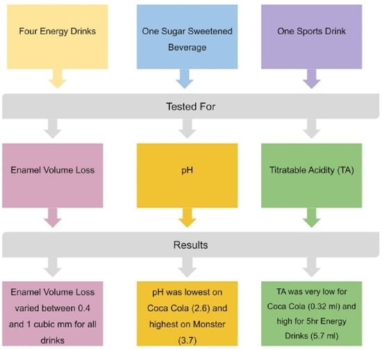
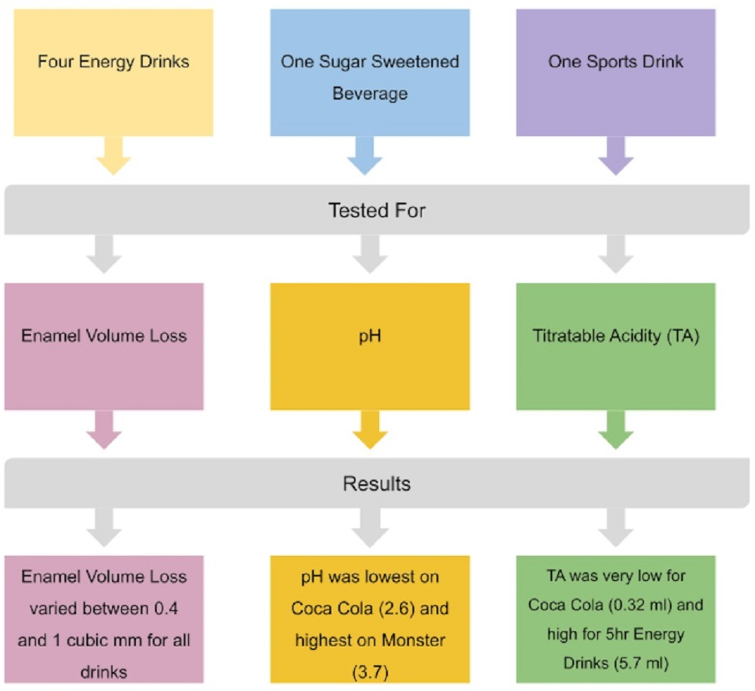
2. Materials and Methods

All teeth for this study were randomly selected from a tooth tissue collection used for pre-clinical education at the School of Dentistry. Ethical approval was obtained by the Internal Review Board with an exempt determination (IRB2024-5).

3. Evaluation of Volume Loss

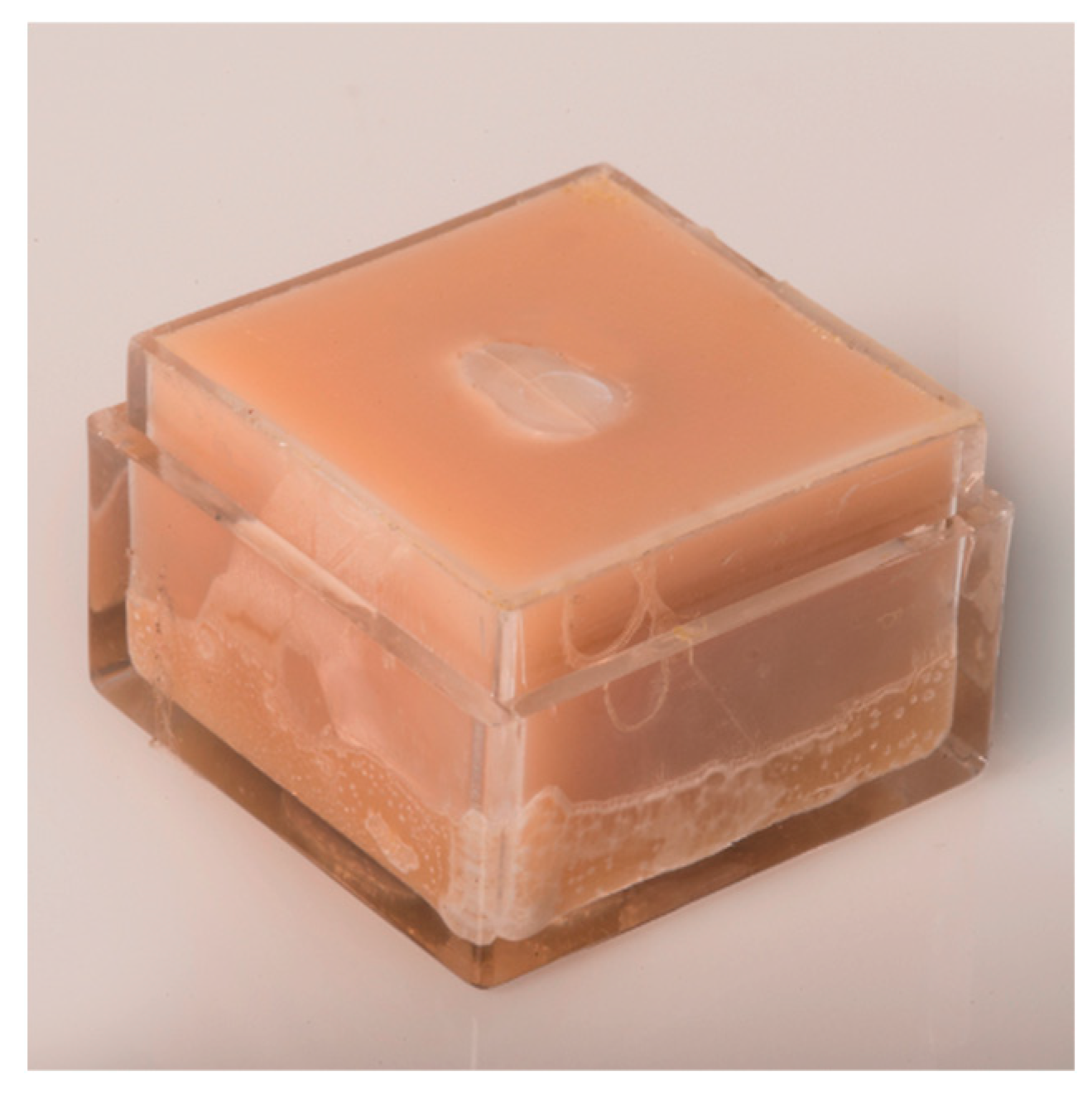
3.1. Sample Preparation

Thirty-six extracted human maxillary central and lateral incisor teeth were used in this study. After removing the roots perpendicular to the long axis of the teeth utilizing the Isomet Low speed diamond saw (Buehler Ltd. Lake Bluff, IL, USA), a flat plane of approximately 4 × 4 mm2 was prepared on the facial surface of each incisor utilizing 240, 320, and 600 µm grit Carbimet Abrasive Strips (Buehler Ltd., USA) on the Handimet Grinder (Buehler Ltd., USA). They were then further polished with Metadi II Diamond Polishing Compound (Buehler Ltd., USA) on a cloth disc resulting in a smoothness of 1.5 µm. Each prepared tooth sample was attached to an acrylic sample holder measuring 25 × 25 × 15 mm filled with Integrity, a bis-acrylic resin (Integrity, Dentsply Caulk, Milford, DE, USA) (Figure 1).

3.2. Scanning

The samples were then sent off to the United Kingdom for baseline measurements using the Proscan 2000 Profilometer (Scantron Ind Products Ltd., Taunton, UK) optical scanner. The sensor used for this study was a CHR contactless optical sensor S16/2.5 with a measuring range of 2500 μm. The lens was a CL2 and the magnifier was an MG140. Data collection was carried out using Proscan software V2.1.1.15B and the surface comparison was carried out using Proform software V1.41.
After the samples were returned from pretreatment scanning, they were covered with adhesive tape in which a 4 mm diameter circle had been punched, exposing the enamel surface area in the center of the sample. All 36 samples were prepared in the same manner.

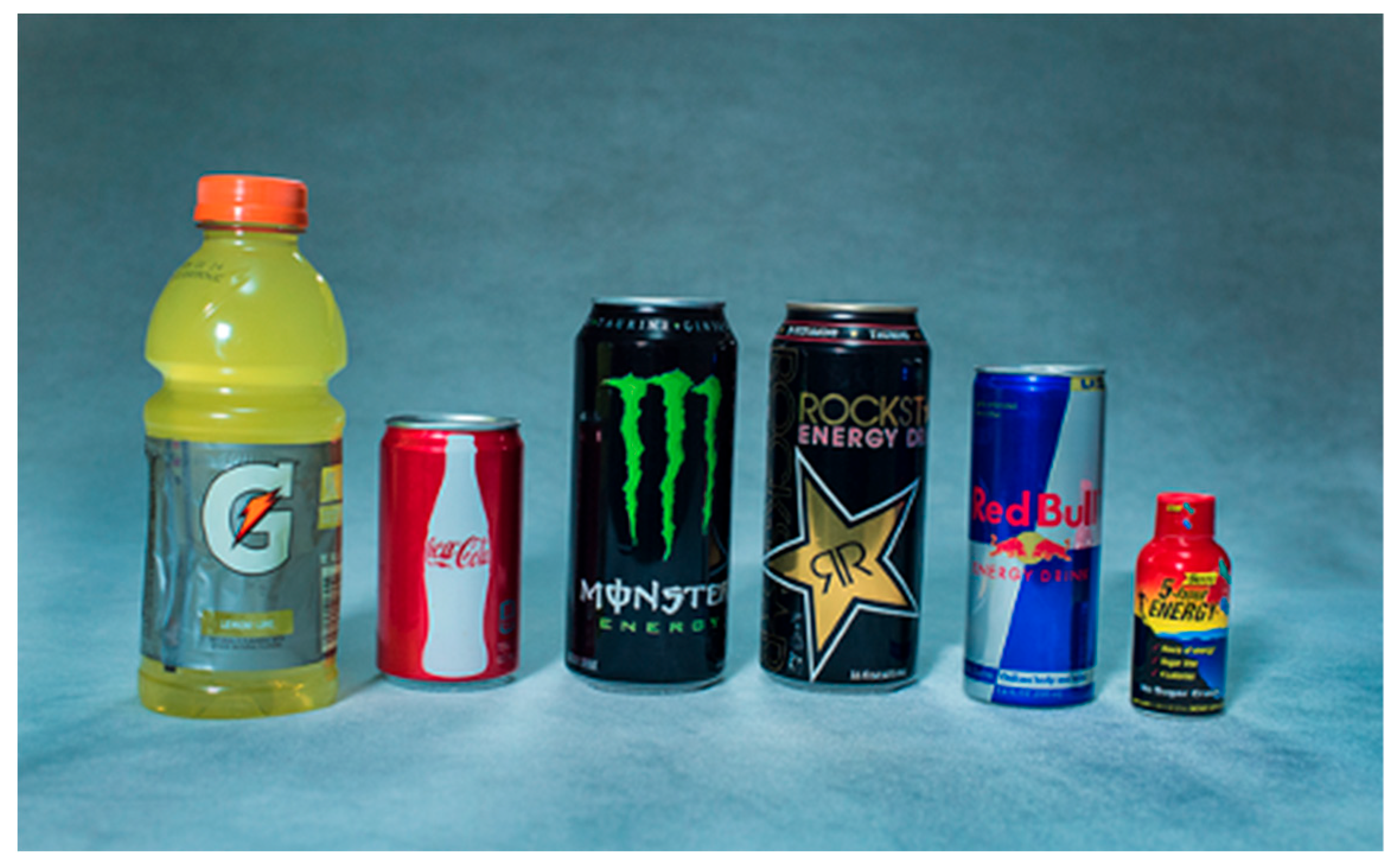
3.3. Drink Selection

The following 4 commercially available EDs were used for the experiment: Monster™, Rockstar™, Red Bull™, and 5-h Energy™. Coca-Cola® was used as an example of an SSB, and Gatorade™ as an example of an SD, for comparison to the energy drinks (Figure 2). Six samples were tested for each beverage for a total of 36 tests. No control sample for exposure in water or a buffered solution was included in this study because previous scientific findings suggest that the tooth surface is not altered when the surrounding solution is in an equilibrium of demineralization and remineralization [35].

3.4. Volume Loss Test

Immediately after the drinks were opened, 100 mL of each drink solution was placed in a 200 mL glass beaker at room temperature (22 °C). Six randomly selected samples of the prepared teeth were separately immersed in the drink solution for four hours (one sample per glass beaker).
During the four-hour cycle, they were stirred with a magnetic stirring device (Cole-Parmer Instrument Corp., Vernon Hills, IL, USA) providing homogeneous exposure of the ingredients to the exposed enamel surface of the sample. The samples were then removed from the solution, rinsed with water, the tape removed, and the samples dried. All samples were again shipped to the United Kingdom for analysis. The post-erosion scanning was done with the same equipment and settings used for the pre-scanning. One calibrated operator did the pre- and post-scanning, as well as the superimposing of the data and software analysis. By comparing the baseline measurements with the post-experiment scans, tooth surface volume loss was calculated. Data collection was carried out using Proscan software V2.1.1.15B and the surface comparison was carried out using Proform software V1.41. (Scantron Ind Products Ltd., Taunton, UK).

3.5. Evaluation of pH

Prior to testing the pH, 100 mL of each drink was poured into a 200 mL glass beaker and set aside for one hour after opening as a degassing process. The pH of each drink was measured using a calibrated Metrohm (700 GPT Titrino, Riverview, FL, USA) machine according to manufacturer’s instructions. A measuring probe of the Metrohm was inserted into the solution and affixed to the beaker so that the probe and the solution were stable during measurement. The measuring probe tested the pH of the solution 3 different times and was recorded at 5 min intervals.

3.6. Evaluation of Titratable Acidity

Titratable acidity is the volume of a buffering solution needed to change the pH of a solution to a different value [36]. Prior to testing for TA, 100 mL of each drink was poured into a 200 mL glass beaker and each drink was set aside for one hour after opening as a degassing process, to dissipate any possible influences from the carbonation.
While the solution was being stirred, drops of 1 mol NaOH buffering solution were added to the drinks at a rate of 1 mm/min and monitored to a pH of 5.5 using Tiano (Metrohm, Vers.3.1, Riverview, FL, USA) software until the pH of 5.5 was reached [1]. The volume of buffering solution needed to reach the pH of 5.5, represents the TA of a solution. Statistical analysis was performed using open-source statistical software “R” from the Free Software Foundation’s GNU General Public License, Boston, MA, USA, 2014.

4. Results

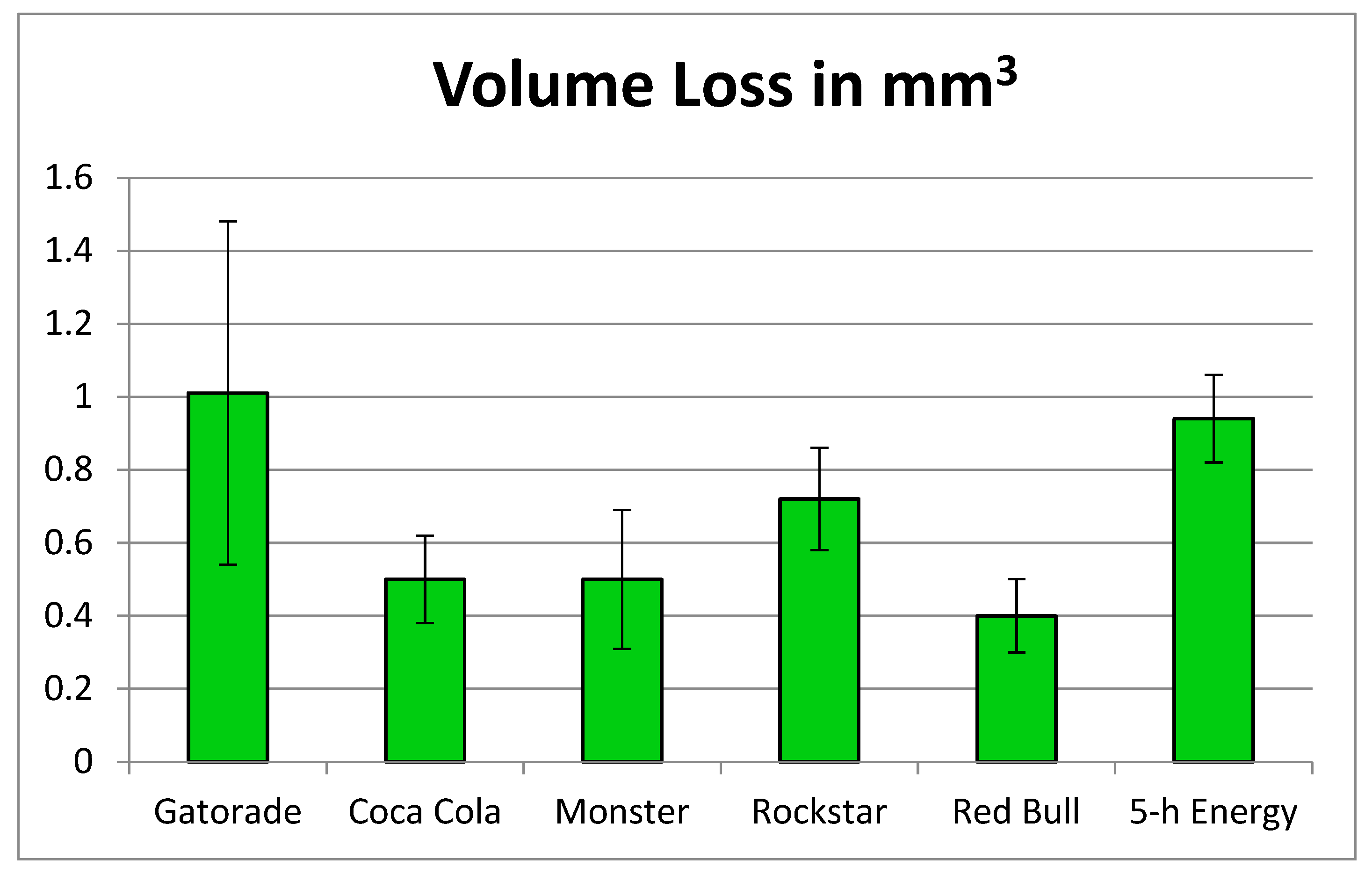
4.1. Volume Loss Evaluation

Figure 3 indicates the enamel volume loss for all drinks tested in this study in mm3 after the artificial erosion process. The volume loss varied from a low of 0.39 mm for Red Bull up to 1.01 mm3 for Gatorade. Data points that were more than 1.5 standard deviations below or above the mean values were eliminated and considered outliers. This applied only to four data points in the volume loss experiment. Figure 4 gives one visual example of the volume loss for each of the drinks that were tested in this study.
All enamel samples tested in this in vitro study showed substantial enamel volume loss. The highest average of volume loss was found for Gatorade™. The tooth volume loss among the three types of drinks were similar but varied, whereas the SD and EDs had a larger volume loss compared to the SSB (Figure 3).
Considering the sample sizes were small, and the samples failed the normality tests, a non-parametric Kruskal–Wallis test was used instead of a one-way ANOVA. The Kruskal–Wallis test resulted in a p-value of 0.001, implying that the six different drinks were not the same in terms of volume loss. It is important to note that data points that were more than 1.5 standard deviations below or above the mean values were eliminated and considered outliers.

4.2. Evaluation of pH

The pH values among the drinks ranged from a pH 2.6 (Coca-Cola®) to a pH 3.7 (Monster™). A noticeably small standard deviation was found (Figure 5). The non-parametric Kruskal–Wallis test for the variable pH resulted in a p-value of 0.005, meaning that the six different drinks were not the same in terms of the variable pH level.

4.3. Evaluation of Titratable Acidity

Figure 6 displays the titratable acidity that represents the volume in milliliters needed to buffer the solution up to a pH of 5.5. A pH of 5.5 was selected as the end point for achieving a buffered solution because, as seen in other studies, this is the value at which the demineralization process of enamel begins [1].
The titratable acidity ranked from Coca-Cola® being the lowest at 0.32 mL, followed by GatoradeTM at 1.38 mL, MonsterTM at 2.64 mL, and RockstarTM at 2.85 mL, the highest being Red BullTM at 2.87 mL and 5-h EnergyTM at 5.70 mL. The volume needed to buffer the 5-h EnergyTM drink was about 18 times higher than the volume needed for Coca-Cola®. This difference may be related to the differences in formulations of these drinks, wherein Coca-Cola® is made with phosphoric acid and the other drinks are made with citric acid. A Kruskal–Wallis test for the variable titratable acidity resulted in a p-value of 0.01, implying that the amount of buffering solution needed to increase the pH to a value of 5.5 was not the same for the six drinks considered.
Among the following energy drinks, MonsterTM, Red BullTM, and RockstarTM, there was no statistically significant difference in regard to titratable acidity. This means that a similar amount of buffering solution was needed to increase the pH to a value of 5.5.

4.4. Correlation Analysis

The correlation coefficient of enamel volume loss versus pH was found to be 0.326 with a p-value of 0.528. This shows a very weak reverse correlation between enamel volume loss and pH, and therefore is not statistically significant. Similarly, the correlation coefficient of enamel volume loss versus the TA was found to be 0.319 with a p-value of 0.537. This indicates a very weak correlation that is also not statistically significant. A significance level of 0.05 was used for all conclusions in this paper. Due to the different results of the beverages tested, the null hypothesis was rejected.

5. Discussion

The purpose of this in vitro study was to evaluate the correlation between pH and titratable acidity of the various drinks and the actual amount of enamel tooth volume or tooth structure loss. Teeth exposed to acidic substances lose structural integrity and physical properties, and acidic substance exposure results in loss of tissue [1]. Our study found that tooth volume loss was not consistently dependent on the pH or the titratable acidity of the various types of drinks, as shown in Figure 3, Figure 4 and Figure 5. Among the three energy drinks, RockstarTM, Monster EnergyTM, and Red BullTM, enamel volume loss varied, while their TA was very similar. With these same energy drinks, higher pH (less acidic) was related to less volume loss (Monster EnergyTM and Red BullTM) and lower pH was related to increased volume loss, as was the case with RockstarTM. With the sports drink, GatoradeTM, there was a moderately high pH, low TA, but high tooth structure loss. With the SSB, Coca-Cola®, there was lower tooth structure loss and very low titratable acidity, despite its low pH which makes it very acidic. These inconsistencies would indicate that the unique formulations of each drink result in varying amounts of tooth surface loss and that there is no dominant factor related to enamel tooth loss among this group. Previous studies have also demonstrated that surface changes induced by energy drinks do not only happen on tooth structures, but also affect the filling materials in the patient’s mouth [37]. All restorative materials tested in Vaidya’s study experienced an altered surface roughness. This can result in the darkening of dental restorations, a sequela that many patients do not consider when deciding to consume an energy drink.
Understanding the chemistry of erosion helps to understand the results of this study [38,39]. At a given pH, increased acid concentration (titratable acidity) results in a greater availability of H+ ions, which requires greater buffering capacity. Thus, as pH falls and the rate of dissolution increases, greater buffering is required to maintain the H+ concentration in the newly demineralized layer [40]. In this study, the actual concentration of acids in each beverage is not known. It should be noted, though, that the Coca-Cola® drink contains phosphoric acid while all other drinks contain citric acid. Since phosphoric acid is a weaker acid than citric acid, this would explain the decreased tooth surface loss with the Coca-Cola® drink. In addition to its high buffering capacity, citric acid may have an additional chelating effect. However, the presence of any and all acids in saliva causes continuing demineralization and reduces beneficial benefits of supersaturation of calcium in saliva by increasing the dissolution of tooth minerals and decreasing any potential remineralization of tooth structure [41].
The variation in results among and between the various drinks could also be explained, in part, by the structural variations found in the different areas of the enamel tooth samples. For example, outer layers of enamel have a more tightly packed prism structure compared to inner enamel. Sample preparations did not account for this variable [42]. Additionally, future studies could determine the surface hardness of the samples, which would provide information about the mineral content of the teeth in an effort to provide a better perspective on the erosion process in relation to the mineralized tissue.
For this experiment, an immersion time for samples of 4 h was chosen. Test scans were made at 30 min, 1, 2, and 4 h prior to the experiments. After reviewing results of the scans, the 4 h scan time appeared to help magnify the differences in quantitative measurements of the actual tooth surface loss. Other studies have used immersion times varying between 3 min [43,44] and 14 days [31,45]. It is important to note that none of these in vitro studies, including this study, would replicate actual in vivo conditions. The manner in which acidic drinks can influence the actual in vivo tooth surface loss also depends on how the drinks are consumed. Drinks that are held in the mouth and rinsed or swished are more harmful than those that are swallowed or gulped [46]. Other studies have suggested that the exposure time of the beverage to the tooth surface has a direct and significant impact on enamel volume loss [43]. This could lead to the assumption that the behaviors of many people, for example tech workers who sit at their computers sipping energy drinks over the course of several minutes or hours, could result in more cases of severe erosive tooth wear [47].
In this study, enamel samples were immersed and stirred at room temperature (22 °C) in each of the various drinks evaluated. It has been shown in vitro that increased temperature and increased flow rate will increase dissolution [48,49]. Therefore, under in vivo conditions, warm, acidic drinks, including energy drinks, that are ‘swished’ or sipped will cause more tooth surface loss [46]. It should also be noted that 5-h Energy™, which is a thicker, more viscous liquid, may remain on the tooth surface for longer periods, thus amplifying tooth surface loss.
This study was conducted with polished enamel samples placed directly in the various acidic solutions. Under in vivo conditions, the saliva would support the formation of a thin acquired pellicle layer on the enamel surface. This semi-permeable, protein-based layer acts as a natural diffusion barrier, inhibiting direct contact of intrinsic or extrinsic acids on the tooth surface [50,51]. Furthermore, under in vivo conditions, saliva also has an important protective influence on dental erosion by diluting, clearing, neutralizing, and buffering the acid, as well as maintaining a healthy biofilm on the tooth surface. It could also aid in the remineralization of the tooth surface by providing calcium, phosphorus, and fluoride [52,53,54]. Patients with hyposalivation and/or dry mouth are at increased risk for erosive changes. Therefore, during routine dental visits, it would be beneficial for dentists to give special attention to the amount and viscosity of the patient’s saliva. Patients should be advised of the importance of consuming adequate liquids and staying well hydrated.
In this in vitro study, all enamel samples showed significant tooth surface loss after being immersed in the various acidic drinks (Figure 6). However, there were various differences among the six drinks regarding enamel loss, pH, and TA. The drinks evaluated in this study each have unique and different ingredients and formulations and, therefore, must be evaluated on an individual basis.
Since the consumption of sports or energy drinks results in serious tooth surface loss, as found in this study, dental professionals are in a unique position to provide counseling and patient education on the risks of consuming these acidic beverages, on the key factors in preventing enamel loss, and the health problems associated with these drinks. Having patients do a dietary intake assessment, can indicate the source and frequency of acid exposure in their diet [46]. According to Lussi et al., four or more dietary acid intakes increase the risk of erosion [55]. Other studies have shown that the frequency of consumption of an acid beverage is an important factor in the development of erosion [56]. Patients should be advised to minimize the use of EDs and decrease acid contact time with teeth by swallowing, not holding or swishing them in their mouth. Dental professionals should advise the patient to rinse with water after use and avoid tooth brushing for one hour after consumption. With an early diagnosis of dental erosion, an individualized prevention plan can be formulated for patients, to help ward off more complicated treatment in the future [47]. The use of a toothpaste containing stannous fluoride will help protect the teeth from these acidic challenges [57].
Our study was done at room temperature. From other studies we have learned that erosion proceeds more rapidly the higher the temperature of that solution [11]. Therefore, it can be assumed that since most drinks are consumed at refrigerator temperature, erosion should be less impactful. If the patient holds the drink longer in the mouth and the temperature increases, there is more potential for erosive damage. Future studies could adjust the temperature of the solutions in which the teeth are immersed from refrigerator temperature to normal body temperature to see how that affects the erosion process.

6. Conclusions

Energy drinks, sugar sweetened beverages, and sport drinks all have the potential to cause enamel tooth surface loss resulting in demineralization. Therefore, the pH of a drink cannot be the sole determinant for choosing a less harmful commercial beverage. A combination of factors including pH (the level of acid) and titratable acidity (the strength of the acid) will affect the tooth surface loss caused by each drink. The proprietary formulations of energy drinks, along with the inclusion of caffeine and other stimulants, place patients at risk for dental erosion and other health issues.

Author Contributions

Conceptualization, W.N. and K.A.S.; Methodology, W.N. and K.A.S.; Validation, S.H.; Formal Analysis, M.B.; Investigation, K.A.S., B.S., N.M.S. and W.N.; Resources, S.H.; Data Curation, M.B.; Writing-Original Draft Preparation, K.A.S., N.M.S., W.N. and B.S.; Writing—Review and Editing, K.A.S. and N.M.S.; Visualization, K.A.S.; Supervision, W.N. and N.M.S.; Project administration, W.N., K.A.S. and N.M.S.; Funding Acquisition, W.N. and K.A.S. All authors have read and agreed to the published version of the manuscript.

Funding

This study was supported by Pilot Project Grant Activity 083 from the University of the Pacific, Arthur A. Dugoni School of Dentistry, San Francisco, CA, USA.

Institutional Review Board Statement

The study was conducted in accordance with the Declaration of Helsinki, and approved by the Institutional Review Board of the University of the Pacific, Stockton, CA, with an exempt determination (IRB2024-5).

Informed Consent Statement

Not applicable.

Data Availability Statement

All data from this study are published in this manuscript.

Acknowledgments

The authors wish to thank Shree Shrestha for assisting with sample preparation in the laboratory, John Draper from the University of the Pacific, Arthur A. Dugoni School of Dentistry, for the help with the images, and Avani Tumuluri for assistance with the references and laboratory procedures.

Conflicts of Interest

The authors declare no conflict of interest.

References

  1. Silva, J.G.; Martins, J.P.; de Sousa, E.B.; Fernandes, N.L.; Meira, I.A.; Sampaio, F.C.; de Oliveira, A.F.; Pereira, A.M. Influence of energy drinks on enamel erosion: In vitro study using different assessment techniques. J. Clin. Exp. Dent. 2021, 11, e1076–e1082. [Google Scholar] [CrossRef]
  2. Ali, F.; Rehman, H.; Babayan, Z.; Stapleton, D.; Joshi, D.D. Energy drinks and their adverse health effects: A systematic review of the current evidence. Postgrad. Med. 2015, 127, 308–322. [Google Scholar] [CrossRef]
  3. Alsunni, A.A. Energy drink consumption: Beneficial and adverse health effects. Int. J. Health Sci. 2015, 4, 468–474. [Google Scholar] [CrossRef]
  4. Statista. Available online: https://www.statista.com/topics/10313/energy-drinks-worldwide/#topicOverview (accessed on 30 November 2023).
  5. Bevsource. Available online: https://www.bevsource.com/news/examining-the-9-latest-trends-in-energy-drinks (accessed on 30 November 2023).
  6. Oregon Consulting Group. Available online: https://business.uoregon.edu/sites/default/files/media/energy-drink-industry-report.pdf (accessed on 30 November 2023).
  7. Investopedia. Available online: https://www.investopedia.com/articles/investing/022315/energy-drinks-industry.asp (accessed on 30 November 2023).
  8. Healthy Food America. Available online: https://www.healthyfoodamerica.org/sugartoolkit_kahuna (accessed on 30 November 2023).
  9. Higgins, J.P.; Babu, K.; Deuster, P.A.; Shearer, J. Energy drinks: A contemporary issues paper. Curr. Sports Med. Rep. 2018, 2, 65–72. [Google Scholar] [CrossRef]
  10. Kumar, G.; Park, S.; Onufrak, S. Perceptions about energy drinks are associated with energy drink intake among US youth. Am. J. Health Promot. 2015, 4, 238–244. [Google Scholar] [CrossRef]
  11. Clapp, O.; Morgan, M.Z.; Fairchild, R.M. The top five selling UK energy drinks: Implications for dental and general health. Br. Dent. J. 2019, 7, 493–497. [Google Scholar] [CrossRef]
  12. Higgins, J.P.; Tuttle, T.D.; Higgins, C.L. Energy beverages: Content and safety. Mayo Clin. Proc. 2010, 11, 1033–1041. [Google Scholar] [CrossRef]
  13. Seifert, S.M.; Seifer, S.A.; Schaechter, J.L.; Bronstein, A.C.; Benson, B.E.; Hershorin, E.R.; Arheart, K.L.; Franco, V.I.; Lipshultz, S.E. An analysis of energy drink toxicity in the National Poison Data System. Clin. Toxicol. 2013, 7, 566–574. [Google Scholar] [CrossRef] [PubMed]
  14. Jackson, D.A.; Cotter, B.V.; Merchant, R.C.; Babu, K.M.; Baird, J.R.; Nirenberg, T. Behavioral and physiologic adverse effects in adolescent and young adult emergency department patients reporting use of energy drinks and caffeine. Clin. Toxicol. 2013, 7, 557–565. [Google Scholar] [CrossRef] [PubMed]
  15. Shah, S.A.; Occiano, A.; Nguyen, T.A.; Chan, A.; Sky, J.C.; Battacharya, M.; O’Dell, K.M.; Shek, A.; Nguyen, N.N. Electrocardiographic and blood pressure effects of energy drinks and Panax ginseng in healthy volunteers: A randomized clinical trial. Int. J. Cardiol. 2016, 218, 318–323. [Google Scholar] [CrossRef] [PubMed]
  16. Kaminer, Y. Problematic use of energy drinks by adolescents. Child Adolesc. Psychiatr. Clin. N. Am. 2010, 19, 643–650. [Google Scholar] [CrossRef]
  17. Duchan, E.; Patel, N.D.; Feucht, C. Energy drinks: A review of use and safety for athletes. Physician Sports Med. 2010, 38, 171–179. [Google Scholar] [CrossRef]
  18. Chrysant, S.G.; Chrysant, G.S. Cardiovascular complications from consumption of high energy drinks: Recent evidence. J. Hum. Hypertens. 2015, 29, 71–76. [Google Scholar] [CrossRef] [PubMed]
  19. Woolsey, C.; Waigandt, A.; Beck, N.C. Athletes and energy drinks: Reported risk-taking and consequences from the combined use of alcohol and energy drinks. J. Appl. Sports Psychol. 2010, 22, 65–71. [Google Scholar] [CrossRef]
  20. Monsterenergy. Available online: https://www.monsterenergy.com/us/en/products/monster-energy/monster-energy (accessed on 30 November 2023).
  21. Seifert, S.M.; Schaechter, J.L.; Hershorin, E.R.; Lipshultz, S.E. Health effects of energy drinks on children and adolescents and young adults. Pediatrics 2011, 127, 511–528. [Google Scholar] [CrossRef] [PubMed]
  22. Trapp, G.S.; Allen, K.L.; O’Sullivan, T.; Robinson, M.; Jacoby, P.; Oddy, W.H. Energy drink consumption among young Australian adults: Associations with alcohol and illicit drug use. Drug Alcohol Depend. 2014, 134, 30–37. [Google Scholar] [CrossRef] [PubMed]
  23. Bonar, E.E.; Cunningham, R.M.; Polshkova, S.; Chermack, S.T.; Blow, F.C.; Walton, W.A. Alcohol and energy use among adolescents seeking emergency department care. Addict. Behav. 2015, 43, 11–17. [Google Scholar] [CrossRef] [PubMed]
  24. Bigard, A.X. Risks of energy drinks in youths. Arch. Pediatr. 2010, 17, 1624–1631. [Google Scholar]
  25. Marczinski, C.A. Can energy drinks increase the desire for more alcohol? Adv. Nutr. 2014, 6, 96–101. [Google Scholar] [CrossRef] [PubMed]
  26. Jaeggi, T.; Lussi, A. Prevalence incidence and distribution of erosion. Monogr. Oral Sci. 2014, 25, 55–73. [Google Scholar]
  27. Ganss, C. Definition of erosion and links to tooth wear. Monogr. Oral Sci. 2006, 20, 9–16. [Google Scholar] [PubMed]
  28. Moazzez, R.; Bartlett, D. Intrinsic causes of erosion. Monogr. Oral Sci. 2014, 25, 180–196. [Google Scholar]
  29. Barbour, M.E.; Lussi, A. Erosion in relation to nutrition and the environment. Monogr. Oral Sci. 2014, 25, 143–154. [Google Scholar] [PubMed]
  30. Lussi, A.; Schlueter, N.; Rakhmatullina, E.; Ganss, C. Dental erosion—An overview with emphasis in chemical and histopathological aspects. Caries Res. 2011, 45 (Suppl. SI), 2–12. [Google Scholar] [CrossRef] [PubMed]
  31. Kitchens, M.; Owens, B.M. Effect of carbonated beverages, coffee, sports and high energy drinks, and bottled water on the in vitro erosion characteristics of dental enamel. J. Clin. Pediatr. Dent. 2007, 31, 153–159. [Google Scholar] [CrossRef] [PubMed]
  32. Featherstone, J.D.; Duncan, J.F.; Cutress, T.W. A mechanism for dental caries based on chemical processes and diffusion phenomena during in vitro caries simulation on human tooth enamel. Arch. Oral Biol. 1979, 24, 101–112. [Google Scholar] [CrossRef]
  33. Lussi, A.; Jaeggi, T.; Schaerer, S. The influence of different factors on in vitro enamel erosion. Caries Res. 1993, 27, 387–393. [Google Scholar] [CrossRef]
  34. Huysmans, M.C.; Chew, H.P.; Ellwood, R.P. Clinical studies of dental erosion and erosive wear. Caries Res. 2011, 45 (Suppl. S1), 60–68. [Google Scholar] [CrossRef]
  35. Abou Neel, E.A.; Aljabo, A.; Strange, A.; Ibrahim, S.; Coathup, M.; Young, A.M.; Bozec, L.; Mudera, V. Demineralization-remineralization dynamics in teeth and bone. Int. J. Nanomed. 2016, 11, 4743–4763. [Google Scholar] [CrossRef]
  36. Attin, T.; Wegehaupt, F.J. Methods for assessment of dental erosion. Monogr. Oral Sci. 2014, 25, 123–142. [Google Scholar]
  37. Vaidya, N.; Kumar, P.; Pathak, K.; Punia, S.K.; Choudhary, A.; Patnana, A.K. Comparative Evaluation of the Influence of Different Sports/Energy Drinks and Alcoholic Beverages on the Surface Roughness of Three Different Flowable Esthetic Restorative Materials: An In Vitro Analysis. J. Int. Soc. Prev. Community Dent. 2020, 5, 585–590. [Google Scholar]
  38. Featherstone, J.D.; Rogers, B.E. Effects of acetic, lactic and other organic acids on the formation of artificial caries lesions. Caries Res. 1981, 15, 377–385. [Google Scholar] [CrossRef] [PubMed]
  39. Featherstone, J.D.; Lussi, A. Understanding the chemistry of dental erosion. Monogr. Oral Sci. 2006, 20, 66–76. [Google Scholar] [PubMed]
  40. Shellis, R.P.; Barbour, M.E.; Jones, S.B.; Addy, M. Effects of pH and acid concentration on erosive dissolution of enamel, dentine, and compressed hydroxyapatite. Eur. J. Oral Sci. 2010, 118, 475–482. [Google Scholar] [CrossRef]
  41. Meurman, J.H.; Ten Cate, J.M. Pathogenesis and modifying factors of dental erosion. Eur. J. Oral Sci. 1997, 104, 199–206. [Google Scholar] [CrossRef]
  42. Mullin, F.; Austin, R.S.; Parkinson, C.R.; Bartlett, D.W. An in situ study to investigate the native clinical resistance of enamel to erosion. J. Dent. 2018, 70, 124–128. [Google Scholar] [CrossRef] [PubMed]
  43. Jager, D.H.; Vieira, A.M.; Ruben, J.L.; Huysmans, M.C. Estimated erosive potential depends on exposure time. J. Dent. 2012, 40, 1103–1108. [Google Scholar] [CrossRef]
  44. Eisenburger, M.; Addy, M. Evaluation of pH and erosion time on demineralization. Clin. Oral Investig. 2001, 5, 108–111. [Google Scholar]
  45. von Fraunhofer, J.A.; Rogers, M.M. Effects of sports drinks and other beverages on dental enamel. Gen. Dent. 2005, 53, 28–31. [Google Scholar]
  46. Johansson, A.K.; Lingström, P.; Imfeld, T.; Birkhed, D. Influence of drinking method on tooth-surface pH in relation to dental erosion. Eur. J. Oral Sci. 2004, 112, 484–489. [Google Scholar] [CrossRef]
  47. Lussi, A.; Hellwig, E. Risk assessment and preventive measures. Monogr. Oral Sci. 2006, 20, 190–199. [Google Scholar]
  48. Barbour, M.E.; Finke, M.; Parker, D.M.; Hughes, J.A.; Allen, G.C.; Addy, M. The relationship between enamel softening and erosion caused by soft drinks at a range of temperatures. J. Dent. 2006, 34, 207–213. [Google Scholar] [CrossRef]
  49. Eisenburger, M.; Addy, M. Influence of liquid temperature and flow rate on enamel erosion and surface softening. J. Oral Rehabil. 2003, 30, 1076–1080. [Google Scholar] [CrossRef]
  50. Hannig, M.; Fiebiger, M.; Güntzer, M.; Döbert, A.; Zimehl, R.; Nekrashevych, Y. Protective effect of the in situ formed short-term salivary pellicle. Arch. Oral Biol. 2004, 49, 903–910. [Google Scholar] [CrossRef] [PubMed]
  51. Vukosavljevic, D.; Custodio, W.; Buzalaf, M.A.; Hara, A.T.; Siqueira, W.L. Acquired pellicle as a modulator for dental erosion. Arch. Oral Biol. 2014, 59, 631–638. [Google Scholar] [CrossRef]
  52. Hara, A.T.; Zero, D.T. The potential of saliva in protecting against dental erosion. Monogr. Oral Sci. 2014, 25, 197–205. [Google Scholar]
  53. Ganss, C.; Lussi, A.; Schlueter, N. The histological features and physical properties of eroded dental hard tissues. Monogr. Oral Sci. 2014, 25, 99–107. [Google Scholar]
  54. Lussi, A.; Carvalho, T.S. Erosive tooth wear: A multifactorial condition of growing concern and Increasing Knowledge. Monogr. Oral Sci. 2014, 25, 1–15. [Google Scholar]
  55. Lussi, A.; Jaeggi, T. Erosion-diagnosis and risk factors. Clin. Oral Investig. 2008, 12 (Suppl. S1), S5–S13. [Google Scholar] [CrossRef]
  56. Søvik, J.B.; Skudutyte-Rysstad, R.; Tveit, A.B.; Sandvik, L.; Mulic, A. Sour Sweets and Acidic Beverage Consumption Are Risk Indicators for Dental Erosion. Caries Res. 2015, 49, 243–250. [Google Scholar] [CrossRef]
  57. West, N.X.; He, T.; Zou, Y.; Biesbrock, A.; Davies, M. Bioavailable gluconate chelated stannous fluoride toothpaste meta-analysis: Effects on dentin hypersensitivity and enamel erosion. J. Dent. 2021, 105, 103566. [Google Scholar] [CrossRef]
Figure 1. Sample embedded in resin block for analysis.
Oral 04 00009 g001
Figure 2. Drinks analyzed in study.
Oral 04 00009 g002
Figure 3. Enamel volume loss after erosive challenge.
Oral 04 00009 g003
Figure 4. These images give one example of the enamel volume loss for each of the drinks tested.
Oral 04 00009 g004
Figure 5. pH values among the drinks tested.
Oral 04 00009 g005
Figure 6. Titratable acidity (buffering capacity) at pH 5.5.
Oral 04 00009 g006
Disclaimer/Publisher’s Note: The statements, opinions and data contained in all publications are solely those of the individual author(s) and contributor(s) and not of MDPI and/or the editor(s). MDPI and/or the editor(s) disclaim responsibility for any injury to people or property resulting from any ideas, methods, instructions or products referred to in the content.

