Oral 2024, 4(2), 163-172; https://doi.org/10.3390/oral4020013 - 07 Apr 2024
Abstract
►
Show Figures
Endodontic treatments are performed to avoid extractions and maintain the natural dentition. Root canal treatments are undertaken to eliminate or prevent an infection within the root canal system. Chemical and mechanical root canal debridement are the main methods used in endodontics to remove
[...] Read more.
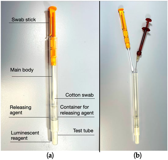
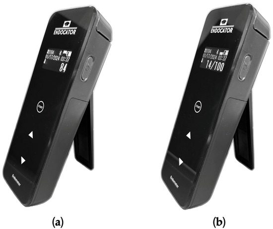
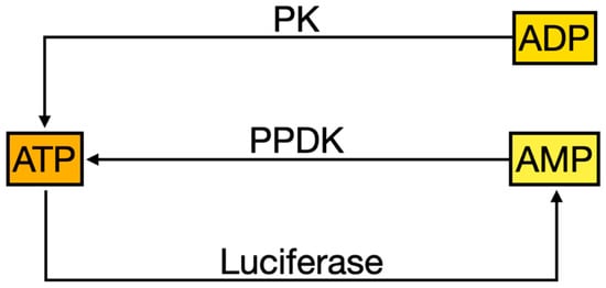
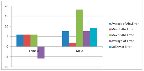
Endodontic treatments are performed to avoid extractions and maintain the natural dentition. Root canal treatments are undertaken to eliminate or prevent an infection within the root canal system. Chemical and mechanical root canal debridement are the main methods used in endodontics to remove necrotic tissue, microorganisms, and microbial byproducts from the canal. However, to date there is no objective method to clinically determine the proper root canal disinfection level and thus proceed with the obturation. Clinicians just rely on their experience and habits or can trust in empirical methods such as the insertion of paper cones inside the canal and then check their appearance after the removal. Even in the in vitro and ex vivo scientific studies there is no objective method to analyze and compare the efficacy of different endodontic chemo-mechanical techniques and materials. The most frequently used method is to visually analyze some areas with a scanning electron microscope (SEM), even if the resulting images are hardly quantifiable and could greatly vary according to the analyzed area. A new device to clinically test the cleanliness of a root canal and display the result in an objective score was recently developed. The device analyzes the luminescence generated by an enzyme cycling method that process the adenosine triphosphate (ATP), adenosine diphosphate (ADP) and adenosine monophosphate (AMP) present in organic residues. The aim of the present in vitro study was to test the efficacy and reliability of this novel device (Endocator) in a controlled in vitro environment, before using it in clinical practice. The device sensitivity was tested on 5 single canal resin blocks. Three consecutive sampling were executed by one operator for each block to test the device repeatability. Results were recorded according to Endoscore (ES) and relative light unit (RLU) scales. Descriptive analysis and comparison between the 5 resin blocks and the 3 consecutive sampling were performed. Only the comparison between the first and third measurements both for ES (p = 0.00115999) and RLU (p = 0.00532749) resulted significant. Endocator was able to determine small variations of canal contamination in a controlled laboratory environment, showing high sensitivity and repeatability.
Full article
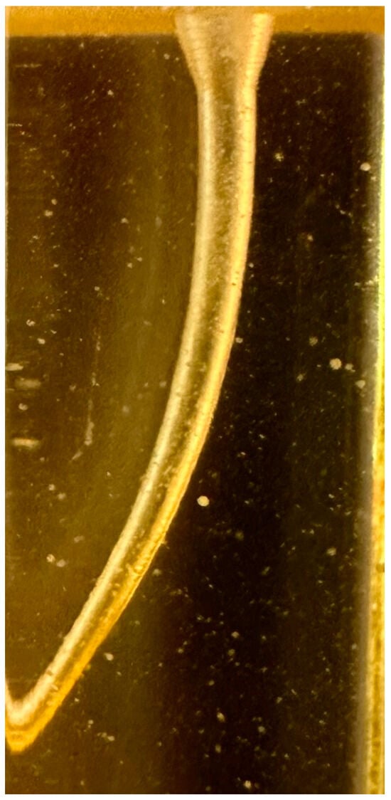
Figure 1


