Next Issue
Volume 3, December
Previous Issue
Volume 3, June


Oral, Volume 3, Issue 3 (September 2023) – 13 articles

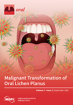
Cover Story (view full-size image): Oral lichen planus (OLP) is classified as a potentially malignant disorder. Systematic reviews collating longitudinal observation studies provide evidence of the rate or proportion of malignant transformation. We conducted an umbrella study of published systematic reviews. Data from eight systematic reviews published between 2014 and 2023 are presented. The reported proportions of malignant transformation ranged from 1.1% to 1.4%. A meta-analysis based on the 10 highest-quality studies yielded a higher proportion of malignant transformation (2.28%). We list some of the limitations found in several of these systematic reviews. Some studies reported an increased risk of malignancy in OLP lesions, demonstrating epithelial dysplasia. In view of the consistent evidence of the risk of oral malignancy, OLP patients should be monitored carefully to detect early cancer development. View this paper
  • Issues are regarded as officially published after their release is announced to the table of contents alert mailing list.
  • You may sign up for e-mail alerts to receive table of contents of newly released issues.
  • PDF is the official format for papers published in both, html and pdf forms. To view the papers in pdf format, click on the "PDF Full-text" link, and use the free Adobe Reader to open them.
Order results
Result details
Select all
Export citation of selected articles as:
13 pages, 2085 KiB  
Article
Real-Time Caries Detection of Bitewing Radiographs Using a Mobile Phone and an Artificial Neural Network: A Pilot Study
by Ming Hong Jim Pun
Oral 2023, 3(3), 437-449; https://doi.org/10.3390/oral3030035 - 05 Sep 2023
Viewed by 2450
Abstract
This pilot study aimed to implement and assess the performance of an experimental artificial intelligence (AI) mobile phone app in the real-time detection of caries lesions on bitewing radiographs (BWRs) with the use of a back-facing mobile phone video camera. The author trained [...] Read more.
This pilot study aimed to implement and assess the performance of an experimental artificial intelligence (AI) mobile phone app in the real-time detection of caries lesions on bitewing radiographs (BWRs) with the use of a back-facing mobile phone video camera. The author trained an EfficientDet-Lite1 artificial neural network using 190 radiographic images from the Internet. The trained model was deployed on a Google Pixel 6 mobile phone and used to detect caries on ten additional Internet BWRs. The sensitivity/precision/F1 scores ranged from 0.675/0.692/0.684 to 0.575/0.719/0.639 for the aggregate handheld detection of caries in static BWRs versus the stationary scanning of caries in a moving video of BWRs, respectively. Averaging the aggregate results, the AI app detected—in real time—62.5% of caries lesions on ten BWRs with a precision of 70.6% using the back-facing mobile phone video camera. When combined with the AI app’s relative ease of use and speed and the potential for global accessibility, this proof-of-concept study could quite literally place AI’s vast potential for improving patient care in dentists’ hands. Full article
Show Figures

Figure 1

17 pages, 1543 KiB  
Review
Oral Cancer Disease among the Poor: A Sri Lankan Context
by Kalpani Senevirathna, Yovanthi Anurangi Jayasinghe, Shalindu Malshan Jayawickrama, Hemantha Amarasinghe and Ruwan Duminda Jayasinghe
Oral 2023, 3(3), 420-436; https://doi.org/10.3390/oral3030034 - 04 Sep 2023
Viewed by 2075
Abstract
Oral cancer (OC) is the 16th most common cancer worldwide. In Sri Lanka, OC is the most prevalent cancer among men and the eighth among women. In most instances, OC is preceded by clinically recognizable disorders appearing on the oral mucosa, termed oral [...] Read more.
Oral cancer (OC) is the 16th most common cancer worldwide. In Sri Lanka, OC is the most prevalent cancer among men and the eighth among women. In most instances, OC is preceded by clinically recognizable disorders appearing on the oral mucosa, termed oral potentially malignant disorders (OPMD). The distribution of OC among low- and middle-income socioeconomic groups contributes to various factors. Poor oral health literacy, a lower quality of higher education, and the economic burden results in neglected oral health. The further economic recession has led to the formation of risk habits, such as chewing betel quid, smoking and the consumption of alcohol, among these groups to minimize stress levels. But with a lack of oral health awareness, the incidences of OC increase in this category. This review elaborates on a few cross-sectional studies conducted in various locations in Sri Lanka, discussing the low awareness of neglected oral health, the usage of tobacco in a smokeless and non-smokeless manner, consuming alcohol, and the chewing of betel nuts. This review aims to overcome the barriers in these low- and middle-income socioeconomic groups in developing nations such as Sri Lanka by creating more awareness and minimizing the incidence and diagnosis and treatment at early stages to improve the quality of life as well as longevity. Full article
(This article belongs to the Special Issue Oral Health in the Global South)
Show Figures

Graphical abstract

9 pages, 1049 KiB  
Conference Report
Oro-Dental Pharmacovigilance in the Digital Age: Promoting Knowledge, Awareness, and Practice in Italy through a Smart Combined System—A Conference at the 30th National Congress of the Italian College of University Professors of Dental Disciplines
by Gaetano La Mantia, Fortunato Buttacavoli, Vera Panzarella, Giuseppe Colella, Annalisa Capuano, Liberata Sportiello, Gaspare Parrinello, Ilaria Morreale, Giacomo Oteri, Giuseppe Bellavia, Vittorio Fusco, Rodolfo Mauceri, Martina Coppini, Monica Bazzano, Giuseppe Seminara, Olga Di Fede and Giuseppina Campisi
Oral 2023, 3(3), 411-419; https://doi.org/10.3390/oral3030033 - 01 Sep 2023
Viewed by 849
Abstract
Adverse drug reactions (ADRs) represent a significant threat to patients’ safety in dentistry, necessitating proactive measures for prevention and treatment. However, identifying ADRs of dental and oral interest can be challenging, and underreporting remains a persistent issue globally. This paper illustrates a smart [...] Read more.
Adverse drug reactions (ADRs) represent a significant threat to patients’ safety in dentistry, necessitating proactive measures for prevention and treatment. However, identifying ADRs of dental and oral interest can be challenging, and underreporting remains a persistent issue globally. This paper illustrates a smart system to help Italian healthcare personnel, including dentists, in identifying and reporting dental and oral ADRs. This educational program is within a larger multi-regional project financed by the Italian Agency of Drugs (AIFA). The proposed system comprises a free online questionnaire on ADRs of dental interest (including specific items about awareness and attitudes), and after the free consultation of an atlas of clinical images of dental and oral ADRs linked with a digital synopsis of drugs potentially related to ADRs. This section of the project was presented in April 2023 at the 30th National Congress of the Italian College of University Professors of Dental Disciplines, and the system appears to hold tremendous potential to improve the knowledge of healthcare professionals on oral and dental ADRs. Full article
Show Figures

Figure 1

9 pages, 242 KiB  
Article
Presurgical Decompensation in Patients Affected by Class III Dentoskeletal Malocclusion: A Comparison between Two Different Bracket Systems
by Andrea Mariani, Roberto Antonio Vernucci, Rosanna Guarnieri, Adriana De Stefano, Roberto Di Giorgio, Ersilia Barbato and Gabriella Galluccio
Oral 2023, 3(3), 402-410; https://doi.org/10.3390/oral3030032 - 01 Sep 2023
Viewed by 666
Abstract
The aim of this study was to analyse if bracket systems could affect the incisors’ decompensation in a group of patients affected by dentoskeletal Class III malocclusion and on the duration of presurgical orthodontics. A single-centre retrospective study was carried out on two [...] Read more.
The aim of this study was to analyse if bracket systems could affect the incisors’ decompensation in a group of patients affected by dentoskeletal Class III malocclusion and on the duration of presurgical orthodontics. A single-centre retrospective study was carried out on two groups of patients affected by dentoskeletal Class III and who underwent orthognathic surgery. Group 1, comprising 25 subjects (13 males, 12 females; mean age, 27.4 y.o.), was treated with interactive self-ligating brackets; group 2, comprising 10 subjects (5 males, 5 females; mean age, 27.2 y.o.), was treated with conventional twin brackets. Bracket prescription and archwire sequence were the same for both groups. Data were collected about age, upper incisors’ axis (UI^ANS-PNS), lower incisors’ axis (IMPA), and the total duration of presurgical orthodontics and were then compared between the two groups. The variation in the upper incisor axis and the duration of presurgical orthodontics was not significant between the groups, while the difference in the IMPA was small but significant. The use of a self-ligating bracket did not influence the decompensation of the upper incisors or the duration of presurgical orthodontics when compared with conventional ligating brackets. The difference in the IMPA was significantly different but clinically negligible. Full article
13 pages, 273 KiB  
Review
Corticotomy-Assisted Orthodontic Treatment: A Literature Review
by Athanasia Patatou, Niovi Iacovou, Paraskevi Zaxaria, Michail Vasoglou and Georgios Vasoglou
Oral 2023, 3(3), 389-401; https://doi.org/10.3390/oral3030031 - 14 Aug 2023
Viewed by 1588
Abstract
The increased number of adults seeking orthodontic treatment has led to the need for faster results as social limitations arise with the use of fixed or even removable appliances. Corticotomy-assisted orthodontic treatment (CAOT) has been proposed as a technique that accelerates tooth movement [...] Read more.
The increased number of adults seeking orthodontic treatment has led to the need for faster results as social limitations arise with the use of fixed or even removable appliances. Corticotomy-assisted orthodontic treatment (CAOT) has been proposed as a technique that accelerates tooth movement and leads to a substantial reduction in treatment time. This review was conducted by searching four databases for original articles about CAOT, review articles and articles about alternative techniques and periodontal effects up to 31 December 2022. The aim of the review was to present the historical and biological background of CAOT, the description of the technique and some alternative and less invasive techniques that have been proposed. Indications, contraindications and periodontal side effects are also discussed. Full article
(This article belongs to the Special Issue Current Issues in Oral Health)
17 pages, 955 KiB  
Article
Tooth-Surface-Specific Effects of MI Varnish™: A 3-Year Randomized Clinical Trial
by Jekaterina Gudkina, Bennett T. Amaechi, Stephen H. Abrams and Anda Brinkmane
Oral 2023, 3(3), 372-388; https://doi.org/10.3390/oral3030030 - 07 Aug 2023
Viewed by 993
Abstract
Aim: Our previous three-year randomized control trial showed that the application of MI Varnish™ (5% NaF/CPP-ACP) every 3 months reduced further caries development in 6- and 12-year-olds over a 3-year period. The purpose of this secondary analysis was to investigate whether MI Varnish™ [...] Read more.
Aim: Our previous three-year randomized control trial showed that the application of MI Varnish™ (5% NaF/CPP-ACP) every 3 months reduced further caries development in 6- and 12-year-olds over a 3-year period. The purpose of this secondary analysis was to investigate whether MI Varnish™ had a differential effect on cumulative caries increment on different tooth surfaces. Methods: Group 1 (n = 48) (6-year-old children) and Group 3 (n = 47) (12-year-old children) received quarterly varnish applications, while Group 2 (n = 48) (6-year-old children) and Group 4 (n = 37) (12-year-old children) did not receive varnish applications. ICDAS caries scoring classified lesions as non-cavitated (n/c) lesions (ICDAS 1 and 2), cavitated (c) lesions (ICDAS II 3–6), non-cavitated lesions around restorations (CARn/c), and cavitated lesions around restorations (CARc). Thus, ‘decayed’ in DFS was calculated as (ICDAS 1–6 + CARn/c + CARc). The Chi-square test, Welch test (paired-t test), risk ratio test, and Pearson correlation coefficient were used for statistical analysis (α = 0.05). Results: After comparing baseline and 36-month data, in group 1, there was a significant (p < 0.01) reduction in caries in occlusal (23.11%) and proximal (21.35%) surfaces and a non-significant reduction in buccal/lingual surfaces (5.28%). In group 2, caries reduction was significant (p < 0.01) in occlusal surfaces (38.52%) but non-significant in proximal (7.78%) and buccal/lingual (7.12%) surfaces. In groups 3 and 4, significant (p < 0.001) increases in caries were observed in proximal (36.03% (group 3)/54.30% (group 4)) and buccal/lingual surfaces (51.02% (group 3)/45.98% (group 4)), and a non-significant increase was observed in occlusal surfaces (11.49% (group 3)/22.01% (group 4)). The relative risk had increased by 4% only on proximal surfaces in 6-year-olds. Conclusions: the application of MI Varnish™ every 3 months demonstrated a caries reduction effect on interproximal and occlusal surfaces among 6- and 12-year-old children. (Trial registration ISRCTN10584414). Full article
Show Figures

Graphical abstract

19 pages, 2334 KiB  
Article
In Vitro Evaluation of Dental Resin Monomers, Triethylene Glycol Dimethacrylate (TEGDMA), and 2-Hydroxyethyl Methacrylate (HEMA) in Primary Human Melanocytes: A Pilot Study
by Shilpi Goenka
Oral 2023, 3(3), 353-371; https://doi.org/10.3390/oral3030029 - 03 Aug 2023
Cited by 4 | Viewed by 1394
Abstract
Triethylene glycol dimethacrylate (TEGDMA) and 2-hydroxyethyl methacrylate (HEMA), two resin monomers often used in dental restorative materials, are leached due to insufficient polymerization and contact cells of the oral cavity. Despite reports on the cytotoxicity of these monomers on different oral cells, their [...] Read more.
Triethylene glycol dimethacrylate (TEGDMA) and 2-hydroxyethyl methacrylate (HEMA), two resin monomers often used in dental restorative materials, are leached due to insufficient polymerization and contact cells of the oral cavity. Despite reports on the cytotoxicity of these monomers on different oral cells, their effects on oral melanocytes remain unknown. This first report provides novel data on the impact of TEGDMA and HEMA monomers on melanocyte viability and functions by utilizing primary human melanocytes from lightly pigmented (HEMn-LP) foreskin as a representative model of oral melanocytes. Results show that TEGDMA induced higher cytotoxicity than HEMA and significant morphological alterations (increased dendricity) in melanocytes at the lowest concentration of 0.25 mM onwards. HEMA achieved similar effects but only at an 8-fold higher concentration (2 mM), while lower concentrations did not induce any change in cellular melanin or morphology. HEMA suppressed intracellular tyrosinase activity at 1 and 2 mM, while TEGDMA had no effect, although none of the monomers altered tyrosinase activity directly in an acellular system. TEGDMA and HEMA did not alter cellular ROS production. TEGDMA suppressed LPS-induced IL-6 cytokine secretion in cells to a greater degree than HEMA, indicating its greater capacity to dampen the immune response. Our findings demonstrate that TEGDMA and HEMA at different concentrations induce cytotoxicity to melanocytes, stimulate their dendricity and impair immune responses, indicative of altered melanocyte homeostasis. Furthermore, it is worth considering alternative monomers in light of the cytotoxicity exhibited by TEGDMA at lower millimolar concentrations compared to HEMA as well as its significant impact on melanocyte differentiation and immune function. Full article
(This article belongs to the Special Issue Advances in Dental Composite Materials)
Show Figures

Figure 1

16 pages, 3521 KiB  
Article
Awareness, Knowledge, and Perception of Tooth-Supported and Implant-Supported Prostheses among Adults in Sana’a City: A Survey-Based Study
by Aqram Al-Fareh, Mohammed Dubais, Ahlam Smran, Shadi El Bahra and Abdulaziz Samran
Oral 2023, 3(3), 337-352; https://doi.org/10.3390/oral3030028 - 01 Aug 2023
Cited by 1 | Viewed by 1019
Abstract
The study aimed to evaluate knowledge, awareness, and perception of tooth-supported and implant-supported prostheses among an adult sample in Sana’a city in Yemen. A cross-sectional descriptive–analytical study was conducted on a convenient sample of 509 participants. All participants were aged 18 years and [...] Read more.
The study aimed to evaluate knowledge, awareness, and perception of tooth-supported and implant-supported prostheses among an adult sample in Sana’a city in Yemen. A cross-sectional descriptive–analytical study was conducted on a convenient sample of 509 participants. All participants were aged 18 years and above with at least 1 missing tooth. The participants visited the public or private hospitals or their outpatient clinics in the municipality of the capital, Sana’a, Yemen. Their chief complaints when visiting the hospitals were not related to dental problems but to general healthcare. The participants who agreed and signed the consent form were interviewed and examined. The data were then recorded and statistically analyzed. The levels of awareness and knowledge of implant-supported prostheses among the 509 participants were low (58.0%, 33.6%, respectively). A significant positive correlation was found between genders (p = 0.003 for males, p = 0.000 for females), but no significant differences were detected between genders related to the awareness and knowledge of tooth-supported prostheses (p = 0.690). Most of the respondents had a low level of awareness, knowledge, and perception of treatment options for tooth replacement modalities. Full article
Show Figures

Figure 1

12 pages, 1506 KiB  
Article
Biochemical Changes in Salivary pH and Its Correlation to Hemoglobin Levels, Calcium and Phosphate Ion Concentrations among Pregnant Women, Tanzania: A Cross-Sectional Study
by Oscar Mbembela, Davis Ngarashi and Kasusu Klint Nyamuryekung’e
Oral 2023, 3(3), 325-336; https://doi.org/10.3390/oral3030027 - 14 Jul 2023
Viewed by 1807
Abstract
Background: Surveys in Tanzania show that pregnant women have a significant burden of oral disease, although there is still little literature on the variation of their intraoral electrolytes. The present study investigated changes in salivary calcium (Ca2+), phosphate (PO43− [...] Read more.
Background: Surveys in Tanzania show that pregnant women have a significant burden of oral disease, although there is still little literature on the variation of their intraoral electrolytes. The present study investigated changes in salivary calcium (Ca2+), phosphate (PO43−), and hydrogen (H) ions during pregnancy and the correlation between salivary pH and hemoglobin levels. Methodology: A cross-sectional study involved sixty (60) pregnant women stratified by trimester and twenty (20) non-pregnant women attending an antenatal clinic at Mnazi Mmoja Hospital. Consecutive sampling was used, saliva was collected, and electrolyte levels were measured. Gestation age was the independent variable, whereas saliva pH and calcium and phosphate ion concentrations and hemoglobin levels were the dependent variables. Results: A difference in calcium concentration (z = −3.145, p = 0.001) and salivary pH (t = −2.49, p = 0.014) was observed between pregnant and non-pregnant women. Kruskal–Wallis tests for saliva Ca2+ and PO43− and ANOVA for saliva pH revealed differences in concentrations of saliva Ca2+, PO43−, and pH levels between trimester groups (saliva Ca2+ (H = 9.91, df = 3, p = 0.019), saliva PO43− (H = 12.36, df = 3, p = 0.006), saliva pH (F (3.76) = (16.42); p < 0.001)). Pearson’s correlation tests showed no association between salivary pH and hemoglobin levels. Conclusions: Pregnancy is associated with a reduction in saliva pH and saliva calcium levels with a progressive increase in the magnitude of reduction from the first trimester to the third trimester. Saliva phosphate reduction was noticed during the third trimester only and saliva pH levels were independent of hemoglobin levels. Full article
Show Figures

Figure 1

9 pages, 876 KiB  
Brief Report
Patients-Reported Oral Manifestations in Coeliac Disease and Inflammatory Bowel Diseases: An Italian Survey
by Giulia Valentini, Silvia D’Agostino, Elisabetta Ferrara and Marco Dolci
Oral 2023, 3(3), 316-324; https://doi.org/10.3390/oral3030026 - 13 Jul 2023
Cited by 1 | Viewed by 1110
Abstract
Inflammatory Bowel Diseases (IBDs), including Ulcerative Colitis (UC), Crohn’s Disease (CD), Irritable Bowel Syndrome (IBS), and Coeliac Disease (CoD), primarily affect the intestinal tract but show some extra-intestinal manifestations affecting the skin, joints, liver, pancreas, and lungs. The aim of this survey was [...] Read more.
Inflammatory Bowel Diseases (IBDs), including Ulcerative Colitis (UC), Crohn’s Disease (CD), Irritable Bowel Syndrome (IBS), and Coeliac Disease (CoD), primarily affect the intestinal tract but show some extra-intestinal manifestations affecting the skin, joints, liver, pancreas, and lungs. The aim of this survey was to define how often these intestinal conditions show oral manifestation. The study involved 46 patients of 56 recruited at the beginning, with a mean age of 42 ± 13.64 and a sex distribution of 67.4% (31) female and 32.6% (15) male. Everyone was sent a questionnaire of four multiple-choice questions about the type of their bowel disease, the time of the diagnosis, the major symptoms which led to the disease discover, and finally their oral symptoms. According to the results, 65.52% referred to oral manifestations, especially canker sores and burning sensation. The oral involvement suggests that multidisciplinary management and therapy are mandatory, with close monitoring and follow-up interfacing with other specialists. Finally, dental practitioners have to be aware of this possible oral effect in IBDs and CoD patients. Full article
Show Figures

Figure 1

9 pages, 5972 KiB  
Case Report
Odontogenic Keratocyst in an Edentulous Patient: Report of an Unusual Case
by Alexandre Perez, Valentina Calcoen and Tommaso Lombardi
Oral 2023, 3(3), 307-315; https://doi.org/10.3390/oral3030025 - 13 Jul 2023
Viewed by 1116
Abstract
The purpose of this study was to report a rare case of an odontogenic keratocyst occurring in the edentulous jaw area. A 64-year-old man presented with a painful swelling of the right posterior mandibular vestibule that prevented him from wearing a complete lower [...] Read more.
The purpose of this study was to report a rare case of an odontogenic keratocyst occurring in the edentulous jaw area. A 64-year-old man presented with a painful swelling of the right posterior mandibular vestibule that prevented him from wearing a complete lower denture. Upon intraoral clinical examination, the patient was totally edentulous and had two removable complete dentures. He had a fistula in the vestibular mucosa of edentulous site 48 that was painful upon palpation. Radiological examination revealed an unilocular radiolucent lesion with a continuous peripheral sclerotic border, centered on both the mandibular angle and right branch. Differential diagnosis mainly included a residual cyst and an odontogenic cystic tumor. The biopsy and the excisional material allowed a diagnosis of an odontogenic keratocyst to be made, the cyst being lined by a uniform parakeratinized squamous epithelium, corrugated in places, showing intercellular edema in places, with a well differentiated basal cell layer ranging from cuboidal to columnar in shape, a relatively thin, inflammation-free fibrous wall, and a cyst lumen that contained varying amounts of desquamated keratin. In this case, the surgical risk was represented by paresthesia of both the inferior alveolar and the lingual nerves. The lesion was enucleated without any complications, and the follow-up after 1 year did not reveal any nerve functional damage. Our case underlines the importance for the clinicians to consider a keratocyst in the differential diagnosis of cyst-like lesions presenting in an edentulous area. Full article
Show Figures

Figure 1

12 pages, 744 KiB  
Systematic Review
Malignant Transformation of Oral Lichen Planus—An Umbrella Study of Systematic Reviews
by Saman Warnakulasuriya, Pablo Ramos-García and Miguel Ángel González-Moles
Oral 2023, 3(3), 295-306; https://doi.org/10.3390/oral3030024 - 03 Jul 2023
Cited by 5 | Viewed by 2143
Abstract
Oral lichen planus (OLP) is classified as a potentially malignant disorder. Systematic reviews collating longitudinal observation studies provide evidence of the rate or proportion of malignant transformation. We conducted an umbrella study of published systematic reviews. An extensive English-language study search was carried [...] Read more.
Oral lichen planus (OLP) is classified as a potentially malignant disorder. Systematic reviews collating longitudinal observation studies provide evidence of the rate or proportion of malignant transformation. We conducted an umbrella study of published systematic reviews. An extensive English-language study search was carried out in several databases to identify relevant articles, providing systematic reviews on the malignant transformation of OLP. Data from eight systematic reviews published between 2014 and 2023 are presented. The reported proportions of malignant transformation ranged from 1.1% to 1.4%. A meta-analysis based on the 10 highest-quality studies yielded a higher proportion of malignant transformation (2.28%). We list some limitations found in several of these systematic reviews. Some studies reported an increased risk of malignancy in OLP lesions, demonstrating epithelial dysplasia. In view of the consistent evidence of the risk of oral malignancy, OLP patients should be monitored carefully to detect early cancer development. Full article
(This article belongs to the Special Issue Oral Lichen Planus: Diagnostic, Prognosis and Management Strategies)
Show Figures

Figure 1

19 pages, 3498 KiB  
Review
Photodynamic Therapy of Oral Cancer and Novel Liposomal Photosensitizers
by Nejat Düzgüneş, Jaroslaw Piskorz, Paulina Skupin-Mrugalska, Metin Yıldırım, Melike Sessevmez and Jennifer Cheung
Oral 2023, 3(3), 276-294; https://doi.org/10.3390/oral3030023 - 25 Jun 2023
Viewed by 1567
Abstract
Photodynamic therapy facilitates the selective destruction of cancer tissue by utilizing a photosensitizer drug, the light near the absorbance wavelength of the drug, and oxygen. Methylene Blue, 5-aminolevulinic acid (the precursor of the photosensitizer, protoporphyrin IX), porphyrin, Foscan, Chlorin e6, and HPPH have [...] Read more.
Photodynamic therapy facilitates the selective destruction of cancer tissue by utilizing a photosensitizer drug, the light near the absorbance wavelength of the drug, and oxygen. Methylene Blue, 5-aminolevulinic acid (the precursor of the photosensitizer, protoporphyrin IX), porphyrin, Foscan, Chlorin e6, and HPPH have been used successfully as photosensitizers in the treatment of oral verrucous hyperplasia, oral leukoplakia, oral lichen planus, and head and neck squamous cell carcinoma. “Theranostic” liposomes can deliver a contrast agent for magnetic resonance imaging and a photosensitizer for the image-guided photodynamic therapy of head and neck cancer. Liposomes incorporating photosensitizers can be targeted to cell surface markers overexpressed on cancer cells. Novel porphyrinoids have been developed in our laboratories that are highly effective as photosensitizers. Tribenzoporphyrazines encapsulated in cationic liposomes have produced IC50 values up to 50 times lower compared to the free photosensitizers. It is anticipated that targeting these drugs to cancer stem cells, using upconversion nanoparticles for the near-infrared irradiation of tumors to activate the photosensitizers, and overcoming tumor hypoxia will enhance the efficacy of photodynamic therapy of tumors accessible to light sources. Full article
(This article belongs to the Special Issue Exclusive Papers of the Editorial Board Members of Oral)
Show Figures

Figure 1

Previous Issue
Next Issue
Back to TopTop