Review

Peripheral T-Cell Lymphomas of the T Follicular Helper Type: Clinical, Pathological, and Genetic Attributes

by
Karthik A. Ganapathi
1,
Kristin H. Karner
2 and
Madhu P. Menon
2,*
1
Department of Laboratory Medicine, University of California, San Francisco, CA 94143, USA
2
ARUP Laboratories and Department of Pathology, University of Utah School of Medicine, Salt Lake City, UT 84108, USA
*
Author to whom correspondence should be addressed.
Hemato 2022, 3(1), 268-286; https://doi.org/10.3390/hemato3010020
Submission received: 7 February 2022 / Revised: 7 March 2022 / Accepted: 8 March 2022 / Published: 21 March 2022

Abstract

:
Follicular helper T-cell (TFH) lymphomas comprise a unique group of T-cell lymphomas that represent neoplastic proliferations of follicular helper T-cells and share genetic, immunophenotypic, morphologic, and clinical features. Angioimmunoblastic T-cell lymphoma (AITL) is the prototypical TFH lymphoma; in addition, the 2017 revised World Health Organization (WHO) 4th edition recognizes two other unique subtypes: follicular T-cell lymphoma (FTCL) and nodal peripheral T-cell lymphoma with the T follicular helper phenotype (PTCL-TFH). This review discusses the morphologic spectrum, immunophenotype, diagnostic mimics/pitfalls, and unique genetic attributes of this category of T-cell lymphomas.

1. Introduction

Follicular helper T-cells (TFHs) are a specialized subset of CD4-positive helper T-cells that are essential for germinal center formation, B-cell maturation, and the development of high-affinity antibodies [1,2]. TFH differentiation from naive CD4-positive helper T-cells is a complex multistep process that is initiated by the interaction between dendritic cells (DCs) and T-cells and mediated by interleukin-6 (IL-6), inducible costimulatory (ICOS), and T-cell receptor (TCR) molecules [3]. The second stage of TFH maturation occurs during T-cell interaction with antigen-specific B-cells in the follicle or interfollicular areas. TFH and B-cell maturation in the follicle is a symbiotic process [4] and is mediated by the BCL6-IRF4-BLIMP1 transcriptional axis [5]. TFH maturation is completed in the germinal center (GC), and the majority of GC TFH cells express CD4, CXCR5, PD1, BCL6, CD10, CXCL13, and ICOS [1,6,7,8]. Mature TFH cells are not confined to the GC; they can exit the follicle and enter a different GC, or return to the same GC, or downregulate BCL6 and become memory TFH cells [1].
The most important function of TFH cells is GC development and function, enabling B-cell maturation and high-affinity antibody production. This critical process is exquisitely regulated and defective TFH function results in suboptimal immune responses to viral infections such as human immunodeficiency virus (HIV) [9]. TFH cells are also implicated in autoimmunity, with increased circulating TFH-like cells seen in some patients with systemic lupus erythematosus (SLE) and Sjögren’s syndrome [10]. TFH cells also seem to play a role in cancer immunity, and it is postulated that they may help maintain ectopic lymphoid structures and potentially affect prognosis [11].
Understanding TFH cell maturation has also allowed the identification of these cells in normal lymphoid tissue and in neoplastic lymphoid proliferations, such as lymphomas. It is now understood that a subset of peripheral T-cell lymphomas (PTCL) represents neoplastic proliferations of TFH cells, the best-characterized of which is angioimmunoblastic T-cell lymphoma (AITL), which has unique clinical and pathologic features [12,13]. Two additional nodal T-cell lymphomas with different morphologic features than AITL, but sharing a TFH cell immunophenotype and genetic and molecular features have been described: follicular T-cell lymphoma (FTCL) and nodal peripheral T-cell lymphoma with the TFH cell phenotype (PTCL-TFH) [14,15,16]. Additionally, cutaneous T-cell proliferations with the TFH cell phenotype have also been described [17].
To reflect this understanding, the 2017 WHO Classification has removed FTCL and PTCL-TFH from the PTCL, not otherwise specified (PTCL, NOS) category and included them with AITL as subtypes of a new broader category, nodal lymphomas of T-follicular helper (TFH) origin (Table 1) [18]. Cutaneous TFH proliferations are not included in this category, as they do not share clinical or molecular features with nodal TFH lymphomas [19].
From a practical and diagnostic standpoint, TFH lymphomas share some features. By flow cytometry, many cases show an aberrant CD3 (dim/−)/CD4+ T-cell population [20]. The most commonly used markers for identifying TFH T-cells in paraffin-embedded tissue are CD10, BCL6, PD1, CXCL13, CXCR5, ICOS, SAP, CD200, and MAF, and the recommendations for assigning a TFH phenotype include expression of at least two, ideally three TFH markers [1,6,7,8,12,13,18,21,22,23,24]. Given the varying sensitivities of immunohistochemical stains, utilizing a panel of markers to identify these T-cell lymphomas in routine practice is a prudent approach [25].

2. Angioimmunoblastic T-Cell Lymphoma

Angioimmunoblastic T-cell lymphoma is the prototypical member of the TFH family of PTCLs [26]. AITLs have unique clinicopathologic and pathologic features. Lymph node biopsies are characterized by a polymorphous infiltrate, effaced architecture, and expanded follicular dendritic cell (FDC) meshworks, which especially encircle the proliferating high endothelial venules (HEVs). In addition to cytogenetic abnormalities, AITLs also demonstrate a unique gene expression signature and mutational profile that is partly shared with other TFH lymphomas (discussed in detail later) [13,15,16,27,28,29].

2.1. Epidemiology and Clinical Features

AITL occurs usually in middle-aged and elderly patients and with a male predominance [30,31]. While it accounts for only 1–2% of non-Hodgkin’s lymphoma, it is one of the more common T-cell lymphomas and constitutes 15–20% of non-cutaneous T-cell lymphomas [32,33].
Patients typically have high-stage (III-IV) disease with frequent bone marrow involvement [34]. They present with diffuse lymphadenopathy, hepatosplenomegaly, skin rashes (with or without pruritus), pleural effusion, ascites, and arthritis [30,31,35,36,37]. Extranodal involvement is usually seen in the liver, spleen, skin, and bone marrow, but other sites such as the lung and gastrointestinal tract can be infrequently involved [31,35,36].
Laboratory studies show polyclonal hypergammaglobulinemia, cold agglutinins, Coombs-positive hemolytic anemia, positive rheumatoid factor, anti-smooth muscle antibodies, and elevated LDH [31,38]. Complete blood counts usually show cytopenia(s) with or without eosinophilia. Patients also demonstrate immune dysfunction, immunodeficiency, and frequent EBV infection. The proliferation of EBV-positive B-cells suggest an etiologic role of EBV in this lymphoma [39,40].
Patients can have a variable clinical course, but the disease tends to be aggressive with a median survival of less than 3 y [41]. Histology or genetics do not influence the outcome [18].

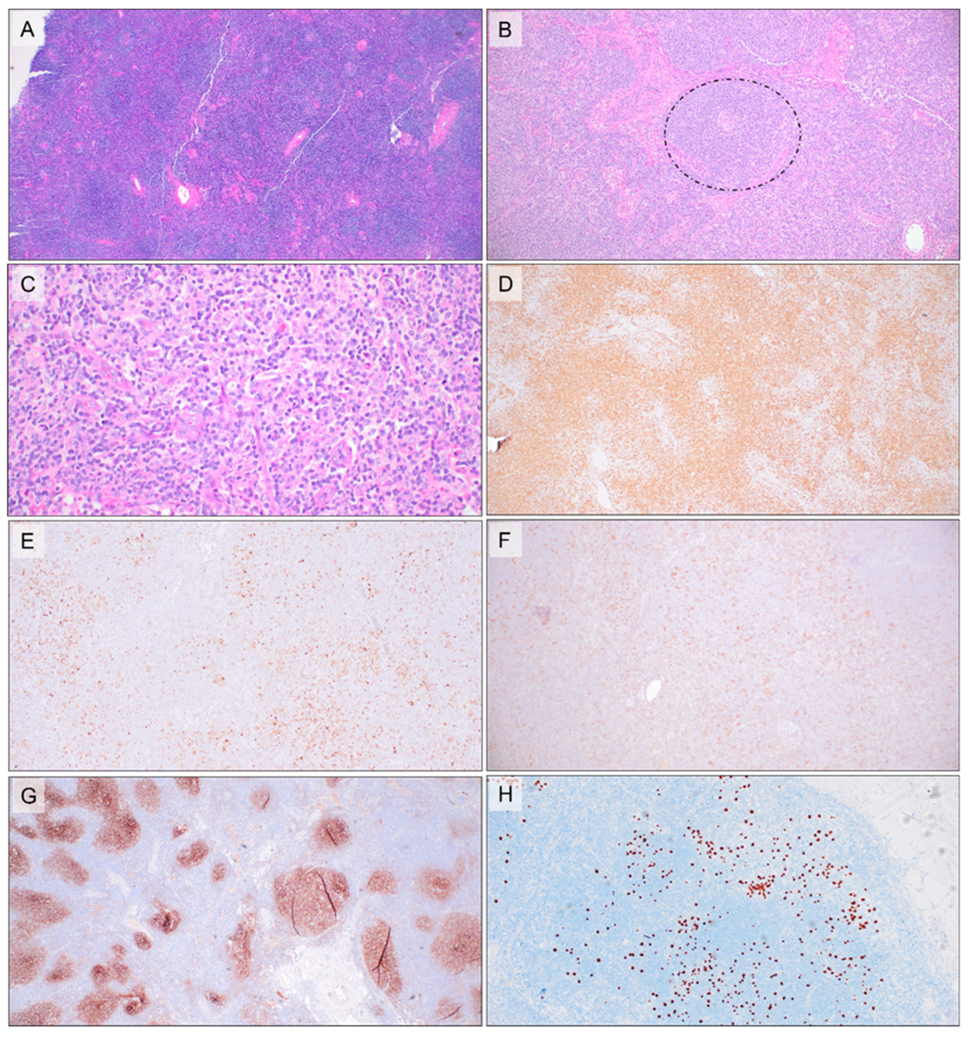
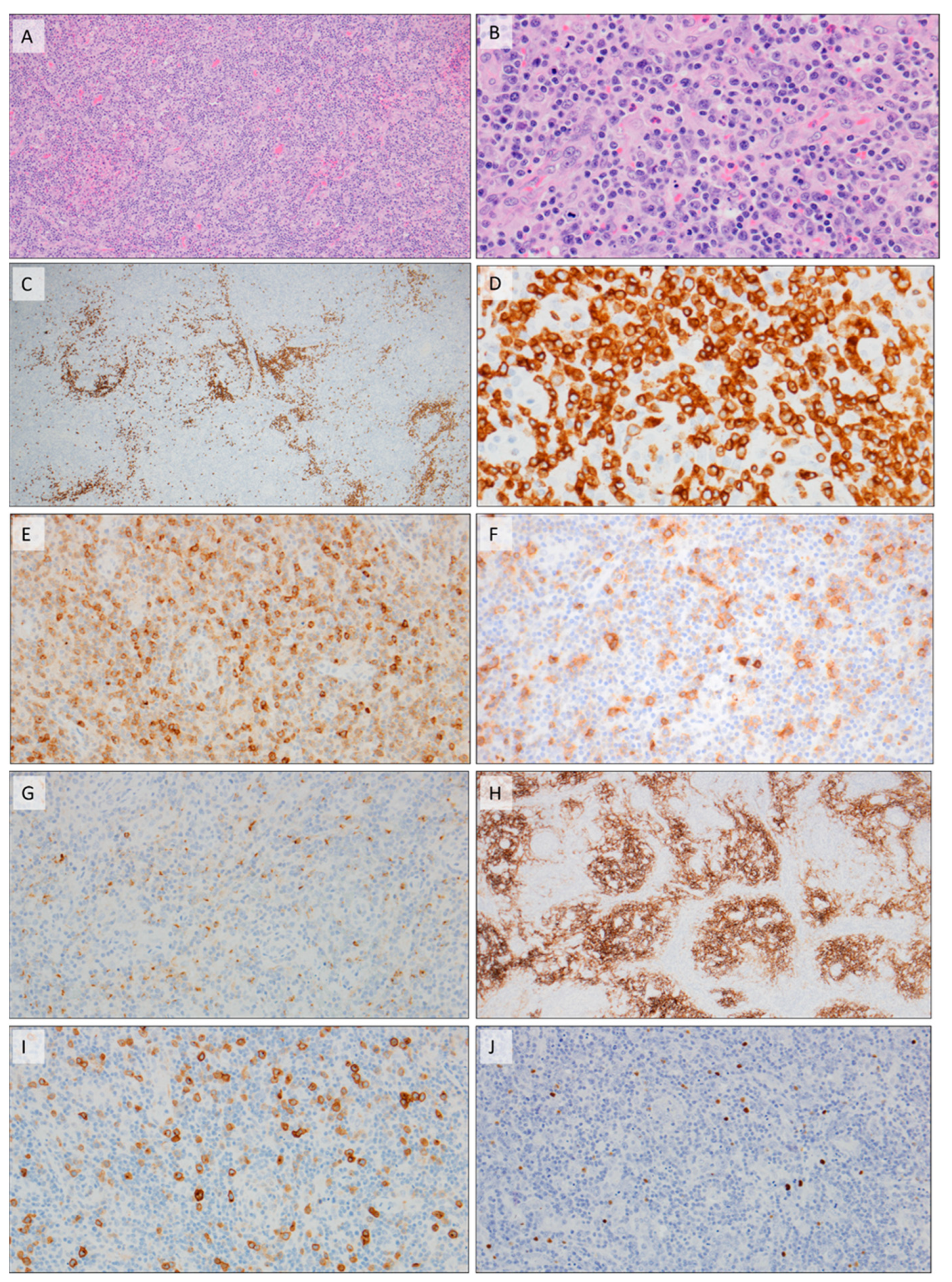
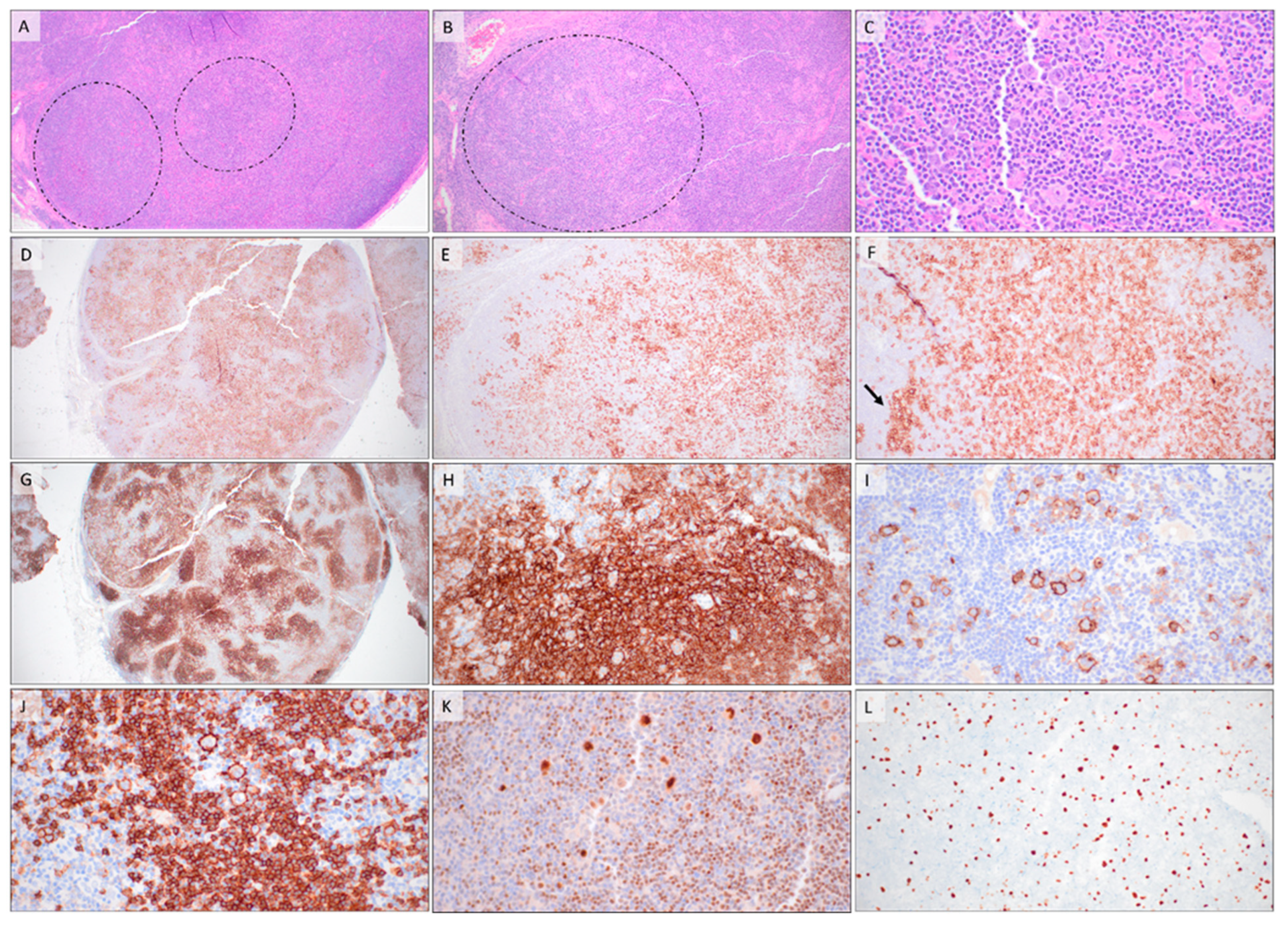
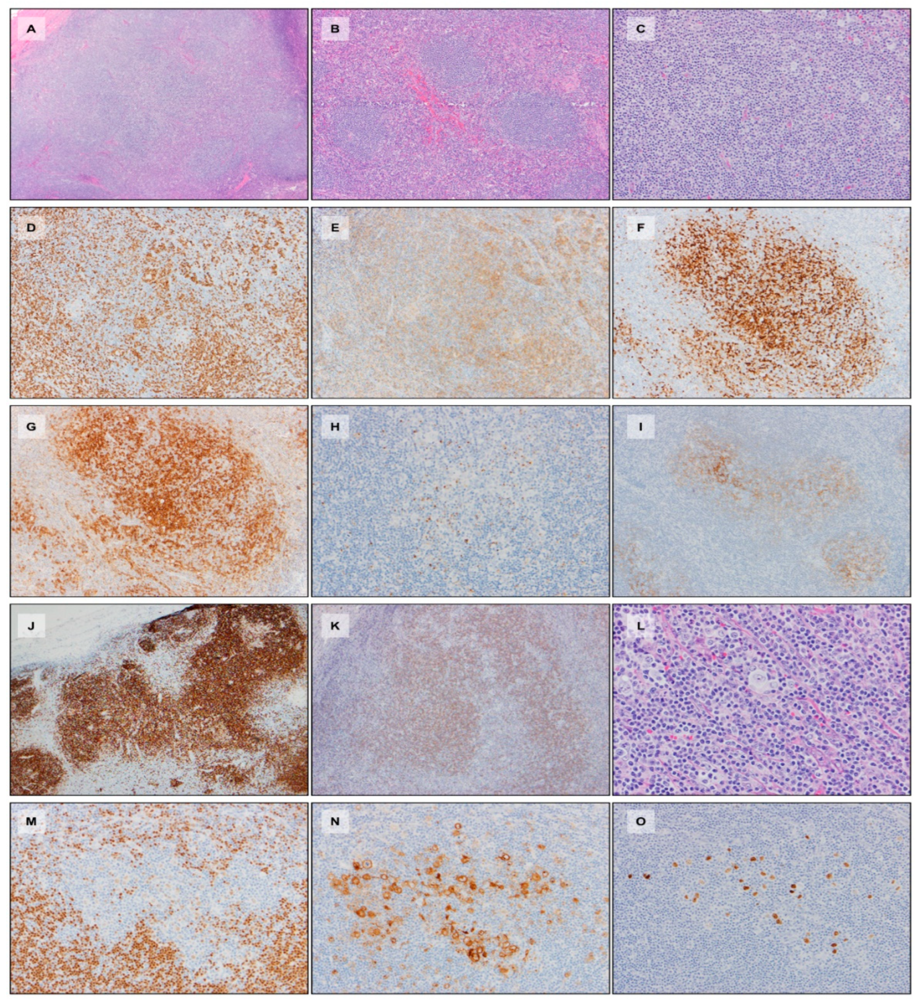
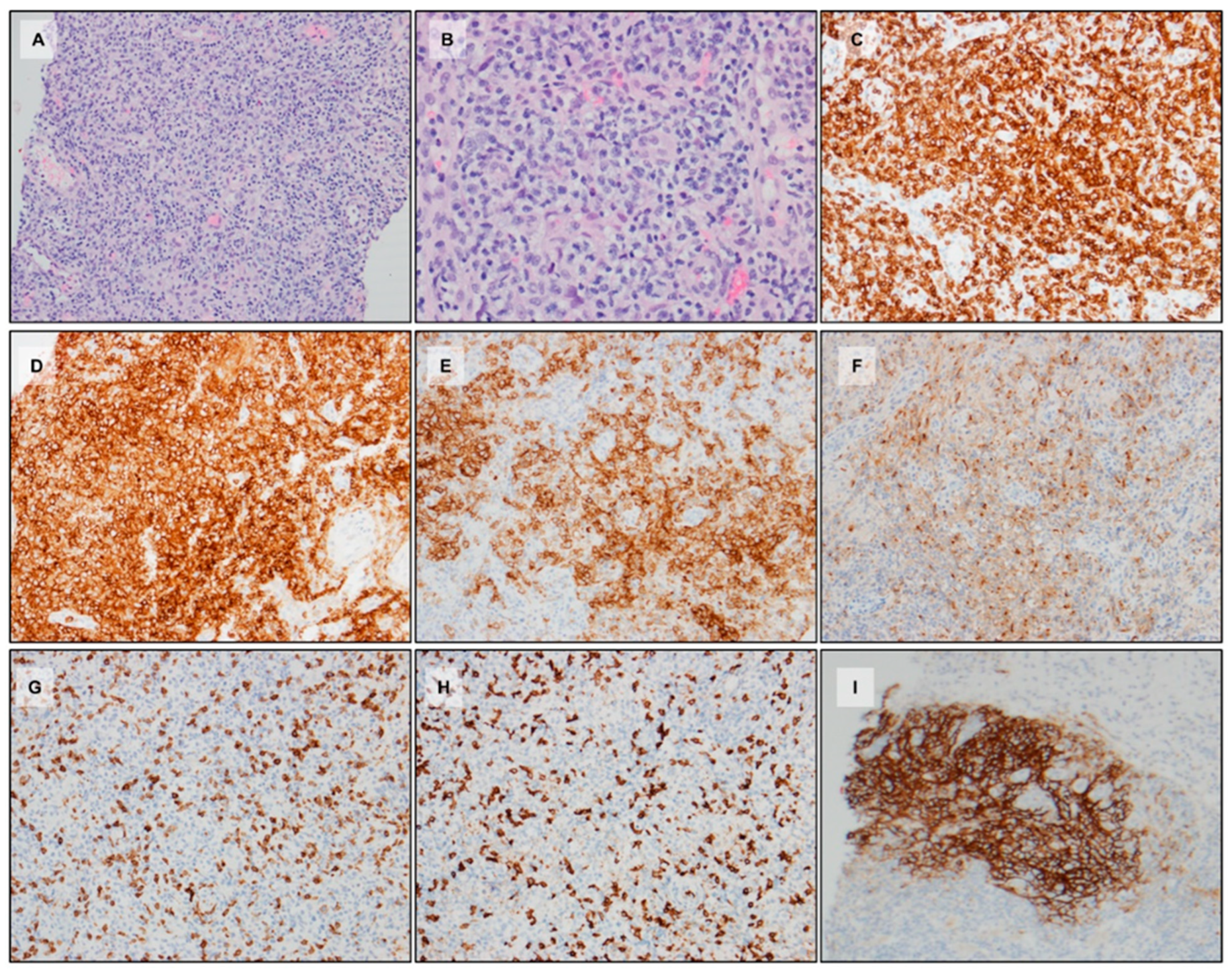
2.2. Morphology and Immunophenotype

Lymph node biopsies are the most common specimens in routine practice that pathologists encounter for AITL diagnosis. An excisional biopsy is the ideal specimen, and diagnoses on small core biopsies might be challenging because of a lack of an architectural context and a paucity of tissue for ancillary tests. A diagnosis of AITL can be especially challenging in extranodal site biopsies.
Lymph node excisional biopsies generally demonstrate effacement of the architecture by a polymorphous infiltrate composed of small, atypical lymphocytes (usually with a clear cytoplasm), plasma cells, macrophages, and scattered larger cells, which appear either immunoblastic or sometimes Hodgkin’s-/Reed–Sternberg cell (HRS)-like. The atypical lymphocytes are clustered especially around proliferating high endothelial venules, which are rather prominent in the biopsies. The subcapsular sinuses tend to be distended. Biopsies can also demonstrate tissue eosinophilia.
Three major patterns have been described in the context of AITL; these are sequentially named Patterns 1, 2, and 3 and are believed to be the histologic progression of the disease (Table 2). A high proportion (up to 17%) of patients present with Pattern 1 [21,42,43,44], which is especially challenging to diagnose because of retention of the architecture, presence of hyperplastic germinal centers, and lack of overt proliferation of high endothelial venules or polymorphous infiltration. The clues to Pattern 1 diagnosis are the absent/attenuated mantle zones surrounded by clear atypical cells, the PD1 staining pattern (circumferential perifollicular PD1 staining TFH cells as opposed to TFH cells either scattered throughout the germinal center or polarized appearing in reactive lymph nodes), and the slight branching of FDC meshworks outside of the intact germinal centers (Table 1 and Figure 1A,D,G). In addition, closer observation might identify focal expansion of the paracortex and a slight increase in HEVs. Any suspicion of a T-cell lymphoproliferative disorder should trigger a molecular or genetic analysis (see below).
Pattern 2 demonstrates partial effacement/partial retention of the architecture (Figure 1B and Figure 2A) with paracortical expansion by a polymorphous infiltrate with neoplastic clear TFH cells, small lymphocytes, plasma cells, macrophages, eosinophils, scattered immunoblasts, and Hodgkin’s-/Reed-Sternberg-like cells (Figure 1B and Figure 2C). Atretic Castleman-like follicles are also seen (Figure 1B and Figure 2A,B). The neoplastic TFH cells are more pronounced and start spilling out of the follicles into the paracortex (Figure 1B) and encircle the HEVs; this is particularly evident with CD3 (Figure 2D) and TFH marker stains, e.g., PD1 stain (Figure 1E and Figure 2E), CD10 (Figure 2E), and CXCL13 (Figure 2F). CD21 demonstrates partial expansion and distortion of FDC meshworks along with some areas, demonstrating retention of the architecture (Figure 1H and Figure 2G). EBER-ish demonstrates scattered EBV+ cells, demonstrating a size range (Figure 2H).
Pattern 3 is the most recognizable pattern of AITL. Lymph nodes demonstrate complete effacement of the architecture (Figure 1C and Figure 3A) by a polymorphous infiltrate (Figure 1C and Figure 3B) composed of mostly atypical clear-appearing neoplastic TFH cells, macrophages, plasma cells, eosinophils, and numerous larger cells, including Hodgkin’s-like cells, representing immunoblasts (Figure 1C and Figure 3B). The neoplastic TFH cells express T-cell markers such as CD3 (Figure 3D) and TFH markers such as PD1 (Figure 1F and Figure 3E) CD10 (Figure 3F), and CXCL13 (Figure 3G). CD21 demonstrates marked expansion and distortion of FDC meshworks (Figure 1I and Figure 3H). CD30 is positive not only in the large HRS-like cells, but also in a subset of the smaller neoplastic TFH cells (Figure 3I). EBER-ish demonstrates scattered EBV-positive cells (Figure 3J).
Immunohistochemical studies are crucial to the diagnosis of AITL, which hinges on the demonstration of a TFH phenotype. An extensive T-cell panel, i.e., CD2, CD3, CD4, CD8, CD5, and CD7, should be used to evaluate immunophenotypic aberrancies. Aberrancies (dimness or loss) of CD3, CD5, and CD7 are most common. The vast majority of the neoplastic TFH cells are CD4-positive. As discussed above, TFH cells express several markers, i.e., CD10, BCL6, PD1, CXCL13, CXCR5, ICOS, SAP, CD200, and MAF, and the recommendations for assigning a TFH phenotype include expression of at least two, ideally three TFH markers [1,6,7,8,12,13,18,21,22,23,24]. Studies have also shown that, while in most cases, a four-marker panel (CD10, PD-1, CXCL13, and BCL6) is adequate, employing ICOS as an additional marker (creating a five-marker panel) greatly improves TFH phenotype detection [25]. An additional consideration is the intensity of these markers, especially PD1 and ICOS, which need to be bright to qualify as TFH markers. In addition, CD10 expression is usually variable and, in most cases, is expressed in a subset of the neoplastic TFH cells. Hence, CD10 needs to be evaluated carefully. Overall, CXCL13 and CD10 are considered most specific, while PD1 and ICOS are considered more sensitive [18]. CD21, CD23, or CD35 can be used to evaluate the FDC meshworks.
An important point worth mentioning is that AITL can relapse with a follicular T-cell lymphoma (FTCL) morphology (discussed below) and vice versa [15]. Figure 4 illustrates one such case of a patient with a previous history of AITL with a subsequent biopsy demonstrating FTCL.

2.3. EBV, B-Cell, and Plasma Cell Proliferations
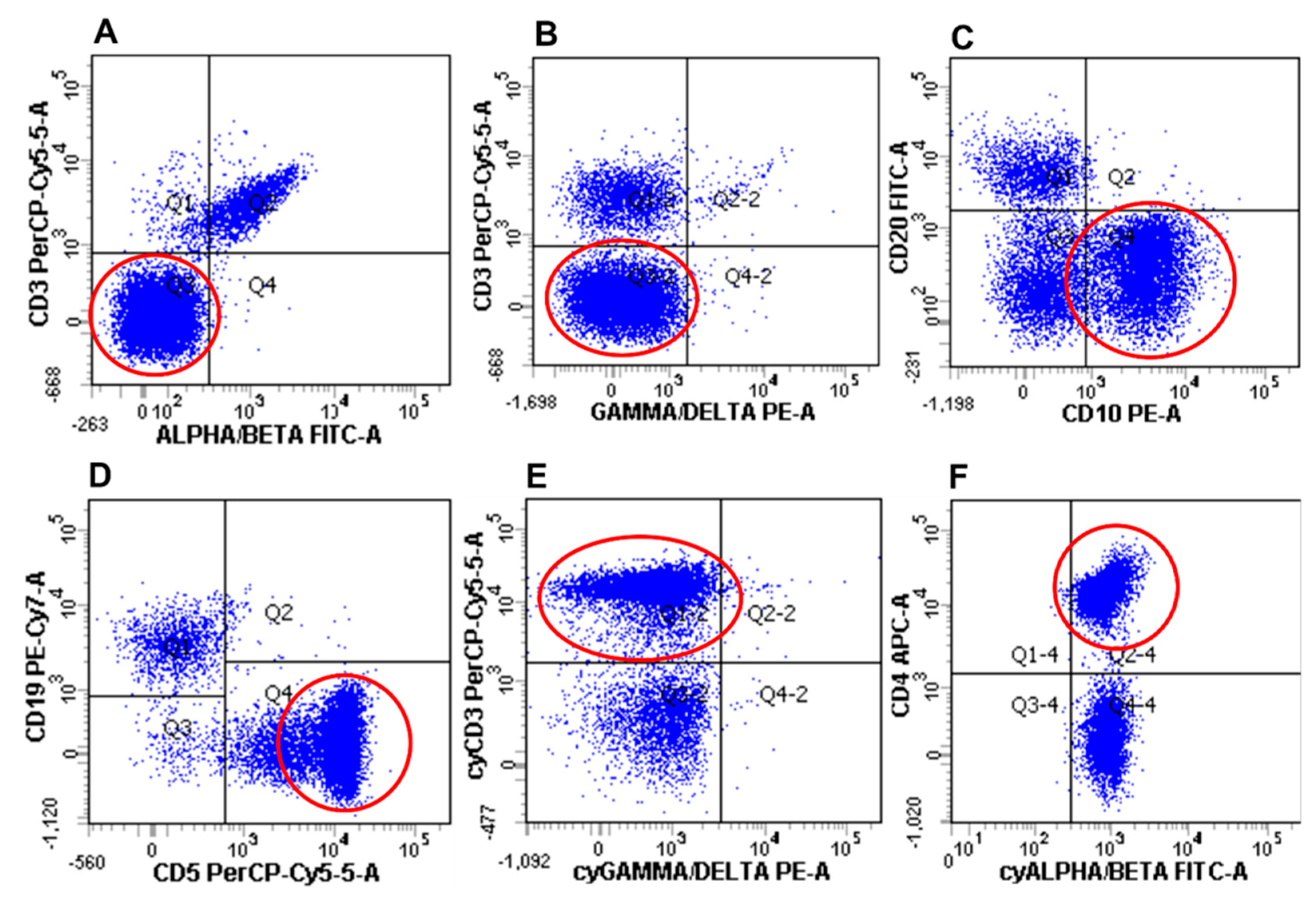
B-cell and plasma cell proliferations (clonal and non-clonal) are a frequent occurrence in AITL [31]. The B-cells can appear to be either immunoblastic or HRS-like. While EBV is associated with the majority of B-cell proliferations in AITLs (more than 80%) [31,40,45], EBV-negative cases have also been reported [46]. In most cases, EBV-positive B-cells are scattered or form small clusters; however, in some cases, sheets of large B-cells are seen, which would prompt the consideration of a second diagnosis of a composite diffuse large B-cell lymphoma [45,47]. Plasmacytosis and plasma cell proliferations are a frequent occurrence in AITL, and most of these are EBV-negative. While most are polyclonal, occasional clonal plasma cell proliferations have also been reported [48,49].

2.4. Flow Cytometry

Flow cytometric studies can be a powerful ancillary tool in the diagnosis of AITL [20,50,51,52,53]. Neoplastic T-cells in AITL frequently demonstrate the downregulation or the absence of surface CD3 along with absent or diminished surface T-cell receptors (Figure 5A,B) [53]. However, in these cases, they generally express cytoplasmic CD3 and cytoplasmic TCR-alpha/beta (Figure 5E,F). In addition, most AITL patients (as opposed to PTCL patients) also seem to demonstrate this atypical surface CD3 dim/neg population to varying extents in the peripheral blood [50,52,53]. A simple T-cell panel employing CD3, CD4, CD10, CD14, CD5, and CD45 to evaluate for surface CD3 dim/neg, CD10+, CD4+ population has been suggested to aid in AITL diagnosis, especially in bone marrow and peripheral blood [20]. Bright PD1 expression by flow cytometry has also been shown to be a useful tool for both the diagnosis and monitoring of AITL [54]. Flow cytometry studies might also demonstrate additional immunophenotypic abnormalities described in AITL, e.g., loss of CD5 or CD7.

2.5. Genetics

Most AITLs demonstrate clonal T-cell receptor rearrangements by PCR; in addition, they also demonstrate clonal immunoglobulin gene rearrangements in 25–30% of cases. The latter usually correspond to EBV-positive B-cell proliferations in most cases [18].
The cytogenetics and mutational spectrum of AITL, as well as the association with clonal hematopoiesis are discussed in detail in a separate section (see below).

2.6. Differential Diagnoses

While other mature T-cell lymphomas including peripheral T-cell lymphoma NOS should be considered in the differential diagnosis, the distinction is perhaps less critical from a treatment standpoint. As mentioned above, the diagnosis of AITL hinges on the demonstration of at least two (ideally three) TFH antigens, as well as the presence of other characteristics’ findings such as a polymorphous infiltrate, aggregates of TFH cells around proliferating HEVs, and scattered EBV+ cells.
Perhaps one of the most challenging differential diagnoses of Pattern 1 AITL is reactive lymphoid hyperplasia [21,42,43,44]. Pattern 1 AITL can often be retrospectively diagnosed in a previous biopsy from a patient with a subsequent Pattern 3 AITL. The main morphologic clues in favor of a Pattern 1 AITL include absent or attenuated mantle zones, the presence of clear atypical cells surrounding the hyperplastic germinal centers (this might not be obvious), focal expansion of the paracortex, and focal increase in HEVs. This should prompt further staining with PD1, which would demonstrate the circumferential grouping of the neoplastic TFH cells and few clusters in the paracortex. CD21 would demonstrate slight branching out of the FDC meshwork. If the findings are suspicious for Pattern 1 AITL, then PCR for T-cell receptor rearrangement and next-generation sequencing studies (NGS) for IDH1, IDH2, DNMT3A, TET2, and RHOA (see below) should be ideally performed. In addition, as discussed above, flow cytometry might demonstrate a CD3dim/neg, CD4+, CD10+ population that is highly suggestive of AITL; if flow cytometry is not available on tissue, it could be attempted on peripheral blood [20,52,53]. Interestingly, several patients present at a clinically advanced stage with a Pattern 1 morphology on a biopsy; this perhaps reflects the morphologic evolution of the disease with the distinct possibility that other lymph nodes in the body might have a more evolved Pattern 3 morphology [42].
Classic Hodgkin lymphoma (CHL) can be a challenging differential diagnosis especially considering the similar polymorphous background and the presence of Hodgkin’s-/Reed–Sternberg (HRS)-like cells, which is common in AITL. Adding to the difficulty is the observation that these large cells are usually CD30-positive and occasionally CD15-positive [46,55]. However, the HRS-like cells in AITL generally tend to preserve the B-cell program more often, and an extensive panel of B-cell markers, i.e., CD20, PAX5, OCT2, BOB1, and CD79a, should be employed (albeit that CD20 and PAX5 downregulation can be seen in HRS-like cells in AITL). In addition, an extensive T-cell IHC panel (CD3, CD2, CD5, CD4, CD8, CD7) should be used in challenging cases to evaluate for immunophenotypic aberrancies in T-cells. Fibrosis is unusual in AITL and is more common in CHL. Cytologic atypia in the surrounding T-cells, proliferating HEVs, FDC expansion beyond follicles, morphologic variation, pleomorphism in the larger cells, etc., would favor AITL. In addition, sheets and aggregates of PD1-positive cells beyond rosetting of HRS-like cells should raise suspicion for AITL and prompt further workup including molecular studies (PCR and NGS; see above). In addition, EBV positivity is usually restricted to the large HRS cells in CHL, while there usually is more variation in the cell size of EBV+ cells in AITL. CD30 staining in smaller lymphocytes, in addition to the larger HRS-like cells, should also prompt consideration of AITL [56]. As mentioned above, flow cytometric detection of a CD3 dim/neg, CD4+, CD10+ population can be particularly helpful in the diagnosis of AITL and other TFH lymphomas, as this population is not seen in CHL [20,50,51,52,53].

3. Follicular T-Cell Lymphoma

3.1. Epidemiology and Clinical Features

Follicular T-cell lymphoma (FTCL) was first described in 1988 [57] and is a rare entity, representing 1–2% of peripheral T-cell lymphomas. It is a disease of older individuals occurring in the sixth decade of life and slightly more common in males. Patients usually present with diffuse lymphadenopathy, and high-stage disease with extranodal and cutaneous involvement has been reported. Rare patients may show immunological abnormalities on laboratory investigations, including positive direct antibody tests (DATs) and hypergammaglobulinemia [15].

3.2. Morphology and Immunophenotype

By morphology, two distinct growth patterns are recognized: a follicular growth pattern that mimics B-cell follicular lymphoma (FL-like) and a progressive transformation of a germinal centers-like pattern (PTGC-like) (Figure 6) [18]. In cases with the FL-like pattern, the neoplastic T-cells are arranged in well-defined nodules that lack the morphological features of normal follicles. Mantle zones can either be preserved or absent. In PTGC-like cases, representing the majority of FTCL, the neoplastic T-cells are seen in small aggregates surrounded by small mantle zone B-cells arranged in large, irregular nodules. Eosinophilic infiltration is uncommon, and unlike in AITL, arborizing high endothelial venules and expanded follicular dendritic cell meshworks are not identified. The neoplastic T-cells range from small to medium with irregular nuclei, coarse to vesicular chromatin, and moderate to abundant pale cytoplasm. While the nuclear features can resemble centrocytes or centroblasts, the pale cytoplasm is a useful distinguishing feature. Scattered B-cell immunoblasts, including some resembling Hodgkin’s/Reed–Sternberg cells, usually surrounded by neoplastic T-cells, are seen in a significant subset of cases.
The neoplastic T-cells in FTCL express pan T-cell antigens including CD2, CD3, CD5, and CD7. An aberrant immunophenotype is seen in the majority of cases with the loss of CD7 most commonly noted [58]. They are positive for CD4 and express multiple TFH markers, with PD1 and ICOS reportedly the most sensitive, followed by CXCL13. CD10 and BCL6 are less sensitive and are expressed in fewer cases. They are negative for CD8, CD56, cytotoxic markers, and EBV. Follicular dendritic cell meshworks are limited to the nodular areas. The proliferative rate, assessed by Ki-67, is variable, with mean values approaching 50%. The transformed immunoblasts are positive for B-cell markers including CD20 and PAX5. CD30 and EBV can be expressed in a subset of cases [58].

3.3. Molecular Studies

Clonal T-cell receptor gene rearrangements are identified in the vast majority of cases; additionally, a subset of cases may show oligoclonal or clonal IgH gene rearrangements [58].

3.4. Differential Diagnoses

While it is rare, a diagnosis of FTCL should not be overlooked when evaluating a lymph node for involvement by lymphoma. FTCL with an FL-like growth pattern can mimic FL, but can be distinguished from FL by the cytologic features of the neoplastic T-cells and the expression of multiple T-cell markers by the abnormal follicles. The PTGC-like variant raises the differential diagnosis of classic Hodgkin’s lymphoma, the lymphocyte-rich variant (CHL-LR), or nodular-lymphocyte-predominant Hodgkin’s lymphoma (NLPHL). In cases resembling CHL or NLPHL, careful examination of the T-cells rosetting the H/RS-like cells or LP cells is mandatory, as in most FTCL cases, these T-cells are neoplastic and express multiple TFH markers, most commonly PD1, ICOS, and CD10 [59,60]. A subset of these neoplastic cells can also express CD30, which can help distinguishing them from reactive T-cells [56].

4. Peripheral T-Cell Lymphoma with TFH Phenotype

Multiple studies have shown that a significant subset of peripheral T-cell lymphomas, morphologically classified as peripheral T-cell lymphoma, not otherwise specified (PTCL, NOS), arise from TFH cells and share genomic features, but are not identical to AITL [16,28,61,62]. This relationship has also been confirmed by the evaluation of TFH marker expression in these lymphomas [14,25,63]. In recognition of these findings and to aid in better classification and therapy, the WHO 2017 Classification recommends that nodal CD4+ T-cell lymphomas be evaluated for the expression of TFH antigens and provisionally classified as peripheral T-cell lymphoma with the TFH phenotype (PTCL-TFH) if they express at least two (ideally three) TFH antigens [18]. The rates of classification as PTCL-TFH appear significantly different when using two or three TFH markers, and large-scale studies are required to help develop stringent criteria for reproducible classification [25].
Morphologically, PTCL-TFH shows diffuse effacement of the nodal architecture by neoplastic T-cells that can range in size from small to large (Figure 7). Unlike AITL, PTCL-TFH lacks the clear cell morphology, polymorphous inflammatory background, and expanded dendritic cell meshworks with high endothelial venule proliferation [14,25] (Figure 4).
PTCL-T TFH shares immunophenotypic features with AITL and FTCL with variable expression of TFH markers. PD1 and ICOS remain the most sensitive, while CXCL13, BCL6, and CD10 are expressed in fewer cases [25].

5. G. Genetics of TFH Lymphomas

T-cell lymphomas of T follicular helper origin show some distinct differences from other peripheral T-cell lymphomas. Angioimmunoblastic T-cell lymphomas have a distinct gene expression profile relating to the unique microenvironment that includes a B-cell signature, angiogenesis/vascular endothelial signature, cytokine signature, and T follicular helper signature [16,29,64]. While PTCL-TFH also shares the TFH signature, it lacks the characteristic microenvironment signature of AITL. Nevertheless, the gene expression profiles of AITL, FTCL, and PTCL-TFH are similar and distinct enough to support the argument that they likely represent a continuum of a single disease [16]. Rodriguez also demonstrated that AITL, but not PTCL-TFH cases that were enriched for the B-cell signature had a more favorable prognosis [61].
Among the genetic mutations identified in T-cell lymphomas, IDH2 R172 mutations seem to be specific for AITL and have only rarely been reported in other PTCL-NOS, while they have been reported in 20–40% of AITL cases [16,29,64,65]. In one study, this mutation was significantly associated with chromosome 5 gains and upregulation of IL4, IL13, and MAPK9 [66].
TET2 mutations and, less frequently, DNMT3A mutations are also enriched in TFH lymphomas (TET2 mutations are seen in approximately 50–75% of cases). Interestingly, these mutations are not only seen in tumor cells, but are present in non-tumor hematopoietic cells as well, suggesting that these lymphomas may have a pre-malignant mutant clone (clonal hematopoiesis of indeterminate potential), much as what has been extensively described and investigated in myeloid malignancies such as myelodysplastic syndrome [16,64]. TET2 mutations have been associated with advanced-stage disease, high IPI scores, and shorter progression-free survival [67].
RHOA mutations, almost always at the G17V hotspot, are frequently identified in AITL and FTCL (60–70%) and essentially always co-occur with TET2 mutations [61,64,68]. While the TET2 mutation is found in the non-tumor hematopoietic cells, the RHOA mutation appears restricted to the tumor cells and likely occurs as a later event. The combination of the two mutations may contribute to AITL-specific pathogenesis. RHOA T19I mutations, albeit less common, have also been reported [69].
Genotype/phenotype correlations have also been described, with AITL carrying RHOA G17V mutations showing increased vasculature and prominent follicular dendritic cell meshworks [70], while those with IDH2 R172 mutations having more prominent large, clear cells and strong TFH marker expression [71].
The cytogenetic profile of AITL is considerably less abnormal than other PTCLs [66]. Chromosome 5 gains are seen in about 40% of AITL and are distinct among hematologic malignancies, which more typically show losses in chromosome 5. Cases that do not have chromosome 5 gains show enrichment of NF-KB and PI3K-AKT pathways and may have a distinct pathogenesis [66]. Additionally, ~20% of FTCL carry a recurrent translocation t(5;9)(q33;q22)(ITK-SYK). This rearrangement is not specific for FTCL and has been described in other peripheral T-cell lymphomas, including AITL [72,73].

6. TFH Lymphomas, Clonal Hematopoiesis of Indeterminate Potential, and Myeloid Neoplasms

TET2 and DNMT3A mutations are frequently noted in clonal hematopoiesis of indeterminate potential (CHIP) [74,75] and myeloid malignancies [76,77,78]. The subsequent discovery of TET2 and DNMT3A mutations in TFH lymphomas has prompted the investigation of the relationship between these seemingly disparate categories of hematologic malignancies. Interestingly, studies have shown that TFH lymphomas can arise from divergent clonal evolution from TET2 and DNMT3A-mutant progenitor cells upon acquisition of RHO and/or IDH2 mutations [79]. Supporting this hypothesis are reports of TFH lymphomas and myeloid malignancies (both de novo and therapy-related) co-occurring in patients and arising from TET2 and DNMT3A-mutated clonal progenitor cells [80,81]. Furthermore, the presence of two or more TET2 mutations and a mutant allele fraction > 15% appears to confer a higher risk for myeloid neoplasms in patients with TFH lymphomas [81,82]. These studies have elucidated the unique biology of TFH lymphomas and highlight the importance of the evaluation for clonal hematopoiesis in these patients, which can have important diagnostic and therapeutic implications.

7. Treatment and Conclusions

TFH lymphomas are an important subtype of peripheral T-cell lymphomas, and the last decade has seen significant advances in understanding their biology and molecular pathogenesis. The identification of shared recurrent mutations in AITL, FTCL, and PTCL-TFH has justified classifying these morphologically unique lymphomas as a single biological category. These discoveries have also provided a wealth of biomarkers for diagnosis and targets for therapy [83]. Currently, the distinction between TFH lymphomas and PTCL, NOS, has no impact on current clinical management, with most patients treated with cyclophosphamide, doxorubicin, vincristine, and prednisone (CHOP) or CHOP-like upfront therapy, but with less-than-optimal response rates. Recent studies have shown improved progression-free survival (PFS) and overall survival (OS) in patients with systemic CD30-positive T-cell lymphomas treated with CHOP-like chemotherapy regimens that replace vincristine with brentuximab vedotin, an anti-CD30 antibody drug conjugate. The majority of these patients had anaplastic large cell lymphoma (ALCL), which is characterized by diffuse and strong CD30 expression. Patients with other T-cell lymphoma subtypes expressing CD30, including AITL, were included; however, due to small sample sizes, efficacy in the non-ALCL subgroups could not be evaluated [84]. Studies evaluating the efficacy of romidepsin, a histone deacetylase inhibitor, in the treatment of TFH lymphomas have shown interesting results. Romidepsin in addition to CHOP showed marginal, but not significant beneficial effects when compared to CHOP in previously untreated patients with TFH lymphoma [85]; however, romidepsin, either alone or in combination with other drugs, had significant benefits in patients with relapsed/refractory TFH lymphoma [86]. These incongruous results warrant further investigation with larger patient cohorts. Finally, the identification of TET2, DNMT3A, and IDH mutations in TFH lymphomas has created opportunities for targeted therapy with hypomethylating agents, such as azacytidine, in combination with current therapy regimens with promising results [87,88].

Author Contributions

Conceptualization, M.P.M.; writing—original draft preparation, K.A.G., K.H.K. and M.P.M.; writing—review and editing, K.A.G. and M.P.M.; visualization, K.A.G. and M.P.M.; supervision, M.P.M. All authors have read and agreed to the published version of the manuscript.

Funding

This research received no external funding.

Institutional Review Board Statement

Not applicable.

Informed Consent Statement

Not applicable.

Data Availability Statement

Not applicable.

Conflicts of Interest

The authors declare no conflict of interest.

References

  1. Crotty, S. T follicular helper cell differentiation, function, and roles in disease. Immunity 2014, 41, 529–542. [Google Scholar] [CrossRef] [PubMed] [Green Version]
  2. Crotty, S. A brief history of t cell help to b cells. Nat. Rev. Immunol. 2015, 15, 185–189. [Google Scholar] [CrossRef] [PubMed]
  3. Goenka, R.; Barnett, L.G.; Silver, J.S.; O’Neill, P.J.; Hunter, C.A.; Cancro, M.P.; Laufer, T.M. Cutting edge: Dendritic cell-restricted antigen presentation initiates the follicular helper t cell program but cannot complete ultimate effector differentiation. J. Immunol. 2011, 187, 1091–1095. [Google Scholar] [CrossRef] [PubMed] [Green Version]
  4. Crotty, S. Follicular helper cd4 t cells (tfh). Annu. Rev. Immunol. 2011, 29, 621–663. [Google Scholar] [CrossRef]
  5. Crotty, S.; Johnston, R.J.; Schoenberger, S.P. Effectors and memories: Bcl-6 and blimp-1 in t and b lymphocyte differentiation. Nat. Immunol. 2010, 11, 114–120. [Google Scholar] [CrossRef]
  6. Crotty, S. T follicular helper cell biology: A decade of discovery and diseases. Immunity 2019, 50, 1132–1148. [Google Scholar] [CrossRef]
  7. Suan, D.; Nguyen, A.; Moran, I.; Bourne, K.; Hermes, J.R.; Arshi, M.; Hampton, H.R.; Tomura, M.; Miwa, Y.; Kelleher, A.D.; et al. T follicular helper cells have distinct modes of migration and molecular signatures in naive and memory immune responses. Immunity 2015, 42, 704–718. [Google Scholar] [CrossRef] [Green Version]
  8. Yu, D.; Vinuesa, C.G. The elusive identity of t follicular helper cells. Trends Immunol. 2010, 31, 377–383. [Google Scholar] [CrossRef]
  9. Cubas, R.A.; Mudd, J.C.; Savoye, A.L.; Perreau, M.; van Grevenynghe, J.; Metcalf, T.; Connick, E.; Meditz, A.; Freeman, G.J.; Abesada-Terk, G., Jr.; et al. Inadequate t follicular cell help impairs b cell immunity during hiv infection. Nat. Med. 2013, 19, 494–499. [Google Scholar] [CrossRef] [Green Version]
  10. Simpson, N.; Gatenby, P.A.; Wilson, A.; Malik, S.; Fulcher, D.A.; Tangye, S.G.; Manku, H.; Vyse, T.J.; Roncador, G.; Huttley, G.A.; et al. Expansion of circulating t cells resembling follicular helper t cells is a fixed phenotype that identifies a subset of severe systemic lupus erythematosus. Arthritis Rheumatol. 2010, 62, 234–244. [Google Scholar] [CrossRef]
  11. Gu-Trantien, C.; Loi, S.; Garaud, S.; Equeter, C.; Libin, M.; de Wind, A.; Ravoet, M.; Le Buanec, H.; Sibille, C.; Manfouo-Foutsop, G.; et al. Cd4(+) follicular helper t cell infiltration predicts breast cancer survival. J. Clin. Investig. 2013, 123, 2873–2892. [Google Scholar] [CrossRef] [PubMed]
  12. Grogg, K.L.; Attygalle, A.D.; Macon, W.R.; Remstein, E.D.; Kurtin, P.J.; Dogan, A. Angioimmunoblastic t-cell lymphoma: A neoplasm of germinal-center t-helper cells? Blood 2005, 106, 1501–1502. [Google Scholar] [CrossRef] [PubMed]
  13. De Leval, L.; Rickman, D.S.; Thielen, C.; Reynies, A.; Huang, Y.L.; Delsol, G.; Lamant, L.; Leroy, K.; Briere, J.; Molina, T.; et al. The gene expression profile of nodal peripheral t-cell lymphoma demonstrates a molecular link between angioimmunoblastic t-cell lymphoma (aitl) and follicular helper t (tfh) cells. Blood 2007, 109, 4952–4963. [Google Scholar] [CrossRef] [PubMed]
  14. Rodriguez-Pinilla, S.M.; Atienza, L.; Murillo, C.; Perez-Rodriguez, A.; Montes-Moreno, S.; Roncador, G.; Perez-Seoane, C.; Dominguez, P.; Camacho, F.I.; Piris, M.A. Peripheral t-cell lymphoma with follicular t-cell markers. Am. J. Surg. Pathol. 2008, 32, 1787–1799. [Google Scholar] [CrossRef]
  15. Huang, Y.; Moreau, A.; Dupuis, J.; Streubel, B.; Petit, B.; Le Gouill, S.; Martin-Garcia, N.; Copie-Bergman, C.; Gaillard, F.; Qubaja, M.; et al. Peripheral t-cell lymphomas with a follicular growth pattern are derived from follicular helper t cells (tfh) and may show overlapping features with angioimmunoblastic t-cell lymphomas. Am. J. Surg. Pathol. 2009, 33, 682–690. [Google Scholar] [CrossRef] [PubMed]
  16. Dobay, M.P.; Lemonnier, F.; Missiaglia, E.; Bastard, C.; Vallois, D.; Jais, J.P.; Scourzic, L.; Dupuy, A.; Fataccioli, V.; Pujals, A.; et al. Integrative clinicopathological and molecular analyses of angioimmunoblastic t-cell lymphoma and other nodal lymphomas of follicular helper t-cell origin. Haematologica 2017, 102, e148–e151. [Google Scholar] [CrossRef] [Green Version]
  17. Beltraminelli, H.; Leinweber, B.; Kerl, H.; Cerroni, L. Primary cutaneous cd4+ small-/medium-sized pleomorphic t-cell lymphoma: A cutaneous nodular proliferation of pleomorphic t lymphocytes of undetermined significance? A study of 136 cases. Am. J. Dermatopathol. 2009, 31, 317–322. [Google Scholar] [CrossRef]
  18. Swerdlow, S.H.; Campo, E.; Harris, N.L.; Jaffe, E.S.; Pileri, S.A.; Stein, H.; Thiele, J.; Arber, D.A.; Hasserjian, R.P.; Le Beau, M.M.; et al. Who Classification of Tumours of Haematopoietic and Lymphoid Tissues (Revised 4th Edition); International Agency for Research on Cancer: Lyon, France, 2017. [Google Scholar]
  19. Beltzung, F.; Ortonne, N.; Pelletier, L.; Beylot-Barry, M.; Ingen-Housz-Oro, S.; Franck, F.; Pereira, B.; Godfraind, C.; Delfau, M.H.; D’Incan, M.; et al. Primary cutaneous cd4+ small/medium t-cell lymphoproliferative disorders: A clinical, pathologic, and molecular study of 60 cases presenting with a single lesion: A multicenter study of the french cutaneous lymphoma study group. Am. J. Surg. Pathol. 2020, 44, 862–872. [Google Scholar] [CrossRef]
  20. Alikhan, M.; Song, J.Y.; Sohani, A.R.; Moroch, J.; Plonquet, A.; Duffield, A.S.; Borowitz, M.J.; Jiang, L.; Bueso-Ramos, C.; Inamdar, K.; et al. Peripheral t-cell lymphomas of follicular helper t-cell type frequently display an aberrant cd3(-/dim)cd4(+) population by flow cytometry: An important clue to the diagnosis of a hodgkin lymphoma mimic. Mod. Pathol. 2016, 29, 1173–1182. [Google Scholar] [CrossRef]
  21. Attygalle, A.; Al-Jehani, R.; Diss, T.C.; Munson, P.; Liu, H.; Du, M.Q.; Isaacson, P.G.; Dogan, A. Neoplastic t cells in angioimmunoblastic t-cell lymphoma express cd10. Blood 2002, 99, 627–633. [Google Scholar] [CrossRef] [Green Version]
  22. Bisig, B.; Thielen, C.; Herens, C.; Gofflot, S.; Travert, M.; Delfau-Larue, M.H.; Boniver, J.; Gaulard, P.; de Leval, L. C-maf expression in angioimmunoblastic t-cell lymphoma reflects follicular helper t-cell derivation rather than oncogenesis. Histopathology 2012, 60, 371–376. [Google Scholar] [CrossRef] [PubMed]
  23. Dorfman, D.M.; Brown, J.A.; Shahsafaei, A.; Freeman, G.J. Programmed death-1 (pd-1) is a marker of germinal center-associated t cells and angioimmunoblastic t-cell lymphoma. Am. J. Surg. Pathol. 2006, 30, 802–810. [Google Scholar] [CrossRef] [PubMed] [Green Version]
  24. Roncador, G.; Garcia Verdes-Montenegro, J.F.; Tedoldi, S.; Paterson, J.C.; Klapper, W.; Ballabio, E.; Maestre, L.; Pileri, S.; Hansmann, M.L.; Piris, M.A.; et al. Expression of two markers of germinal center t cells (sap and pd-1) in angioimmunoblastic t-cell lymphoma. Haematologica 2007, 92, 1059–1066. [Google Scholar] [CrossRef] [PubMed] [Green Version]
  25. Basha, B.M.; Bryant, S.C.; Rech, K.L.; Feldman, A.L.; Vrana, J.A.; Shi, M.; Reed, K.A.; King, R.L. Application of a 5 marker panel to the routine diagnosis of peripheral t-cell lymphoma with t-follicular helper phenotype. Am. J. Surg. Pathol. 2019, 43, 1282–1290. [Google Scholar] [CrossRef]
  26. Dogan, A.; Attygalle, A.D.; Kyriakou, C. Angioimmunoblastic t-cell lymphoma. Br. J. Haematol. 2003, 121, 681–691. [Google Scholar] [CrossRef] [PubMed]
  27. Piccaluga, P.P.; Agostinelli, C.; Califano, A.; Carbone, A.; Fantoni, L.; Ferrari, S.; Gazzola, A.; Gloghini, A.; Righi, S.; Rossi, M.; et al. Gene expression analysis of angioimmunoblastic lymphoma indicates derivation from t follicular helper cells and vascular endothelial growth factor deregulation. Cancer Res. 2007, 67, 10703–10710. [Google Scholar] [CrossRef] [Green Version]
  28. Piccaluga, P.P.; Fuligni, F.; De Leo, A.; Bertuzzi, C.; Rossi, M.; Bacci, F.; Sabattini, E.; Agostinelli, C.; Gazzola, A.; Laginestra, M.A.; et al. Molecular profiling improves classification and prognostication of nodal peripheral t-cell lymphomas: Results of a phase iii diagnostic accuracy study. J. Clin. Oncol. 2013, 31, 3019–3025. [Google Scholar] [CrossRef]
  29. Iqbal, J.; Wright, G.; Wang, C.; Rosenwald, A.; Gascoyne, R.D.; Weisenburger, D.D.; Greiner, T.C.; Smith, L.; Guo, S.; Wilcox, R.A.; et al. Gene expression signatures delineate biological and prognostic subgroups in peripheral t-cell lymphoma. Blood 2014, 123, 2915–2923. [Google Scholar] [CrossRef]
  30. Mourad, N.; Mounier, N.; Briere, J.; Raffoux, E.; Delmer, A.; Feller, A.; Meijer, C.J.; Emile, J.F.; Bouabdallah, R.; Bosly, A.; et al. Clinical, biologic, and pathologic features in 157 patients with angioimmunoblastic t-cell lymphoma treated within the groupe d’etude des lymphomes de l’adulte (gela) trials. Blood 2008, 111, 4463–4470. [Google Scholar] [CrossRef]
  31. Xie, Y.; Jaffe, E.S. How i diagnose angioimmunoblastic t-cell lymphoma. Am. J. Clin. Pathol. 2021, 156, 1–14. [Google Scholar] [CrossRef]
  32. De Leval, L.; Gisselbrecht, C.; Gaulard, P. Advances in the understanding and management of angioimmunoblastic t-cell lymphoma. Br. J. Haematol. 2010, 148, 673–689. [Google Scholar] [CrossRef] [PubMed]
  33. De Leval, L.; Parrens, M.; Le Bras, F.; Jais, J.P.; Fataccioli, V.; Martin, A.; Lamant, L.; Delarue, R.; Berger, F.; Arbion, F.; et al. Angioimmunoblastic t-cell lymphoma is the most common t-cell lymphoma in two distinct french information data sets. Haematologica 2015, 100, e361–e364. [Google Scholar] [CrossRef] [PubMed] [Green Version]
  34. Cho, Y.U.; Chi, H.S.; Park, C.J.; Jang, S.; Seo, E.J.; Huh, J. Distinct features of angioimmunoblastic t-cell lymphoma with bone marrow involvement. Am. J. Clin. Pathol. 2009, 131, 640–646. [Google Scholar] [CrossRef] [PubMed] [Green Version]
  35. Federico, M.; Rudiger, T.; Bellei, M.; Nathwani, B.N.; Luminari, S.; Coiffier, B.; Harris, N.L.; Jaffe, E.S.; Pileri, S.A.; Savage, K.J.; et al. Clinicopathologic characteristics of angioimmunoblastic t-cell lymphoma: Analysis of the international peripheral t-cell lymphoma project. J. Clin. Oncol. Off. J. Am. Soc. Clin. Oncol. 2013, 31, 240–246. [Google Scholar] [CrossRef] [PubMed] [Green Version]
  36. Lachenal, F.; Berger, F.; Ghesquieres, H.; Biron, P.; Hot, A.; Callet-Bauchu, E.; Chassagne, C.; Coiffier, B.; Durieu, I.; Rousset, H.; et al. Angioimmunoblastic t-cell lymphoma: Clinical and laboratory features at diagnosis in 77 patients. Medicine 2007, 86, 282–292. [Google Scholar] [CrossRef] [PubMed]
  37. Siegert, W.; Nerl, C.; Agthe, A.; Engelhard, M.; Brittinger, G.; Tiemann, M.; Lennert, K.; Huhn, D. Angioimmunoblastic lymphadenopathy (aild)-type t-cell lymphoma: Prognostic impact of clinical observations and laboratory findings at presentation. The kiel lymphoma study group. Ann. Oncol. 1995, 6, 659–664. [Google Scholar] [CrossRef]
  38. Dunleavy, K.; Wilson, W.H.; Jaffe, E.S. Angioimmunoblastic t cell lymphoma: Pathobiological insights and clinical implications. Curr. Opin. Hematol. 2007, 14, 348–353. [Google Scholar] [CrossRef]
  39. Anagnostopoulos, I.; Hummel, M.; Finn, T.; Tiemann, M.; Korbjuhn, P.; Dimmler, C.; Gatter, K.; Dallenbach, F.; Parwaresch, M.R.; Stein, H. Heterogeneous epstein-barr virus infection patterns in peripheral t-cell lymphoma of angioimmunoblastic lymphadenopathy type. Blood 1992, 80, 1804–1812. [Google Scholar] [CrossRef] [Green Version]
  40. Weiss, L.M.; Jaffe, E.S.; Liu, X.F.; Chen, Y.Y.; Shibata, D.; Medeiros, L.J. Detection and localization of epstein-barr viral genomes in angioimmunoblastic lymphadenopathy and angioimmunoblastic lymphadenopathy-like lymphoma. Blood 1992, 79, 1789–1795. [Google Scholar] [CrossRef] [Green Version]
  41. Pautier, P.; Devidas, A.; Delmer, A.; Dombret, H.; Sutton, L.; Zini, J.M.; Nedelec, G.; Molina, T.; Marolleau, J.P.; Brice, P. Angioimmunoblastic-like t-cell non hodgkin’s lymphoma: Outcome after chemotherapy in 33 patients and review of the literature. Leuk. Lymphoma 1999, 32, 545–552. [Google Scholar] [CrossRef]
  42. Attygalle, A.D.; Kyriakou, C.; Dupuis, J.; Grogg, K.L.; Diss, T.C.; Wotherspoon, A.C.; Chuang, S.S.; Cabecadas, J.; Isaacson, P.G.; Du, M.Q.; et al. Histologic evolution of angioimmunoblastic t-cell lymphoma in consecutive biopsies: Clinical correlation and insights into natural history and disease progression. Am. J. Surg. Pathol. 2007, 31, 1077–1088. [Google Scholar] [CrossRef]
  43. Ree, H.J.; Kadin, M.E.; Kikuchi, M.; Ko, Y.H.; Go, J.H.; Suzumiya, J.; Kim, D.S. Angioimmunoblastic lymphoma (aild-type t-cell lymphoma) with hyperplastic germinal centers. Am. J. Surg. Pathol. 1998, 22, 643–655. [Google Scholar] [CrossRef]
  44. Rodriguez-Justo, M.; Attygalle, A.D.; Munson, P.; Roncador, G.; Marafioti, T.; Piris, M.A. Angioimmunoblastic t-cell lymphoma with hyperplastic germinal centres: A neoplasia with origin in the outer zone of the germinal centre? Clinicopathological and immunohistochemical study of 10 cases with follicular t-cell markers. Mod. Pathol. 2009, 22, 753–761. [Google Scholar] [CrossRef] [PubMed] [Green Version]
  45. Zettl, A.; Lee, S.S.; Rudiger, T.; Starostik, P.; Marino, M.; Kirchner, T.; Ott, M.; Muller-Hermelink, H.K.; Ott, G. Epstein-barr virus-associated b-cell lymphoproliferative disorders in angloimmunoblastic t-cell lymphoma and peripheral t-cell lymphoma, unspecified. Am. J. Clin. Pathol. 2002, 117, 368–379. [Google Scholar] [CrossRef] [PubMed]
  46. Nicolae, A.; Pittaluga, S.; Venkataraman, G.; Vijnovich-Baron, A.; Xi, L.; Raffeld, M.; Jaffe, E.S. Peripheral t-cell lymphomas of follicular t-helper cell derivation with hodgkin/reed-sternberg cells of b-cell lineage: Both ebv-positive and ebv-negative variants exist. Am. J. Surg. Pathol. 2013, 37, 816–826. [Google Scholar] [CrossRef] [PubMed]
  47. Willenbrock, K.; Brauninger, A.; Hansmann, M.L. Frequent occurrence of b-cell lymphomas in angioimmunoblastic t-cell lymphoma and proliferation of epstein-barr virus-infected cells in early cases. Br. J. Haematol. 2007, 138, 733–739. [Google Scholar] [CrossRef] [PubMed]
  48. Balague, O.; Martinez, A.; Colomo, L.; Rosello, E.; Garcia, A.; Martinez-Bernal, M.; Palacin, A.; Fu, K.; Weisenburger, D.; Colomer, D.; et al. Epstein-barr virus negative clonal plasma cell proliferations and lymphomas in peripheral t-cell lymphomas: A phenomenon with distinctive clinicopathologic features. Am. J. Surg. Pathol. 2007, 31, 1310–1322. [Google Scholar] [CrossRef]
  49. Huppmann, A.R.; Roullet, M.R.; Raffeld, M.; Jaffe, E.S. Angioimmunoblastic t-cell lymphoma partially obscured by an epstein-barr virus-negative clonal plasma cell proliferation. J. Clin. Oncol. 2013, 31, e28–e30. [Google Scholar] [CrossRef] [Green Version]
  50. Serke, S.; van Lessen, A.; Hummel, M.; Szczepek, A.; Huhn, D.; Stein, H. Circulating cd4+ t lymphocytes with intracellular but no surface cd3 antigen in five of seven patients consecutively diagnosed with angioimmunoblastic t-cell lymphoma. Cytometry 2000, 42, 180–187. [Google Scholar] [CrossRef]
  51. Stacchini, A.; Demurtas, A.; Aliberti, S.; Francia di Celle, P.; Godio, L.; Palestro, G.; Novero, D. The usefulness of flow cytometric cd10 detection in the differential diagnosis of peripheral t-cell lymphomas. Am. J. Clin. Pathol. 2007, 128, 854–864. [Google Scholar] [CrossRef]
  52. Singh, A.; Schabath, R.; Ratei, R.; Stroux, A.; Klemke, C.D.; Nebe, T.; Florcken, A.; van Lessen, A.; Anagnostopoulos, I.; Dorken, B.; et al. Peripheral blood scd3(-) cd4(+) t cells: A useful diagnostic tool in angioimmunoblastic t cell lymphoma. Hematol. Oncol. 2014, 32, 16–21. [Google Scholar] [CrossRef] [PubMed]
  53. Loghavi, S.; Wang, S.A.; Medeiros, L.J.; Jorgensen, J.L.; Li, X.; Xu-Monette, Z.Y.; Miranda, R.N.; Young, K.H. Immunophenotypic and diagnostic characterization of angioimmunoblastic t-cell lymphoma by advanced flow cytometric technology. Leuk. Lymphoma 2016, 57, 2804–2812. [Google Scholar] [CrossRef] [PubMed] [Green Version]
  54. Yabe, M.; Gao, Q.; Ozkaya, N.; Huet, S.; Lewis, N.; Pichardo, J.D.; Moskowitz, A.J.; Horwitz, S.M.; Dogan, A.; Roshal, M. Bright pd-1 expression by flow cytometry is a powerful tool for diagnosis and monitoring of angioimmunoblastic t-cell lymphoma. Blood Cancer J. 2020, 10, 32. [Google Scholar] [CrossRef] [PubMed] [Green Version]
  55. Quintanilla-Martinez, L.; Fend, F.; Moguel, L.R.; Spilove, L.; Beaty, M.W.; Kingma, D.W.; Raffeld, M.; Jaffe, E.S. Peripheral t-cell lymphoma with reed-sternberg-like cells of b-cell phenotype and genotype associated with epstein-barr virus infection. Am. J. Surg. Pathol. 1999, 23, 1233–1240. [Google Scholar] [CrossRef]
  56. Hartmann, S.; Goncharova, O.; Portyanko, A.; Sabattini, E.; Meinel, J.; Kuppers, R.; Agostinelli, C.; Pileri, S.A.; Hansmann, M.L. Cd30 expression in neoplastic t cells of follicular t cell lymphoma is a helpful diagnostic tool in the differential diagnosis of hodgkin lymphoma. Mod. Pathol. 2019, 32, 37–47. [Google Scholar] [CrossRef]
  57. Van den Oord, J.J.; de Wolf-Peeters, C.; O’Connor, N.T.; de Vos, R.; Tricot, G.; Desmet, V.J. Nodular t-cell lymphoma. Report of a case studied with morphologic, immunohistochemical, and DNA hybridization techniques. Arch. Pathol. Lab. Med. 1988, 112, 133–138. [Google Scholar]
  58. Hu, S.; Young, K.H.; Konoplev, S.N.; Medeiros, L.J. Follicular t-cell lymphoma: A member of an emerging family of follicular helper t-cell derived t-cell lymphomas. Hum. Pathol. 2012, 43, 1789–1798. [Google Scholar] [CrossRef]
  59. Sakakibara, A.; Suzuki, Y.; Kato, H.; Yamamoto, K.; Sakata-Yanagimoto, M.; Ishikawa, Y.; Furukawa, K.; Shimada, K.; Kohno, K.; Nakamura, S.; et al. Follicular t-cell lymphoma mimicking lymphocyte-rich classic hodgkin lymphoma: A case report of a diagnostic pitfall. J. Clin. Exp. Hematop. JCEH 2021, 61, 97–101. [Google Scholar] [CrossRef]
  60. Moroch, J.; Copie-Bergman, C.; de Leval, L.; Plonquet, A.; Martin-Garcia, N.; Delfau-Larue, M.H.; Molinier-Frenkel, V.; Belhadj, K.; Haioun, C.; Audouin, J.; et al. Follicular peripheral t-cell lymphoma expands the spectrum of classical hodgkin lymphoma mimics. Am. J. Surg. Pathol. 2012, 36, 1636–1646. [Google Scholar] [CrossRef]
  61. Rodriguez, M.; Alonso-Alonso, R.; Tomas-Roca, L.; Rodriguez-Pinilla, S.M.; Manso-Alonso, R.; Cereceda, L.; Borregon, J.; Villaescusa, T.; Cordoba, R.; Sanchez-Beato, M.; et al. Peripheral t-cell lymphoma: Molecular profiling recognizes subclasses and identifies prognostic markers. Blood Adv. 2021, 5, 5588–5598. [Google Scholar] [CrossRef]
  62. Manso, R.; Gonzalez-Rincon, J.; Rodriguez-Justo, M.; Roncador, G.; Gomez, S.; Sanchez-Beato, M.; Piris, M.A.; Rodriguez-Pinilla, S.M. Overlap at the molecular and immunohistochemical levels between angioimmunoblastic t-cell lymphoma and a subgroup of peripheral t-cell lymphomas without specific morphological features. Oncotarget 2018, 9, 16124–16133. [Google Scholar] [CrossRef] [PubMed] [Green Version]
  63. Agostinelli, C.; Hartmann, S.; Klapper, W.; Korkolopoulou, P.; Righi, S.; Marafioti, T.; Piccaluga, P.P.; Patsouris, E.; Hansmann, M.L.; Lennert, K.; et al. Peripheral t cell lymphomas with follicular t helper phenotype: A new basket or a distinct entity? Revising karl lennert’s personal archive. Histopathology 2011, 59, 679–691. [Google Scholar] [CrossRef] [PubMed]
  64. Attygalle, A.D. Nodal t-cell lymphomas with a t-follicular helper cell phenotype. Diagn. Histopathol. 2018, 24, 227–236. [Google Scholar] [CrossRef]
  65. Cairns, R.A.; Iqbal, J.; Lemonnier, F.; Kucuk, C.; de Leval, L.; Jais, J.P.; Parrens, M.; Martin, A.; Xerri, L.; Brousset, P.; et al. Idh2 mutations are frequent in angioimmunoblastic t-cell lymphoma. Blood 2012, 119, 1901–1903. [Google Scholar] [CrossRef] [Green Version]
  66. Heavican, T.B.; Bouska, A.; Yu, J.; Lone, W.; Amador, C.; Gong, Q.; Zhang, W.; Li, Y.; Dave, B.J.; Nairismagi, M.L.; et al. Genetic drivers of oncogenic pathways in molecular subgroups of peripheral t-cell lymphoma. Blood 2019, 133, 1664–1676. [Google Scholar] [CrossRef] [Green Version]
  67. Lemonnier, F.; Couronne, L.; Parrens, M.; Jais, J.P.; Travert, M.; Lamant, L.; Tournillac, O.; Rousset, T.; Fabiani, B.; Cairns, R.A.; et al. Recurrent tet2 mutations in peripheral t-cell lymphomas correlate with tfh-like features and adverse clinical parameters. Blood 2012, 120, 1466–1469. [Google Scholar] [CrossRef] [Green Version]
  68. Sakata-Yanagimoto, M.; Enami, T.; Yoshida, K.; Shiraishi, Y.; Ishii, R.; Miyake, Y.; Muto, H.; Tsuyama, N.; Sato-Otsubo, A.; Okuno, Y.; et al. Somatic rhoa mutation in angioimmunoblastic t cell lymphoma. Nat. Genet. 2014, 46, 171–175. [Google Scholar] [CrossRef]
  69. Yoon, S.E.; Cho, J.; Kim, Y.J.; Ko, Y.H.; Park, W.Y.; Kim, S.J.; Kim, W.S. Comprehensive analysis of clinical, pathological, and genomic characteristics of follicular helper t-cell derived lymphomas. Exp. Hematol. Oncol. 2021, 10, 33. [Google Scholar] [CrossRef]
  70. Ondrejka, S.L.; Grzywacz, B.; Bodo, J.; Makishima, H.; Polprasert, C.; Said, J.W.; Przychodzen, B.; Maciejewski, J.P.; Hsi, E.D. Angioimmunoblastic t-cell lymphomas with the rhoa p.Gly17val mutation have classic clinical and pathologic features. Am. J. Surg. Pathol. 2016, 40, 335–341. [Google Scholar] [CrossRef]
  71. Steinhilber, J.; Mederake, M.; Bonzheim, I.; Serinsoz-Linke, E.; Muller, I.; Fallier-Becker, P.; Lemonnier, F.; Gaulard, P.; Fend, F.; Quintanilla-Martinez, L. The pathological features of angioimmunoblastic t-cell lymphomas with idh2(r172) mutations. Mod. Pathol. 2019, 32, 1123–1134. [Google Scholar] [CrossRef]
  72. Attygalle, A.D.; Feldman, A.L.; Dogan, A. Itk/syk translocation in angioimmunoblastic t-cell lymphoma. Am. J. Surg. Pathol. 2013, 37, 1456–1457. [Google Scholar] [CrossRef] [PubMed]
  73. Streubel, B.; Vinatzer, U.; Willheim, M.; Raderer, M.; Chott, A. Novel t(5;9)(q33;q22) fuses itk to syk in unspecified peripheral t-cell lymphoma. Leukemia 2006, 20, 313–318. [Google Scholar] [CrossRef] [PubMed]
  74. Genovese, G.; Kahler, A.K.; Handsaker, R.E.; Lindberg, J.; Rose, S.A.; Bakhoum, S.F.; Chambert, K.; Mick, E.; Neale, B.M.; Fromer, M.; et al. Clonal hematopoiesis and blood-cancer risk inferred from blood DNA sequence. N. Engl. J. Med. 2014, 371, 2477–2487. [Google Scholar] [CrossRef] [Green Version]
  75. Jaiswal, S.; Fontanillas, P.; Flannick, J.; Manning, A.; Grauman, P.V.; Mar, B.G.; Lindsley, R.C.; Mermel, C.H.; Burtt, N.; Chavez, A.; et al. Age-related clonal hematopoiesis associated with adverse outcomes. N. Engl. J. Med. 2014, 371, 2488–2498. [Google Scholar] [CrossRef] [PubMed] [Green Version]
  76. Abdel-Wahab, O.; Mullally, A.; Hedvat, C.; Garcia-Manero, G.; Patel, J.; Wadleigh, M.; Malinge, S.; Yao, J.; Kilpivaara, O.; Bhat, R.; et al. Genetic characterization of tet1, tet2, and tet3 alterations in myeloid malignancies. Blood 2009, 114, 144–147. [Google Scholar] [CrossRef] [PubMed]
  77. Ley, T.J.; Ding, L.; Walter, M.J.; McLellan, M.D.; Lamprecht, T.; Larson, D.E.; Kandoth, C.; Payton, J.E.; Baty, J.; Welch, J.; et al. Dnmt3a mutations in acute myeloid leukemia. N. Engl. J. Med. 2010, 363, 2424–2433. [Google Scholar] [CrossRef] [Green Version]
  78. Papaemmanuil, E.; Gerstung, M.; Malcovati, L.; Tauro, S.; Gundem, G.; Van Loo, P.; Yoon, C.J.; Ellis, P.; Wedge, D.C.; Pellagatti, A.; et al. Clinical and biological implications of driver mutations in myelodysplastic syndromes. Blood 2013, 122, 3616–3627; quiz 3699. [Google Scholar] [CrossRef]
  79. Yao, W.Q.; Wu, F.; Zhang, W.; Chuang, S.S.; Thompson, J.S.; Chen, Z.; Zhang, S.W.; Clipson, A.; Wang, M.; Liu, H.; et al. Angioimmunoblastic t-cell lymphoma contains multiple clonal t-cell populations derived from a common tet2 mutant progenitor cell. J. Pathol. 2020, 250, 346–357. [Google Scholar] [CrossRef] [Green Version]
  80. Tiacci, E.; Venanzi, A.; Ascani, S.; Marra, A.; Cardinali, V.; Martino, G.; Codoni, V.; Schiavoni, G.; Martelli, M.P.; Falini, B. High-risk clonal hematopoiesis as the origin of aitl and npm1-mutated aml. N. Engl. J. Med. 2018, 379, 981–984. [Google Scholar] [CrossRef]
  81. Lewis, N.E.; Petrova-Drus, K.; Huet, S.; Epstein-Peterson, Z.D.; Gao, Q.; Sigler, A.E.; Baik, J.; Ozkaya, N.; Moskowitz, A.J.; Kumar, A.; et al. Clonal hematopoiesis in angioimmunoblastic t-cell lymphoma with divergent evolution to myeloid neoplasms. Blood Adv. 2020, 4, 2261–2271. [Google Scholar] [CrossRef]
  82. Cheng, S.; Zhang, W.; Inghirami, G.; Tam, W. Mutation analysis links angioimmunoblastic t-cell lymphoma to clonal hematopoiesis and smoking. Elife 2021, 10, e66395. [Google Scholar] [CrossRef] [PubMed]
  83. Mulvey, E.; Ruan, J. Biomarker-driven management strategies for peripheral t cell lymphoma. J. Hematol. Oncol. 2020, 13, 59. [Google Scholar] [CrossRef] [PubMed]
  84. Horwitz, S.; O’Connor, O.A.; Pro, B.; Illidge, T.; Fanale, M.; Advani, R.; Bartlett, N.L.; Christensen, J.H.; Morschhauser, F.; Domingo-Domenech, E.; et al. Brentuximab vedotin with chemotherapy for cd30-positive peripheral t-cell lymphoma (echelon-2): A global, double-blind, randomised, phase 3 trial. Lancet 2019, 393, 229–240. [Google Scholar] [CrossRef] [Green Version]
  85. Bachy, E.; Camus, V.; Thieblemont, C.; Sibon, D.; Casasnovas, R.O.; Ysebaert, L.; Damaj, G.; Guidez, S.; Pica, G.M.; Kim, W.S.; et al. Romidepsin plus chop versus chop in patients with previously untreated peripheral t-cell lymphoma: Results of the ro-chop phase iii study (conducted by lysa). J. Clin. Oncol. 2022, 40, 242–251. [Google Scholar] [CrossRef] [PubMed]
  86. Ghione, P.; Faruque, P.; Mehta-Shah, N.; Seshan, V.; Ozkaya, N.; Bhaskar, S.; Yeung, J.; Spinner, M.A.; Lunning, M.; Inghirami, G.; et al. T follicular helper phenotype predicts response to histone deacetylase inhibitors in relapsed/refractory peripheral t-cell lymphoma. Blood Adv. 2020, 4, 4640–4647. [Google Scholar] [CrossRef]
  87. Ma, H.; O’Connor, O.A.; Marchi, E. New directions in treating peripheral t-cell lymphomas (ptcl): Leveraging epigenetic modifiers alone and in combination. Expert Rev. Hematol. 2019, 12, 137–146. [Google Scholar] [CrossRef]
  88. Ma, H.; O’Connor, O.A.; Marchi, E. Management of angioimmunoblastic t-cell lymphoma (aitl) and other t follicular helper cell lymphomas (tfh ptcl). Semin. Hematol. 2021, 58, 95–102. [Google Scholar] [CrossRef]
Figure 1. Morphologic patterns of AITL. Pattern 1: (A) intact architecture, hyperplastic germinal centers, attenuated/absent mantle zones, slight paracortical expansion, and slight proliferation of high endothelial venules; (D) circumferential perifollicular PD1-positive TFH cells with only occasional clustering around vessels; (G) intact CD21+ FDC meshworks with only slight expansion into the paracortex. Pattern 2: (B) Partial effacement of architecture, proliferation of HEVs, and paracortical expansion by a polymorphous infiltrate with atypical clear cells, small lymphocytes, plasma cells, macrophages, eosinophils, frequent immunoblasts, and rare Hodgkin/Reed–Sternberg-like cells; (E) increase in PD1-positive TFH cells extending out of the follicle into the paracortex with accumulation around the HEVs; (H) CD21+ FDC meshworks start expanding and encircling the HEVs. Pattern 3: (C) Total effacement of architecture, polymorphous infiltration with diffuse neoplastic clear-appearing T-cells, small lymphocytes, macrophages, plasma cells, eosinophils, immunoblasts, and scattered Hodgkin’s-/Reed–Sternberg-like cells. Marked proliferation of HEVs is seen with neoplastic clear-appearing T-cells clustered around them; (F) PD1-positive neoplastic T-cells are diffusely present; (I) CD21+ FDC meshworks are markedly expanded and encircle the blood vessels. Image created in Biorender by M.M.
Hemato 03 00020 g001
Figure 2. AITL, Pattern 2. (A) The lymph node architecture is partially effaced with the presence of atretic follicles, 20×; (B) Castleman like-follicles surrounded by atypical TFH cells, 200×; (C) partially effaced areas with polymorphous infiltration and the presence of neoplastic T-cells. The neoplastic T-cells are positive for (D) CD3, (E) PD1, and (F) CD10. (G) CD21 shows partial FDC meshworks (right) and expanded distorted FDC meshworks (left). (H) EBER-ish demonstrates scattered EBV-positive cells.
Hemato 03 00020 g002
Figure 3. AITL, Pattern 3. (A) The lymph node architecture is completely effaced; (B) polymorphous infiltrate composed of neoplastic T-cells, macrophages, plasma cells, few eosinophils, and scattered immunoblasts; (C) CD20 demonstrates few retained B-cell areas with some follicles compressed at the periphery. The neoplastic T-cells are positive for (D) CD3, (E) PD1, (F) CD10, and (G) CXCL13. (H) CD21 shows expanded distorted FDC meshworks. (I) CD30 demonstrates positivity not only in the larger immunoblastic/Hodgkin’s-like cells, but also in the surrounding smaller neoplastic T-cells. (J) EBER-ish demonstrates scattered EBV-positive cells.
Hemato 03 00020 g003
Figure 4. Follicular-T-cell-lymphoma-like relapse of AITL. (A) The lymph node demonstrates a nodular architecture. The nodules are composed of mostly small lymphocytes and macrophages (B) reminiscent of progressive transformation of germinal centers (PTGCs) and nodular lymphocyte predominant Hodgkin’s lymphoma (NLPHL), with scattered larger lymphocyte predominant (LP), Hodgkin’s/Reed–Sternberg-like (HRS), and immunoblasts (C). PD1-positive neoplastic T-cells are diffusely increased (D,F), albeit that in some of the nodules, they form rosettes around the larger cells (E). CD21 shows expanded distorted FDC meshworks (G), which also surround the HEVs (H). The larger HRS and LP-like cells are positive for CD30 (I), CD20 (J), and OCT2 (K). EBER-ish demonstrates scattered EBV-positive cells, both large and small (L).
Hemato 03 00020 g004
Figure 5. Flow cytometric findings in AITL The neoplastic T-cells are negative for surface CD3 and surface TCRalpha/beta (A) and surface TCR gamma/delta (B), but positive for CD10 (C), CD5 (D), cytoplasmic CD3 (E), cytoplasmic TCR alpha/beta (F), and CD4 (F).
Hemato 03 00020 g005
Figure 6. Follicular T-cell lymphoma with a PTGC-like pattern. (A) The lymph node architecture is effaced by atypical nodules containing (B) small mature lymphocytes resembling mantle zone cells and admixed medium-sized lymphocytes with clear cytoplasm. (C) Scattered Hodgkin’s/Reed–Sternberg-like cells are noted. The neoplastic T-cells are positive for (D) CD3, (E) CD4, (F) CD10, (G) PD1, and (H) CXCL13. (I) CD21 shows dendritic cell meshworks within the nodules that are composed predominantly of mantle zone B-cells positive for (J) CD20 and (K) IgD. (L) The H/RS-like cells are positive for (M) PAX5 (weak), (N) CD30, and (O) EBER.
Hemato 03 00020 g006
Figure 7. Peripheral T-cell lymphoma with the TFH phenotype. (A,B) The interfollicular areas are expanded by a diffuse proliferation of predominantly medium-sized cells with irregular nuclei, mature chromatin, and variable amounts of cytoplasm. The atypical cells are positive for (C) CD3, (D) CD4, (E) PD1, and (F) CXCL13 and negative for (G) CD7 and (H) CD8. (I) CD21 shows relatively intact dendritic cell meshworks.
Hemato 03 00020 g007
Table 1. 2017 WHO classification of T follicular helper cell (TFH) lymphomas.
Angioimmunoblastic T-Cell Lymphoma and Other Nodal Lymphomas of T Follicular Helper (TFH) Cell Origin
Angioimmunoblastic T-cell lymphoma
Follicular T-cell lymphoma
Nodal peripheral T-cell lymphoma with the TFH phenotype
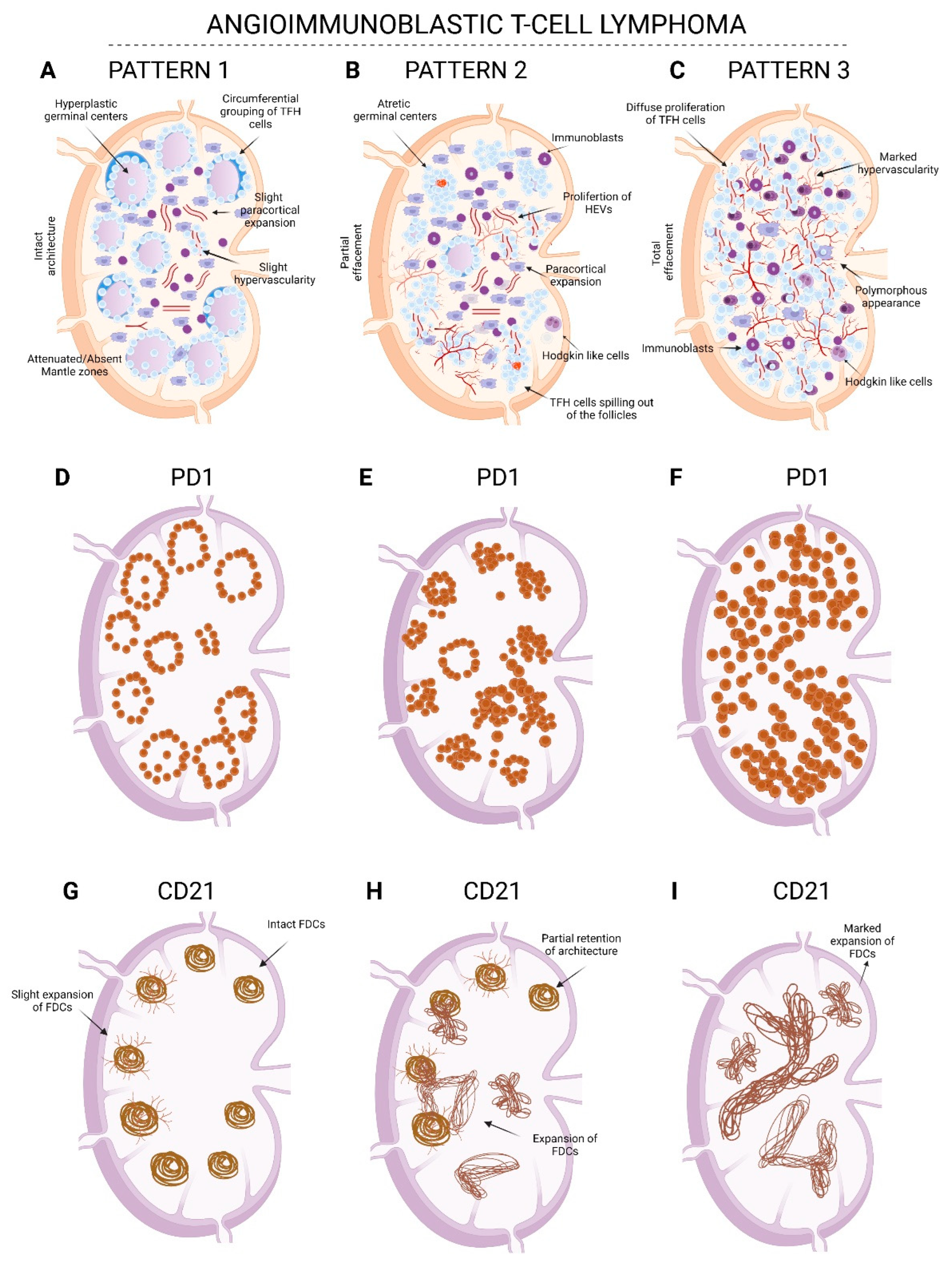
Table 2. Morphologic patterns of angioimmunoblastic T-cell lymphoma.
Pattern 1Pattern 2Pattern 3
ArchitecturePreservedPartially effacedTotally effaced
FolliclesHyperplastic
germinal centers
Absent/attenuated mantle zones
Atretic/regressed
Castleman-like follicles
Absent or occasional compressed follicles seen at the periphery (especially highlighted with a CD20 stain)
ParacortexFocal areas of paracortical expansion and perifollicular polymorphous infiltrate
focal slight increase in HEVs
Areas with polymorphous infiltration with immunoblasts and Hodgkin’s-like cells
Proliferation of HEVs in the expanded areas
Marked polymorphous diffuse infiltration with immunoblasts and Hodgkin’s-like cells
Marked proliferation of HEVs
TFH cellPerifollicular with occasional paracortical clusteringParacortical aggregates spilling out of follicles with grouping around HEVsDiffuse proliferation of TFH cells with grouping around HEVs
FDC
meshworks (CD21 or CD23 IHC)
Intact with slight branching outPartially intact with areas demonstrating expansion and encircling of HEVsMarked expansion with encircling around HEVs
FDC, follicular dendritic cells; HEV, high endothelial venule; IHC, immunostain; TFH, follicular helper T-cells.
Publisher’s Note: MDPI stays neutral with regard to jurisdictional claims in published maps and institutional affiliations.

