Review

Indolent T- and NK-Cell Lymphoproliferative Disorders of the Gastrointestinal Tract: Current Understanding and Outstanding Questions

by
Craig R. Soderquist
* and
Govind Bhagat
*
Department of Pathology and Cell Biology, Columbia University Irving Medical Center, 630 West 168th Street, New York, NY 10032, USA
*
Authors to whom correspondence should be addressed.
Hemato 2022, 3(1), 219-231; https://doi.org/10.3390/hemato3010018
Submission received: 1 February 2022 / Revised: 8 March 2022 / Accepted: 8 March 2022 / Published: 10 March 2022

Abstract

:
Indolent T- and NK-cell lymphoproliferative disorders of the gastrointestinal tract are uncommon clonal neoplasms that have a protracted clinical course and limited response to therapy. In recent years, advances in the immunophenotypic, genetic, and clinical characterization of these disorders have led to increased awareness and a better understanding of disease pathogenesis. However, many questions remain unanswered, including those concerning the cell(s) of origin, inciting immune or environmental factors, and the molecular pathways underlying disease progression and transformation. In this review, we discuss recent findings regarding the immunophenotypic and genomic spectrum of these lymphoproliferative disorders and highlight unresolved issues.

1. Introduction

Accumulating evidence over the past two decades has led to the recognition of rare indolent T- and NK-cell lymphoproliferative disorders (LPDs) occurring in the gastrointestinal (GI) tract and rarely other organs that have unique clinical, morphologic, immunophenotypic, and genetic features. Carbonnel et al. reported the first case of “low grade” small intestinal T-cell lymphoma in 1994 involving the duodenum of a 28 year-old man with a 7-year history of diarrhea and weight loss [1]. Since then, over 70 additional cases have been reported in a variety of GI sites, all showing similar patterns of mucosal infiltration by bland appearing small-sized lymphocytes (Figure 1A,B,D,E; Table 1), indolent clinical behavior, and poor response to chemotherapy [2,3,4,5,6,7,8,9,10,11,12,13,14,15,16,17,18,19,20,21,22,23,24,25,26,27,28,29]. The vast majority of these LPDs persisted as organ-confined disease for years, at times with regional lymph node involvement; however, dissemination outside the GI tract has been documented in some cases, and transformation to aggressive lymphomas has rarely been observed. Although early series described predominantly CD4+ cases (Figure 1C), many subsequent studies have reported CD8+ cases (Figure 1F), and less commonly CD4/CD8 and CD4+/CD8+ LPDs. The clonal nature of these diseases was established and confirmed by investigators from the 1990s through the early 2010s, but no recurrent chromosomal changes were identified. In recent years, high throughput DNA- and RNA-based sequencing approaches have provided greater insights into the genetic bases of these LPDs and recurrent alterations in multiple gene classes and pathways are now recognized. The knowledge gained thus far has led to the inclusion of these diseases within a provisional category in the 2017 revised 4th edition of the WHO classification of lymphoid neoplasms, termed “indolent T-cell LPD of the GI tract” (ITLPD-GI) [30].
In addition to LPDs of T-cell lineage, indolent LPDs of natural killer (NK) cells also occur in the GI tract. The first case (“atypical NK-cell proliferation of the gastrointestinal tract”) was reported by Vega et al. in 2006 [31], followed by two series from the United States and Japan describing morphologically similar lymphoproliferations, albeit with distinct clinical presentations and sites of involvement, designated “NK-cell enteropathy” (NKCE) and “lymphomatoid gastropathy” [32,33]. Over the years, nearly 50 cases of indolent NK-cell LPDs have been reported, as isolated case reports or small series [34,35,36,37,38,39,40,41,42,43,44,45,46], further refining the clinical, morphologic, and immunophenotypic features of these diseases, which are currently considered to represent a single entity, referred to as NKCE in this manuscript. In contrast to ITLPD-GI, the lesional cells of NKCE are medium- to large-sized and show mild pleomorphism (Figure 1J,K; Table 1) but lack the histopathologic features of extranodal NK/T-cell lymphomas, i.e., angioinvasion/angiodestruction and EBV infection [31,32,33,34,35,36,37,38,39,40,41,42,43,44,45,46]. Until recently, it wasn’t clear whether NKCE represented reactive or neoplastic proliferations, in part due to the challenges associated with demonstrating clonality of NK-cells; however, a recent study identified recurrent mutations in a small number of cases [46], supporting the neoplastic character of at least some if not all cases of NKCE.
Despite advances in our understanding of the pathobiology of ITLPD-GI and NKCE, many questions remain. The cell(s) of origin of these lymphoid proliferations have not been established and the (micro)environmental and immunological factors associated with disease initiation as well as those related to disease progression and transformation are unknown. Alterations of molecular pathways and signaling networks, as a consequence of mutations and structural genetic alterations, remain to be deciphered, and optimal strategies for disease monitoring need to be defined. Here we review recent progress in the immunophenotypic and genetic characterization of ITLPD-GI and NKCE and call attention to some of the lingering questions.

2. Indolent T-Cell Lymphoproliferative Disorder of the Gastrointestinal Tract (ITLPD-GI)

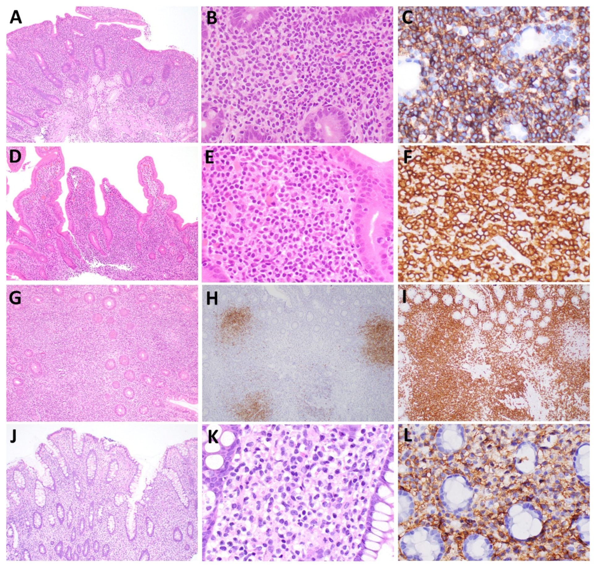
2.1. Immunophenotype

ITLPD-GI encompasses immunophenotypically heterogeneous diseases. Of the >70 cases reported to date, 34 are CD4+ [2,3,5,8,10,14,16,17,22,23,24,26,27,28], 29 are CD8+ [4,5,6,10,11,12,13,15,18,19,25,27,29], 5 are CD4/CD8 (“double-negative”, DN) [7,10,25,27], and 3 are CD4+/CD8+ (“double-positive,” DP) [5,9,27] (Table 1). Comprehensive immunophenotypic analyses have not been performed in all cases; however, virtually all cases express CD2 and CD3, and partial downregulation or loss of CD5 and/or CD7 has been reported in ~25% of cases. CD103 is generally negative, though variable expression has been described in 2 CD4+ cases [14,17] and 3 CD8+ cases [10,27] and partial weak CD56 expression has been observed in one CD8+ case [27]. T-cell receptor (TCR) αβ or βF1 is positive in all analyzed cases and T-follicular helper cell (TFH) markers (CD10, BCL6, PD1, CXCL13) are usually negative, although weak positivity for CD10 and CXCL13 was reported in one DN case [10] and PD1 expression in two cases, each exhibiting DN and DP phenotypes [27]. Expression of the regulatory T-cell (Treg) marker FOXP3 was negative in all cases tested [21,27]. The cytotoxic marker TIA1 is frequently expressed by CD8+ cases, and CD8+ and DN cases display variable granzyme B and perforin expression [4,5,6,7,8,10,12,15,18,19,21,25,27]. Aberrant CD20 expression has been reported in five cases (2 CD4+, 1 CD8+, 2 DN) [7,10,15,16]. Epstein–Barr virus (EBV)-encoded RNA (EBER) is negative in the T-cells, though rare admixed EBV+ cells, probably B-cells, have been reported in two cases [2,10]. The Ki-67 proliferation index is inherently low (usually <5%). CD30 and/or MUM1 expression has been reported in transformed cases, but not during the indolent phase of disease [5,21,22].
Figure 1. Histopathologic and immunophenotypic features of ITLPD-GI and NKCE. (AC) CD4+ ITLPD-GI: (A) A duodenal biopsy shows expansion of the lamina propria by a dense lymphocytic infiltrate that extends into the submucosa, accompanied by villous atrophy and crypt hyperplasia. (B) The lymphocytes are small and have round or mildly irregular nuclei, condensed chromatin, inconspicuous nucleoli, and scant cytoplasm. (C) The vast majority of lymphocytes express CD4. (DF) CD8+ ITLPD-GI: (D) The lymphocytic infiltrate within the lamina propria mildly distends the lower portions of the villi; no villous atrophy or crypt hyperplasia is evident. (E) The lymphocytes are small and mature appearing and do not show significant atypia. (F) In this case, the lymphocytes are CD8-positive. (GI) Reactive B-cell follicles: (G) Scattered, small lymphoid follicles can be seen in some cases, which are comprised of (H) B-cells (CD20+) and surrounded by neoplastic (I) T-cells (CD3+). (JL) NKCE: (J) A colonic biopsy shows an infiltrate of lymphoid cells within the lamina propria that displaces the crypts. (K) The lymphoid cells are medium- to large-sized and have ovoid or irregular nuclei, fine chromatin, indistinct or small nucleoli, and moderate or abundant cytoplasm. (L) The cells express CD56.
Hemato 03 00018 g001

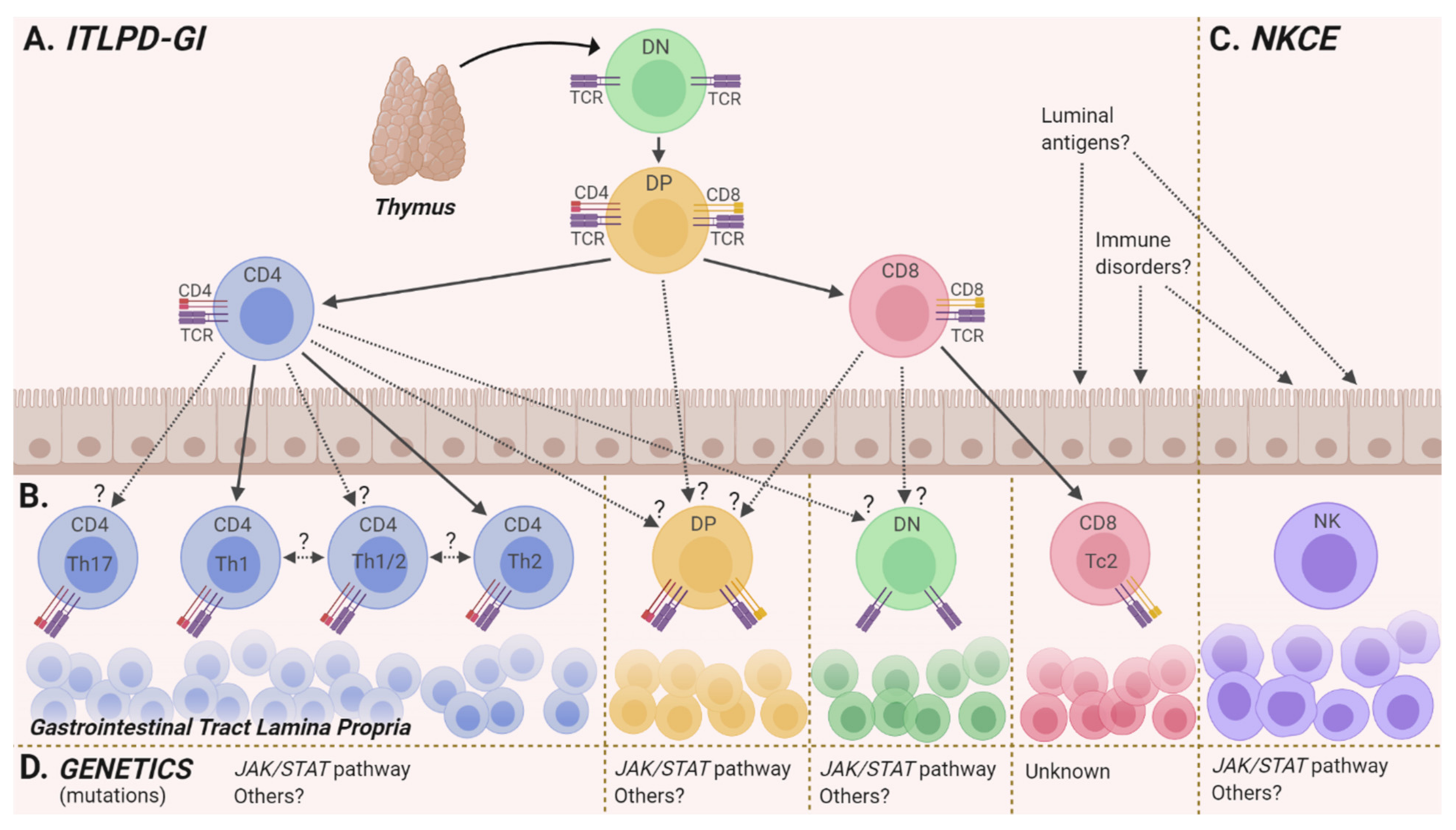
2.2. Cell(s) of Origin

Lamina propria (LP) helper T-cells (Th) are believed to be the cell of origin of CD4+ ITLPD-GI. CD4+ T-cells comprise the largest LP T-lymphocyte subset (~70%) [47] and they represent a diverse family, including Th type 1 and 2 (Th1, Th2) cells, T-follicular helper (TFH) cells, and regulatory T-cells (Tregs), amongst others [48,49]. Absence of TFH and Treg markers by CD4+ ITLPD-GI argues against their derivation from these T-cell lineages. By immunohistochemistry, CD4+ cases show heterogeneous expression of T-bet (TBX21) and GATA3 [27], which are master transcription factors governing Th1 vs. Th2 cell development [50]. Th1, Th2, and hybrid Th1/2 profiles have each been observed (Figure 2A,B), and in one case with longitudinal data, a phenotypic shift from a Th1/Th2 to Th2 profile was noted over time [27]. The etiology and significance of this phenotypic variability is unknown. Studies have shown that Th cells, both within and outside the GI tract, are not restricted to singular fates and can display phenotypic plasticity even after lineage specification [50]. It is presently unclear whether T-bet+/GATA3+ ITLPD-GI derive from bifunctional Th1/2 cells that develop directly from naïve T-cells in certain immune/inflammatory conditions [51] or committed Th subsets, which have been reprogrammed to adopt a mixed Th1/Th2 phenotype [52]. Inter-tumoral differences in the frequency of T-bet and GATA3 expression favors the latter supposition. The impact of lineage-specific transcription factor co-expression on cellular identity and function, however, remains poorly understood. Importantly, since RORγt (transcriptional regulator of Th17 differentiation) can be co-expressed with either T-bet or GATA3 in certain pathological conditions, induction of a Th17 program by some CD4+ ITLPD-GI cannot be ruled out (Figure 2A,B).
CD8+ ITLPD-GI are favored to originate from lamina propria CD8+ T-cells, which constitute a smaller subset of lamina propria T-cells (~30%) [47]. Similar to their CD4+ counterparts, LP CD8+ T-cells are subclassified based on their cytokine expression profiles into type 1 effector (Tc1) and type 2 effector (Tc2) T-cells. Immunohistochemical analysis has shown that most CD8+ ITLPD-GI express GATA3, suggesting an origin from Tc2 T-cells [27,53] (Figure 2A,B).
The cellular derivation of the DN and DP ITLPD-GI cases is not known. DN and DP T-cells represent uncommon GI mucosal T-cell subsets, and most of the knowledge regarding origin, differentiation, and function of these cells is inferred from their peripheral blood (PB) counterparts or from murine studies. LP DN T-cells have not been functionally characterized, but PB DN T-cells have been shown to derive from activated CD4+ or CD8+ T-cell subsets, which have downregulated these antigens due to chronic antigenic stimulation in response to infectious agents or in autoimmune/inflammatory conditions [54,55] (Figure 2A,B). LP DP T-cells have also not been characterized in humans; however in rhesus macaques (Macaca mulatta), LP DP T-cells have been shown to represent terminally differentiated effector memory T-cells with innate cytotoxic activity [56], which are inferred to play roles in viral immune responses, similar to human PB DP T-cells [57] (Figure 2A,B). It remains to be determined whether DN and DP ITLPD-GI arise from CD4+ or CD8+ T-cell subsets that have silenced or upregulated CD4 or CD8 expression (Figure 2A,B). In one reported DN case, the authors hypothesized that CD4 expression had been lost during disease progression; however, this supposition was not confirmed by TCR clonality analysis of early and late samples [22]. The lineage of the DN case expressing two TFH antigens (CD10 and CXCL13) is unclear, given the co-expression of TIA1 and perforin [10]. Expression of PD-1 by DN and DP ITLPDs (in the absence of other TFH markers) could be a manifestation of an “exhausted” cell state due to chronic activation/antigen stimulation [58].
Interrogation of gene expression programs and chromatin accessibility states of ITLPD-GI by transcriptome (e.g., RNA-seq [59]) and chromatin profiling (e.g., ATAC-seq [60]), and comparisons with normal mucosal and/or PB T-cell subsets, could help delineate the cell(s) of origin of the different immunophenotypic subtypes and unravel the transcriptional and epigenetic mechanisms responsible for disease heterogeneity.

2.3. Genetics

2.3.1. Clonality, Karyotyping, and Chromosome Microarray Analyses

All cases analyzed demonstrate clonal T-cell receptor gene (TR) rearrangements [2,3,4,5,6,7,8,9,10,12,14,15,17,19,20,21,22,23,24,25,26,27,28,29]. Karyotype analysis of isolated cases has identified a few non-recurrent chromosome abnormalities [2,13,27]. In a CD4+ case, Carbonnel et al. detected a translocation t(4q27; 16p13) involving the interleukin-2 (IL2) gene on chromosome 4q27 and TNF Receptor Superfamily Member 17 (TNFRSF17), also known as B-cell maturation antigen (BCMA), on chromosome 16p13 [1,2,61]. In another CD4+ case, a bone marrow sample showed a balanced translocation t(9;17)(p24;q21) that later was shown to represent a STAT3-JAK2 fusion [27]. Chromosome microarray studies have revealed a multitude of non-recurrent copy number changes in 10 of the 13 analyzed cases [21,26]. Large segmental chromosomal gains and losses were seen in some cases, encompassing numerous genes, and no minimal commonly altered region has been uncovered till now. Of interest, however, some of the altered loci (1p13, 4q27, 16p13, 17q21) harbor genes with potential relevance to ITLPD-GI pathogenesis, which might be deregulated as a consequence of the copy number aberrations, e.g., loss of SOCS1, TNIP3, and CD58, and gain of STAT3.

2.3.2. Next-Generation Sequencing (NGS)

JAK/STAT Pathway Alterations

Recent RNA- and DNA-based sequencing studies identified frequent genetic abnormalities in ITLPD-GI, with an enrichment of alterations involving the JAK/STAT pathway (Figure 2D). Recurrent STAT3-JAK2 rearrangements were observed in 5/11 (45%) CD4+ cases, but not in any CD8+ (0/12, 0%), DN (0/3, 0%), or DP (0/2, 0%) cases (one case listed in the EAHP workshop report by Montes-Moreno et al. was included in the study of Soderquist et al.) [5,10,27]. The rearrangements generate fusion transcripts joining the first 21 coding exons of STAT3 with the last 9 exons of JAK2. The resultant protein retains many key functional domains of STAT3, including the Src homology 2 (SH2) domain, and JAK2, including the JAK homology 1 (JH1) tyrosine kinase domain, and activates STAT5.
In some cases lacking STAT3-JAK2 rearrangements, genetic alterations in other components of the JAK/STAT pathway were detected. Specifically, activating STAT3 point mutations were seen in 3 cases; 1/4 (25%) CD4+ cases, 1/1 (100%) DP case, and 1/2 (50%) DN cases, but in none of the 10 CD8+ cases assessed [6,10,25,27]. The reported STAT3 mutations, D661Y and S614R, are well-characterized SH2 domain hotspot mutations, which enhance dimerization, nuclear transport, and activation of the STAT3 transcription factor, leading to heightened JAK/STAT signaling in response to cytokine stimulation [62,63]. Deletion of SOCS1, a negative regulator of the JAK family proteins [64], was also reported in one CD4+ case [27].

IL2 Structural Alterations

Rearrangements or deletions involving the 3′ untranslated region (UTR) of the IL2 gene, which encodes an important T-cell cytokine that signals via multiple pathways, including the JAK/STAT pathway [65], have been identified in two CD8+ cases (2/4, 50%), but not in any CD4+ (0/4, 0%), DP 0/1 (%), or DN (0/1, 05) cases [27]. Mapping of these structural alterations showed that they result in loss of most or all of the 3′ UTR AU-rich regulatory elements (AREs) which serve as binding sites for components of the mRNA degradation machinery and play a key role in mRNA stability [66]. In cultured T-cells, deletion of certain IL2 3′ UTR AREs resulted in longer mRNA half-life [67], demonstrating a potential mechanism wherein 3′ UTR alterations could modulate turnover and concentration of IL2 transcripts. The functional significance of these structural abnormalities (genes and signaling pathways impacted) in ITLPD-GI, however, is not clear.

Other Altered Genes and Pathways

Beyond JAK/STAT signaling, other pathways/gene classes are also altered in ITLPD-GI. Putative loss-of-function mutations in epigenetic modifier genes, including genes involved in DNA methylation (TET2, DNMT3A) and histone modification (KMT2D, EZH2), have been identified in five cases, including 3/5 (60%) CD4+ cases, 1/1 (100%) double-negative case, and 1/1 (100%) double-positive case, but not in five CD8+ cases analyzed [6,10,27]. While these specific variants have not been functionally characterized, mutations in epigenetic modifiers are pervasive in hematopoietic neoplasia and are known to disrupt CpG methylation, chromatin structure, and gene expression. Additionally, non-recurrent alterations have been observed in genes affecting NF-κB signaling. One CD4+ case harbored a frameshift TNFAIP3 mutation and one CD8+ case demonstrated a small inversion disrupting TNIP3 (also known as TNFAIP3 Interacting Protein 3). Since both TNFAIP3 and TNIP are negative regulators of NF-κB signaling, these presumed loss-of-function mutations are predicted to result in aberrant NF-κB activation.

2.3.3. Genetic Stability and Evolution

Longitudinal genetic data have only been reported for five cases to date [27]. Four of those five cases, including two with JAK/STAT pathway and epigenetic modifier mutations, showed stable genomic profiles for many years, which tracked the indolent disease course. In contrast, the ITLPD of one patient treated with multiple chemotherapeutic regimens over the years, including antimetabolite drugs, which eventually transformed to aggressive lymphoma, demonstrated genetic evolution over time.
Mutations targeting epigenetic modifiers and the JAK/STAT pathway likely represent early events in ITLPD-GI pathogenesis, as has been proposed in other hematopoietic neoplasms [68,69,70], whereas mutations targeting other pathways, particularly those predisposing to genomic instability, e.g., TP53, may be late events. The implications of the sequence of acquisition and interplay of somatic lesions, as well as the role of chemotherapy, in fostering clonal evolution and disease progression await clarification.

2.4. Environmental and Immunologic Factors

To date, no inherited, environmental, or immunologic factors have been definitively linked to ITLPD-GI and disease-specific triggers are unknown. Some observations, however, suggest a potential role of chronic antigenic stimulation and/or immune dysregulation. An underrecognized histologic feature of ITLPD-GI is the presence of reactive lymphoid follicles (Figure 1G–I). Whether these structures are related to disease pathogenesis or represent epiphenomenon (“reactive”) change, is not known. Prior and/or concurrent inflammatory or autoimmune diseases, including Crohn’s disease, ulcerative colitis, autoimmune enteropathy, and rheumatoid arthritis have been reported in a few patients [6,12,22,25,26,27], and viral infections, either gastrointestinal (e.g., HHV6), extra-intestinal (e.g., HSV), or systemic (e.g., HTLV-1) have also been described [25,26].
The contributions of immune dysregulation and/or suppression in triggering or modifying the course of ITLPD-GI is not known, since many patients have been treated with a variety of immunomodulatory agents, including TNF inhibitors (infliximab, adalimumab, and certolizumab), mycophenolate mofetil, methotrexate, and 6-mercaptopurine, either before or after ITLPD-GI diagnosis [6,12,19,22,25,27]. Two ITLPD-GI cases were also reported following solid organ transplant (liver and kidney) [12,17].
It is possible that persistent antigenic stimulation amplifies signaling cascades normally utilized by mucosal immune subsets, resulting in augmented survival and proliferation, which can increase the likelihood of incurring genetic lesions. It is unclear whether some immunomodulatory agents have mutagenic effects in addition to impairing tumor immunosurveillance, which allows the unrestrained expansion of neoplastic T-cell clones.
Table 1. Pathologic and genetic characteristics of ITLPD-GI and NKCE.
ITLPD-GINKCE
Site of involvementGI tract: Small intestine (84%, 53/63), colon (48%, 30/63), stomach (38%, 24/63), oral cavity (5 cases), esophagus (2 cases)
Abdominal lymph nodes: enlarged (47%, 22/47), biopsy confirmed involvement (12 cases)
Other: Bone marrow (9 cases), blood, liver, peripheral lymph nodes
GI tract: Stomach (73%, 35/48), small intestine (31%, 15/48), colon (27%, 13/48)
Other: Gallbladder, cystic duct lymph node, esophagus, vagina *
CytomorphologySmall size, round, oval or mildly irregular nuclei, condensed chromatin, inconspicuous nucleoli, scant/moderate cytoplasmMedium/large size, ovoid or irregular nuclei, fine chromatin, inconspicuous nucleoli, moderate pale cytoplasm with occasional azurophilic granules
ImmunophenotypeCD4+: 48% (34/71), CD8+: 41% (29/71), DN: 7% (5/71), DP: 4% (3/71)
Typical: CD2+, CD3+, CD5+, CD7+, TCRαβ+, CD103, CD10, BCL6, PD1, CXCL13, FOXP3, CD30, MUM1, MATK
Variant: CD5 (14%, 6/42), CD7 (24%, 9/38), CD103+ (5 cases), CD20+ (5 cases), CD56+ (1 case), PD1+ (2 cases), CXCL13+ and CD10+ (1 case)
Cytotoxic markers:
CD8: TIA1+ (96%), GrzB+ (30%)
CD4: TIA+ (0%), GrzB+ (0%)
Ki-67 index: <10%
Typical: sCD3, cCD3+, CD5, CD2+, CD7+, CD56+, CD4, CD8, TIA1+, GrzB+
Variant: CD2 (30%, 7/23), CD7 (1 case), CD8+ (11%, 5/46)
Ki-67 index: <40%
Other histopathologic
features
Typical: Diffuse or nodular infiltrate largely confined to the lamina propria
Other: Small clusters of lymphocytes infiltrating crypt or villous epithelium, scattered B-cell follicles, and occasionally granulomas
Well-circumscribed infiltrate within the lamina propria, often surrounded by a rim of polymorphous inflammatory cells
Absence of angioinvasion/angiodestruction
Chromosome/
genomic structural alterations
      CD4    
Translocations:
 STAT3-JAK2 (45%, 5/11)
  IL2-TNFRSF17
Gains/Losses:
Gains:
Chr: 1p, 1q, 8q, 13q, 15q, 17q, 19q, X
Losses:
Chr: 1p, 3q, 4q, 7q, 9p, 10p, 15q, 16p, 19p, 19q, 20q, X
    CD8  
Structural alterations:
 IL2 3′UTR-RHOH
 IL2 3′UTR del/IL2-TNIP3 
None reported
Mutations      CD4    
JAK/STAT pathway:
 STAT3, SOCS1 del
Epigenetics:
 TET2, KMT2D, EZH2
Other:
 TNFAIP3, DIS3
    CD8  
JAK/STAT pathway:
 None reported
Other:
 MCM5
JAK/STAT pathway:
 JAK3 (27%, 3/11)
Other:
 PTPRS, AURKB, AXL, ERBB4, IGF1R, PIK3CB,
CUL3, CHEK2, RUNX1T1, CIC, SMARCB1, SETD5
DN: double-negative (CD4/CD8); DP: double-positive (CD4+/CD8+); GI: gastrointestinal; GrzB: granzyme B. * Indolent NK-cell proliferations can occasionally be seen outside the GI tract. Two IL2 alterations were observed in one case: a deletion spanning the majority of the 3′ UTR (“IL2 3′ UTR del”) and an inversion involving IL2 and TNIP3 (“IL2-TNIP3”).

3. NK-Cell Enteropathy (NKCE)

3.1. Immunophenotype

The neoplastic cells demonstrate a phenotype typical of NK-cells: CD3(cytoplasmic)+, CD3(surface), CD7+, CD5, and CD56+ (Figure 1L, Table 1), with variable downregulation/loss of CD2. They often express one or more cytotoxic granule proteins (TIA1, granzyme-B, and/or perforin) and usually lack CD4 and CD8 expression; however, four CD8+ cases have been reported [31,32,33,34,35,36,37,38,39,40,41,42,43,44,45,46]. The Ki-67 labeling index is generally <40%, but higher proliferation indices (up to 90%) have been reported in a few cases. In situ hybridization for EBER is always negative.

3.2. Cell of Origin

It is not known whether mucosal tissue-resident or circulating NK-cells are the normal counterparts of NKCE (Figure 2C). Mucosal NK-cells normally account for a small fraction of lymphoid cells in the GI tract [71,72,73,74]. Various subpopulations of cells with NK attributes reside within the LP, including conventional NK-cells, tissue-resident NK-cells, and NK-like innate lymphoid cells; however, knowledge of the function and distribution of these populations is limited [71,72,73,74]. These cells are believed to play roles in combatting viruses and other intracellular pathogens via secretion of cytotoxic proteins and IFN-γ [72,74]. The occurrence of two cases of indolent NK-cell LPDs in the gallbladder, one in a cystic duct lymph node and one in the vagina [38,43,45,75], suggests that either extra-intestinal (and rarely extra-GI sites) harbor unique tissue-specific NK-cell populations or provide a microenvironment conducive to the growth and proliferation of NK-cells that traffic to these locations.

3.3. Genetics

Until recently, there was no evidence that NKCE represented a clonal or neoplastic disorder. In keeping with NK-cell derivation, the LPDs show an absence of clonal TR gene rearrangements [31,32,33,34,35,40,41,42,45]. Heterogeneous killer-cell immunoglobulin-like receptor (KIR) expression was reported in a single case [31]. In 2019, Xiao et al. identified identical somatic JAK3 K563_C565del mutations in 3 out of 10 (30%) NKCE cases [46] (Figure 2D, Table 1). This in-frame deletion of three amino acids is predicted to disrupt a portion of the JH2 pseudokinase domain responsible for modulating inhibitory feedback and suppression of JAK kinase activity. While the K563_C565del mutation has not been functionally characterized, similar mutations in the JH2 domain in other lymphoid neoplasms have been shown to result in upregulation of JAK activity [76]. Immunohistochemical analysis showed p-STAT5 staining in all NKCE cases tested, irrespective of JAK3 mutations, suggesting ubiquitous activation of the JAK/STAT pathway in NKCE [46]. In addition, an assortment of non-recurrent somatic variants were identified, but their pathogenic significance is presently uncertain [46].

3.4. Environmental and Immunologic Factors

No instigating factors for NKCE are known, though similar to ITLPD-GI, chronic antigenic stimulation and dysregulated immune signaling have been implicated. In cases from Japan, many of the gastric cases had co-existing H. pylori infection, and a history of gastric cancer was noted in others; however, no definitive causal links between NKCE and these diseases have been identified [33,34,36,39,41,42,45,46]. In two cases, anti-gliadin antibodies were reported in the absence of histologic evidence of celiac disease [31,43].

4. Disease Monitoring of ITLPD-GI and NKCE

While the majority of ITLPD-GI persist for years without evidence of disease dissemination outside the GI tract, imaging reveals enlarged abdominal lymph nodes in ~50% of cases [2,4,13,16,17,19,23,24,25,26,27,28], and biopsy-confirmed nodal involvement has been reported in ~15% of cases [2,10,13,26,27] (Table 1). Spread to bone marrow and peripheral blood, amongst other sites, is considered uncommon [2,4,7,13,19,21,22,24,26,27]. NKCE can also persist for years to decades, sometimes showing a relapsing/remitting course, though unlike ITLPD-GI, disease dissemination outside the GI tract has not been documented [31,32,33,34,35,36,37,38,39,40,41,42,43,44,45,46]. It is important to note, however, that either systemic evaluation was not documented in many studies of ITLPD-GI and NKCE or information regarding the assays used to investigate presence of disease outside the GI tract was not provided.
Figure 2. Pathogenetic aspects of ITLPD-GI and NKCE: (A) During “normal” T-cell development, immature double-negative (“DN”) thymocytes give rise to double-positive (“DP”) thymocytes in the thymus, which subsequently express either CD4 or CD8 following positive and negative selection. (B) Mature T-cells migrate to the gastrointestinal tract lamina propria. CD4+ ITLPD-GI manifest Th1, Th2, or hybrid Th1/2 profiles; acquisition of a Th17 fate has not been excluded. CD8+ cases predominantly display a Tc2 profile. It is not known whether the rare DP cases derive from DP thymocytes, which migrate directly to the intestinal mucosa, or from single-positive T-cells which up-regulate CD4+ or CD8+. DN ITLPD-GI may derive from activated CD4+ or CD8+ T-cells that have downregulated the T-cell co-receptors. (C) The cell of origin of NKCE (tissue-resident or circulating NK-cells) is unclear. The inciting factors for ITLPD-GI and NKCE (local or systemic) have not been elucidated. (D) Genetic alterations in the JAK/STAT signaling pathway have been identified in most immunophenotypic subsets of ITLPD-GI and in NKCE.
Hemato 03 00018 g002
Currently, the optimal strategies and frequency of testing for extra-GI disease in these indolent LPDs are unclear. When present, the burden of ITLPD-GI in the peripheral blood is generally low, therefore high analytic sensitivity of the assay is paramount. Multiplex polymerase chain reaction (PCR) analysis for TR rearrangement, which has utility in the evaluation of ITLPD-GI but not NKCE, shows a limit of detection (LOD) of ~5% of T-cells (range 1–10%), though comparison with involved GI biopsy results can improve confidence at the lower end [77]. NGS-based clonality methods are more sensitive (LOD ~2.5%) and can more accurately identify and track disease-associated clonotypes, as well as distinguish them from background non-neoplastic clonal expansions due to other etiologies [78]. Flow cytometry can detect immunophenotypically aberrant T- and NK-cell populations, but a substantial proportion of ITLPD-GI and NKCE do not harbor overt immunophenotypic abnormalities. Finally, targeted NGS analysis of PB circulating DNA to detect disease-associated mutations (“liquid biopsy”), although not yet attempted in ITLPD-GI or NKCE, has been reported to be equally or more sensitive than standard PCR analysis in other lymphoid malignancies harboring mutations [79]. Periodic imaging is recommended, and bone marrow biopsy may also be considered; however, invasive procedures should perhaps be used more sparingly (e.g., at initial diagnosis or on suspicion of disease progression). For each of these modalities, additional studies are required to establish the appropriate assessment intervals.

5. Summary

Progress in the clinicopathologic, immunophenotypic, and molecular characterization of ITLPD-GI and NKCE has increased awareness of these rare and underrecognized disorders, furthered our understanding of disease pathogenesis, and confirmed their neoplastic nature; however, many questions remain. While targeted genomic profiling studies have identified recurrent alterations in many cases of ITLPD-GI and NKCE, unbiased whole exome or genome sequencing analyses are required to delineate the genetic landscape of these diseases and decipher their clonal architecture. The etiology of the different immunophenotypic subsets of ITLPD-GI is still a mystery and it remains to be seen whether similar diseases in some animals can serve as surrogates for human ITLPD-GI and provide clues about disease-initiating factors [80]. The use of newer multi-omic approaches integrating genetic, epigenetic, transcriptomic, and metabolomic data may unmask novel therapeutic targets. Finally, it is hoped that incorporation of newer disease monitoring tools will allow a better understanding of the natural course of indolent T- and NK-cell LPDs.

Author Contributions

C.R.S. and G.B.: conceptualization, methodology, formal analysis, investigation, data curation, writing—original draft preparation, writing—review and editing. All authors have read and agreed to the published version of the manuscript.

Funding

This research received no external funding.

Conflicts of Interest

Neither author has any conflict of interest to disclose.

References

  1. Carbonnel, F.; Lavergne, A.; Messing, B.; Tsapis, A.; Berger, R.; Galian, A.; Nemeth, J.; Brouet, J.C.; Rambaud, J.C. Extensive small intestinal T-cell lymphoma of low-grade malignancy associated with a new chromosomal translocation. Cancer 1994, 73, 1286–1291. [Google Scholar] [CrossRef]
  2. Carbonnel, F.; D’Almagne, H.; Lavergne, A.; Matuchansky, C.; Brouet, J.C.; Sigaux, F.; Beaugerie, L.; Nemeth, J.; Coffin, B.; Cosnes, J.; et al. The clinicopathological features of extensive small intestinal CD4 T cell infiltration. Gut 1999, 45, 662–667. [Google Scholar] [CrossRef] [PubMed]
  3. Egawa, N.; Fukayama, M.; Kawaguchi, K.; Hishima, T.; Hayashi, Y.; Funata, N.; Ibuka, T.; Koike, M.; Miyashita, H.; Tajima, T. Relapsing oral and colonic ulcers with monoclonal T-cell infiltration. A low grade mucosal T-lymphoproliferative disease of the digestive tract. Cancer 1995, 75, 1728–1733. [Google Scholar] [CrossRef]
  4. Leventaki, V.; Manning, J.T.; Luthra, R.; Mehta, P.; Oki, Y.; Romaguera, J.E.; Medeiros, L.J.; Vega, F. Indolent peripheral T-cell lymphoma involving the gastrointestinal tract. Hum. Pathol. 2014, 45, 421–426. [Google Scholar] [CrossRef] [PubMed]
  5. Sharma, A.; Oishi, N.; Boddicker, R.L.; Hu, G.; Benson, H.K.; Ketterling, R.P.; Greipp, P.T.; Knutson, D.L.; Kloft-Nelson, S.M.; He, R.; et al. Recurrent STAT3-JAK2 fusions in indolent T-cell lymphoproliferative disorder of the gastrointestinal tract. Blood 2018, 131, 2262. [Google Scholar] [CrossRef] [Green Version]
  6. Edison, N.; Belhanes-Peled, H.; Eitan, Y.; Guthmann, Y.; Yeremenko, Y.; Raffeld, M.; Elmalah, I.; Trougouboff, P. Indolent T-cell lymphoproliferative disease of the gastrointestinal tract after treatment with adalimumab in resistant Crohn’s colitis. Hum. Pathol. 2016, 57, 45–50. [Google Scholar] [CrossRef]
  7. Wang, X.; Ng, C.-S.; Chen, C.; Yu, G.; Yin, W. An unusual case report of indolent T-cell lymphoproliferative disorder with aberrant CD20 expression involving the gastrointestinal tract and bone marrow. Diagn. Pathol. 2018, 13, 82. [Google Scholar] [CrossRef]
  8. Zanelli, M.; Zizzo, M.; Sanguedolce, F.; Martino, G.; Soriano, A.; Ricci, S.; Ruiz, C.C.; Annessi, V.; Ascani, S. Indolent T-cell lymphoproliferative disorder of the gastrointestinal tract: A tricky diagnosis of a gastric case. BMC Gastroenterol. 2020, 20, 336. [Google Scholar] [CrossRef]
  9. Guo, L.; Wen, Z.; Su, X.; Xiao, S.; Wang, Y. Indolent T-cell lymphoproliferative disease with synchronous diffuse large B-cell lymphoma. Medicine 2019, 98, e15323. [Google Scholar] [CrossRef]
  10. Montes-Moreno, S.; King, R.L.; Oschlies, I.; Ponzoni, M.; Goodlad, J.R.; Dotlic, S.; Traverse-Glehen, A.; Ott, G.; Ferry, J.A.; Calaminici, M. Update on lymphoproliferative disorders of the gastrointestinal tract: Disease spectrum from indolent lymphoproliferations to aggressive lymphomas. Virchows Arch. 2020, 476, 667–681. [Google Scholar] [CrossRef]
  11. Kohri, M.; Tsukasaki, K.; Akuzawa, Y.; Tanae, K.; Takahashi, N.; Saeki, T.; Okamura, D.; Ishikawa, M.; Maeda, T.; Kawai, N.; et al. Peripheral T-cell lymphoma with gastrointestinal involvement and indolent T-lymphoproliferative disorders of the gastrointestinal tract. Leuk. Res. 2020, 91, 106336. [Google Scholar] [CrossRef] [PubMed]
  12. Soon, G.; Wang, S. Indolent T-cell lymphoproliferative disease of the gastrointestinal tract in a renal transplant patient: Diagnostic pitfalls and clinical challenges. Pathology 2017, 49, 547–550. [Google Scholar] [CrossRef] [PubMed]
  13. Lee, J.; Park, K.; Kim, K.H.; Bang, H.I.; Yoon, S.Y.; Choi, I.H. Diagnostic challenges of indolent peripheral T cell lymphoma: A case report and literature review. Medicine 2020, 99, e22657. [Google Scholar] [CrossRef] [PubMed]
  14. Hirakawa, K.; Fuchigami, T.; Nakamura, S.; Daimaru, Y.; Ohshima, K.; Sakai, Y.; Ichimaru, T. Primary gastrointestinal T-cell lymphoma resembling multiple lymphomatous polyposis. Gastroenterology 1996, 111, 778–782. [Google Scholar] [CrossRef]
  15. Saggini, A.; Baciorri, F.; Di Prete, M.; Zizzari, A.G.; Anemona, L. Oral manifestation of indolent T-cell lymphoproliferative disorder of the gastrointestinal tract: A potential diagnostic pitfall. J. Cutan. Pathol. 2020, 47, 494–496. [Google Scholar] [CrossRef]
  16. Nagaishi, T.; Yamada, D.; Suzuki, K.; Fukuyo, R.; Saito, E.; Fukuda, M.; Watabe, T.; Tsugawa, N.; Takeuchi, K.; Yamamoto, K.; et al. Indolent T cell lymphoproliferative disorder with villous atrophy in small intestine diagnosed by single-balloon enteroscopy. Clin. J. Gastroenterol. 2019, 12, 434–440. [Google Scholar] [CrossRef] [Green Version]
  17. Goto, R.; Kawamura, N.; Watanabe, M.; Koshizuka, Y.; Shiratori, S.; Ara, M.; Honda, S.; Mitsuhashi, T.; Matsuno, Y.; Shimamura, T.; et al. Post-transplant indolent T cell lymphoproliferative disorder in living donor liver transplantation: A case report. Surg. Case Rep. 2020, 6, 147. [Google Scholar] [CrossRef]
  18. Takahashi, N.; Tsukasaki, K.; Kohri, M.; Akuzawa, Y.; Saeki, T.; Okamura, D.; Ishikawa, M.; Maeda, T.; Kawai, N.; Matsuda, A.; et al. Indolent T-cell lymphoproliferative disorder of the stomach successfully treated by radiotherapy. J. Clin. Exp. Hematop. 2020, 60, 7–10. [Google Scholar] [CrossRef] [Green Version]
  19. Wu, J.; Li, L.-G.; Zhang, X.-Y.; Wang, L.-L.; Zhang, L.; Xiao, Y.-J.; Xing, X.-M.; Lin, D.-L. Indolent T cell lymphoproliferative disorder of the gastrointestinal tract: An uncommon case with lymph node involvement and the classic Hodgkin’s lymphoma. J. Gastrointest. Oncol. 2020, 11, 812–819. [Google Scholar] [CrossRef]
  20. Ranheim, E.A.; Jones, C.; Zehnder, J.L.; Warnke, R.; Yuen, A. Spontaneously relapsing clonal, mucosal cytotoxic T-cell lymphoproliferative disorder: Case report and review of the literature. Am. J. Surg. Pathol. 2000, 24, 296–301. [Google Scholar] [CrossRef]
  21. Margolskee, E.; Jobanputra, V.; Lewis, S.K.; Alobeid, B.; Green, P.H.R.; Bhagat, G. Indolent Small Intestinal CD4+ T-cell Lymphoma Is a Distinct Entity with Unique Biologic and Clinical Features. PLoS ONE 2013, 8, e68343. [Google Scholar] [CrossRef] [PubMed] [Green Version]
  22. Perry, A.M.; Bailey, N.G.; Bonnett, M.; Jaffe, E.S.; Chan, W.C. Disease Progression in a Patient With Indolent T-Cell Lymphoproliferative Disease of the Gastrointestinal Tract. Int. J. Surg. Pathol. 2019, 27, 102–107. [Google Scholar] [CrossRef] [PubMed]
  23. Zivny, J.; Banner, B.F.; Agrawal, S.; Pihan, G.; Barnard, G.F. CD4+T-cell lymphoproliferative disorder of the gut clinically mimicking celiac sprue. Dig. Dis. Sci. 2004, 49, 551–555. [Google Scholar] [CrossRef] [PubMed]
  24. Svrcek, M.; Garderet, L.; Sebbagh, V.; Rosenzwajg, M.; Parc, Y.; Lagrange, M.; Bennis, M.; Lavergne-Slove, A.; Fléjou, J.-F.; Fabiani, B. Small intestinal CD4+ T-cell lymphoma: A rare distinctive clinicopathological entity associated with prolonged survival. Virchows Arch. 2007, 451, 1091–1093. [Google Scholar] [CrossRef] [PubMed]
  25. Perry, A.M.; Warnke, R.A.; Hu, Q.; Gaulard, P.; Copie-Bergman, C.; Alkan, S.; Wang, H.-Y.; Cheng, J.X.; Bacon, C.M.; Delabie, J.; et al. Indolent T-cell lymphoproliferative disease of the gastrointestinal tract. Blood 2013, 122, 3599–3606. [Google Scholar] [CrossRef] [Green Version]
  26. Malamut, G.; Meresse, B.; Kaltenbach, S.; Derrieux, C.; Verkarre, V.; Macintyre, E.; Ruskone–Fourmestraux, A.; Fabiani, B.; Radford–Weiss, I.; Brousse, N.; et al. Small Intestinal CD4+ T-Cell Lymphoma Is a Heterogenous Entity With Common Pathology Features. Clin. Gastroenterol. Hepatol. 2014, 12, 599–608. [Google Scholar] [CrossRef] [PubMed]
  27. Soderquist, C.R.; Patel, N.; Murty, V.V.; Betman, S.; Aggarwal, N.; Young, K.H.; Xerri, L.; Leeman-Neill, R.; Lewis, S.K.; Green, P.H.; et al. Genetic and phenotypic characterization of indolent T-cell lymphoproliferative disorders of the gastrointestinal tract. Haematologica 2020, 105, 1895–1906. [Google Scholar] [CrossRef] [Green Version]
  28. Sena Teixeira Mendes, L.; DAttygalle, A.; Cunningham, D.; Benson, M.; Andreyev, J.; Gonzales-de-Castro, D.; Wotherspoon, A. CD4-positive small T-cell lymphoma of the intestine presenting with severe bile-acid malabsorption: A supportive symptom control approach. Br. J. Haematol. 2014, 167, 265–269. [Google Scholar] [CrossRef] [Green Version]
  29. Tsutsumi, Y.; Inada, K.-I.; Morita, K.; Suzuki, T. T-cell lymphomas diffusely involving the intestine: Report of two rare cases. Jpn. J. Clin. Oncol. 1996, 26, 264–272. [Google Scholar] [CrossRef] [Green Version]
  30. Swerdlow, S.H.; Campo, E.; Harris, N.L.; Jaffe, E.S.; Pileri, S.A.; Stein, H.; Thiele, J.; Arber, D.A.; Hasserjian, R.P.; Le Beau, M.M.; et al. (Eds.) World Health Organization Classification of Tumours of Haematopoietic and Lymphoid Tissues; IARC: Lyon, France, 2016. [Google Scholar]
  31. Vega, F.; Chang, C.-C.; Schwartz, M.R.; Preti, H.A.; Younes, M.; Ewton, A.; Verm, R.; Jaffe, E.S. Atypical NK-cell proliferation of the gastrointestinal tract in a patient with antigliadin antibodies but not celiac disease. Am. J. Surg. Pathol. 2006, 30, 539–544. [Google Scholar] [CrossRef]
  32. Mansoor, A.; Pittaluga, S.; Beck, P.L.; Wilson, W.H.; Ferry, J.A.; Jaffe, E.S. NK-cell enteropathy: A benign NK-cell lymphoproliferative disease mimicking intestinal lymphoma: Clinicopathologic features and follow-up in a unique case series. Blood 2011, 117, 1447–1452. [Google Scholar] [CrossRef] [PubMed] [Green Version]
  33. Takeuchi, K.; Yokoyama, M.; Ishizawa, S.; Terui, Y.; Nomura, K.; Marutsuka, K.; Nunomura, M.; Fukushima, N.; Yagyuu, T.; Nakamine, H.; et al. Lymphomatoid gastropathy: A distinct clinicopathologic entity of self-limited pseudomalignant NK-cell proliferation. Blood 2010, 116, 5631–5637. [Google Scholar] [CrossRef] [PubMed]
  34. Tanaka, T.; Megahed, N.; Takata, K.; Asano, N.; Niwa, Y.; Hirooka, Y.; Goto, H. A case of lymphomatoid gastropathy: An indolent CD56-positive atypical gastric lymphoid proliferation, mimicking aggressive NK/T cell lymphomas. Pathol. Res. Pract. 2011, 207, 786–789. [Google Scholar] [CrossRef] [PubMed]
  35. McElroy, M.K.; Read, W.L.; Harmon, G.S.; Weidner, N. A unique case of an indolent CD56-positive T-cell lymphoproliferative disorder of the gastrointestinal tract: A lesion potentially misdiagnosed as natural killer/T-cell lymphoma. Ann. Diagn. Pathol. 2011, 15, 370–375. [Google Scholar] [CrossRef]
  36. Isom, J.A.; Arroyo, M.R.; Reddy, D.; Joshi-Guske, P.; Al-Quran, S.Z.; Li, Y.; Allan, R.W. NK cell enteropathy: A case report with 10 years of indolent clinical behaviour. Histopathology 2018, 73, 345–350. [Google Scholar] [CrossRef]
  37. Wang, R.; Kariappa, S.; Toon, C.W.; Varikatt, W. NK-cell enteropathy, a potential diagnostic pitfall of intestinal lymphoproliferative disease. Pathology 2019, 51, 338–340. [Google Scholar] [CrossRef]
  38. Dargent, J.-L.; Tinton, N.; Trimech, M.; de Leval, L. Lymph node involvement by enteropathy-like indolent NK-cell proliferation. Virchows Arch. 2021, 478, 1197–1202. [Google Scholar] [CrossRef]
  39. Terai, T.; Sugimoto, M.; Uozaki, H.; Kitagawa, T.; Kinoshita, M.; Baba, S.; Yamada, T.; Osawa, S.; Sugimoto., K. Lymphomatoidgastropathy mimicking extranodal NK/T cell lymphoma, nasal type: A case report. World J. Gastroenterol. 2012, 18, 2140–2144. [Google Scholar] [CrossRef]
  40. Koh, J.; Go, H.; Lee, W.A.; Jeon, Y.K. Benign Indolent CD56-Positive NK-Cell Lymphoproliferative Lesion Involving Gastrointestinal Tract in an Adolescent. Korean J. Pathol. 2014, 48, 73. [Google Scholar] [CrossRef] [Green Version]
  41. Ishibashi, Y.; Matsuzono, E.; Yokoyama, F.; Ohara, Y.; Sugai, N.; Seki, H.; Miura, A.; Fujita, J.; Suzuki, J.; Fujisawa, T.; et al. A case of lymphomatoid gastropathy: A self-limited pseudomalignant natural killer (NK)-cell proliferative disease mimicking NK/T-cell lymphomas. Clin. J. Gastroenterol. 2013, 6, 287–290. [Google Scholar] [CrossRef]
  42. Takata, K.; Noujima-Harada, M.; Miyata-Takata, T.; Ichimura, K.; Sato, Y.; Miyata, T.; Naruse, K.; Iwamoto, T.; Tari, A.; Masunari, T.; et al. Clinicopathologic Analysis of 6 Lymphomatoid Gastropathy Cases. Am. J. Surg. Pathol. 2015, 39, 1259–1266. [Google Scholar] [CrossRef]
  43. Xia, D.; A Morgan, E.; Berger, D.; Pinkus, G.S.; Ferry, J.A.; Zukerberg, L.R. NK-Cell Enteropathy and Similar Indolent Lymphoproliferative Disorders. Am. J. Clin. Pathol. 2018, 151, 75–85. [Google Scholar] [CrossRef] [PubMed]
  44. Yamamoto, J.; Fujishima, F.; Ichinohasama, R.; Imatani, A.; Asano, N.; Harigae, H. A case of benign natural killer cell proliferative disorder of the stomach (gastric manifestation of natural killer cell lymphomatoid gastroenteropathy) mimicking extranodal natural killer/T-cell lymphoma. Leuk. Lymphoma 2011, 52, 1803–1805. [Google Scholar] [CrossRef] [PubMed]
  45. Hwang, S.H.; Park, J.S.; Jeong, S.H.; Yim, H.; Han, J.H. Indolent NK cell proliferative lesion mimicking NK/T cell lymphoma in the gallbladder. Hum. Pathol. Case Rep. 2016, 5, 39–42. [Google Scholar] [CrossRef] [Green Version]
  46. Xiao, W.; Gupta, G.K.; Yao, J.; Jang, Y.J.; Xi, L.; Baik, J.; Sigler, A.; Kumar, A.; Moskowitz, A.J.; Arcila, M.E.; et al. Recurrent somatic JAK3 mutations in NK-cell enteropathy. Blood 2019, 134, 986–991. [Google Scholar] [CrossRef]
  47. Jahnsen, F.L.; Farstad, I.N.; Aanesen, J.P.; Brandtzaeg, P. Phenotypic distribution of T cells in human nasal mucosa differs from that in the gut. Am. J. Respir. Cell Mol. Biol. 1998, 18, 392–401. [Google Scholar] [CrossRef]
  48. Gibbons, D.L.; Spencer, J. Mouse and human intestinal immunity: Same ballpark, different players; Different rules, same score. Mucosal Immunol. 2011, 4, 148–157. [Google Scholar] [CrossRef]
  49. Cosorich, I.; McGuire, H.M.; Warren, J.; Danta, M.; King, C. CCR9 expressing T helper and T follicular helper cells exhibit site-specific identities during inflammatory disease. Front. Immunol. 2019, 10, 2899. [Google Scholar] [CrossRef] [Green Version]
  50. Zhu, J.; Paul, W.E. Heterogeneity and plasticity of T helper cells. Cell Res. 2009, 20, 4–12. [Google Scholar] [CrossRef]
  51. Peine, M.; Rausch, S.; Helmstetter, C.; Fröhlich, A.; Hegazy, A.N.; Kühl, A.A.; Grevelding, C.G.; Höfer, T.; Hartmann, S.; Löhning, M. Stable T-bet+GATA-3+ Th1/Th2 Hybrid Cells Arise In Vivo, Can Develop Directly from Naive Precursors, and Limit Immunopathologic Inflammation. PLoS Biol. 2013, 11, e1001633. [Google Scholar] [CrossRef] [Green Version]
  52. Hegazy, A.N.; Peine, C.; Helmstetter, C.; Panse, I.; Fröhlich, A.; Bergthaler, A.; Flatz, L.; Pinschewer, D.D.; Radbruch, A.; Löhning, M. Interferons Direct Th2 Cell Reprogramming to Generate a Stable GATA-3+T-bet+ Cell Subset with Combined Th2 and Th1 Cell Functions. Immunity 2010, 32, 116–128. [Google Scholar] [CrossRef] [PubMed]
  53. Fox, A.; Harland, K.; Kedzierska, K.; Kelso, A. Exposure of human CD8+ T cells to type-2 cytokines impairs division and differentiation and induces limited polarization. Front. Immunol. 2018, 9, 1141. [Google Scholar] [CrossRef] [PubMed]
  54. Priatel, J.J.; Utting, O.; Teh, H.-S. TCR/self-antigen interactions drive double-negative T cell peripheral expansion and differentiation into suppressor cells. J. Immunol. 2001, 167, 6188–6194. [Google Scholar] [CrossRef] [PubMed] [Green Version]
  55. Brandt, D.; Hedrich, C.M. TCRαβ+CD3+CD4CD8 (double negative) T cells in autoimmunity. Autoimmun. Rev. 2018, 17, 422–430. [Google Scholar] [CrossRef]
  56. Pahar, B.; Lackner, A.A.; Veazey, R.S. Intestinal double-positive CD4+CD8+ T cells are highly activated memory cells with an increased capacity to produce cytokines. Eur. J. Immunol. 2006, 36, 583–592. [Google Scholar] [CrossRef]
  57. Nascimbeni, M.; Shin, E.-C.; Chiriboga, L.; Kleiner, D.; Rehermann, B. Peripheral CD4(+)CD8(+) T cells are differentiated effector memory cells with antiviral functions. Blood 2004, 104, 478–486. [Google Scholar] [CrossRef]
  58. Keir, M.E.; Francisco, L.M.; Sharpe, A.H. PD-1 and its ligands in T-cell immunity. Curr. Opin. Immunol. 2007, 19, 309–314. [Google Scholar] [CrossRef]
  59. Hrdlickova, R.; Toloue, M.; Tian, B. RNA-Seq methods for transcriptome analysis. Wiley Interdiscip. Rev. RNA 2017, 8, e1364. [Google Scholar] [CrossRef] [Green Version]
  60. Sun, Y.; Miao, N.; Sun, T. Detect accessible chromatin using ATAC-sequencing, from principle to applications. Hereditas 2019, 156, 29. [Google Scholar] [CrossRef] [Green Version]
  61. Laâbi, Y.; Gras, M.P.; Carbonnel, F.; Brouet, J.C.; Berger, R.; Larsen, C.J.; Tsapis, A. A new gene, BCM, on chromosome 16 is fused to the interleukin 2 gene by a t(4;16)(q26;p13) translocation in a malignant T cell lymphoma. EMBO J. 1992, 11, 3897–3904. [Google Scholar] [CrossRef]
  62. Koskela, H.L.; Eldfors, S.; Ellonen, P.; Van Adrichem, A.J.; Kuusanmäki, H.; Andersson, E.; Lagström, S.; Clemente, M.J.; Olson, T.; Jalkanen, S.E.; et al. Somatic STAT3 mutations in large granular lymphocytic leukemia. N. Engl. J. Med. 2012, 366, 1905–1913. [Google Scholar] [CrossRef] [PubMed] [Green Version]
  63. Ettersperger, J.; Montcuquet, N.; Malamut, G.; Guegan, N.; Lastra, S.L.; Gayraud, S.; Reimann, C.; Vidal, E.; Cagnard, N.; Villarese, P.; et al. Interleukin-15-Dependent T-Cell-like Innate Intraepithelial Lymphocytes Develop in the Intestine and Transform into Lymphomas in Celiac Disease. Immunity 2016, 45, 610–625. [Google Scholar] [CrossRef] [PubMed]
  64. Liau, N.P.D.; Laktyushin, A.; Lucet, I.S.; Murphy, J.M.; Yao, S.; Whitlock, E.; Callaghan, K.; Nicola, N.A.; Kershaw, N.J.; Babon, J.J. The molecular basis of JAK/STAT inhibition by SOCS1. Nat. Commun. 2018, 9, 1558. [Google Scholar] [CrossRef] [PubMed]
  65. Ross, S.; Cantrell, D.A. Signaling and Function of Interleukin-2 in T Lymphocytes. Annu. Rev. Immunol. 2018, 36, 411–433. [Google Scholar] [CrossRef] [PubMed]
  66. Myer, V.E.; Fan, X.C.; Steitz, J.A. Identification of HuR as a protein implicated in AUUUA-mediated mRNA decay. EMBO J. 1997, 16, 2130–2139. [Google Scholar] [CrossRef]
  67. Chen, C.-Y.; Del Gatto–Konczak, F.; Wu, Z.; Karin, M. Stabilization of interleukin-2 mRNA by the c-Jun NH2-terminal kinase pathway. Science 1998, 280, 1945–1949. [Google Scholar] [CrossRef]
  68. Schwartz, F.H.; Cai, Q.; Fellmann, E.; Hartmann, S.; Mäyränpää, M.I.; Karjalainen-Lindsberg, M.-L.; Sundström, C.; Scholtysik, R.; Hansmann, M.-L.; Küppers, R. TET2 mutations in B cells of patients affected by angioimmunoblastic T-cell lymphoma. J. Pathol. 2017, 242, 129–133. [Google Scholar] [CrossRef]
  69. Soderquist, C.R.; Lewis, S.K.; Gru, A.A.; Vlad, G.; Williams, E.S.; Hsiao, S.; Mansukhani, M.M.; Park, D.C.; Bacchi, C.E.; Alobeid, B.; et al. Immunophenotypic Spectrum and Genomic Landscape of Refractory Celiac Disease Type II. Am. J. Surg. Pathol. 2021, 45, 905–916. [Google Scholar] [CrossRef]
  70. Cording, S.; Lhermitte, L.; Malamut, G.; Berrabah, S.; Trinquand, A.; Guegan, N.; Villarese, P.; Kaltenbach, S.; Meresse, B.; Khater, S.; et al. Oncogenetic landscape of lymphomagenesis in coeliac disease. Gut 2021, 71, 497–508. [Google Scholar] [CrossRef]
  71. Yu, X.; Vargas, J.; Green, P.H.; Bhagat, G. Innate Lymphoid Cells and Celiac Disease: Current Perspective. Cell. Mol. Gastroenterol. Hepatol. 2021, 11, 803–814. [Google Scholar] [CrossRef]
  72. Shi, F.-D.; Ljunggren, H.-G.; La Cava, A.; Van Kaer, L. Organ-specific features of natural killer cells. Nat. Rev. Immunol. 2011, 11, 658–671. [Google Scholar] [CrossRef] [PubMed]
  73. Grégoire, C.; Chasson, L.; Luci, C.; Tomasello, E.; Geissmann, F.; Vivier, E.; Walzer, T. The trafficking of natural killer cells. Immunol. Rev. 2007, 220, 169–182. [Google Scholar] [CrossRef] [PubMed] [Green Version]
  74. Panda, S.K.; Colonna, M. Innate lymphoid cells in mucosal immunity. Front. Immunol. 2019, 10, 861. [Google Scholar] [CrossRef] [PubMed] [Green Version]
  75. Krishnan, R.; Ring, K.; Williams, E.; Portell, C.; Jaffe, E.S.; Gru, A.A. An Enteropathy-like Indolent NK-Cell Proliferation Presenting in the Female Genital Tract. Am. J. Surg. Pathol. 2020, 44, 561. [Google Scholar] [CrossRef]
  76. Kiel, M.J.; Velusamy, T.; Rolland, D.; Sahasrabuddhe, A.A.; Chung, F.; Bailey, N.G.; Schrader, A.; Li, B.; Li, J.Z.; Ozel, A.B.; et al. Integrated genomic sequencing reveals mutational landscape of T-cell prolymphocytic leukemia. Blood 2014, 124, 1460–1472. [Google Scholar] [CrossRef] [Green Version]
  77. Van Dongen, J.J.M.; Langerak, A.W.; Brüggemann, M.; Evans, P.A.S.; Hummel, M.; Lavender, F.L.; Delabesse, E.; Davi, F.; Schuuring, E.; García-Sanz, R.; et al. Design and standardization of PCR primers and protocols for detection of clonal immunoglobulin and T-cell receptor gene recombinations in suspect lymphoproliferations: Report of the BIOMED-2 Concerted Action BMH4-CT98-3936. Leukemia 2003, 17, 2257–2317. [Google Scholar] [CrossRef] [Green Version]
  78. Van den Brand, M.; Rijntjes, J.; Möbs, M.; Steinhilber, J.; van der Klift, M.Y.; Heezen, K.C.; Kroeze, L.I.; Reigl, T.; Porc, J.; Darzentas, N.; et al. Next-Generation Sequencing–Based Clonality Assessment of Ig Gene Rearrangements: A Multicenter Validation Study by EuroClonality-NGS. J. Mol. Diagn. 2021, 23, 1105–1115. [Google Scholar] [CrossRef]
  79. Rossi, D.; Spina, V.; Bruscaggin, A.; Gaidano, G. Liquid biopsy in lymphoma. Haematologica 2019, 104, 648. [Google Scholar] [CrossRef] [Green Version]
  80. Freiche, V.; Cordonnier, N.; Paulin, M.V.; Huet, H.; Turba, M.E.; Macintyre, E.; Malamut, G.; Cerf-Bensussan, N.; Molina, T.J.; Hermine, O.; et al. Feline low-grade intestinal T cell lymphoma: A unique natural model of human indolent T cell lymphoproliferative disorder of the gastrointestinal tract. Lab. Investig. 2021, 101, 794–804. [Google Scholar] [CrossRef]
Publisher’s Note: MDPI stays neutral with regard to jurisdictional claims in published maps and institutional affiliations.

