Review

Sex-Dependent Skin Aging and Rejuvenation Strategies

by
Marta Gerasymchuk
1,
Gregory Ian Robinson
1,2,
Nataliia Vardinska
3,
Samuel Abiola Ayedun
4,
Sandra Chinwe Alozie
5,
John Wesley Robinson
6,
Olga Kovalchuk
1,* and
Igor Kovalchuk
1,*
1
Department of Biological Sciences, University of Lethbridge, Lethbridge, AB T1K 3M4, Canada
2
Gies College of Business, University of Illinois Urbana-Champaign, Champaign, IL 61820, USA
3
Healthy & Happy Health Care Center, 01033 Kyiv, Ukraine
4
Department of Internal Medicine, Kubwa General Hospital, Kubwa, FCT Abuja 901101, Nigeria
5
Department of Internal Medicine, Asokoro District Hospital, Asokoro, FCT Abuja 900231, Nigeria
6
Department of Biology, Western University, London, ON N6A 3K7, Canada
*
Authors to whom correspondence should be addressed.
Dermato 2023, 3(3), 196-223; https://doi.org/10.3390/dermato3030016
Submission received: 24 March 2023 / Revised: 20 May 2023 / Accepted: 25 July 2023 / Published: 4 August 2023
(This article belongs to the Special Issue Antioxidants and Skin Health)

Abstract

:
The skin, the largest external organ, serves as the primary defensive barrier against various environmental factors such as ultraviolet exposure, pollution, dietary habits, pathogens, and chemical compounds. Consequently, the skin reflects our age through visible signs of aging, such as wrinkles, age spots, dullness, and sagging. This review explores the gender-related aspects of cutaneous aging and the associated dermatological conditions. It highlights the different manifestations of aging in females and males that become evident after the age of 12, emphasizing the susceptibility to conditions such as seborrheic eczema, acne, and rosacea. Treatment strategies often vary between genders due to these disparities. While men tend to experience accelerated skin aging, most anti-aging products and strategies primarily target females. However, there has been a recent shift in men’s priorities, leading to increased interest in maintaining a youthful appearance and seeking cosmetic treatments. The manuscript covers a comprehensive range of modern dermal anti-aging and rejuvenation procedures, including plastic surgery, bio-revitalization methods, lasers, microneedling, and topical treatments. Additionally, it explores promising natural and synthetic therapeutics for combating age-related skin changes. The focus is on understanding the physiological aspects of gender-related cutaneous structure and aging to guide effective and tailored approaches in dermatological practice.

1. Introduction

Aging is a gradual process characterized by a progressive loss of adaptation and the accumulation of wear and tear throughout life, leading to an increased risk of morbidity and mortality [1]. However, aging is a natural phenomenon that all humans will experience [1].
The skin is the largest external organ in the human body and acts as the first line of defense against various external factors that can be harmful, such as ultraviolet (UV) exposure, environmental pollution, pathogens, and chemical compounds [2,3]. Due to the skin’s reduced ability to adapt to these stressors, the skin displays signs of aging, including wrinkles, age spots, dullness, and sagging texture. Facial skin, in particular, exhibits the first signs of aging, such as fine lines and shallow wrinkles at the outer corners of the eyes, commonly known as crow’s feet. Although aging is an inevitable biological process, it is associated with various cutaneous and systemic pathologies, including degenerative disorders and benign and malignant neoplasms [4]. Understanding the physiological and pathological processes occurring in the skin, including the interplay of gender differences and biological mechanisms like extracellular matrix (ECM) protein synthesis, ECM degradation, molecular aspects of metabolic activity, and cell cycle regulation, is crucial for the development of anti-aging therapies [5].
Initially, the physiological properties of the skin, such as hydration of the stratum corneum, transepidermal water loss (TEWL), sebum content, and pH value, appear similar between males and females. This holds true for both sexes during infancy, childhood, and early adolescence. However, as hormonal production begins during puberty, differences in the skin become more pronounced [6,7,8]. Moreover, there is a recognized gender-associated predisposition to certain dermatological conditions, such as seborrheic eczema, acne, and rosacea, leading to variations in treatment strategies for males and females [9]. Thus, the differences in skin structure, physiology, and susceptibility to specific diseases based on gender play a significant role in the development of skin pathologies and require distinct therapeutic approaches.
While it is well known that proper nutrition, sleep, and physical activity are crucial for maintaining healthy-looking skin, aging is an ongoing process that affects all tissues and organs. However, its manifestations vary depending on factors such as genetics, ethnicity, sex hormones, lifestyle, and environmental factors [10]. Therefore, understanding sex-specific differences is essential for the development of cosmetics and skin care products tailored specifically for females or males to address pathological skin conditions. This integrative review aims to provide an overview of the physiological differences in skin aging between men and women, discuss common dermatological diseases and factors associated with premature aging, and consider the impact of gender and age on rejuvenation, anti-aging strategies, therapy for specific skin disorders, lifestyle modifications, and therapeutic options that can slow down the aging process.

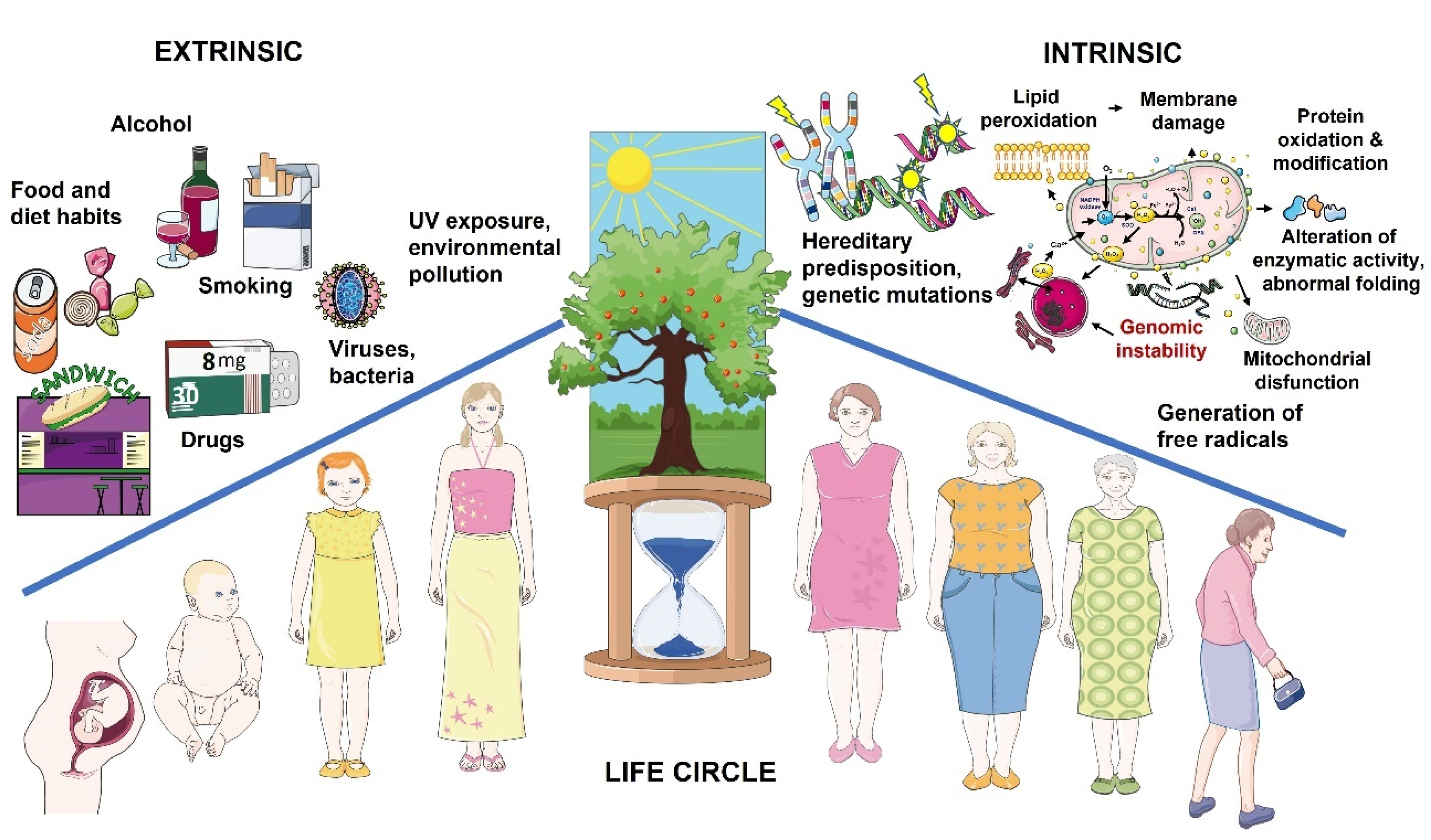
2. Main Hallmarks of Cutaneous Aging

Aging can be understood as the accumulation of changes over time in response to stress, lifestyle, and environmental factors, [NO_PRINTED_FORM] ultimately leading to irreparable damage and maladaptation of cells, ECM, cell communication, intercellular signaling, and more. These changes contribute to age-related diseases and mortality (Figure 1) [1,11,12].
Cutaneous aging is a complex process involving: intrinsic aging, also known as chronological aging, which is genetically determined; and extrinsic aging, which is caused by environmental factors such as chronic sun exposure, known as photoaging [13]. Aging is characterized by the gradual development of wrinkles, sagging skin, and drooping. Intrinsically aged skin appears dry and exhibits fine wrinkles, but it remains relatively smooth and light [14]. In contrast, extrinsically photoaged skin presents with thickened layers, a “leathery” appearance, rough wrinkles, pigmentation (referred to as “age spots”, or actinic lentigines), and capillary telangiectasia [15,16].

3. Skin Functions and Structure
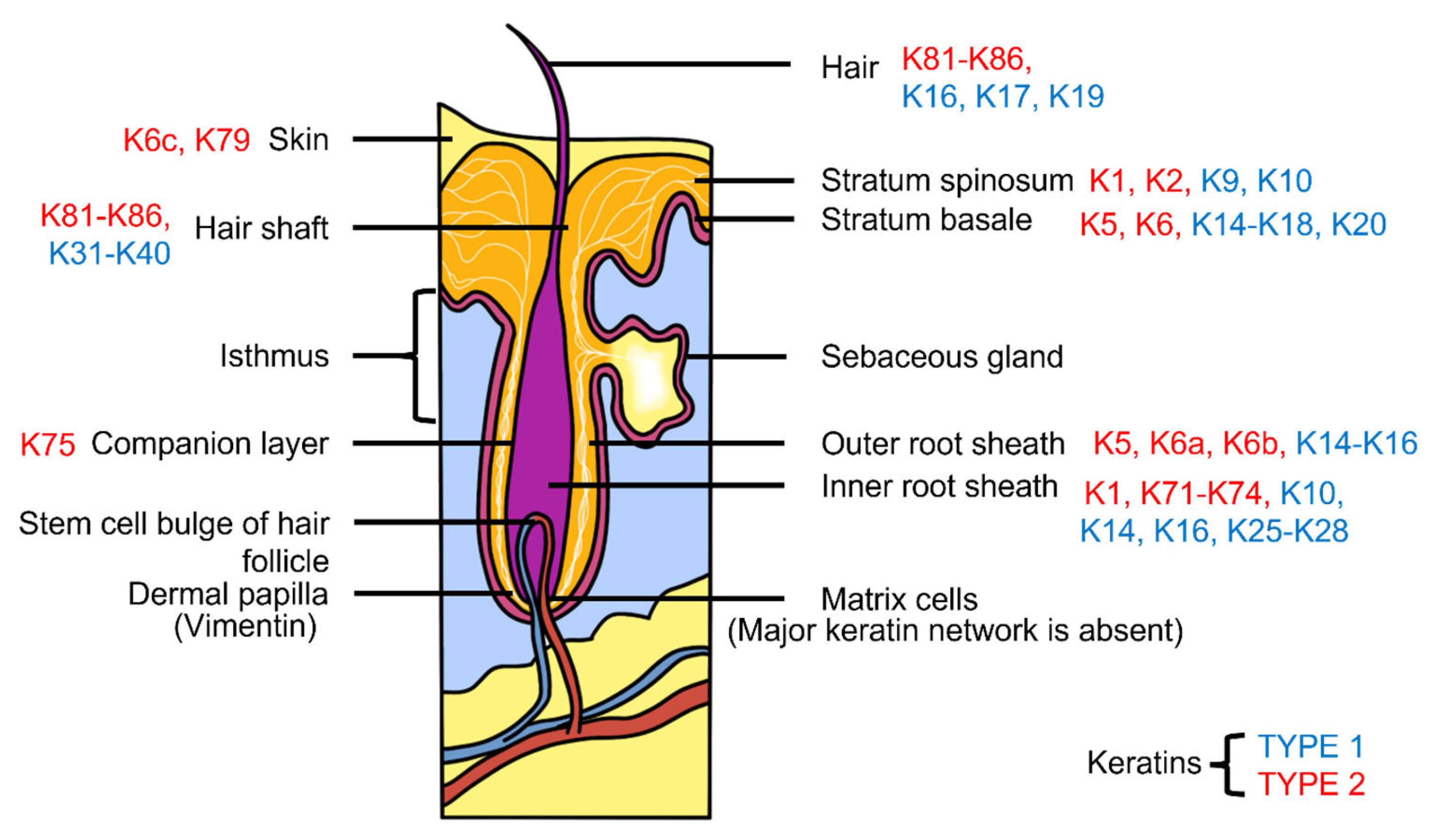
The skin, as the outermost and most prominent defensive organ in the human body, plays a crucial role in maintaining homeostasis. It achieves this through various functions, including the prevention of percutaneous loss of electrolytes, fluids, and proteins, the regulation of temperature, the mediation of immune activity, and sensory perception.
The outermost layer of the epidermis, known as the stratum corneum, serves two essential functions. Firstly, it acts as a barrier, providing waterproofing and physical protection against environmental factors such as UV radiation, temperature extremes, microbial invasion, allergens, and chemical irritants. Secondly, it plays a role in maintaining the body’s water balance, as the human body consists of approximately 60–70% water. Additionally, the skin carries out vital processes such as cell proliferation, differentiation, stem cell maintenance, and programmed cell death, which contribute to the regeneration and healing of wounds. Cutaneous immune surveillance is also a key function, involving the synthesis and release of pro- and anti-inflammatory mediators, including cytokines, chemokines, and trophic factors, as well as the antimicrobial activity of the sebum. The skin has an endocrine role as well, being responsible for synthesizing vitamin D, steroids, and peptide hormones. It expresses functional hormone receptors and enzymes involved in hormonal synthesis and metabolism. Exocrine functions encompass the production and release of sweat and sebum, which are essential for thermoregulation, physical barrier formation, and antimicrobial activity.
The skin plays a vital role in maintaining body temperature through insulation, control of heat loss mechanisms (such as radiation, convection, and conduction), vasoregulation (where vasodilation promotes heat loss and vasoconstriction preserves body heat), evaporation, and piloerection (which assists in insulation). Cutaneous motor function involves the regulation of blood vessel dilation, constriction, and piloerection [3].
Furthermore, the skin contributes to body homeostasis by preventing the loss of fluid, electrolytes, and proteins. It facilitates the transport of respiratory gases and nutrients between skin layers and to/from the skin surface. Additionally, it has the ability to absorb topically applied medications. The skin also acts as a sensory perception organ, responsible for sensing heat, cold, touch, pressure, vibration, pain, and itch (which are related to tissue injury). It releases neuropeptides that regulate local vasculature, immune responses, inflammation, and trophic functions (Figure 2) [14,17].
The skin is composed of three distinct but functionally interdependent layers: the epidermis, dermis, and hypodermis. The thickness of these cutaneous layers varies across different regions of the body and is determined by the unique structural characteristics of the epidermis and dermis. For instance, “thick skin” is typically found on the palms of the hands and soles of the feet. It is characterized by being hairless and having an additional layer in the epidermis called the stratum lucidum. On the other hand, the skin on the face and most other parts of the body is considered “thin” as it lacks the stratum lucidum layer in the epidermis.

3.1. Epidermis

The epidermis, derived from the ectoderm, serves as the outermost layer of the skin [3]. It is comprised of various cell types, including keratinocytes, Langerhans cells, melanocytes, and neuroendocrine (Merkel) cells, and lacks blood vessels. The arrangement of the epidermal cells varies depending on their maturation stage and location in the body, typically forming four to five layers (Figure 2). These layers, from internal to external, are the stratum basale, stratum spinosum, stratum granulosum, stratum lucidum (absent in the “thin skin”), and stratum corneum (SC) [3].
Notably, the SC functions differently depending on gender and age (Table 1). It plays a crucial role in maintaining skin hydration, TEWL, and water retention, preventing skin cracking. The SC is comprised of corneocytes, which are anucleated keratinocytes that have reached the final stage of differentiation. These corneocytes retain keratin filaments within a filaggrin matrix, and the cornified lipid envelope replaces the keratinocyte plasma membrane [18].
The epidermis undergoes renewal every two weeks through the activity of stem cells within the basal layer, also known as the germinativum. These highly proliferative stem cells support the production of differentiated progeny, ensuring the maintenance of a robust external barrier. The predominant cell type in the epidermis and its appendages is the keratinocyte. Keratinocytes produce various types of keratins, secrete lipids that regulate the epidermal water barrier, and synthesize vitamin D from cholesterol precursors under the influence of UV radiation [18]. Keratins, the primary structural proteins in the epidermis, form intermediate filaments that are categorized into type I and type II keratins (Figure 3) [19]. Alongside actin microfilaments and microtubules, keratin filaments contribute to the cytoskeletons of vertebrate epithelial cells, providing mechanical anchoring and strength to keratinocytes. They are organized into stabilized cell-cell adhesions called desmosomes as well as cell-substratum adhesions known as hemidesmosomes (Figure 2). Disruptions in these adhesive components and mutations in keratin genes can lead to cellular fragility, making the skin prone to rupture under physical stress and resulting in blistering skin disorders such as epidermolysis bullosa [20].
Keratinocytes are produced by mitotically active stem cells. As they move toward the skin’s surface, they gradually undergo changes and transform into flattened corneocytes. Each stage of differentiation brings about morphofunctional and metabolic modifications, corresponding to the formation of a new epidermal layer. Stem cells in the germinative layer primarily express keratins 5 and 14 [18]. During the transition from basal to spinous type, keratinocytes enter a postmitotic state and start expressing keratins 1 and 10. This transition is accompanied by an upregulation of desmosomes, resulting in a spiny-like appearance. Simultaneously, the structure of desmosomes undergoes changes during differentiation [21,22,23].
The epidermal differentiation complex, located on chromosome 1q21, encompasses proteins essential for forming a barrier that prevents fluid loss. Tight junctions in the granular layer establish cell-cell adhesions to restrict the flow of fluids and ions (Figure 2). Eventually, granular cells die, leading to the formation of cornified envelopes that subsequently shed. Melanocytes produce melanin, which protects against UV radiation and induces pigmentation in basal keratinocytes. Females typically have lighter skin color compared to males due to differences in melanin, hemoglobin, and carotene [21]. Hormonal changes, such as estrogen and testosterone, can also influence skin color. In early adolescence, girls may have darker skin; both sexes tend to have equal pigmentation in middle adolescence; and boys may become darker than girls in late adolescence [21,22,23].
Langerhans cells, dendritic cells located in the stratum spinosum, play a crucial role in maintaining immune defense. These cells, derived from CD34-positive stem cells, express MHC I and MHC II molecules and are part of the mononuclear phagocytic system [18]. Recent studies show that cutaneous immune cells express transient receptor potential channels of the vanilloid subtypes (TRPV) 1, 2, 3, and 4, as well as cannabinoid receptor 2 (CB2) (Figure 2) [24]. Merkel cells, on the other hand, are tactile epithelial cells found in the stratum basale. They act as mechanoreceptors for light touch and contribute to sensory functions through their interaction with the free nerve endings in the skin. Merkel cells are connected to neighboring keratinocytes via desmosomes and are mostly concentrated in the fingertips, palms, soles, and oral and genital mucosa [3,18].
Table 1. Differences in dermal properties between women and men.
Skin PropertiesNewborn/ToddlersPuberty Group/Teenagers18–30 Years Old30–50 Years Old50–70 Years Old70 Years Old and Older
HydrationA, B, ♀ = ♂ [7]F, FA, ♀ = ♂ [8]F*, C, N* A*, ♀ < ♂; H*, ♀ > ♂ [9]; F*, FA, ♀ < ♂ [8]; FA, ♀ < ♂ [25]F, N*, A*, ♀ < ♂; C, H*, ♀ > ♂ [9]; F, FA, ♀ < ♂ [8]F*, C, A, H, ♀ > ♂; N, ♀ < ♂ [9]; F*, FA, ♀ < ♂ [8]; A, ♀ > ♂ (baseline); ♀ < ♂* (after mineral water therapy) [26]F*, C*, N, A, H*, ♀ > ♂ [9]; F, ♀ = ♂ FA, ♀ > ♂ [8]
TEWLA, B, ♀ = ♂ [7] F, ♀ > ♂; C*, A*, N*, H, ♀ < ♂ [9]; FA, ♀ < ♂ [25]F*, C, N*, A*, ♀ > ♂; H ♀ < ♂ [9]F, C, N, ♀ < ♂; A*, H, ♀ > ♂ [9]; A, ♀ < ♂* (baseline and after mineral water therapy) [26]F, ♀ < ♂; C, N, A*, H*, ♀ > ♂ [9]
pHA, B, ♀ = ♂ [7]F, FA, ♀ > ♂ [8]F* N*, A*, C*, H*, ♀ > ♂ [9]; F*, FA, ♀ > ♂ [8]; FA*, ♀ > ♂ [25]F*, C*, N*, A*, H*, ♀ > ♂ [9]; F*, FA*, ♀ > ♂ [8]F*, N*, A*, C*, H*, ♀ > ♂ [9]; F, FA, ♀ > ♂ [8]F, C*, N*, A*, H*, ♀ < ♂ [9]; F, FA, ♀ < ♂ [8]
ElasticityF, CN, H, ♀ = ♂ [6]F, CN, H, ♀ = ♂ [6]; A, ♀ = ♂ [27]♀ = ♂ [22]; A, ♀ = ♂ [27];A, ♀ = ♂ [27]A*, ♀ < ♂ (baseline and after mineral water therapy) [26]; A, ♀ = ♂ [27]F, CN, H, ♀ = ♂ [6]; A, ♀ = ♂ [27]; ♀ = ♂ [22]
Thickness♀ = ♂ [22]A, ♀ < ♂ [27]; ♀ < ♂ [22]A, ♀ < ♂ [27]; ♀ < ♂ [22]A, ♀ < ♂ [27]; ♀ < ♂ [22]A, ♀ < ♂ [27]; ♀ < ♂ [22]A, ♀ < ♂ [27]; ♀ < ♂ [22]
SebumN/AF, ♀ > ♂, FA, ♀ > ♂ [8]F, C*, ♀ < ♂ [9]; F, ♀ < ♂*, FA, ♀ < ♂* [8], FA, ♀ < ♂ [25]F, C*, ♀ < ♂ [9]; F, ♀ < ♂*, FA, ♀ > ♂ [8]F*, C*, ♀ < ♂ [9]; F*, FA, ♀ < ♂ [8]F*, C*, ♀ < ♂ [9];
F*, FA, ♀ < ♂ [8]
WrinklesN/AN/AC*, F*, EC*, G*, UE, LE*, NG, MC*, ♀ < ♂ [28]C, F*, EC*, G, UE, LE*, NG*, MC*, ♀ < ♂ [28]C, F*, EC*, G, UE, LE*, NG*, MC*, ♀ < ♂ [28]C, F*, LE, MC*, ♀ < ♂, G*, UE, EC, NG, ♀ > ♂ [28]
Wrinkle roughnessN/AN/AF*, EC*, LE*, NG, MC*, ♀ < ♂ [28]F*, EC*, LE*, NG*, MC*, ♀ < ♂ [28]F*, EC*, LE*, NG*, MC*, ♀ < ♂ [28]F*, EC*, LE*, NG, MC*, ♀ < ♂ [28]
♀, female; ♂, male; *, significant changes; A, forearm; B, buttocks; C, cheek; CN, cantus; EC, eye corner; F, forehead; FA, flexor forearm; G, glabella; H, hand; LE, lower eyelid; MC, mouth corner; N, neck; N/A, non applicable; NG, nasolabial groove; TEWL, transepidermal water loss; UE, upper eyelid.

3.2. Differences between Male and Female Epidermal Structure and Functions

The stratum corneum (SC) serves as the primary exposed layer and acts as the body’s first line of defense against external exposures such as the sun, chemicals, wind, and temperature. Interestingly, the reaction of female and male skin to these factors varies depending on age. A study by Liu (2012) found no significant changes in SC hydration after sun exposure in either young or aged males, compared to their age-matched control subjects. In contrast, in young females, baseline hydration of the outermost layer did not differ significantly between sun-exposed and non-sun-exposed conditions. However, on both the forehead and dorsal hand of aged females, hydration was significantly lower in sun-exposed individuals compared to non-sun-exposed subjects [29]. Surprisingly, the rehydration capacity of the SC also differs based on sex and age. Several studies have shown that male skin is more prone to hydration [26] (Table 1). After the application of moisturizer, the gender difference becomes evident on the dorsal hand of sun-exposed aged groups, with higher hydration levels observed in aged males compared to aged females (46.81 ± 1.97 for aged males and 37.52 ± 2.62 for aged females; p < 0.01). Age-related changes in corneal layer hydration were observed on the dorsal hand of sun-exposed young and aged females (54.10 ± 2.68 and 37.52 ± 2.62; p < 0.0001, respectively), as well as on the canthus of non-sun-exposed young and aged males (57.10 ± 2.93 and 48.05 ± 2.76; p < 0.05, respectively) [2,13,22,23,29,30].
Interestingly, female hormones influence the composition of SC sphingolipids. In females, the composition of these lipids varies significantly with age, whereas in males, it remains relatively constant. From the prepubertal period to adulthood, there is a substantial increase in ceramides 1 and 2, accompanied by a decrease in ceramides 3 and 6 [30]. These lipids play a crucial role in maintaining the water-permeability barrier function of the human epidermis [22].
Differences in the adhesive properties of the SC have been observed to be age- and hormone-related. Measurements of the time required for blisters to form by controlled suction in vivo indicate that dermal-epidermal separation is slower in females than males from ages 15 to 69. This disparity is more pronounced in the age ranges of 15–39 years compared to 40–69 years [31]. Moreover, blistering time serves as a marker for dermo-epidermal adhesion and reflects the structural and mechanical integrity of the deep cutaneous layers. It confirms the association between age and reduced dermo-epidermal adhesion, which, in turn, contributes to the increased vulnerability of aged skin to shear-type and other injuries [32].

3.3. Dermis

The dermis is located beneath the epidermis and contains various structures such as hair, hair follicles, blood vessels, muscles, sensory neurons, and sweat glands. It is comprised of two layers of connective tissue: the papillary and reticular layers. The papillary layer, located close to the epidermis, is thinner and consists of loose connective areolar tissue. It contains fibroblasts, phagocytes, lymphatic capillaries, nerve fibers, and touch receptors called Meissner corpuscles [3]. This layer provides mechanical support and supplies nutrients to the epidermis. Fibroblasts play a vital role in producing ECM components, including collagen bundles, thin elastic fibers, hyaluronan (also known as hyaluronic acid), and others, which form the dermal scaffold [3].
On the other hand, the reticular layer is thicker and composed of dense connective tissue with thicker collagen bundles and coarse elastic fibers. It is richly supplied with blood vessels as well as sensory and sympathetic nerves. Elastin fibers in the reticular layer contribute to dermal elasticity and movement. Collagen fibers, important for maintaining structural integrity and tensile strength, also have the ability to bind water and support skin hydration [18].

3.4. Differences between Male and Female Dermal Structure and Functions

The rate of collagen loss with age is similar for both genders, although the total dermal collagen content is lower in females compared to males at all ages [22]. In the postmenopausal period, skin becomes thinner due to a reduction in cutaneous collagen content, primarily in the type III to type I collagen ratio [33,34]. Recent studies have shown that hormonal replacement therapy with estrogen and testosterone for 2–10 years increases the mean hydroxyproline content and collagen content significantly. The collagen content was elevated by 48% (p < 0.01) compared to non-treated subjects [34]. In females who received hormone replacement therapy for a median duration of 8 years, the portion of type III collagen increased by 25.4% (p < 0.01). Furthermore, after six months of estrogen implant therapy, the median cutaneous type III collagen increased from 19.9% to 22.5% (p < 0.01) [33].
The dermis, which determines dermal thickness, is the thickest layer of the skin. It is worth noting that men generally have thicker skin than women, except for the lower back, across all age ranges from 5 to 90 years. Interestingly, while males experience gradual cutaneous thinning with age, females’ dermal thickness remains constant until the fifth decade and then decreases with age [22]. Furthermore, it has been shown that men’s skin is about 16% (p < 0.001) thicker than women’s skin in age groups between 15 and 65. However, in younger and older age groups, women have thicker skin, and the skin of the five-year-old age group is thicker than that of the 90-year-old age group [27].
Previous studies have demonstrated that there are no significant differences between males and females in terms of cutaneous elasticity and extensibility parameters, with gradual decreases in extensibility after 65 years. This decline is attributed to the deterioration and loss of certain structural components in the fibrous network of the adult dermis, including alterations in collagen cross-linking, reductions in glycosaminoglycans, and displacement of the interstitial fluid through the fibrous network with age [27]. Another study by Xin et al. (2010) showed that the cutaneous resonance running time (CRRT), mainly influenced by collagen fibers in the papillary layers of the dermis, is negatively correlated with skin stiffness and positively correlated with SC hydration. The study found that CRRT is more prevalent on the hands compared to the forehead and canthus. Although age-related changes in CRRTs are less dramatic on the forehead and canthus, they still correlate negatively with age in all measured locations in both males and females [6].
Cutaneous functional properties such as elasticity, friction, and SC hydration do not show significant differences between males and females in both young (0–15) and elderly (over 70) age groups (Table 1). During the first two years of life, no differences in dermal TEWL, pH, or capacitance (corresponding to SC hydration) were observed based on sex or age. However, the skin of infants aged 8–24 months demonstrated functional signs of immaturity, which may be attributed to increased permeability and lower defense against chemical and microbial agents [7].
Another study examined different dermal mechanical properties across various age groups (20–74) and both genders. Cutaneous elasticity was measured three times on the cheek, neck, and dorsum of the hand to represent areas prone to extrinsic aging, while the volar forearm was selected as a predominantly sun-protected region. The results revealed a significant negative correlation between gross elasticity and age for both men (r = 0.811, p < 0.001) and women (r = 0.782, p < 0.001) in all tested areas, indicating an age-associated reduction in elasticity. On the other hand, the skin’s ability to recover its elasticity after stretching was found to be more strongly affected by the aging process compared to dermal firmness. Moreover, the absolute parameters of skin distensibility (the capacity to swell) and immediate distensibility showed a significant negative correlation with age, with reductions of up to 50% observed in both genders. As a result, the measurements for males were significantly higher than those for females. The elastic properties of female skin experienced a sharp decline after the age of 30, while the age-related reduction in dermal results remained consistent in male skin [35].
Nerve endings, sweat glands, and sebaceous glands are all present in the dermis (Figure 3). The quantity and functional activity of these components influence dermal sensitivity, oiliness, and sweat production. Female skin is typically more sensitive than male skin, while men tend to sweat more than women [22]. Furthermore, male skin is characterized by increased sebum production [22].

3.5. Hypodermis

The deepest layer of the skin, known as the hypodermis or subcutaneous fascia, is located beneath the dermis. It consists of adipocytes arranged in adipose lobules, as well as blood and lymphatic vessels, sensory neurons, sweat glands, fibroblasts, and skin appendages such as hair follicles (Figure 3). The color of the hypodermis can range from light to dark yellow, depending on the amount of carotene pigment present. The thickness of the hypodermis varies depending on location, hormonal status, genetics, and functional requirements.
In males, the hypodermis tends to be thicker in areas such as the lower back, abdomen, arms, and shoulders, primarily due to higher testosterone levels. On the other hand, in females, it is wider in the buttock (gluteus) muscles, hips, and thighs, which can be attributed to a higher amount of estrogen and increased lipoprotein lipase activity. Sex-based differences in the skin become more apparent after the age of 12, with subcutaneous fat increasing more than threefold in both sexes while internal fat mass increases less than two-fold. Importantly, females continue to accumulate more subcutaneous fat in the lower part of the body after the age of 12, whereas males do not experience the same increase [22].
The thickness of the hypodermis can vary across different regions of the body, with areas like the eyelids and external genitalia having a thickness of less than 1 mm. Together with other layers of the skin, the hypodermis plays a role in providing protection, insulation, and maintaining the shape of the body. Fibroblasts within the hypodermis produce ECM components that contribute to these functions. However, as aging occurs, the sponge-like architecture of the hypodermis deteriorates, leading to a loss of adipose and connective tissue that connects the bones and muscles to the dermis. This can result in a decrease in hypodermal thickness and ultimately contribute to a sagging appearance.

3.6. Hormones and Physiopathological Aspects of Gender-Related Cutaneous Aging

Gender-specific aspects of de rmal physiology also play a role in the predisposition and severity of certain skin conditions. Numerous studies summarized in Table 1 clearly indicate that the aging process differs between males and females. Hormonal changes occur as we age, including a decrease in estrogen after menopause and a decline in testosterone in aging men. Adrenal hormones such as dehydroepiandrosterone (DHEA) and dehydroepiandrosterone sulfate (DHEAS) also decrease significantly with age. Studies have shown that supplementation with these hormones can lead to a dramatic increase in sebum production, cutaneous hydration, and skin thickness [36]. In men, the decline of testosterone is associated with the sexual dimorphism observed in aging, with testosterone levels decreasing as much as 0.4–2% annually after the age of 30. Importantly, males are more susceptible to the effects of free radicals and experience higher levels of oxidative stress compared to females. In contrast, females are enriched with estrogen, which acts as an innate antioxidant defense and regulates the expression of antioxidant molecules like superoxide dismutase [37]. Furthermore, studies have demonstrated that men experience a decline of 3% to 6% in peak aerobic capacity per decade starting in their 30s and a decline of more than 20% per decade in their 70s and beyond. This steeper decline in aerobic and pulmonary function with age [38] contributes to accelerated cutaneous aging [38].
Physiologically, females have a thinner epidermis and dermis compared to males. They have less prominent facial musculature, a lower density of hair follicles, lower sebum and sweat production, and a decreased ratio of muscle to subcutaneous tissue [37]. Consequently, the loss of hypodermal adipose tissue with age leads to deeper expression lines in men, while women typically develop superficial wrinkles (rhytides). Additionally, the reduction in androgen levels during aging results in epidermal thinning and flattening of the dermo-epidermal junctions, leading to cutaneous atrophy, diminished elasticity, impaired metabolic and reparative abilities, increased susceptibility to shear stress, reduced nutrient transfer, compromised skin barrier function, and alterations in hair follicles [37].
Hormonal changes primarily occur in a gender-specific manner, but exposure to external factors such as sunlight, lifestyle choices, and environmental pollutants can also accelerate the aging process. The cumulative exposure to these external factors increases with age. The interplay between extrinsic and intrinsic factors not only influences the speed of the aging process but also affects the predisposition to certain skin diseases such as acne vulgaris, actinic keratosis, eczema, and psoriasis and can exacerbate the penetrance of genetic conditions associated with premature aging, such as Hutchinson-Gilford Progeria syndrome and Werner syndrome (Table 2).

4. Treatments That Improve Cutaneous Appearance

Throughout history, humans have been in search of the mythical “Elixir of Youth”, striving to extend their longevity and preserve a youthful appearance for as long as possible. Over time, we have developed various methods and techniques, ranging from diets and exercise regimens to pharmaceutical compounds and a wide array of cosmetic procedures, all aimed at slowing down the aging process. Despite the significant progress made in the field of anti-aging therapeutics and rejuvenation techniques, we are still in pursuit of groundbreaking therapies or extraordinary remedies that can truly rejuvenate the skin or the entire organism [4].
Strategies targeting anti-aging focus on preserving skin health and minimizing the visible signs of aging. Facial aging, in particular, is a multifaceted process involving muscle deterioration, loss of fat and volume, as well as changes in bony support [4]. Modern approaches to dermal anti-aging and rejuvenation can be categorized into invasive, micro-invasive, minimally invasive, and non-invasive methods. These encompass surgical procedures, such as plastic surgery, as well as various techniques for skin revitalization and rejuvenation, including the augmentation and restoration of different skin layers using lasers, microneedling, injections, peels, and a multitude of topical treatments. Therefore, in this discussion, we will focus on popular anti-aging and rejuvenation procedures as well as promising natural and synthetic compounds used in topical and systemic therapies within the field of dermatology [4,52].

4.1. Invasive Procedures

Invasive procedures primarily involve plastic surgery rejuvenation treatments, with a focus on face-lift practices. These procedures aim to lift and tighten sagging skin on the face and neck. Typically performed under general anesthesia, they involve the excision of excessively loose dermal tissue. A facelift addresses issues such as nasolabial folds, midface drooping, a double chin, and loose neck skin, effectively reducing the visible signs of aging. Recovery from a facelift usually takes approximately two weeks. While complications are rare, they may include hematoma formation within 24 h of surgery, scarring (often associated with hormonal/vitamin imbalances or a predisposition to keloids), nerve injury resulting in temporary or permanent loss of sensation or facial muscle movement (lasting from a few months to a year), and potentially temporary hair loss around the incision sites due to damaged hair follicles, which may regenerate over time [53,54].
Another popular surgical procedure in rejuvenation medicine is blepharoplasty, which involves the removal of excess skin from the eyelids. This excess skin and the accumulation of fat result from weakened muscles and stretched eyelids. This condition leads to sagging eyebrows, drooping upper lids, and the appearance of under-eye bags. In addition to giving an older and tired appearance, it can also narrow the visual field and even cause partial blindness. Blepharoplasty is typically performed on an outpatient basis under local anesthesia, and the recovery period can take up to three weeks. Complications are uncommon but can include infection, bleeding, eye muscle or nerve injury, scarring, temporary blurred vision, or loss of eyesight requiring follow-up surgery [53,54].
Compared to injections, the positive outcomes of surgical rejuvenation procedures such as facelifts and blepharoplasty generally last for several years, whereas the effects of injections typically endure for anywhere from eight months to two years.

4.2. Gender Dominance in Anti-Aging and Rejuvenation Surgical Services

Historically, females have been at the forefront as customers in the beauty industry, seeking anti-aging products and services. As a result, the market has seen a proliferation of goods and services catering to their needs, with substantial cosmetology industries dedicated to maintaining the well-being of facial and body skin, largely targeting women.
However, over the last three decades, there has been a notable shift in attitude towards aesthetics and the pursuit of a youthful appearance among men of all ages, ethnicities, and cultures. This shift is evident in the increased interest in in-office cosmetic treatments, moving away from over-the-counter men’s-specific cosmetics. According to the American Society for Aesthetic Plastic Surgery (ASAPS), there has been a remarkable 325% increase in the number of cosmetic procedures performed on men since 1997. In 2015 alone, men accounted for nearly 10% (approximately 1.2 million) of all plastic surgeries conducted that year [37]. However, as per the ASAPS statistical data from 2021, women still make up approximately 94% of all procedures [52]. Nowadays, both genders desire a youthful, fit, and attractive appearance. Interestingly, the ASAPS 2021 report highlights that liposuction rates were the highest among both males and females compared to other plastic surgery procedures (Table 3). Additionally, abdominoplasty was in high demand by both genders. However, blepharoplasty ranked third among surgical procedures for males, although it was nearly seven times less frequent than in females.

4.3. Micro-Invasive Procedures

Micro-invasive procedures encompass a wide range of techniques and treatments, including chemical peelings, dermal augmentation and contouring, injectable skin biostimulation, and rejuvenation. These procedures also focus on preventing dynamic wrinkles, correcting static wrinkles, utilizing visible light devices like intense pulsed light (IPL), restoring lost fat and volume, using ablative and nonablative lasers for photorejuvenation, and using radiofrequency. Most of these methods are performed in the office and aim to “resurface” the epidermis. This involves removing damaged skin layers and replacing them with remodeled and deeper layers while stimulating the formation of new collagen [4]. The advantages and disadvantages of commonly used micro-invasive skin rejuvenation strategies are summarized in Table 4.

4.4. Energy-Based Dermal Rejuvenation

4.4.1. Intense Pulse Light Devices

Broadband light, also known as IPL, has been a prominent treatment option in skin rejuvenation for approximately two decades. It utilizes light waves with a broad range of wavelengths, typically between 500 and 1200 nm, which are absorbed by various components of the skin [55]. One of the advantages of IPL is its versatility, achieved by applying different filters to the same device, allowing for the treatment of various targets. Through selective photothermolysis, IPL can specifically target epidermal and/or dermal regions while minimizing damage to the surrounding tissues. This approach reduces the risk of widespread thermal injury and preserves neighboring cells. IPL is commonly used for managing vascular lesions (such as cavernous hemangiomas, facial telangiectasias, and poikiloderma of Civatte), pigmented lesions and dyschromia, rhytids, and skin tightening, as well as hair removal [55]. However, it is important to note that there are still some limitations to the IPL. Long-term complications and their impact on age-associated conditions like actinic or seborrheic keratosis are yet to be fully understood. Furthermore, the potential effects of IPL on gene expression have not been determined.

4.4.2. Laser Treatment

Lasers can be broadly characterized as ablative, non-ablative, and fractional lasers [56]. They utilize light energy at varying frequencies to achieve specific clinical outcomes. By employing selective thermolysis and targeted cell stimulation, lasers can spare neighboring tissues. These devices are commonly employed for skin rejuvenation, hair removal, and tattoo removal. However, it is important to note that there are potential complications associated with laser use, including scarring, skin pigment changes, erythema, and infection [57].
In the realm of anti-aging treatments, lasers that target specific layers of the dermis play a crucial role by stimulating new collagen formation and tightening the skin. Non-ablative lasers, such as Nd: YAG and diode lasers, achieve their effects by heating water molecules and collagen in the dermal layer of skin, improving wrinkles and photodamage while preserving the integrity of the epidermis. On the other hand, ablative lasers like carbon dioxide (CO2) lasers (wavelength 10,600 nm) and Er: YAG lasers (2940 nm) destroy the epidermis, which can be associated with complications such as infections, erythema, and scarring [56,57].
Recently, the Food and Drug Administration (FDA) has approved new laser-based devices for dermatological and cosmetic purposes, including Aerolase Neo and Cutera’s AviClear lasers. The Aerolase Neo Elite, for instance, operates at a long wavelength of 1064 nm with a short pulse duration of 650 microseconds. This versatile 1064 nanometer Nd:YAG laser allows for a wide range of dermatological applications. It passes through the epidermis quickly, preventing the overheating of melanocytes and reducing the risk of hyperpigmentation. The treatment is virtually painless due to the rapid pulse duration. Furthermore, its deep penetrating property helps decrease sebum production in acne, reduce inflammation, and prevent future occurrences [58,59].
AviClear, on the other hand, utilizes a laser system with a distinct wavelength of 1726 nm. This particular wavelength enables selective photothermolysis of the sebaceous gland, offering a safe and effective approach for treating acne vulgaris [60].

4.4.3. Radiofrequency Microneedling

Radiofrequency (RF) is a non-ablative technology that utilizes thermal heating systems, in contrast to lasers, which rely on light sources. RF has the ability to penetrate deep into the skin, leading to dermal remodeling, skin tightening, and neocollagenesis. However, it is important to note that in some cases, deep penetration into the dermis and subcutaneous tissue can result in visible fat reduction, which may not be desired [61]. RF microneedling combines traditional microneedling with RF energy to enhance skin tone, texture, wrinkles, fine lines, and skin laxity. It also aids in reducing blemishes such as acne scars and promotes the production of new collagen, resulting in skin resurfacing [62]. The procedure is relatively painless and yields quick results by stimulating collagen production [62].
Unlike lasers, microneedling RF devices offer the advantage of adjustable treatment depths. The latest advancements in RF microneedling technology allow for precise control of the depth of exposure, even down to a tenth of a millimeter. This eliminates pain and minimizes potential damage to the skin’s surface [62,63].
Table 4. Micro-invasive dermal anti-aging and rejuvenation strategies.
Procedure/Compounds Properties Benefits Limitation Adverse Effects
Energy-based dermal rejuvenation
Lasers and radiofrequency (RF) [64] RF is a non-ablative technology that uses an electric current to target photodamage, unlike lasers that use a light source RF and lasers can be applied to multiple skin colors and types. It is minimally invasive or non-invasive and can focus high energy to treat various skin conditions Therapeutic standards and operation specifications for RF therapy have not been unified Erythema, edema and mild to moderate pain during treatment
Injectable dermal rejuvenation techniques
Dermal fillers (collagen, hyaluronic acid (HA), autologous fat) [65,66,67] HA is a GAG naturally found in the extracellular matrix. HA is the most commonly used filler Removes wrinkles and restores facial volume to create a rejuvenated look Patients requiring a large volume of fillers (advanced skin laxity, deep rhytids) may have to deal with costly procedures Intravascular occlusion, Nicolau syndrome, vision loss [65,66], hematomas, asymmetry, hypersensitivity, hyperpigmentation, infection, extrusion, or migration of the implant, neuropraxia, glabellar or perioral skin necrosis [67]
Botulinum neurotoxin (Botox) [68] Botox blocks acetylcholine release resulting in paralysis of muscles. Following Botox injection, paralysis occurs within 24 h to 2 weeks and lasts 3–6 months Improves wrinkles and nasolabial folds; treats hyperhidrosis, lichen simplex, and dyshidrotic eczema It has little to no effect on dry skin, pigmentation disorders, and vascular abnormalities; has no effect on skin texture; and cannot discontinue the skin aging process Bleeding, swelling, erythema, and pain at injection sites. Over time, the effect of Botox gradually resolves, resulting in reduced muscle paralysis
Chemical peeling
Light (e.g., alpha hydroxyl and salicylic acid peel), medium (e.g., TCA 30%), and deep peels (e.g., phenol) [4,57]Peels remove the uppermost layers of the skin, depending on the depth of the peel Instant result—smooth, radiant, and young-looking skin; removal of dead skin cells; cleaning up clogged pores and reduction of their size; decrease in facial wrinkles and fine lines; improvement of cutaneous texture and tone; boosting collagen production and promotion of healthy cell growth Increased sun sensitivity; carbolic acid (phenol) can damage kidneys, liver, and heart muscle, causing arrhythmias, which limits its application in patients with corresponding pathologies Hyperpigmentation, solar lentigines, risk of post-operative infections, and especially herpetic ones [4]
GAG, glycosaminoglycan; HA, hyaluronic acid; RF, radiofrequency; TCA, trichloroacetic acid.

4.5. Injectable Cutaneous Rejuvenation Options and Dermal Fillers

The core concept of cutaneous rejuvenation and anti-aging strategies revolves around enhancing the biosynthesis of essential ECM components such as collagen, elastin, and HA by dermal fibroblasts. Microinjections in the superficial dermis offer a means to achieve this effect, utilizing products that contain either a single active ingredient or a combination of biocompatible and absorbable compounds. These revitalizers encompass a range of substances, including amino acids, autologous cultured fibroblasts, growth factors, HA, hormones, homeopathic products, minerals, nutrients, vitamins, and more [4]. Mesotherapy and microneedling techniques are commonly employed for the application of these rejuvenation and anti-aging injectable compounds.
Mesotherapy involves the administration of multiple chemical injections using short, fine needles. These injections target the subcutaneous or intradermal layers as well as the subcutaneous fat, delivering pharmaceutical and homeopathic medications, plant extracts, vitamins, proteins, and other bioactive substances. On the other hand, microneedling, also known as percutaneous collagen induction (PCI) or collagen induction therapy, utilizes the physical trauma caused by needle penetration to stimulate skin regeneration [69]. Unlike mesotherapy, microneedling does not remove the epidermis or create open wounds, as it leaves the stratum corneum and basement membrane intact. Consequently, microneedling offers a shorter healing phase compared to laser or dermabrasion techniques, and it can be used safely on all skin types with a reduced risk of photosensitivity, infection, and pigmentation changes [69].

4.5.1. Dermal Fillers

Dermal fillers have become increasingly popular as a skin rejuvenation strategy. The quest for the perfect filler began over 100 years ago when paraffin injections were used for cosmetic purposes, resulting in the development of paraffinomas. However, in 1893, Neuber introduced a modified procedure involving the transfer of autologous fat, which is still practiced today. In the 1960s, medical-grade liquid silicone was successfully used in thousands of patients in the USA and elsewhere, although it is currently not approved by the FDA [67].
Dermal fillers are soft, gel-like products that are injected within or beneath the skin to enhance its physical features, such as soft tissue augmentation, wrinkle plumping, and line smoothing on the face. These fillers can be categorized into: (a) autologous fillers (cultured human fibroblasts, fat); (b) collagen-based fillers (derived from bovine or human tissue culture); (c) HA fillers (non-animal stabilized or viscoelastic HA produced through bacterial fermentation); (d) synthetic or pseudo-synthetic implants (such as calcium hydroxylapatite microspheres suspended in aqueous polysaccharide gel, alkyl-imide gel polymer, polymethacrylate microspheres, poly-L-lactic acid, and silicone) [4]. The filler materials can be either absorbable or non-absorbable and are grouped based on the duration of their effects, ranging from temporary to semi-permanent (1–2 years) or permanent materials (over two years). An example of a non-absorbable or permanent filler is Polymethylmethacrylate beads (PMMA), which is a non-biodegradable, synthetic polymer [66,67]. Allergic reactions are a significant adverse effect of using bovine collagen, requiring two tests to be conducted three weeks apart to assess possible allergic reactions.
When it comes to absorbable materials, a significant limitation of these fillers is their temporary effect, as most of them can be absorbed by the body over time. Examples of absorbable materials approved by the FDA [70] include:
Hyaluronic Acid, which is naturally found in the skin. It is a glycosaminoglycan biopolymer that creates a fluid matrix for collagen and elastic fibers to build upon. HA swells as a polysaccharide and provides a smoothing or filling effect when combined with water. It can be sourced from bacteria or chemically modified for a lasting impact on the skin, with effects ranging from 6 to 12 months [57,66]. Several clinical trials (NCT05609617, NCT02679924, NCT03461198, NCT02974985, NCT03677258, and NCT04088006, see Table S1) are currently focused on improving, prolonging, and comparing the effects of HA with other fillers, such as collagens.
Calcium hydroxylapatite (CaHA) is naturally found in bones and teeth and is commonly used to fill wrinkles on the face and hands [4]. CaHA is injected into a gel-like solution to create a filling effect. The results of this material typically last around 18 months. The synthetic analog of CaHA approved by the FDA and successfully used over the past 20 years is Radiesse™ (Bioform Inc., San Mateo, CA, USA). This facial filler is a sterile, latex-free, nonpyrogenic, semisolid, cohesive subdermal injectable implant with synthetic CaHA as its principal component. It is supplied in 1.3-mL and 0.3-mL disposable syringes with Luer-lock fittings [71]. One downside of using CaHA material is that it obscures underlying structures in X-rays [71].
Poly-L-lactic acid (PLLA) is a biodegradable synthetic polymer that is often administered through a series of injections over several months. It is a relatively long-lasting filler material, with effects lasting approximately 24 months [66]. Unlike other fillers, PLLA has a delayed but progressive volumizing effect that begins following the initial response and lasts one week or less. The mechanism of action involves hydration, loss of cohesion and molecular weight, solubilization and phagocytosis of PLLA by the host’s macrophages, and degradation into lactic acid microspheres, which are eliminated through respiratory excretion, leaving behind crystals that stimulate collagen production and trigger a granulomatous reaction. Ongoing inflammatory responses promote resorption and the formation of fibrous connective tissue around the foreign body, leading to dermal fibroplasia and the intended cosmetic effect [4]. A recent clinical trial (NCT01307865, see Table S1) supported the efficacy of injectable PLLA (Sculptra/Sculptra Aesthetic; Valeant) as a long-lasting volumizing agent, with an increase in midfacial volume that remained at least one year after treatment [72].
In general, dermal fillers are popular among both females and males and rank second in demand after neurotoxins (Table 5).

4.5.2. Neurotoxins

Neurotoxins are highly popular injectable cosmetic treatments used to improve the appearance of facial lines, folds, and volume loss associated with aging. Botulinum toxin (BoTN) is a protease neurotoxin produced by the spore-forming, Gram-positive, obligate anaerobic bacterium Clostridium botulinum. It exists in seven different isoforms: BoNT-A, B, C, D, E, F, and G. Among the isoforms, BoNT-A and BoNT-B are commercially used and are commonly known as Botox [73]. According to the ASAPS, Botox cosmetic injections were the most popular minimally invasive procedure in the United States in 2021, with 3,474,160 procedures for females and 155,882 procedures for males (Table 5), surpassing the usage of dermal fillers by more than double [52].
Botox A has found its place in dermatology for cosmetic purposes due to its ability to reduce the appearance of facial wrinkles. It works by blocking the release of acetylcholine, substance P, noradrenaline, vasoactive intestinal peptide, and calcitonin gene-related peptide at the neuromuscular junction, resulting in muscle paralysis [74]. Paralysis occurs within 24 h to 2 weeks after a Botox injection and typically lasts for 3 to 6 months. Botox can be used to improve wrinkles around the lips (smoker’s lines), marionette lines, elevate the eyebrows, reduce nasolabial folds, platysmal bands in the neck, and more. Additionally, Botox is applied to treat non-cosmetical issues such as strabismus, blepharospasm, cervical dystonia, lichen simplex, acne vulgaris, dyshidrotic eczema hyperhidrosis, and synkinesis following facial surgery [68,73]. While there are numerous beneficial effects of BoNT, further studies are needed to explore its long-term efficacy and optimal injecting protocols. Clinicians worldwide are continuously studying different applications of BoNT in clinical trials, such as NCT02580370, NCT00986570, NCT00856414, NCT01529788, and more (Table S1).
The recommended dose of Botox for cosmetic purposes is typically 20 units, injected 1–1.5 cm from the superior, lateral, or inferior margins of the orbits. Common adverse effects associated with Botox injections include swelling, purpura, and ecchymosis. However, these can be managed by applying ice compression to the injection site before and after the procedure. After the injection, the injection sites should not be manipulated for at least 2 to 3 h, and patients are advised to maintain an upright posture or remain standing for 3 to 4 h [4,57].
Despite its widespread use for cosmetic purposes, Botox is not effective in preventing other signs of aging such as pigmentation disorders, dry skin, and vascular lesions. Moreover, it is contraindicated in patients who are allergic to the toxin, have conditions like Myasthenia gravis, Lambert–Eaton syndrome, Amyotrophic lateral sclerosis, or multiple sclerosis, and is not recommended for use in children, breastfeeding women, or pregnant women [57,68].

4.5.3. Chemical Peeling

Unlike mesotherapy and microneedling, chemical peeling is a topical treatment that involves the application of chemicals to induce epidermal and dermal injury. This process stimulates growth and exfoliation, effectively reversing skin degeneration caused by various factors, including time, extrinsic elements, and intrinsic factors [69]. Epidermolysis resulting from chemical peeling leads to smoother skin, particularly through the lysis of the stratum corneum, thereby facilitating cutaneous resurfacing, rejuvenation, or remodeling.
Chemical peels have been utilized as rejuvenation techniques for centuries, with their origin dating back to ancient Egyptian times [69,75]. During Cleopatra’s era, individuals achieved aesthetically improved and smoother skin through the application of animal oils, alabaster, salt, and sour milk (which contains lactic acid and alpha-hydroxy acid as the main active components). Subsequently, the Greeks and Romans utilized poultices containing mustard, sulfur, and corrosive sublimate of limestone. They also employed pumice, frankincense, myrrh, and tree resins to lighten the skin and eliminate freckles and wrinkles. To induce light exfoliation, Turkish practices involved singeing the skin with fire, while Indian women mixed pumice with urine for cutaneous application [76].
Modern dermatology and cosmetology continue to utilize compounds for rejuvenation and anti-aging practices, albeit with significant changes over time. Chemical exfoliation substances are categorized based on their depth of penetration into the skin, and they can be classified as follows [4,57,77]:
  • Light peels: These peels exfoliate the epidermal layers without extending beyond the basal layer. Examples of substances in this category include α-hydroxy acids (such as glycolic acid at concentrations of 20–70%, lactic acid, pyruvic acid, malic acid, and tartaric acid), β-hydroxy acids (salicylic acid at concentrations of 10–30%), carbon dioxide snow, Jessner’s solution, lipo-hydroxy acids, resorcinol, retinoic acid, Unna paste, 5-fluorouracil, retinoic acid, and trichloroacetic acid (TCA) at concentrations of 10–30%;
  • Medium peels: These peels reach a depth of approximately 0.45 mm into the upper reticular dermis. Examples of peeling agents in this category include TCA at concentrations of 35–50% combined with glycolic acid at 70% or Jessner’s solution, as well as TCA at 35% combined with solid CO2;
  • Deep peels: These peels penetrate the lower reticular dermis, reaching the basal layer where melanocytes, which synthesize and store melanin pigment, are located. Melanin is stored in melanosomes, which are membrane-bound organelles. Stimulated melanocytes release melanin to the surrounding keratinocytes, helping to maintain balanced dermal pigmentation. Deep peels that expose or remove the papillary or reticular layer of the dermis effectively reduce wrinkles (rhytids). Deep peels typically contain TCA concentrations exceeding 50% and may involve combinations of croton oil and phenol, such as the Baker–Gordon formula.
The depth of peeling is also influenced by the concentration of substances, the pH of the solution, and the duration of application. It directly correlates with potential side effects, including hyperpigmentation, solar lentigines, and the risk of postoperative infections [4] (Table 4).

4.5.4. Non-Invasive Aesthetic Strategies

Non-invasive aesthetic strategies encompass various agents that can be used as anti-aging cream, serum, or oil components for topical application, as well as medications for internal use. These agents often contain antioxidants and cell regulators that can be applied alone or in combination with physical influences like light, temperature, and massage as part of regular or daily care routines.
Antioxidants play a crucial role in preventing or slowing down cell damage caused by free radicals, unstable molecules that contribute to aging and disease by damaging cells. Topical antioxidants offer protection against environmental stressors such as UV radiation and pollution, and they can also improve the appearance of fine lines, wrinkles, and age spots [78]. Commonly used antioxidants include vitamins, polyphenols, and flavonoids, which help prevent or reduce the degradation of ECM components like collagen and elastin by eliminating free radicals’ in the tissues (Table 6). Additionally, cell regulators such as retinols, peptides, and growth factors directly influence collagen production and metabolism, contributing to their anti-aging effects [4].
Botanical extracts have a long history of use in skin care as topical anti-aging products due to their abundance of bioactive compounds like polyphenols and flavonoids. Examples of commonly used botanicals in anti-aging formulations include green tea, aloe vera, chamomile, and lavender. Botanicals can be used alone or in combination with other anti-aging ingredients such as antioxidants, retinol, and vitamin C. Hormone-related therapies, particularly estrogen therapy, are also explored for their systemic benefits in improving skin dryness, texture, elasticity, and reducing wrinkles in estrogen-deficient skin [79] (Table 6). Furthermore, natural remedies associated with mushrooms have shown promise in anti-aging formulations, as mushroom extracts contain veratric acid with anti-oxidant and anti-inflammatory properties [80]. Mushrooms have been found to aid in wound healing and offer various cosmetic benefits, including redness reduction, soothing effects, moisturization, antioxidant protection, nourishment, wrinkle reduction, and skin brightening. Commonly used mushrooms in skincare include shiitake (Lentinula edodes), reishi (Ganoderma lucidum), and chaga (Inonotus obliquus) [81].
These non-invasive anti-aging and rejuvenation strategies are highly sought after by both genders due to their effectiveness, low risk of severe adverse effects, easy accessibility, and relatively low costs. A summary of the advantages and disadvantages of the most prominent non-invasive treatments is presented in Table 6.
Table 6. Non-invasive agents with anti-aging and rejuvenation properties.
Treatment Benefits Limitation Adverse Effects
Antioxidants for topical use
Topical retinoids (vitamin A, tretinoin, and tazarotene) [4,12,57,82]Improve mottled hyperpigmentation, fine wrinkles, roughness, and lentigines via compaction of the stratum corneum and epidermal thickening; improve synthesis and deposition of GAGs; induce collagen biosynthesis; and reduce the expression of MMP 1 (collagenase 1) [4] Beneficial effects of topical retinoids are only evident over a long period of time, which often leads to early discontinuation [82] Skin irritation, erythema, dermatitis, pruritus, peeling, stinging, or burning, and sensitivity
Ascorbic acid (AA) [4,83,84] Catalyzes the hydroxylation reaction in collagen synthesis, thereby increasing collagen synthesis and reducing degradation. AA reduces pigmentation by decreasing melanin formation; daily topical application of 3% vitamin C over four months led to a significant increase in the density of dermal papillae 831; 5–15% was proven to have an anti-aging skin effect by inducing the production of collagens 1 and 3 and modulating MMP1 [4] L-ascorbic acid is an unstable molecule, hydrophilic and charged, that reduceS penetration into the skin; albeit, lowering the pH to <3.5 improves penetration [84]. Exposed to light and heat, AA inactivates, while air oxidize extremely fast to dehydroascorbic acid Allergy
Topical and systemic endocrinological therapies
Vital hormones (DHEA, DHEAS, testosterone, estrogen,
TSH, T3, T4) [79,85,86,87]
Improves cutaneous hydration and reduces skin atrophy [79,85]; improves sebum production and cutaneous hydration with less progressive skin thinning in older patients [86]. 50 mg of DHEA daily for a year reduced bone mineral density loss in 140 postmenopausal women between the age of 60–79 years [87]; estrogen face creams contained 0.3% estriol (slightly superior results) or 0.01% estradiol for 6 months showed improvement in cutaneous vascularization, firmness and elasticity, hydration, and reduction in wrinkle depth and pore size in perimenopausal women [79] Use of hormonal therapy more than 5 years is associated with increased thromboembolic risk, breast, ovarian and endometrial cancers [86] Increases thromboembolic risks; breast, ovarian, endometrial cancers
Botanicals
Green tea polyphenols [4,88] An increase in the minimal erythema dose decreases the number of Langerhans cells and reduces DNA damage in the skin before UV exposure [4] Reported inhibitory effects on metabolizing enzymes; hepatotoxicity [88]Erythema and papular lesions [88]
Rosmarinus officinalis [89,90] Antimicrobial, antimycotic, antiviral, anti-inflammatory, anti-mutagen, and antioxidative effects; inhibition of UV-induced MMP-1 [89]; treatment of cellulitis [90]Biodegradation of active components depends on cosmetic product m (oil vs. methanol/ethanol extracts) [90]Allergic contact dermatitis
Mushroom treatments
Mushroom extracts (ethanolic extracts prepared from Agaricus bisporus, Pleurotus ostreatus, and Lentinula edodes [80,81] Anti-inflammatory, anti-tyrosinase, antioxidant, and antibacterial Lack of evidence-based clinical trials Allergy
AA, ascorbic acid; DHEA, dehydroepiandrosterone; GAGs, glycosaminoglycans; GH, growth hormone; IGF-1, insulin-like growth factor-1; melatonin (nocturnal), MMP1, Matrix metalloproteinase 1; T3, triiodothyronine; T4, thyroxin; TSH, thyroid stimulating hormone; UV, ultraviolet.
Ref. [91] Non-invasive procedures for whole-body rejuvenation primarily focus on enhancing body shape through cryolipolysis, which is a controlled nonsurgical cooling technique used for localized fat reduction. One popular cryolipolysis treatment is CoolSculpting, which safely and effectively targets lipid-rich fat cells in specific areas such as the arms, flanks, thighs, back, abdomen, and face. This treatment leads to the apoptosis of adipocytes, and results can typically be seen within a month, with the desired effects becoming more noticeable over several months as the metabolization and elimination of fat cells occur [91].

4.6. Herbal and Myco-Based Remedies in Dermatology and Cosmetology

It is widely known that plants serve as sources of numerous biologically active substances and have been used as remedies since early human history. These active biological compounds can be extracted from various plant parts, such as flowers, leaves, roots, or the entire plant, and utilized in different formulations. Plants and their extracts have been employed in both conventional and alternative medicine for treating multiple diseases, and they also play a common role in anti-aging and rejuvenation practices.
For example, green tea (Camellia sinensis) contains catechin and epigallocatechin gallate, which have been found to inhibit collagenase and elastase, demonstrating their potential in preventing collagen and elastin degradation [92]. These polyphenols also exhibit potent antioxidant activity (Table 6). Aloe vera (Aloe barbadensis) contains aloins that have shown strong anti-collagenase activity in vitro [92]. Additionally, Finnish Chaga, a mushroom commonly found in Eastern and Northern Europe, possesses powerful antioxidant properties that protect against signs of aging, including wrinkles. Research on Inonotus obliquus (Chaga mushroom) extract has indicated its potential as a skin-lightening therapy by reducing melanin production [81].
In recent years, anti-aging and rejuvenation strategies have started exploring the potential of cannabis plants, which contain over 140 active medical ingredients known as phytocannabinoids (pCBs). Although current medical use of cannabis products often surpasses the existing scientific evidence, research has shown promising results in the therapeutic benefits of cannabis for various conditions affecting humans. The therapeutic potential of cannabinoids has been extensively studied in neurodegenerative disorders [93], gastrointestinal and liver diseases [94], cancer [93], cardiovascular disorders [95], inflammatory processes, obesity [96], musculoskeletal disorders [97], ischemia/reperfusion injury [98], pain management [93], and anti-allergic effects [99]. These compelling findings have sparked the interest of researchers, clinicians in dermatology and gerontology medicine, and the beauty industry in exploring the versatile therapeutic potential of cannabis.
Cannabinoids can be broadly categorized into three main classes: (i) pCBs derived from the C. sativa plant, such as delta-9-tetrahydrocannabinol (THC) and cannabidiol (CBD); (ii) endocannabinoids (eCBs), which are endogenously produced in the human body, including 2-AG and AEA; and (iii) synthetic cannabinoids (sCBs), which are artificially synthesized molecules that bind to endogenous CB receptors. Examples of sCBs include WIN55, which produces effects similar to THC; AM-1221, a potent and selective agonist for CB2 receptors; and dronabinol and Nabilone, synthetic analogs of THC that act as agonists of CB1 and CB2 receptors [100]. Interestingly, CB1 and CB2 receptors are expressed in epidermal keratinocytes, cutaneous nerves, sebaceous cells, eccrine sweat glands, mast cells, and macrophages [101]. Furthermore, CBs are recognized as transcriptional repressors that can regulate cell proliferation and differentiation [102]. Collectively, these findings suggest that cannabinoids hold promise in the treatment of dermatological pathologies. Positive results from pre-clinical research are rapidly advancing towards randomized controlled trials to further support the positive experiences observed with commercially available cannabinoid preparations (Table 7).
Recent in vitro studies have demonstrated that low doses (0.5 μM to 2.0 μM) of pCBs can promote cell growth, reduce senescence, and enhance wound healing in naturally aged and stress-induced premature senescence dermal fibroblast cell lines. These compounds also increase mRNA levels of cell cycle regulators and genes associated with ECM production [78,121]. Interestingly, when combined with triacetylresveratrol, an acetylated form of resveratrol found in red wine, pCBs have demonstrated enhanced viability of aged cells, accelerated wound healing, and particularly effective stimulation of collagen and elastin production [78]. Additionally, topical application of cannabis extracts in hydrogels has exhibited improvements in cutaneous hydration, reduction of oxidative stress markers, and suppression of MMPs, collagenase, and elastase activity [122]. These findings suggest that cannabis compounds hold potential in the fields of anti-aging and rejuvenation [123] (Figure 2).
Eugenol (EU), also known as 4-allyl-2 methoxyphenol, is a safe botanical compound found in many medicinal herbs. EU possesses several pharmacological properties, including antioxidant, neuroprotective, antimicrobial, anti-inflammatory, and anti-carcinogenic actions [124]. Recent studies have highlighted the potent antioxidant activity of EU, which can delay aging and age-related disorders by inhibiting lipid peroxidation. Animal studies have demonstrated that EU upregulates p53 and p21 mRNA expression levels, enhances brain antioxidant capacity, and attenuates aging-related oxidative alterations [125]. Moreover, EU has been shown to effectively ameliorate UVB-induced photoaging by suppressing MMP secretion and activating Nrf2/ARE signaling [126]. Overall, EU holds promise as a natural anti-aging product.
Psychedelics remain one of the most intriguing compounds due to their elusive effects. While mushrooms are the primary natural sources of psychedelics, certain plants also produce psychedelic substances. Psilocybin, a serotonergic hallucinogen, metabolizes into psilocin and is found in numerous mushrooms worldwide [127,128]. Acting as a 5-HT2A receptor agonist, psilocybin induces distortions in perception, thoughts, emotions, and self-boundary dissolution. It has also been observed to affect animal behavior in various paradigms, including drug discrimination, head twitch response, prepulse inhibition of startle, exploratory behavior, and interval timing [129]. Interestingly, recent data suggests that psilocybin can have long-lasting beneficial effects on mental health [130], metabolic disorders [131], and even influence leucocyte telomere length (a predictor of mortality and age-related diseases) [132]. It also exhibits anti-inflammatory properties [127]. Notably, 5-HT receptors, including 5-HT2 and 5-HT7, are found in the skin (Figure 3), and blocking these receptors ameliorated symptoms of atopic dermatitis and reduced serotonergic itch [133]. Consequently, psilocybin may have the ability to slow down aging at an epigenetic level, enhance well-being and quality of life during the aging process, and potentially serve as a novel geroprotective and neurorestorative pharmaceutical agent. Additionally, there is speculation that topical application of psilocybin could have positive effects in treating certain dermatological diseases and may play a role in rejuvenation strategies (Figure 3).
While these newly discovered natural products show great potential for anti-aging and rejuvenation, it is essential to conduct well-designed clinical trials to confirm their efficacy as therapeutics. Furthermore, future development of medicinal formulations and commercial beauty products should take into account the accumulated knowledge about gender differences in dermal physiology and the aging process.

5. Conclusions and Future Directions

Humanity has always sought remedies for anti-aging, rejuvenation, and life extension, but unfortunately, there has been little success in anti-aging therapeutics. The skin, being the first organ that displays signs of aging, serves as a reflection of our age, albeit in different ways depending on gender. Numerous studies have shown that men’s skin tends to age faster than women’s due to functional, hormonal, and physiological differences. These facts are crucial to consider when developing anti-aging and rejuvenation products and treatments tailored to specific gender-related cutaneous pathologies. Studies need to be conducted comparing the responses of male and female skin to various interventions.
Furthermore, new directions in anti-aging practices should involve researching the molecular biology of aging, focusing on understanding the mechanisms behind the aging process, and exploring pharmacological and environmental interventions that can extend health spans or affect other aspects of aging. Promising avenues for future anti-aging remedies include natural products derived from hemp plants and mushrooms.
It is important to note that all known strategies for preventing dermal aging and rejuvenation procedures should be complemented with a “healthy daily routine”. While we cannot prevent aging altogether, there are several ways to maintain our health as we age. Therefore, an essential aspect of rejuvenation practices should involve avoiding external factors that contribute to aging, making lifestyle corrections, and abstaining from unhealthy habits on a regular basis. Dietary supplementation with natural herbal and fungal extracts with documented anti-oxidative and anti-inflammatory properties will also extend the healthy longevity of the skin.

Supplementary Materials

The following supporting information can be downloaded at: https://www.mdpi.com/article/10.3390/dermato3030016/s1. Table S1: Skin aging.

Author Contributions

Conceptualization, M.G.; writing, reviewing, and editing, M.G., G.I.R., N.V., S.A.A., S.C.A., J.W.R., I.K. and O.K. All authors have read and agreed to the published version of the manuscript.

Funding

This research received no external funding.

Institutional Review Board Statement

Not applicable.

Informed Consent Statement

Not applicable.

Data Availability Statement

Not applicable.

Acknowledgments

We acknowledge the use of the Servier Medical Art Commons Attribution 3.0 Unported License (http://smart.servier.com (accessed on 15 September 2022)) to generate Figure 1 and Figure 2.

Conflicts of Interest

The authors declare no conflict of interest.

References

  1. Boraldi, F.; Annovi, G.; Tiozzo, R.; Sommer, P.; Quaglino, D. Comparison of ex vivo and in vitro human fibroblast ageing models. Mech. Ageing Dev. 2010, 131, 625–635. [Google Scholar] [CrossRef] [PubMed] [Green Version]
  2. del Río, C.; Millán, E.; García, V.; Appendino, G.; DeMesa, J.; Muñoz, E. The endocannabinoid system of the skin. A potential approach for the treatment of skin disorders. Biochem. Pharmacol. 2018, 157, 122–133. [Google Scholar]
  3. Gerasymchuk, M.; Cherkasova, V.; Kovalchuk, O.; Kovalchuk, I. The role of microRNAs in organismal and skin aging. Int. J. Mol. Sci. 2020, 21, 5281. [Google Scholar] [CrossRef] [PubMed]
  4. Ganceviciene, R.; Liakou, A.I.; Theodoridis, A.; Makrantonaki, E.; Zouboulis, C.C. Skin anti-aging strategies. Derm.-Endocrinol. 2012, 4, 308–319. [Google Scholar] [CrossRef] [PubMed] [Green Version]
  5. Lago, J.C.; Puzzi, M.B. The effect of aging in primary human dermal fibroblasts. PLoS ONE 2019, 14, e0219165. [Google Scholar] [CrossRef] [Green Version]
  6. Xin, S.; Man, W.; Fluhr, J.W.; Song, S.; Elias, P.M.; Man, M.-Q. Cutaneous resonance running time varies with age, body site and gender in a normal Chinese population. Ski. Res. Technol. 2010, 16, 413–421. [Google Scholar] [CrossRef] [Green Version]
  7. Giusti, F.; Martella, A.; Bertoni, L.; Seidenari, S. Skin barrier, hydration, and pH of the skin of infants under 2 years of age. Pediatr. Dermatol. 2001, 18, 93–96. [Google Scholar] [CrossRef]
  8. Man, M.; Xin, S.; Song, S.; Cho, S.; Zhang, X.; Tu, C.; Feingold, K.; Elias, P. Variation of skin surface pH, sebum content and stratum corneum hydration with age and gender in a large chinese population. Ski. Pharmacol. Physiol. 2009, 22, 190–199. [Google Scholar] [CrossRef] [Green Version]
  9. Luebberding, S.; Krueger, N.; Kerscher, M. Skin physiology in men and women: In vivo evaluation of 300 people including TEWL, SC hydration, sebum content and skin surface pH. Int. J. Cosmet. Sci. 2013, 35, 477–483. [Google Scholar] [CrossRef]
  10. Rahrovan, S.; Fanian, F.; Mehryan, P.; Humbert, P.; Firooz, A. Male versus female skin: What dermatologists and cosmeticians should know. Int. J. Womens Dermatol. 2018, 4, 122–130. [Google Scholar] [CrossRef]
  11. Gerasymchuk, M.; Robinson, G.I.; Kovalchuk, O.; Kovalchuk, I. Modeling of the senescence-associated phenotype in human skin fibroblasts. Int. J. Mol. Sci. 2022, 23, 7124. [Google Scholar] [CrossRef]
  12. Shin, J.-W.; Kwon, S.-H.; Choi, J.-Y.; Na, J.-I.; Huh, C.-H.; Choi, H.-R.; Park, K.-C. Molecular mechanisms of dermal aging and antiaging approaches. Int. J. Mol. Sci. 2019, 20, 2126. [Google Scholar] [CrossRef] [PubMed] [Green Version]
  13. Dudonné, S.; Coutière, P.; Woillez, M.; Mérillon, J.-M.; Vitrac, X. DNA macroarray study of skin aging-related genes expression modulation by antioxidant plant extracts on a replicative senescence model of human dermal fibroblasts. Phytother. Res. 2011, 25, 686–693. [Google Scholar] [CrossRef] [PubMed]
  14. Tobin, D.J. Introduction to skin aging. J. Tissue Viability 2017, 26, 37–46. [Google Scholar] [CrossRef] [PubMed] [Green Version]
  15. Berneburg, M.; Plettenberg, H.; Medve-König, K.; Pfahlberg, A.; Gers-Barlag, H.; Gefeller, O.; Krutmann, J. Induction of the photoaging-associated mitochondrial common deletion in vivo in normal human skin. J. Investig. Dermatol. 2004, 122, 1277–1283. [Google Scholar] [CrossRef] [Green Version]
  16. Kang, S.M.; Han, S.; Oh, J.-H.; Lee, Y.M.; Park, C.-H.; Shin, C.-Y.; Lee, D.H.; Chung, J.H. A synthetic peptide blocking TRPV1 activation inhibits UV-induced skin responses. J. Dermatol. Sci. 2017, 88, 126–133. [Google Scholar] [CrossRef] [Green Version]
  17. Cavinato, M.; Koziel, R.; Romani, N.; Weinmüllner, R.; Jenewein, B.; Hermann, M.; Dubrac, S.; Ratzinger, G.; Grillari, J.; Schmuth, M.; et al. UVB-induced senescence of human dermal fibroblasts involves impairment of proteasome and enhanced autophagic activity. J. Gerontol. Ser. A Biol. Sci. Med. Sci. 2017, 72, 632–639. [Google Scholar] [CrossRef] [Green Version]
  18. Kang, S. Fitzpatrick’s Dermatology; McGraw-Hill Education: New York, NY, USA, 2019. [Google Scholar]
  19. Jacob, J.T.; Coulombe, P.A.; Kwan, R.; Omary, M.B. Types I and II keratin intermediate filaments. Cold Spring Harb. Perspect. Biol. 2018, 10, a018275. [Google Scholar] [CrossRef] [Green Version]
  20. Fuchs, E. Keratins and the skin. Annu. Rev. Cell Dev. Biol. 1995, 11, 123–153. [Google Scholar] [CrossRef]
  21. Kalla, A.K.; Tiwari, S.C. Sex differences in skin colour in man. Acta Genet. Med. Gemellol. 1970, 19, 472–476. [Google Scholar] [CrossRef] [Green Version]
  22. Tur, E. Physiology of the skin-differences between women and men. Clin. Dermatol. 1997, 15, 5–16. [Google Scholar] [PubMed]
  23. Roh, K.-Y.; Kim, D.; Ha, S.-J.; Ro, Y.-J.; Kim, J.-W.; Lee, H.-J. Pigmentation in Koreans: Study of the differences from caucasians in age, gender and seasonal variations. Br. J. Dermatol. 2001, 144, 94–99. [Google Scholar] [CrossRef] [PubMed]
  24. Mahmoudinoodezh, H.; Telukutla, S.R.; Bhangu, S.K.; Bachari, A.; Cavalieri, F.; Mantri, N. The transdermal delivery of therapeutic cannabinoids. Pharmaceutics 2022, 14, 438. [Google Scholar] [CrossRef] [PubMed]
  25. Jacobi, U.; Gautier, J.; Sterry, W.; Lademann, J. Gender-related differences in the physiology of the stratum corneum. Dermatology 2005, 211, 312–317. [Google Scholar] [CrossRef]
  26. Mac-Mary, S.; Creidi, P.; Marsaut, D.; Courderot-Masuyer, C.; Cochet, V.; Gharbi, T.; Guidicelli-Arranz, D.; Tondu, F.; Humbert, P. Assessment of effects of an additional dietary natural mineral water uptake on skin hydration in healthy subjects by dynamic barrier function measurements and clinic scoring. Ski. Res. Technol. 2006, 12, 199–205. [Google Scholar]
  27. Escoffier, C.; de Rigal, J.; Rochefort, A.; Vasselet, R.; Lévêque, J.L.; Agache, P.G. Age-related mechanical properties of human skin: An in vivo study. J. Investig. Dermatol. 1989, 93, 353–357. [Google Scholar] [CrossRef]
  28. Tsukahara, K.; Hotta, M.; Osanai, O.; Kawada, H.; Kitahara, T.; Takema, Y. Gender-dependent differences in degree of facial wrinkles. Ski. Res. Technol. 2013, 19, e65–e71. [Google Scholar] [CrossRef]
  29. Liu, Z.; Song, S.; Luo, W.; Elias, P.M.; Man, M.Q. Sun-induced changes of stratum corneum hydration vary with age and gender in a normal Chinese population. Ski. Res. Technol. 2012, 18, 22–28. [Google Scholar] [CrossRef]
  30. Denda, M.; Horii, I.; Takahashi, M.; Hara, M.; Tagami, H. Age-and sex-dependent change in stratum corneum sphingolipids. Arch. Dermatol. Res. 1993, 285, 415–417. [Google Scholar]
  31. Kiistala, U. Dermal-epidermal separation. I. The influence of age, sex and body region on suction blister formation in human skin. Ann. Clin. Res. 1972, 4, 10–22. [Google Scholar]
  32. Hatje, L.K.; Richter, C.; Blume-Peytavi, U.; Kottner, J. Blistering time as a parameter for the strength of dermoepidermal adhesion: A systematic review and meta-analysis. Br. J. Dermatol. 2015, 172, 323–330. [Google Scholar] [CrossRef] [PubMed]
  33. Savvas, M.; Bishop, J. Type III collagen content in the skin of postmenopausal women receiving oestradiol and testosterone implants. Br. J. Obs. Gynaecol. 1993, 100, 154–156. [Google Scholar] [CrossRef]
  34. Brincat, M.; Moniz, C.F.; Studd, J.W.; Darby, A.J.; Magos, A.; Cooper, D. Sex hormones and skin collagen content in postmenopausal women. Proc. R. Soc. Med. 1983, 287, 989–993. [Google Scholar] [CrossRef] [Green Version]
  35. Luebberding, S.; Krueger, N.; Kerscher, M. Mechanical properties of human skin in vivo: A comparative evaluation in 300 men and women. Ski. Res. Technol. 2014, 20, 127–135. [Google Scholar] [CrossRef]
  36. Tur, E.; Maibach, H.I. Gender and Dermatology; Springer: Cham, Switzerland, 2019. [Google Scholar] [CrossRef]
  37. Sadick, N.S. The pathophysiology of the male aging face and body. Dermatol. Clin. 2018, 36, 1–4. [Google Scholar]
  38. Fleg, J.L.; Morrell, C.H.; Bos, A.G.; Brant, L.J.; Talbot, L.A.; Wright, J.G.; Lakatta, E.G. Accelerated longitudinal decline of aerobic capacity in healthy older adults. Circulation 2005, 112, 674–682. [Google Scholar] [CrossRef] [Green Version]
  39. Tan, A.U.; Schlosser, B.J.; Paller, A.S. A review of diagnosis and treatment of acne in adult female patients. Int. J. Womens Dermatol. 2018, 4, 56–71. [Google Scholar] [CrossRef] [PubMed]
  40. Quan, T. Molecular Mechanisms of Skin Aging and Age-Related Diseases; CRC Press: Abingdon, UK, 2016. [Google Scholar] [CrossRef]
  41. Marques, E.; Chen, T.M. Actinic Keratosis; StatPearls Publishing: Treasure Island, FL, USA, 2022. Available online: https://www.ncbi.nlm.nih.gov/books/NBK557401/ (accessed on 14 January 2023).
  42. Kapur, S.; Watson, W.; Carr, S. Atopic dermatitis. Allergy Asthma Clin. Immunol. 2018, 14 (Suppl. S2), 5. [Google Scholar] [CrossRef] [Green Version]
  43. Chen, W.; Mempel, M.; Schober, W.; Behrendt, H.; Ring, J. Gender difference, sex hormones, and immediate type hypersensitivity reactions. Allergy Eur. J. Allergy Clin. Immunol. 2008, 63, 1418–1427. [Google Scholar] [CrossRef] [PubMed]
  44. Parisi, R.; Iskandar, I.Y.K.; Kontopantelis, E.; Augustin, M.; Griffiths, C.E.M.; Ashcroft, D.M. National, regional, and worldwide epidemiology of psoriasis: Systematic analysis and modelling study. BMJ 2020, 369, m1590. [Google Scholar] [CrossRef]
  45. Raharja, A.; Mahil, S.K.; Barker, J.N. Psoriasis: A brief overview. Clin. Med. J. R. Coll. Physicians Lond. 2021, 21, 170–173. [Google Scholar] [CrossRef] [PubMed]
  46. Blake, S.C.; Daniel, B.S. Cutaneous lupus erythematosus: A review of the literature. Int. J. Womens Dermatol. 2019, 5, 320–329. [Google Scholar] [CrossRef] [PubMed]
  47. Kumar Sinha, J.; Ghosh, S.; Raghunath, M. Progeria: A rare genetic premature ageing disorder. Indian J. Med. Res. 2014, 139, 667–674. [Google Scholar]
  48. Gerasymchuk, M. Genomic instability and aging: Causes and consequences. In Genome Stability; Academic Press: Cambridge, MA, USA, 2021; pp. 533–553. [Google Scholar] [CrossRef]
  49. Croteau, D.L.; Popuri, V.; Opresko, P.L.; Bohr, V.A. Human RecQ helicases in DNA repair, recombination, and replication. Annu. Rev. Biochem. 2014, 83, 519–552. [Google Scholar] [CrossRef] [Green Version]
  50. Lehmann, A.R. DNA repair-deficient diseases, xeroderma pigmentosum, Cockayne syndrome and trichothiodystrophy. Biochimie 2003, 85, 1101–1111. [Google Scholar] [CrossRef]
  51. Bensenouci, S.; Louhibi, L.; de Verneuil, H.; Mahmoudi, K.; Saidi-Mehtar, N. Diagnosis of Xeroderma pigmentosum groups A and C by detection of two prevalent mutations in west algerian population: A rapid genotyping tool for the frequent XPC mutation c.1643-1644delTG. Biomed. Res. Int. 2016, 2016, 2180946. [Google Scholar] [CrossRef] [Green Version]
  52. The Aesthetic Society. Aesthetic Plastic Surgery National Databank for 2020–2021. 2021. Available online: https://cdn.theaestheticsociety.org/media/statistics/2021-TheAestheticSocietyStatistics.pdf (accessed on 14 January 2023).
  53. Santosa, K.B.; Oliver, J.D.; Thompson, G.; Beil, R.J. Perioperative management of the facelift patient. Clin. Plast. Surg. 2019, 46, 625–639. [Google Scholar] [CrossRef]
  54. Charafeddine, A.H.; Drake, R.; McBride, J.; Zins, J.E. Facelift: History and anatomy. Clin. Plast. Surg. 2019, 46, 505–513. [Google Scholar] [PubMed]
  55. Goldberg, D.J. Current trends in intense pulsed light. J. Clin. Aesthet. Dermatol. 2012, 5, 45–53. [Google Scholar]
  56. Beigvand, H.H.; Razzaghi, M.; Rostami-Nejad, M.; Rezaei-Tavirani, M.; Safari, S.; Rezaei-Tavirani, M.; Mansouri, V.; Heidari, M.H. Assessment of laser effects on skin rejuvenation. J. Lasers Med. Sci. 2020, 11, 212–219. [Google Scholar]
  57. Sator, P.G. Skin treatments and dermatological procedures to promote youthful skin. Clin. Interv. Aging 2006, 1, 51–56. [Google Scholar] [CrossRef] [PubMed]
  58. Kesty, K.; Goldberg, D.J. 650 usec 1064nm Nd:YAG laser treatment of acne: A double-blind randomized control study. J. Cosmet. Dermatol. 2020, 19, 2295–2300. [Google Scholar] [CrossRef] [PubMed]
  59. Gold, M.; Manturova, N.; Kruglova, L.; Ikonnikova, E. Treatment of moderate to severe acne and scars with a 650-microsecond 1064-nm laser and isotretinoin. J. Drugs Dermatol. 2020, 19, 646–651. [Google Scholar] [CrossRef] [PubMed]
  60. Scopelliti, M.G.; Kothare, A.; Karavitis, M. A novel 1726-nm laser system for safe and effective treatment of acne vulgaris. Lasers Med. Sci. 2022, 37, 3639–3647. [Google Scholar] [CrossRef] [PubMed]
  61. Preissig, J.; Hamilton, K.; Markus, R. Current laser resurfacing technologies: A review that delves beneath the surface. Semin. Plast. Surg. 2012, 26, 109–116. [Google Scholar] [CrossRef] [Green Version]
  62. Chandrashekar, B.; Sriram, R.; Mysore, R.; Bhaskar, S.; Shetty, A. Evaluation of microneedling fractional radiofrequency device for treatment of acne scars. J. Cutan. Aesthet. Surg. 2014, 7, 93–97. [Google Scholar] [CrossRef]
  63. Nilforoushzadeh, M.A.; Alavi, S.; Heidari-Kharaji, M.; Hanifnia, A.R.; Mahmoudbeyk, M.; Karimi, Z.; Kahe, F. Biometric changes of skin parameters in using of microneedling fractional radiofrequency for skin tightening and rejuvenation facial. Ski. Res. Technol. 2020, 26, 859–866. [Google Scholar]
  64. Lyu, J.J.; Liu, S.X. Radiofrequency in facial rejuvenation. Int. J. Dermatol. Venereol. 2022, 5, 94–100. [Google Scholar] [CrossRef]
  65. Andre, P.; Haneke, E. Nicolau syndrome due to hyaluronic acid injections. J. Cosmet. Laser Ther. 2016, 18, 239–244. [Google Scholar]
  66. Akinbiyi, T.; Othman, S.B.; Familusi, O.; Calvert, C.; Card, E.B.B.; Percec, I. Better results in facial rejuvenation with fillers. Plast. Reconstr. Surg. Glob. Open 2020, 8, e2763. [Google Scholar] [CrossRef]
  67. Murray, C.A.; Zloty, D.; Warshawski, L. The evolution of soft tissue fillers in clinical practice. Dermatol. Clin. 2005, 23, 343–363. [Google Scholar] [PubMed]
  68. Satriyasa, B.K. Botulinum toxin (Botox) a for reducing the appearance of facial wrinkles: A literature review of clinical use and pharmacological aspect. Clin. Cosmet. Investig. Dermatol. 2019, 12, 223–228. [Google Scholar] [CrossRef] [PubMed] [Green Version]
  69. Lee, J.C.; Daniels, M.A.; Roth, M.Z. Mesotherapy, microneedling, and chemical peels. Clin. Plast. Surg. 2016, 43, 583–595. [Google Scholar] [CrossRef] [PubMed]
  70. US Food and Drug Administration. FDA-Approved Dermal Fillers. 2020. Available online: https://www.fda.gov/medical-devices/aesthetic-cosmetic-devices/fda-approved-dermal-fillers (accessed on 14 January 2023).
  71. Jacovella, P.F. Calcium hydroxylapatite facial filler (RadiesseTM): Indications, technique, and results. Clin. Plast. Surg. 2006, 33, 511–523. [Google Scholar] [CrossRef] [PubMed]
  72. Chen, H.H.; Javadi, P.; Daines, S.M.; Williams, E.F. Quantitative assessment of the longevity of poly-L-lactic acid as a volumizing filler using 3-dimensional photography. JAMA Facial Plast. Surg. 2015, 17, 39–43. [Google Scholar] [CrossRef] [Green Version]
  73. Lewandowski, M.; Świerczewska, Z.; Barańska-rybak, W. Off-label use of Botulinum toxin in dermatology—Current state of the art. Molecules 2022, 27, 3143. [Google Scholar]
  74. Iranmanesh, B.; Khalili, M.; Mohammadi, S.; Amiri, R.; Aflatoonian, M. Employing microbotox technique for facial rejuvenation and face-lift. J. Cosmet. Dermatol. 2022, 21, 4160–4170. [Google Scholar] [CrossRef]
  75. Lee, K.C.; Wambier, C.G.; Soon, S.L.; Sterling, J.B.; Landau, M.; Rullan, P.; Brody, H.J. Basic chemical peeling: Superficial and medium-depth peels. J. Am. Acad. Dermatol. 2019, 81, 313–324. [Google Scholar] [CrossRef]
  76. Brody, H.J.; Monheit, G.D.; Resnik, S.S.; Alt, T.H. A history of chemical peeling. Dermatol. Surg. 2000, 26, 405–409. [Google Scholar] [CrossRef]
  77. O’Connor, A.A.; Lowe, P.M.; Shumack, S.; Lim, A.C. Chemical peels: A review of current practice. Australas. J. Dermatol. 2018, 59, 171–181. [Google Scholar] [CrossRef] [Green Version]
  78. Gerasymchuk, M.; Robinson, G.I.; Kovalchuk, O.; Kovalchuk, I. The effects of nutrient signaling regulators in combination with phytocannabinoids on the senescence-associated phenotype in human dermal fibroblasts. Int. J. Mol. Sci. 2022, 23, 8804. [Google Scholar]
  79. Schmidt, J.; Binder, M.; Macheiner, W.; Kainz, C.; Gitsch, G.; Bieglmayer, C. Treatment of skin ageing symptoms in perimenopausal females with estrogen compounds. A pilot study. Maturitas 1994, 20, 25–30. [Google Scholar]
  80. Schmidt, J.; Binder, M.; Macheiner, W.; Kainz, C.; Gitsch, G.; Bieglmayer, C. Development of mushroom-based cosmeceutical formulations with anti-inflammatory, anti-tyrosinase, antioxidant, and antibacterial properties. Molecules 2016, 21, 1372. [Google Scholar]
  81. Yan, Z.F.; Yang, Y.; Tian, F.H.; Mao, X.X.; Li, Y.; Li, C.T. Inhibitory and acceleratory effects of Inonotus obliquus on tyrosinase activity and melanin formation in b16 melanoma cells. Evid.-Based Complement. Altern. Med. 2014, 2014, 259836. [Google Scholar]
  82. Mukherjee, S.; Date, A.; Patravale, V.; Korting, H.C.; Roeder, A.; Weindl, G. Retinoids in the treatment of skin aging: An overview of clinical efficacy and safety. Clin. Interv. Aging 2006, 1, 327–348. [Google Scholar]
  83. Al-Niaimi, F.; Yi Zhen Chiang, N. Topical vitamin C and the skin: Mechanisms of action and clinical applications. J. Clin. Aesthet. Dermatol. 2017, 10, 14–17. [Google Scholar] [PubMed]
  84. Pinnell, S.R.; Yang, H.; Omar, M.; Riviere, N.M.; DeBuys, H.V.; Walker, L.C.; Wang, Y.; Levine, M. Topical L-ascorbic acid: Percutaneous absorption studies. Dermatol. Surg. 2001, 27, 137–142. [Google Scholar]
  85. Borda, L.J.; Wong, L.L.; Tosti, A. Bioidentical hormone therapy in menopause: Relevance in dermatology. Dermatol. Online J. 2019, 25, 1. [Google Scholar]
  86. Baulieu, E.-E.; Thomas, G.; Legrain, S.; Lahlou, N.; Roger, M.; Debuire, B.; Faucounau, V.; Girard, L.; Hervy, M.-P.; Latour, F.; et al. Dehydroepiandrosterone (DHEA), DHEA sulfate, and aging: Contribution of the DHEAge Study to a sociobiomedical issue. Proc. Natl. Acad. Sci. USA 2000, 97, 4279–4284. [Google Scholar]
  87. Daniell, H.W. Oral dehydroepiandrosterone might prevent frequent tears in atrophic skin: A case report. JAAD Case Rep. 2017, 3, 534–535. [Google Scholar]
  88. Bedrood, Z.; Rameshrad, M.; Hosseinzadeh, H. Toxicological effects of Camellia sinensis (green tea): A review. Phytother. Res. 2018, 32, 1163–1180. [Google Scholar] [PubMed]
  89. Cronin, H.; Draelos, Z.D. Top 10 botanical ingredients in 2010 anti-aging creams. J. Cosmet. Dermatol. 2010, 9, 218–225. [Google Scholar] [PubMed]
  90. de Macedo, L.M.; Santos, É.M.D.; Militão, L.; Tundisi, L.L.; Ataide, J.A.; Souto, E.B.; Mazzola, P.G. Rosemary (Rosmarinus officinalis L., syn Salvia rosmarinus Spenn.) and its topical applications: A review. Plants 2020, 9, 651. [Google Scholar]
  91. McKeown, D.J.; Payne, J. Significant improvement in body contour with multiple cycles of CoolSculpting: Results of a prospective study. Dermatol. Ther. 2021, 34, e14850. [Google Scholar]
  92. Thring, T.S.; Hili, P.A.; Naughton, D.P. Anti-collagenase, anti-elastase and anti-oxidant activities of extracts from 21 plants. BMC Complement. Altern. Med. 2009, 9, 27. [Google Scholar]
  93. MacCallum, C.A.; Russo, E.B. Practical considerations in medical cannabis administration and dosing. Eur. J. Intern. Med. 2018, 49, 12–19. [Google Scholar]
  94. Izzo, A.A.; Camilleri, M. Emerging role of cannabinoids in gastrointestinal and liver diseases: Basic and clinical aspects. Gut 2008, 57, 1140–1155. [Google Scholar]
  95. Pacher, P.; Mukhopadhyay, P.; Mohanraj, R.; Godlewski, G.; Bátkai, S.; Kunos, G. Modulation of the endocannabinoid system in cardiovascular disease: Therapeutic potential and limitations. Hypertension 2008, 52, 601–607. [Google Scholar]
  96. Scherer, T.; Buettner, C. The dysregulation of the endocannabinoid system in diabesity-a tricky problem. J. Mol. Med. 2009, 87, 663–668. [Google Scholar] [CrossRef] [Green Version]
  97. Xing, C.; Zhuang, Y.; Xu, T.-H.; Feng, Z.; Zhou, X.E.; Chen, M.; Wang, L.; Meng, X.; Xue, Y.; Wang, J.; et al. Cryo-EM structure of the human cannabinoid receptor CB2-Gi signaling complex. Cell 2020, 180, 645–654.e13. [Google Scholar]
  98. Pacher, P.; Haskó, G. Endocannabinoids and cannabinoid receptors in ischaemia-reperfusion injury and preconditioning. Br. J. Pharmacol. 2008, 153, 252–262. [Google Scholar]
  99. Husni, A.S.; McCurdy, C.R.; Radwan, M.M.; Ahmed, S.A.; Slade, D.; Ross, S.A.; ElSohly, M.A.; Cutler, S.J. Evaluation of phytocannabinoids from high-potency Cannabis sativa using in vitro bioassays to determine structure-activity relationships for cannabinoid receptor 1 and cannabinoid receptor 2. Med. Chem. Res. 2014, 23, 4295–4300. [Google Scholar]
  100. Sivesind, T.E.; Maghfour, J.; Rietcheck, H.; Kamel, K.; Malik, A.S.; Dellavalle, R.P. Cannabinoids for the treatment of dermatologic conditions. JID Innov. 2022, 2, 100095. [Google Scholar]
  101. Lim, M.; Kirchhof, M. Dermatology-related uses of medical cannabis promoted by dispensaries in Canada, Europe, and the United States. J. Cutan. Med. Surg. 2018, 23, 178–184. [Google Scholar] [PubMed]
  102. Ramot, Y.; Sugawara, K.; Zákány, N.; Toth, B.I.; Bíró, T.; Paus, R. A novel control of human keratin expression: Cannabinoid receptor 1-mediated signaling down-regulates the expression of keratins K6 and K16 in human keratinocytes in vitro and in situ. PeerJ 2013, 1, e40. [Google Scholar] [PubMed] [Green Version]
  103. Oláh, A.; Markovics, A.; Szabó-Papp, J.; Szabó, P.T.; Stott, C.; Zouboulis, C.C.; Bíró, T. Differential effectiveness of selected non-psychotropic phytocannabinoids on human sebocyte functions implicates their introduction in dry/seborrhoeic skin and acne treatment. Exp. Dermatol. 2016, 25, 701–707. [Google Scholar]
  104. Kemény, L.; Bíró, T.; Abels, C.; Gertsch, J.; Nicolussi, S.; Tubak, V.; Soeberdt, M.; Ambrus, L.; Oláh, A. Inhibition of fatty acid amide hydrolase exerts cutaneous anti-inflammatory effects both in vitro and in vivo. Exp. Dermatol. 2016, 25, 328–330. [Google Scholar]
  105. Yuan, C.; Wang, X.-M.; Guichard, A.; Tan, Y.-M.; Qian, C.-Y.; Yang, L.-J.; Humbert, P. N-palmitoylethanolamine and N-acetylethanolamine are effective in asteatotic eczema: Results of a randomized, double-blind, controlled study in 60 patients. Clin. Interv. Aging 2014, 9, 1163–1169. [Google Scholar]
  106. Maghfour, J.; Rundle, C.W.; Rietcheck, H.R.; Dercon, S.; Lio, P.; Mamo, A.; Runion, T.M.; Fernandez, J.; Kahn, J.; Dellavalle, R.P.; et al. Assessing the effects of topical cannabidiol in patients with atopic dermatitis. Dermatol. Online J. 2021, 27, 15. [Google Scholar]
  107. Oláh, A.; Szabó-Papp, J.; Soeberdt, M.; Knie, U.; Dähnhardt-Pfeiffer, S.; Abels, C.; Bíró, T. Echinacea purpurea-derived alkylamides exhibit potent anti-inflammatory effects and alleviate clinical symptoms of atopic eczema. J. Dermatol. Sci. 2017, 88, 67–77. [Google Scholar]
  108. Chalmers, S.; Garcia, S.; Draganski, A.; Doerner, J.; Friedman, A.; Friedman, J.; Putterman, C. Topical endocannabinoid ad-ministration protects MRL-Lpr/Lpr mice from cutaneous lupus erythematosus. In Proceedings of the 2018 ACR/ARHP Annual Meeting, Chicago, IL, USA, 19–24 October 2018; Volume 70. [Google Scholar]
  109. Ardigò, M.; Franceschini, C.; Campione, E.; Cosio, T.; Lanna, C.; Bianchi, L.; Milani, M. Efficacy of a topical product containing purified omental lipids and three anti-itching compounds in the treatment of chronic pruritus/prurigo nodularis in elderly subjects: A prospective, assessor-blinded, 4-week trial with transepidermal water loss and optical coherence tomography assessments. Clin. Cosmet. Investig. Dermatol. 2020, 13, 1051–1058. [Google Scholar] [PubMed]
  110. Eberlein, B.; Eicke, C.; Reinhardt, H.W.; Ring, J. Adjuvant treatment of atopic eczema: Assessment of an emollient containing N-palmitoylethanolamine (ATOPA study). J. Eur. Acad. Dermatol. Venereol. 2008, 22, 73–82. [Google Scholar] [PubMed]
  111. Robinson, E.S.; Alves, P.; Bashir, M.M.; Zeidi, M.; Feng, R.; Werth, V.P. Cannabinoid reduces inflammatory cytokines, tumor necrosis factor-α, and type I interferons in dermatomyositis in vitro. J. Investig. Dermatol. 2017, 137, 2445–2447. [Google Scholar] [CrossRef] [Green Version]
  112. Chelliah, M.P.; Zinn, Z.; Khuu, P.; Teng JM, C. Self-initiated use of topical cannabidiol oil for epidermolysis bullosa. Pediatr. Dermatol. 2018, 35, e224–e227. [Google Scholar]
  113. Luca, T.; Di Benedetto, G.; Scuderi, M.R.; Palumbo, M.; Clementi, S.; Bernardini, R.; Cantarella, G. The CB1/CB2 receptor agonist WIN-55,212-2 reduces viability of human Kaposi’s sarcoma cells in vitro. Eur. J. Pharmacol. 2009, 616, 16–21. [Google Scholar] [CrossRef]
  114. Armstrong, J.L.; Hill, D.S.; McKee, C.S.; Hernandez-Tiedra, S.; Lorente, M.; Lopez-Valero, I.; Anagnostou, M.E.; Babatunde, F.; Corazzari, M.; Redfern, C.P.F.; et al. Exploiting cannabinoid-induced cytotoxic autophagy to drive melanoma cell death. J. Investig. Dermatol. 2015, 135, 1629–1637. [Google Scholar]
  115. Gęgotek, A.; Atalay, S.; Rogowska-Wrzesińska, A.; Skrzydlewska, E. The effect of cannabidiol on UV-induced changes in intracellular signaling of 3D-cultured skin keratinocytes. Int. J. Mol. Sci. 2021, 22, 1501. [Google Scholar] [CrossRef]
  116. Singh, A.; Singh, A.; Sand, J.M.; Bauer, S.J.; Hafeez, B.B.; Meske, L.; Verma, A.K. Topically applied Hsp90 inhibitor 17AAG inhibits UVR-induced cutaneous squamous cell carcinomas. J. Investig. Dermatol. 2015, 135, 1098–1107. [Google Scholar] [CrossRef]
  117. Friedman, A.J.; Momeni, K.; Kogan, M. Topical cannabinoids for the management of Psoriasis Vulgaris: Report of a case and review of the literature. J. Drugs Dermatol. 2020, 19, 795. [Google Scholar]
  118. Vincenzi, C.; Tosti, A. Efficacy and tolerability of a shampoo containing broad-spectrum cannabidiol in the treatment of scalp inflammation in patients with mild to moderate scalp psoriasis or seborrheic dermatitis. Ski. Appendage Disord. 2020, 6, 355–361. [Google Scholar]
  119. Spiera, R.; Hummers, L.; Chung, L.; Frech, T.M.; Domsic, R.; Hsu, V.; Furst, D.E.; Gordon, J.; Mayes, M.; Simms, R.; et al. Safety and Efficacy of Lenabasum in a Phase II, Randomized, Placebo-Controlled Trial in Adults With Systemic Sclerosis. Arthritis Rheumatol. 2020, 72, 1350–1360. [Google Scholar] [CrossRef] [PubMed]
  120. Grant, J.E.; Odlaug, B.L.; Chamberlain, S.R.; Kim, S.W. Dronabinol, a cannabinoid agonist, reduces hair pulling in trichotillomania: A pilot study. Psychopharmacology 2011, 218, 493–502. [Google Scholar]
  121. Gerasymchuk, M.; Robinson, G.I.; Groves, A.; Haselhorst, L.; Nandakumar, S.; Stahl, C.; Kovalchuk, O.; Kovalchuk, I. Phytocannabinoids stimulate rejuvenation and prevent cellular senescence in human dermal fibroblasts. Cells 2022, 11, 3939. [Google Scholar]
  122. Zagórska-Dziok, M.; Bujak, T.; Ziemlewska, A.; Nizioł-Łukaszewska, Z. Positive effect of Cannabis sativa L. Herb extracts on skin cells and assessment of cannabinoid-based hydrogels properties. Molecules 2021, 26, 802. [Google Scholar]
  123. Martinelli, G.; Magnavacca, A.; Fumagalli, M.; Dell’agli, M.; Piazza, S.; Sangiovanni, E. Cannabis sativa and skin health: Dissecting the role of phytocannabinoids. Planta Med. 2022, 88, 492–506. [Google Scholar] [CrossRef]
  124. Eleleemy, M.; Amin, B.; Nasr, M.; Sammour, O. A succinct review on the therapeutic potential and delivery systems of Eugenol. Arch. Pharm. Sci. Ain Shams Univ. 2020, 4, 290–311. [Google Scholar] [CrossRef]
  125. El-Far, A.H.; Mohamed, H.H.; Elsabagh, D.A.; Mohamed, S.A.; Noreldin, A.E.; Al Jaouni, S.K.; Alsenosy, A.A. Eugenol and carvacrol attenuate brain d-galactose-induced aging-related oxidative alterations in rats. Environ. Sci. Pollut. Res. 2022, 29, 47436–47447. [Google Scholar]
  126. Hwang, E.; Lin, P.; Ngo HT, T.; Yi, T.H. Clove attenuates UVB-induced photodamage and repairs skin barrier function in hairless mice. Food Funct. 2018, 9, 4936–4947. [Google Scholar] [CrossRef]
  127. Zanikov, T.; Gerasymchuk, M.; Gojani, E.G.; Robinson, G.I.; Asghari, S.; Groves, A.; Haselhorst, L.; Nandakumar, S.; Stahl, C.; Cameron, M.; et al. The effect of combined treatment of psilocybin and eugenol on lipopolysaccharide-induced brain inflammation in mice. Molecules 2023, 28, 2624. [Google Scholar] [CrossRef]
  128. Lowe, H.; Toyang, N.; Steele, B.; Valentine, H.; Grant, J.; Ali, A.; Ngwa, W.; Gordon, L. The therapeutic potential of psilocybin. Molecules 2021, 26, 2948. [Google Scholar]
  129. Halberstadt, A.L. Recent advances in the neuropsychopharmacology of serotonergic hallucinogens. Behav. Brain Res. 2015, 277, 99–120. [Google Scholar] [CrossRef] [PubMed] [Green Version]
  130. Yu, C.-L.; Liang, C.-S.; Yang, F.-C.; Tu, Y.-K.; Hsu, C.-W.; Carvalho, A.F.; Stubbs, B.; Thompson, T.; Tsai, C.-K.; Yeh, T.-C.; et al. Trajectory of antidepressant effects after single-or two-dose administration of psilocybin: A systematic review and multivariate meta-analysis. J. Clin. Med. 2022, 11, 938. [Google Scholar] [CrossRef] [PubMed]
  131. Huang, J.; Pham, M.; Panenka, W.J.; Honer, W.G.; Barr, A.M. Chronic treatment with Psilocybin decreases changes in body weight in a rodent model of obesity. Front. Psychiatry 2022, 13, 891512. [Google Scholar] [CrossRef] [PubMed]
  132. Germann, C.B. The Psilocybin-Telomere Hypothesis: An empirically falsifiable prediction concerning the beneficial neuropsychopharmacological effects of psilocybin on genetic aging. Med. Hypotheses 2020, 134, 109406. [Google Scholar] [CrossRef]
  133. Park, H.-J.; Ahn, S.; Lee, H.; Hahm, D.; Kim, K.; Yeom, M. Acupuncture ameliorates not only atopic dermatitis-like skin inflammation but also acute and chronic serotonergic itch possibly through blockade of 5-HT2 and 5-HT7 receptors in mice. Brain Behav. Immun. 2021, 93, 399–408. [Google Scholar] [CrossRef]
Figure 1. The main two groups of factors that trigger aging. This figure was created using images from Servier Medical Art Commons Attribution 3.0 Unported License (http://smart.servier.com, accessed on 15 September 2022).
Dermato 03 00016 g001
Figure 2. Skin structure with an allocation of targeted receptors for new trends in rejuvenation treatments. Schematic diagram showing the structural components of the normal human skin. Cutaneous receptors, which might be potential targets for the natural compounds in non-invasive rejuvenation treatments, are shown in blue. 1, Stratum corneum; 2, Stratum lucidum; 3, Stratum granulosum; 4, Stratum spinosum; 5, Stratum basale (germinativum); 6, Basal lamina; 7, Papillary dermis; 8, Reticular dermis; 9, Stem cell bulge of a hair follicle; 5-HT, hydroxytryptamine receptor; CB1/2 cannabinoid receptors 1 and 2; CBD, cannabidiol; MT1/2, melatonin receptors 1 and 2; PPAR-γ, peroxisome proliferator-activated receptor gamma; Psi, psilocybin; THC, ∆-9-tetrahydrocannabinol; TRPA1, transient receptor potential ankyrin subtype 1; TRPM8, transient receptor potential cation channel subfamily M (melastatin) member 8; TRPV1/2/3/4, transient receptor potential channels of the vanilloid subtype 1, 2, 3, and 4. Dashed arrows indicate targets for potential rejuvenation treatments. This figure was created using images from Servier Medical Art Commons Attribution 3.0 Unported License (http://smart.servier.com, accessed on 15 September 2022).
Dermato 03 00016 g002
Figure 3. Keratin family members’ expression in the skin and appendages. Keratins (K) are shown as Type 1 in blue and Type 2 in red.
Dermato 03 00016 g003
Table 2. Gander-associated dermal diseases and premature aging conditions.
Disease Etiology Gender Predisposition Age Pathogenesis Cutaneous Manifestations Management Adverse Effects
Skin diseases with gender predisposition
Acne Vulgaris [39] Cutibacterium acnes (formerly known as Propionibacterium) ♀ > ♂ Adolescent Increased androgen production during puberty; genetics, diets, drugs—predisposing factors [39] Papules, pustules, cysts, nodules, comedones Topical: salicylic acid, benzoyl peroxide, retinoids, glycolic acid; antibiotics: Doxycycline Scarring, major depressive disorder, poor self-image
Actinic (solar) keratosis [40,41] Sun exposure—UV radiation accumulated over time Elderly—as a result of cumulative lifetime exposure The intraepidermal proliferation of dysplastic keratinocytes Hyperkeratotic scaly clusters of papules and patches Cryotherapy, shave excision, 5-Fluorouracil cream, Imiquimod Often precancerous conditions evolve into squamous cell carcinoma
Atopic Dermatitis (eczema) [42,43] Multifactorial—immune dysregulation (IgE) and environmental factors Adult ♀;
preschool children show insignificant gender difference [43]
Approximately 45% of all cases begin within the first 6 months of life, 60%—the first year of life, 85%—before 5 years of age Filaggrin gene mutation, which leads to TEWL and pH alteration Xerosis, itchy and lichenified (thickened) skin Topical corticosteroid, calcineurin inhibitors Lichenification of the skin, increased cutaneous markings
Bullous Pemphigoid (BP) [18,40]Autoantibodies against hemidesmosomes—BP antigen 180 (BP180 or type XVII collagen) and BP230 [40]♀ > ♂ >60 years Triggered by trauma, UV irradiation, burns and drugs Itchy papules, patches, vesicles, or bullae Oral and topical corticosteroids, Tacrolimus, Doxycycline, Azathioprine Bacterial superinfection
Ichthyosis Vulgaris [18] Loss of function in the Filaggrin gene ♀ > ♂ Neonates Filaggrin mutation leads to impaired epidermal hydration, decreased hydrolysis of DHEA and DHEAS Reduced skin moisturization, scaly skin, xerosis (skin dryness), lichenification Long baths to remove scales, keratolytics (e.g., urea, alpha-hydroxy acid, salicylic acid Skin allergies, bacterial super infection
Psoriasis [44,45] Immune-mediated ♀ = ♂, but earlier onset in ♀Bimodal distribution: 30–39 years and 60–69 years; 10 years earlier in women [44] Often multifactorial, but genetics is a primary contributor in early-onset (<40 years) plaque appearance Pink plaques, pustules, and silvery-white scales affecting the extensor surfaces (elbows and knees), trunk and scalp Topical corticosteroids, vitamin D analog, phototherapy, psoralen and UV A radiation; Methotrexate, Cyclosporine, Dimethyl fumarate and Apremilast [45] Psoriatic arthropathy, cardiovascular and hepatic diseases
Senile Purpura [40] Trauma to blood vessels in the skin ♀ = ♂ Elderly Increased vessel fragility due to atrophy of the dermis Dark purple well-demarcated macules Sunscreen, vitamin K, topical retinoids Resolves spontaneously
Systemic Lupus Erythematosus (SLE) [46] Autoantibodies directed against ANA, dsDNA Reproductive age (middle-aged women) Immune dysregulation results in increased cytokine activity, T and B-cells activation, and immune complex deposition, which leads to cell death and accumulation of cellular debris [46] Malar rash, erythematous macules and papules typically in sun-exposed areas with nasolabial sparing Sunscreens, topical corticosteroids, hydroxychloroquine, dapsone, oral vitamin A derivatives, Methotrexate, Mycophenolate mofetil, Azathioprine Skin scarring, arthropathy, renal failure, cardiovascular complications
Premature aging diseases
Hutchinson-Gilford Progeria syndrome (HGPS) [47] Mutation in LMNA and ZMPSTE24 genes that results in impaired Lamin A ♀ = ♂ Infants De novo point mutations. These children look normal at birth but develop symptoms within a year Atrophied, wrinkled and dry skin; alopecia Anticancer medications—Farnesyltransferase inhibitors (FTIs), associated with increased prelamin A [47] Atherosclerosis, osteoporosis, decreased life expectancy (≈13 years)
Rothmund-Thomson syndrome (RTS) or poikiloderma congenitale [48,49]Mutation in a RecQ-like helicase RECQL4 and ANAPC1♀ = ♂ Infants (3–6 months) RTS is an autosomal recessive with a defect of the DNA repair [49]Growth deficiency, gray hair, cataracts, atrophic skin areas with hypopigmentation/hyperpigmentation changes and telangiectasias (poikiloderma), osteosarcomas, skin cancers Laser treatment of telangiectasias; surgical cure of cataracts; complementary cancer therapy; growth hormone; sunscreens. Osteosarcoma, basal cell carcinoma, squamous cell carcinoma, and intraepidermal carcinoma (Bowen disease) cataracts, bone fractures
Werner syndrome (WS) [48,49]WRN mutation ♀ = ♂The early third decade of lifeWS is an autosomal recessive disorder with a defected telomere maintenance, DNA recombination and repairAtrophic skin, loss of cutaneous firmness, wrinkles, thin gray hair, hair loss all over the body, osteoporosis, type II diabetes, autoimmunity, skin and muscle atrophy, poor wound healing, cataracts, atherosclerosis, hypogonadism, cancer [49] Vitamin C, management of symptoms and complications Skin ulcers, dental pathologies, diabetes, infertility, cancer nd myocardial infarction; life expectancy (≈40–50)
Xeroderma pigmentosum (XP) [48,50,51]XPA and XPC genes mutation♀ = ♂ Early infancy, around 1–2 years XP is an autosomal recessive disorder accompanied by defected NER pathway, which enables them to repair DNA damage caused by UV light due to poor CPD repair [50,51]Hypersensitivity to UV exposure, pigment alterations, high incidence of skin and eye cancer and neurological abnormalities. Other skin manifestations including poikiloderma, skin atrophy, telangiectasia, actinic keratoses, angiomas, and keratoacanthomas Vitamin D, sunscreen with an SPF 50 and higher, UV-blocking sunglasses, avoidance of cigarette smoke; surgical treatment (excision) of precancerous and cancerous lesions Increased incidence in childhood melanoma—10,000-fold
♀, female; ♂, male; ANA, antinuclear antibody; BP, Bullous Pemphigoid; CPD, cyclobutane pyrimidine dimer; DHEA, dehydroepiandrosterone; DHEAS, dehydroepiandrosterone sulfate; dsDNA, double-stranded DNA; FTIs, Farnesyltransferase inhibitors; NER, nucleotide excision repair; RTS, Rothmund-Thomson syndrome; SPF, sun protection factor; TEWL, transepidermal water loss; UV, ultraviolet; WS, Werner syndrome; XP, Xeroderma pigmentosum.
Table 3. Gender- and age-based demand in surgical procedures in America.
Surgical ProceduresTotal ProceduresSurgical ProceduresTotal Procedures
WomenMen
Liposuction458,628Liposuction30,806
Breast augmentation:362,346Gynecomastia (male breast reduction)22,467
Abdominoplasty (tummy tuck)234,696Blepharoplasty18,688
Mastopexy (breast lift)165,968Rhinoplasty (nose)10,487
Removal replacement of breast implants146,731Abdominoplasty (tummy tuck)7335
Blepharoplasty130,489Facelift5061
Top surgical—by age
Surgical proceduresCountAge% of surgical procedures
Breast augmentation162,70017–3525%
Liposuction251,50036–5041%
Liposuction108,10051–7031%
Blepharoplasty15,00071+3%
Data extracted from the 2021 report of the Aesthetic Plastic Surgery National Databank published at https://www.theaestheticsociety.org/media/procedural-statistics (accessed on 14 January 2023).
Table 5. Gender- and age-based demand for non-surgical rejuvenation procedures in America.
Non-Surgical ProceduresTotal ProceduresNon-Surgical ProceduresTotal Procedures
WomenMen
Neurotoxins3,474,160Neurotoxins155,882
Dermal fillers1,777,989Dermal fillers69,450
Skin treatment (hydrofacials, chemical peels, etc.)1,323,811Skin treatment (hydrofacials, chemical peels, etc.)47,999
Hair removal423,861Skin treatment (Combination lasers)28,824
Skin treatment (Combination lasers)400,255Hair removal26,430
Skin tightening374,030Fat reduction22,513
Percentage of non-surgical—by age
AgePercentage of procedures
17–3517%
36–5041%
51–7037%
71+5%
Data extracted from the 2021 report of the Aesthetic Plastic Surgery National Databank published at https://www.theaestheticsociety.org/media/procedural-statistics (accessed on 14 January 2023).
Table 7. Treatments of skin disorders based on the targeting of the cutaneous endocannabinoid system.
Disease or Pathologic ConditionTarget Receptor(s)/Components of ECSTreatmentTested Object/Type of Study
Acne vulgarisTRPV4CBDIn vitro (human immortalized SZ95 sebocytes) [24,103]
Allergic contact dermatitisCB1, CB2, FAAHWOBE440, WOBE479In vitro (primary normal human epidermal keratinocytes and immortalized (HPV-KER) human epidermal keratinocytes); in vivo (NC/Tnd mice) [104]
Asteatotic eczema (dermatitis)PPAR-αPEA/AEAClinical trial (double-blind, randomized study—66 participants) [105]
Atopic dermatitisCB1, CB2Ec. Extract, CBDIn vitro (HaCaT keratinocytes); Clinical trial (pre-post observational study) [106,107]
Cutaneous Lupus ErythematosusCB1, CB2AEA-npIn vivo (MRL-Lpr/Lpr mice) [108]
Chronic pruritusPPAR-αPEAClinical trials:
(1) open application observation—22 participants; (2) multinational, multicentre, observational, non-controlled, prospective cohort study—2456 participants [109,110]
DermatomyositiCB2Ajulemic acid/lenabasum, a CB2R agonistIn vitro (peripheral blood mononuclear cells) [111]
Epidermolysis bullosCB2, TRPV1Cannabidiol oil3 clinical case reports [112]
Kaposi SarcomaCB1, CB2CB1/CB2 agonist WIN-55,212–2In vitro (human Kaposi’s sarcoma cell line KS-IMM) [113]
MelanomaCB1, CB2Sativex (1:1 ratio of THC and CBD)In vitro (melanoma cell lines CHL-1, A375, and SK-MEL-28) [114]
PhotodamageCB1, CB2, HSP90CBD, 17AAGIn vitro (human epidermal keratinocytes, CDD 1102 KERTr); In vivo (SKH-1 hairless mice) [115,116]
PsoriasisCB1, CB2THC distillate, CBD shampooClinical case report of a 33-year-old male patient; Clinical trial (50—participants) [117,118]
Systemic sclerosisCB2Oral lenabasum (an agonist of CB2)Clinical trial (randomized, double-blind, placebo-controlled, phase II study—42 participants) [119]
TrichotillomaniaCB1, CB2Dronabinol 2.5–15 mg/dayClinical trial (pilot study—14 subjects) [120]
CB1, cannabinoid receptor 1; CB2, cannabinoid receptor 2; CBD, cannabidiol; Ec. Extract, Echinacea purpurea-derived alkylamides; FAAH, fatty acid amide hydrolase; HSP90, heat shock protein 90; 17AAG, HSP90 inhibitor; PEA, N-Palmitoylethanolamine; PPAR-α, peroxisome-proliferative-activated receptor-α; THC, delta-9-tetrahydrocannabinol; WOBE440 and WOBE479, FAAH inhibitors; TRPV1, transient receptor potential channels of the vanilloid subtype 1, TRPV4, transient receptor potential channels of the vanilloid subtype 4.
Disclaimer/Publisher’s Note: The statements, opinions and data contained in all publications are solely those of the individual author(s) and contributor(s) and not of MDPI and/or the editor(s). MDPI and/or the editor(s) disclaim responsibility for any injury to people or property resulting from any ideas, methods, instructions or products referred to in the content.

