
Severe Parasite Co-Infection in a Captive Bactrian Camel: Case Report


Abstract
:1. Introduction
2. Materials and Methods
2.1. Case History
2.2. Laboratory Investigations
3. Results
4. Discussion
5. Conclusions
Author Contributions
Funding
Institutional Review Board Statement
Data Availability Statement
Conflicts of Interest
References
- Hare, J. Camelus ferus Przewalski, 1878. The IUCN Red List of Threatened Species. 2008. Available online: https://www.iucnredlist.org/species/63543/12689285 (accessed on 6 October 2023).
- Parsani, H.R.; Singh, V.; Momin, R.R. Common parasitic diseases of camel. Vet. World 2008, 1, 317–318. [Google Scholar]
- Hamed, M.I. Ivermectin resistance in intestinal parasites of camels in a private farm at Assiut, Egypt. Comp. Clin. Path. 2018, 27, 1221–1226. [Google Scholar] [CrossRef]
- Guowu, Z.; Kai, Z.; Xifeng, W.; Chunhui, J.; Chengcheng, N.; Yue, Z.; Jun, Q.; Qingling, M.; Xingxing, Z.; Kuojun, C.; et al. Occurrence of gastrointestinal parasites in camels in the Tianshan Mountains pastoral area in China. J. Vet. Res. 2020, 64, 509–515. [Google Scholar] [CrossRef] [PubMed]
- Panayotova-Pencheva, M.S. Experience in the Ivermectin treatment of internal parasites in zoo and captive wild animals: A review. Der Zoolog. Garten 2016, 85, 280–308. [Google Scholar] [CrossRef]
- Abbas, I.E.; El-Alfy, E.; Al-Araby, M.; Al-Kappany, Y.; El-Seadaw, R.; Dubey, J.P. Prevalence of Eimeria species in camels (Camelus dromedarius) from Egypt and variability in structure of Eimeria cameli oocysts. J. Parasitol. 2019, 105, 395–400. [Google Scholar] [CrossRef]
- Radfar, M.H.; Gowhari, M.A. Common gastrointestinal parasites of indigenous camels (Camelus dromedarius) with traditional husbandry management (free-ranging system) in central deserts of Iran. J. Parasit. Dis. 2013, 37, 225–230. [Google Scholar] [CrossRef]
- Dubey, J.P.; Schuster, R.K. A review of coccidiosis in Old World camels. Vet. Parasitol. 2018, 262, 75–83. [Google Scholar] [CrossRef]
- El-Khabaz, K.A.S.; Abdel-Hakeem, S.S.; Arfa, M.I. Protozoan and helminthes parasites endorsed by imported camels (Camel dromedaries) to Egypt. J. Parasitol. Dis. 2019, 43, 607–615. [Google Scholar] [CrossRef]
- Metwally, D.M.; Al-Otaibi, T.T.; Albasyouni, S.A.; El-Khadragy, M.F.; Alajmi, R.A. Prevalence of eimeriosis in the one-humped camels (Camelus dromedarius) from Riyadh and Al-Qassim, Saudi Arabia. Peer J. 2020, 8, e10347. [Google Scholar] [CrossRef]
- Kvapil, P.; Kastelic, M.; Dovč, A.; Bártová, E.; Čížek, P.; Lima, N.; Štrus, Š. An eight-year survey of the intestinal parasites of carnivores, hoofed mammals, primates, ratites and reptiles in the Ljubljana zoo in Slovenia. Folia Parasitol. 2017, 64, 013. [Google Scholar] [CrossRef]
- Maesano, G.; Capasso, M.; Ianniello, D.; Cringoli, G.; Rinaldi, L. Parasitic infections detected by flotac in zoo mammals from Warsaw, Poland. Acta Parasitol. 2014, 59, 343–353. [Google Scholar] [CrossRef] [PubMed]
- Nosal, P.; Kowal, J.; Kornaś, S.; Wyrobisz, A.; Skotnicki, J.; Basiaga, M.; Plucińska, N.E. Endoparasites of exotic ungulates from the Giraffidae and Camelidae families kept ex situ. Ann. Parasitol. 2016, 62, 67–70. [Google Scholar] [CrossRef] [PubMed]
- Di Filippo, M.M.; Berrilli, F.; De Liberato, C.; Di Giovanni, V.; D’Amelio, S.; Friedrich, K.G.; Cavallero, S. Molecular characterization of Trichuris spp. from captive animals based on mitochondrial markers. Parasitol. Int. 2020, 75, 102043. [Google Scholar] [CrossRef] [PubMed]
- Eleni, C.; Di Cesare, A.; Cavicchio, P.; Tonnicchia, M.C.; Meoli, R.; di Regalbono, A.F.; Paoletti, B.; Pietrobelli, M.; De Liberato, C. Fatal Angiostrongylus dujardini infection in callitrichid monkeys and suricates in an Italian zoological garden. Parasitol. Int. 2016, 65, 333–335. [Google Scholar] [CrossRef]
- Kotelnikov, G.A. Gelminthological Animal and Environment Investigation; Kolos: Moscow, Russia, 1984; p. 208. (In Russian) [Google Scholar]
- Yagoub, I.A. Coccidiosis in Sudanese camels (Camelus dromedarius): 1-First record and description of Eimeria spp. Harboured by Camels in the Eastern Region of Sudan. J. Protozool. 1989, 36, 422–423. [Google Scholar] [CrossRef] [PubMed]
- Thienpont, D.; Rochette, F.; Vanparijs, O. Diagnosing Helminthiasis through Coprological Examination, 3rd ed.; Jansen Animal Health: Beerse, Belgium, 2003; p. 215. [Google Scholar]
- Foreyt, W.J. Veterinary Parasitology Reference Manual, 5th ed.; Blackwell Publishing: Hoboken, NJ, USA; Iowa State University Press: Ames, IA, USA, 2001; p. 235. [Google Scholar]
- Walker, P.G. Camelid Medicine Cabinet. Available online: https://icinfo.vet.ohio-state.edu/sites/camelid-sta.osumc.edu/files/documents/Camelid_Medicine_Cabinet.pdf (accessed on 6 October 2023).
- Banaja, A.A.; Guandour, A.M. A review of parasites of camels (Camelus dromedarius) in Saudi Arabia. Science 1994, 6, 75–86. [Google Scholar] [CrossRef]
- Dubey, J.P. A review of coccidiosis in South American camelids. Parasitol. Res. 2018, 117, 1999–2013. [Google Scholar] [CrossRef]
- Zanzani, S.A.; Villa, L.; Gazzonis, A.L.; Cartagena, D.; Mortarino, M.; Bonacina, E.; Guadagnini, D.; Allievi, C.; Manfredi, M.T. The prophylactic effect of ivermectin treatments on nematode infections of mammals in a Faunistic Park (Northern Italy). Animals 2022, 12, 1124. [Google Scholar] [CrossRef]
- Osman, F.; Mustafa, H.; Sayed, G. Clinico- hematological and biochemical changes in camels infected with gastro-intestinal parasites. J. Anim. Sci. Adv. 2015, 5, 1245–1252. [Google Scholar] [CrossRef]
- Bekele, J.T.; Aregawi, W.G.; Wegi, F.G.; Geletu, A.S.; Tesfamariam, W. Epidemiological investigation of gastrointestinal parasites of dromedary camels in Administrative Zone Three of Afar Region, Ethiopia. Vet. Med. Int. 2022, 2022, 8433997. [Google Scholar] [CrossRef]
- Geraghty, V.; Mooney, J.; Pike, K. A study of parasitic infections in mammals and birds at the Dublin Zoological Gardens. Vet. Res. Commtm. 1982, 5, 343–348. [Google Scholar] [CrossRef] [PubMed]
- Fagiolini, M.; Lia, R.P.; Laricchiuta, P.; Cavicchio, P.; Mannella, R.; Cafarchia, C.; Otranto, D.; Finotello, R.; Perrucci, S. Gastrointestinal parasites in mammals of two Italian zoological gardens. J. Zoo Wildl. Med. 2010, 41, 662–670. [Google Scholar] [CrossRef] [PubMed]
- Atanaskova, E.; Kochevski, Z.; Stefanovska, J.; Nikolovski, G. Endoparasites in wild animals at the zoological garden in Skopje, Macedonia. J. Threat. Taxa 2011, 3, 1955–1958. [Google Scholar] [CrossRef]
- Matevsky, S. Helminths and helminthoses in zoo animals in Sofia. Verh. Erkrg. Zootiere 1988, 30, 173–176. (In Russian) [Google Scholar]
- Tajik, J.; Nourollahi Fard, S.R.; Paidar, A.; Anousheh, S.; Dehghani, E. Balantidiasis in a dromedarian camel. Asian Pac. J. Trop. Dis. 2013, 3, 409–412. [Google Scholar] [CrossRef]
- Hussein, A.G.; Al-Samarai, F.R.; Asal, S.N. Impact of age and gender of camels (Camelus dromedarius) and their breeders on the prevalence of Balantiium coli. J. Biosci. Agric. Res. 2016, 8, 734–738. [Google Scholar] [CrossRef]
- Osman, S.A. Balantidiasis in dromedary camels: Prevalence, haematology and treatment outcomes. J. Camel Pract. Res. 2019, 26, 81–85. [Google Scholar] [CrossRef]
- Saidi, R.; Mimoune, N.; Chaibi, R.; Baazizi, R.; Abdelouahed, K.; Khelef, D.; Kaidi, R. Camel gastrointestinal parasites in southern Algeria. Vet. Stanica 2022, 53, 283–294. [Google Scholar] [CrossRef]
- Bouragba, M.; Laatamna, A.K.; Cheddad, F.E.; Baroudi, D.; Houali, K.; Hakem, A. Gastrointestinal parasites of dromedary camel (Camelus dromedarius) in Algeria. Vet. World 2020, 13, 1635–1640. [Google Scholar] [CrossRef]
- Esteban-Sanchez, L.; Panayotova-Pencheva, M.; Qablan, M.; Modrý, D.; Hofmannova, L.; Ponce-Gordo, F. Question of agent of camel balantidiosis solved: Molecular identity, taxonomic solution and epidemiological considerations. Vet. Parasitol. 2023, 321, 109984. [Google Scholar] [CrossRef]
- Scullion, F.T. An investigation into the parasite burden available to Grant’s zebras (Hippotigris burchelli) and Bactrian camels (Camelus bactrianus) in the Dublin Zoo. J. Zoo An. Med. 1982, 13, 156–160. [Google Scholar] [CrossRef]
- Eo, K.Y.; Kwak, D.; Kwon, O.D. Severe whipworm (Trichuris spp.) infection in the dromedary (Camelus dromedarius). J. Zoo Wildl. Med. 2014, 45, 190–192. [Google Scholar] [CrossRef] [PubMed]
- AL-Tayib, O. Zoonotic balantidiasis in camel from Saudi Arabia. Sch. Acad. J. Biosci. 2014, 2, 445–447. [Google Scholar]
- Locklear, T.R.; Videla, R.; Breuer, R.M.; Mulon, P.-Y.; Passmore, M.; Mochel, J.P.; Gerhold, R.; Schaefer, J.J.; Smith, J.S. Presentation, clinical pathology abnormalities, and identification of gastrointestinal parasites in camels (Camelus bactrianus and Camelus dromedarius) presenting to two North American veterinary teaching hospitals. A Retrospective Study: 1980–2020. Front. Vet. Sci. 2021, 8, 651672. [Google Scholar] [CrossRef]
- Paul, B.T.; Jesse, F.F.A.; Chung, E.L.T.; Che’Amat, A.; Mohd Lila, M.A. Risk factors and severity of gastrointestinal parasites in selected small ruminants from Malaysia. Vet. Sci. 2020, 7, 208. [Google Scholar] [CrossRef] [PubMed]


| Parasite | Present Case | Other Sources | ||||
|---|---|---|---|---|---|---|
| Range | Mean ± SD | Range | Hosts | References | ||
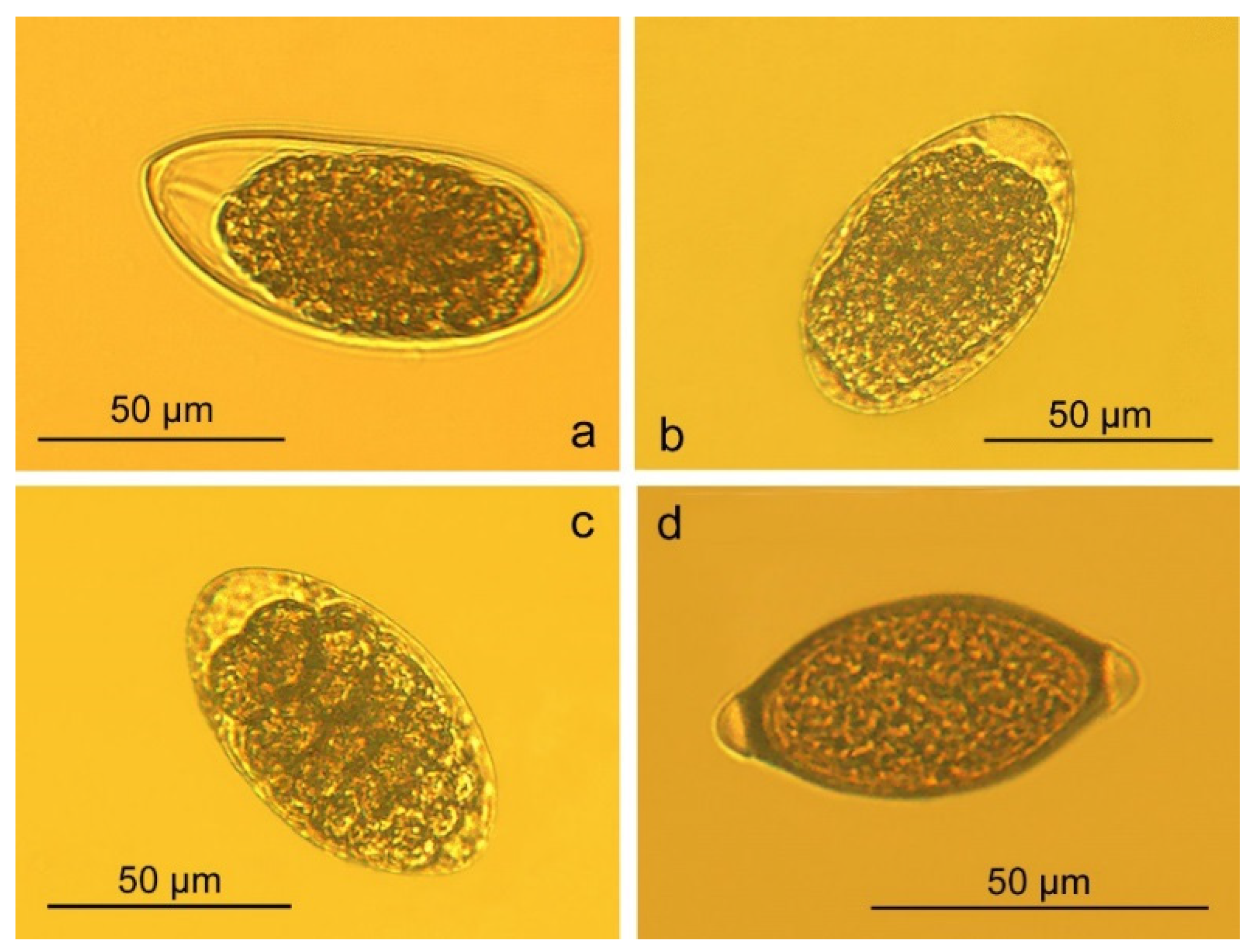
| Trichostrongylus sp. eggs | L | 81–94 | 86.18 ± 4.96 | 70–125 | Cattle, Sheep, Goats | Thienpont et al. [18] Foreyt [19] |
| W | 37–46 | 42.45 ± 2.62 | 30–55 | |||
| Haemonchus sp. eggs | L | 72–90 | 83 ± 4.94 | 62–95 | Cattle, Sheep, Goats | Thienpont et al. [18] Foreyt [19] |
| W | 44–50 | 47.33 ± 1.72 | 36–50 | |||
| Oesophagostomum sp. eggs | L | 76–85 | 81 ± 3.24 | 65–120 | Cattle, Sheep, Goats | Thienpont et al. [18] Foreyt [19] |
| W | 41–48 | 44.22 ± 2.05 | 40–60 | |||
| Trichuris sp. eggs | L | 62–72 | 66.8 ± 3.83 | 70–80 | Cattle, Sheep, Goats | Thienpont et al. [18] Foreyt [19] |
| W | 27–32 | 30.2 ± 2.17 | 30–42 | |||
| Trichostrongylus sp. L1 | L | 480–676 | 602.33 ± 106.68 | 650–770 | Ruminants | Kotelnikov [16] |
| W | 20–23 | 21.33 ± 1.53 | - | |||
| Dicrocoelium sp. eggs | L | 37–40 | 38.33 ± 1.21 | 38–45 | Cattle, Sheep | Thienpont et al. [18] |
| W | 23–24 | 23.33 ± 0.52 | 22–30 | |||
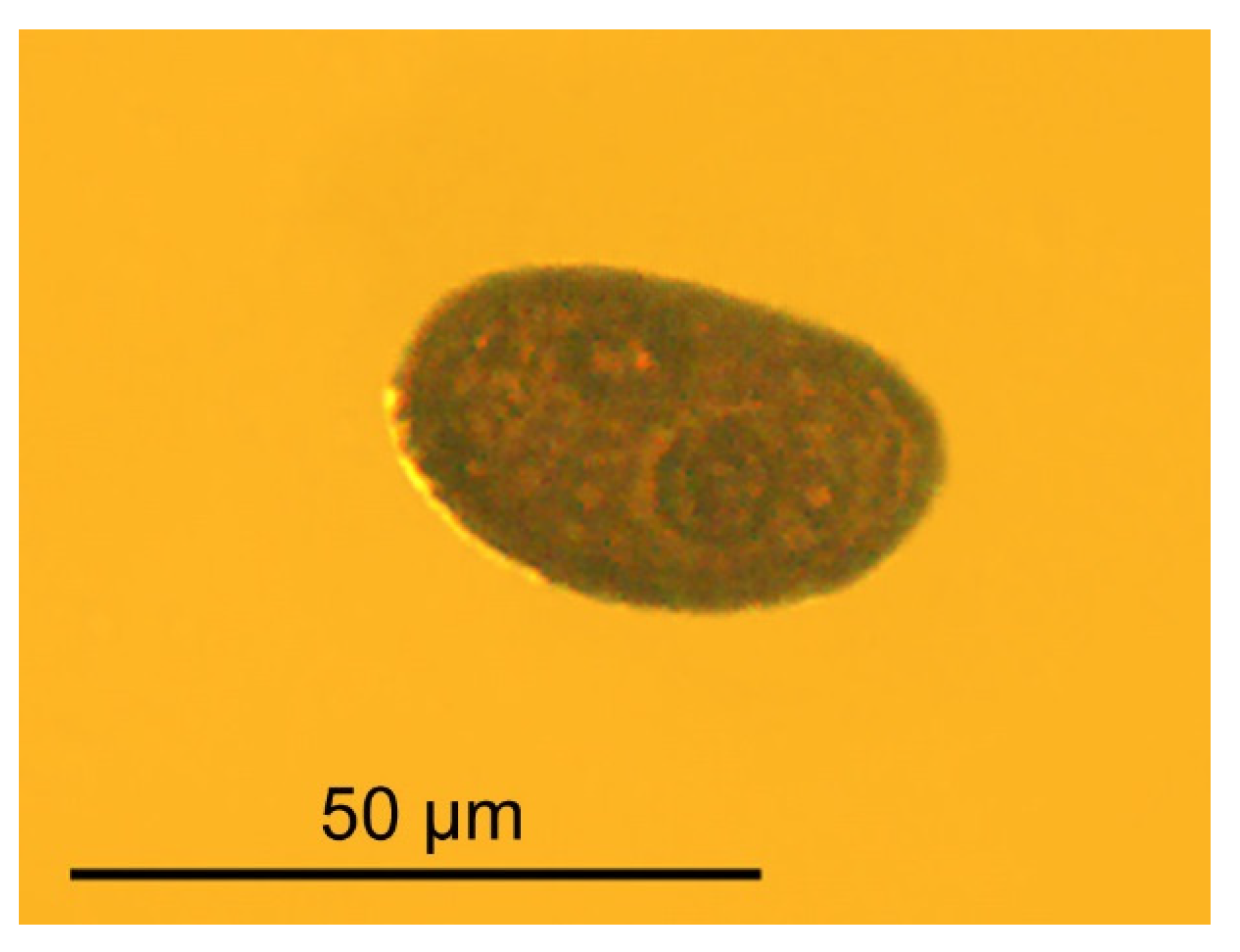
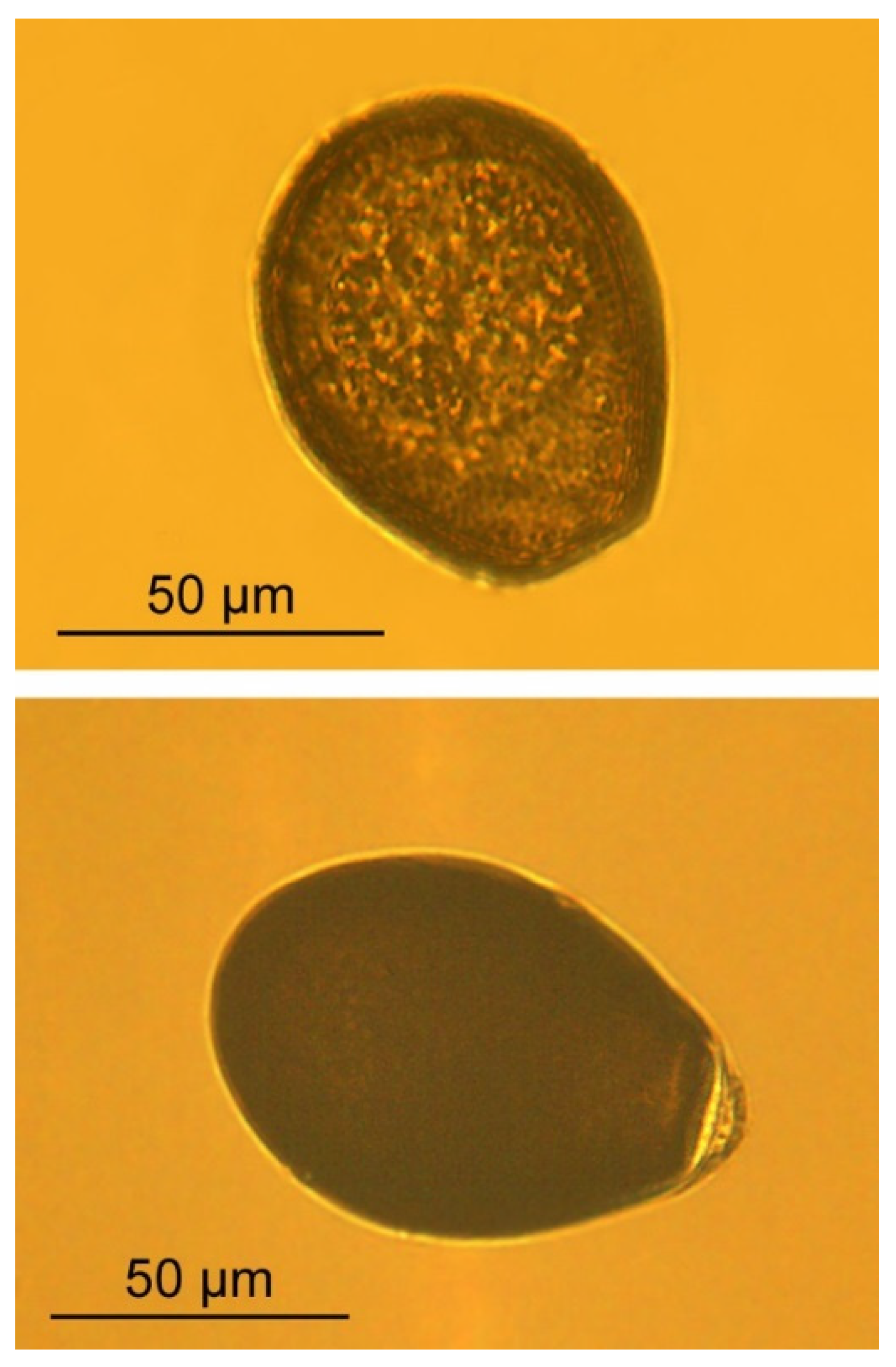
| Eimeria cameli oocysts | L | 76–96 | 86.62 ± 6.05 | 70–100 | Dromedary camels | Abbas et al. [6] Yagoub [17] |
| W | 56–70 | 63.10 ± 3.65 | 52.5–73.8 | |||
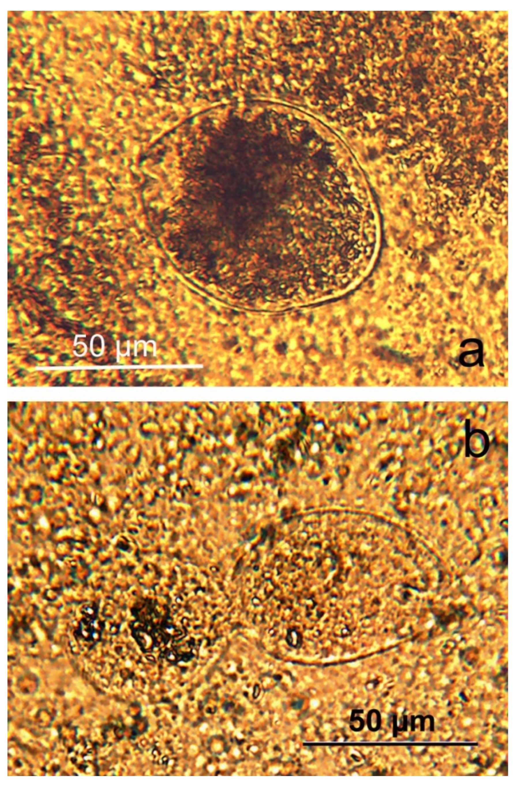
| Buxtonella cameli trophozoite | L | 64–86 | 74.5 ± 6.29 | 30–150 | Pigs | Foreyt [19] |
| W | 51–72 | 61.9 ± 5.15 | - | |||
Disclaimer/Publisher’s Note: The statements, opinions and data contained in all publications are solely those of the individual author(s) and contributor(s) and not of MDPI and/or the editor(s). MDPI and/or the editor(s) disclaim responsibility for any injury to people or property resulting from any ideas, methods, instructions or products referred to in the content. |
© 2023 by the authors. Licensee MDPI, Basel, Switzerland. This article is an open access article distributed under the terms and conditions of the Creative Commons Attribution (CC BY) license (https://creativecommons.org/licenses/by/4.0/).
Share and Cite
Panayotova-Pencheva, M.; Ponce-Gordo, F. Severe Parasite Co-Infection in a Captive Bactrian Camel: Case Report. J. Zool. Bot. Gard. 2023, 4, 728-737. https://doi.org/10.3390/jzbg4040051
Panayotova-Pencheva M, Ponce-Gordo F. Severe Parasite Co-Infection in a Captive Bactrian Camel: Case Report. Journal of Zoological and Botanical Gardens. 2023; 4(4):728-737. https://doi.org/10.3390/jzbg4040051
Chicago/Turabian StylePanayotova-Pencheva, Mariana, and Francisco Ponce-Gordo. 2023. "Severe Parasite Co-Infection in a Captive Bactrian Camel: Case Report" Journal of Zoological and Botanical Gardens 4, no. 4: 728-737. https://doi.org/10.3390/jzbg4040051