Article

Respiratory Infection by Cyathostoma (Hovorkonema) americana in a Population of Burrowing Owls (Athene cunicularia)—A Potential Case of Zoo–Wildlife Cross-Transmission

1
Zoomarine, 8201-864 Guia, Portugal
2
CIISA—Centre for Interdisciplinary Research in Animal Health, Faculty of Veterinary Medicine, University of Lisbon, 1300-477 Lisbon, Portugal
3
Associate Laboratory for Animal and Veterinary Sciences (AL4AnimalS), 1300-477 Lisbon, Portugal
*
Author to whom correspondence should be addressed.
These authors contributed equally to this work.
J. Zool. Bot. Gard. 2023, 4(4), 788-799; https://doi.org/10.3390/jzbg4040056
Submission received: 24 October 2023 / Revised: 15 November 2023 / Accepted: 29 November 2023 / Published: 5 December 2023

Abstract

:
A population of burrowing owls (Athene cunicularia) under professional care at Zoomarine Portugal presented with sudden respiratory clinical signs. Clinical management included a thorough diagnosis plan, including in-house fecal analysis that revealed the presence of ovoid unioperculate eggs. In the postmortem examination of one hyperacute dyspneic specimen, adult nematode parasites were collected and identified based on their morphology as Cyathostoma (Hovorkonema) americana. Even after a broad-spectrum deworming protocol as part of the treatment and metaphylaxis approach, the incidence of parasitic reinfection was high. The complete clinical resolution was only accomplished after the identification and management of the possible focus of infection, a wild population of cattle egrets (Bubulcus ibis) that frequently congregated above the owls’ habitat. To the authors’ best knowledge, this is the first report of infection by Cyathostoma (Hovorkonema) americana in burrowing owls. Although nematodes of the family Syngamidae are not commonly included in the differential diagnosis of infectious respiratory agents of birds of the order Strigiformes, this report highlights the possibility of opportunistic parasitism in a zoological context, especially where there is a continued proximity to free-ranging avifauna.

1. Introduction

Burrowing owls (Athene cunicularia) have a wide distribution on the continents of North and South America, and their conservation status is defined by the International Union of Conservation of Nature (IUCN) as of least concern, although there is a decreasing population trend [1,2]. Given the important role of these birds in maintaining a balance in the populations of their prey and also serving as prey themselves for other birds of prey, the decline of some populations may be causing a ripple effect throughout the ecosystem [3,4]. Due to seasonal changes on the population density of their prey, burrowing owls have adaptable food habits. Approximately 90% of burrowing owls’ diet is based on arthropods, including grasshoppers, crickets, and beetles, although they also eat small mammals, such as mice, small birds, and ground squirrels [3,5,6]. The main threat to these owls is the loss of habitat, mainly due to human activity [2,3,4]. Burrowing owls’ habitats consist of open-canopied areas, with sparse ground vegetation. These habitats may include agricultural lands, grasslands, prairies, plains, and deserts [3,4].
These small birds of prey have diurnal habits and use burrows in the ground for refuge [3]. They may dig their own burrows or use others abandoned by other animals such as prairie dogs, ground squirrels, badgers, tortoises, coyotes, and foxes. These burrows may reach more than 3 m in length, angled downwards [3,4].
Burrowing owls live 6–8 years in the wild and up to 10 years under professional care in captivity [3,7]. In the wild, two-thirds do not live to adulthood, with mortality frequently happening between fledging and the end of first year of age. The causes of mortality at a young age include low prey density, inexperience in capturing food, predators, and parasitic infections [3].
This species of owl is found in several zoological institutions. A burrowing owl population of 11 resident individuals at Zoomarine Portugal inhabited an outdoor walkthrough enclosure adapted for a variety of species of birds and reptiles. As part of the park’s zoological collection, these individuals were included in a preventative medicine program and had no relevant clinical history until the sudden and sequential development of respiratory clinical signs.
The subfamily Syngaminae Baylis & Daubney, 1926 includes parasitic nematode organisms found in the respiratory systems of both avian and mammalian hosts, with representatives such as Boydinema, Cyathostoma, and Syngamus for birds and Mammomonogamus and Rodentogamus for mammals. Notably, the genus Cyathostoma was established by Blanchard in 1849 and stands out as the most extensive, boasting a population exceeding 20 distinct species [8]. Cyathostoma sp. were reported in several bird species and, even though the occurrence of these nematodes is generally subclinical, heavy infections are often associated with secondary infections and death [8,9,10,11,12,13,14]. The ecology of Cyathostoma sp. parasites is important for veterinary and conservation reasons and is still a challenging taxonomy and a contentious topic [8].
In the most recent system of Syngaminae proposed by Lichtenfels in 1980 [15], despite being more than 40 years old, the genus Cyathostoma is divided into two subgenera based on the structure of the copulatory bursa and spiculae length. On the one hand, Cyathostoma (Cyathostoma) (Blanchard, 1849) exhibits a dorsal ray that extends beyond the end of the copulatory bursa, forming characteristic thorn-like projections and spicules that measure 0.08–0.4 mm. On the other hand, Cyathostoma (Hovorkonema) Turemuratov, 1963 features a dorsal ray that does not extend beyond the end of the copulatory bursa, and spicules are within the range of 0.45–0.8 mm.
Cyathostoma (Hovorkonema) americana was first described by Chapin in 1925, based on material collected from a red-tailed hawk (Buteo jamaicensis Gmelin, 1788) in Virginia (USA), and, while there are occasional subsequent reports of this species in birds of prey [8,10,11,13,14,16,17,18], there are no available reports of C. (Hovorkonema) americana in A. cunicularia.
This study aims to describe the diagnostic approach for the respiratory infection in a population of A. cunicularia held in a zoo, primarily focusing on parasite identification, exploring the evidence of cross-transmission, and investigating opportunistic parasite infections within the unique environment of a zoological setting.

2. Materials and Methods
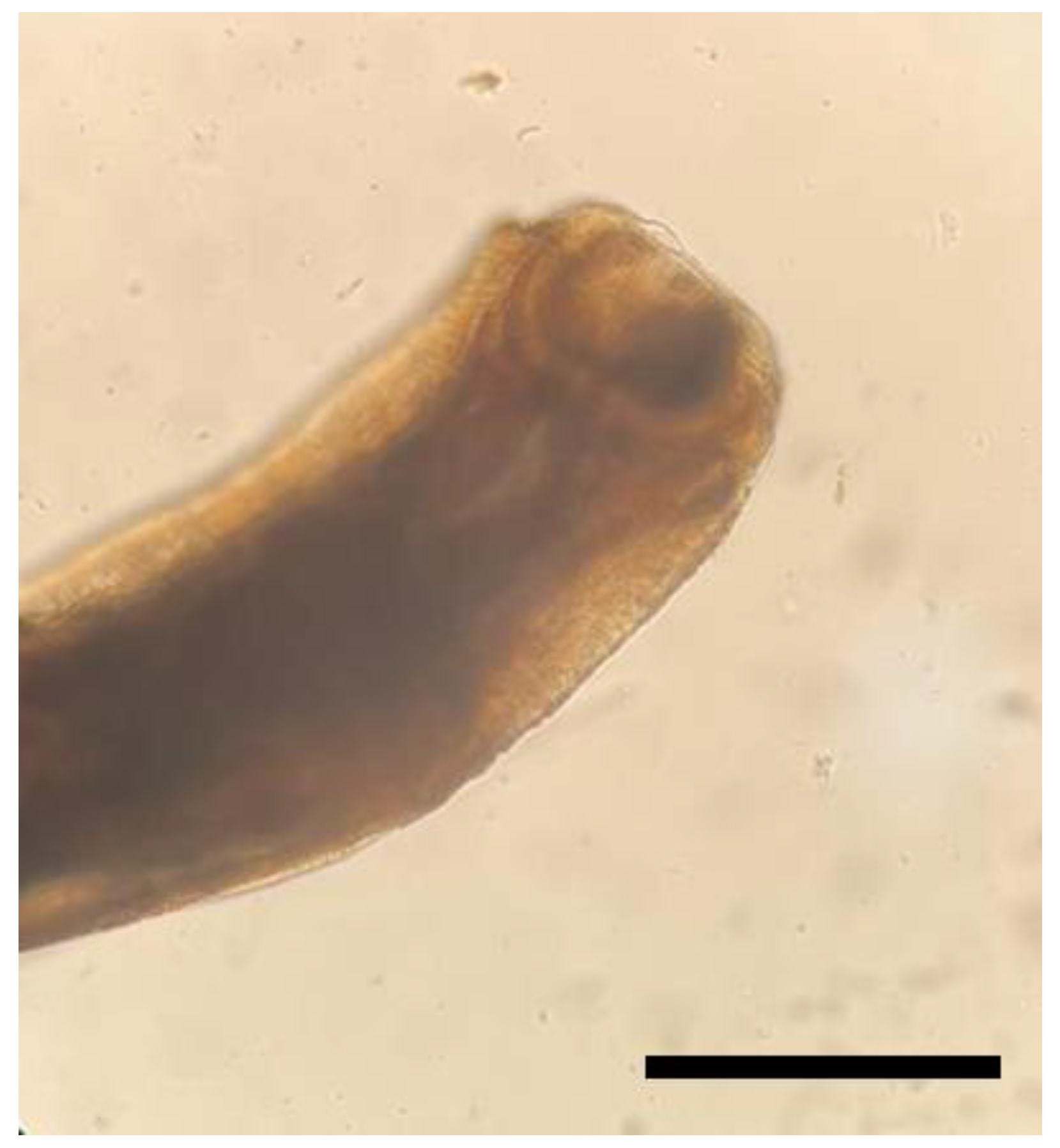
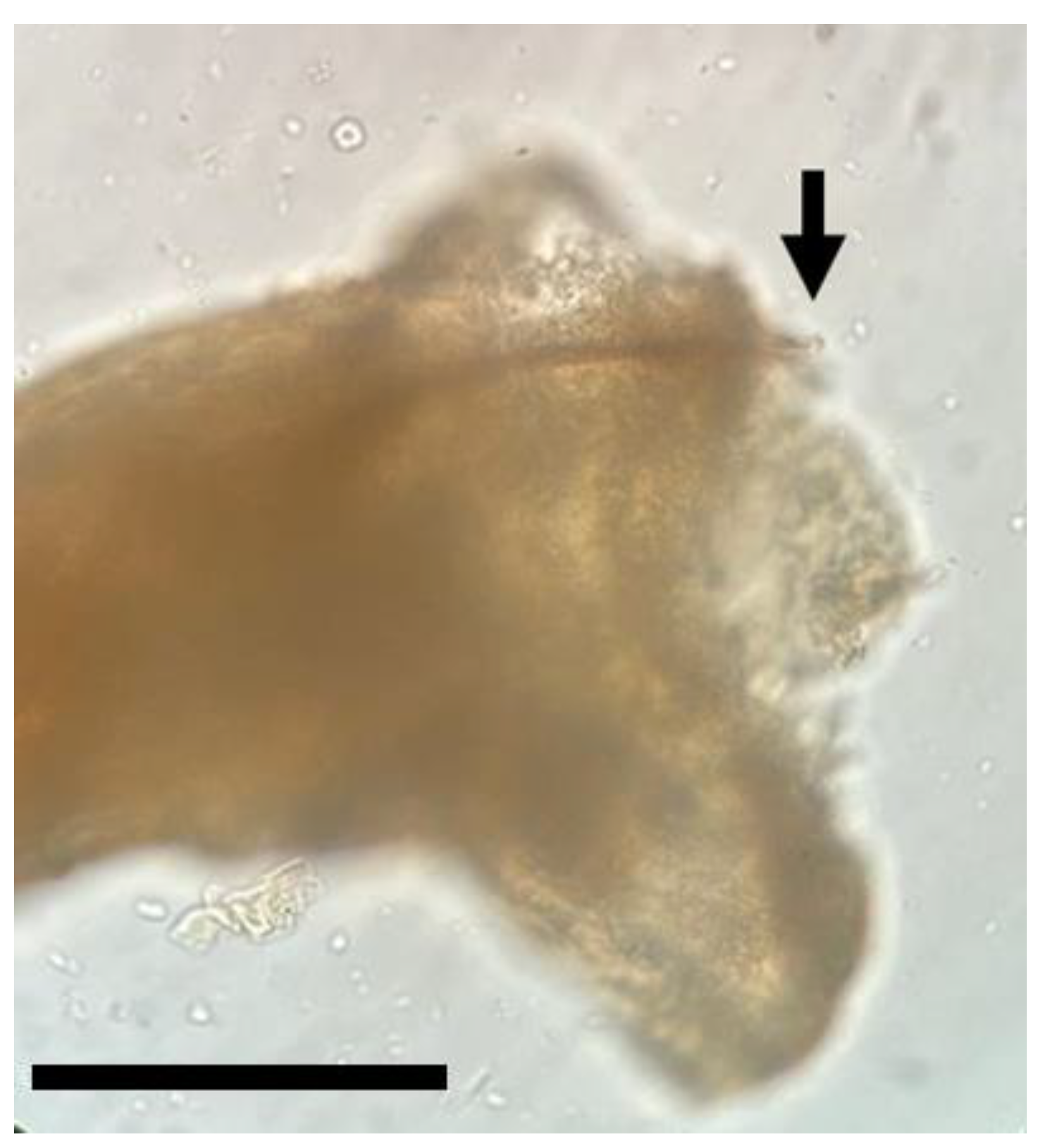
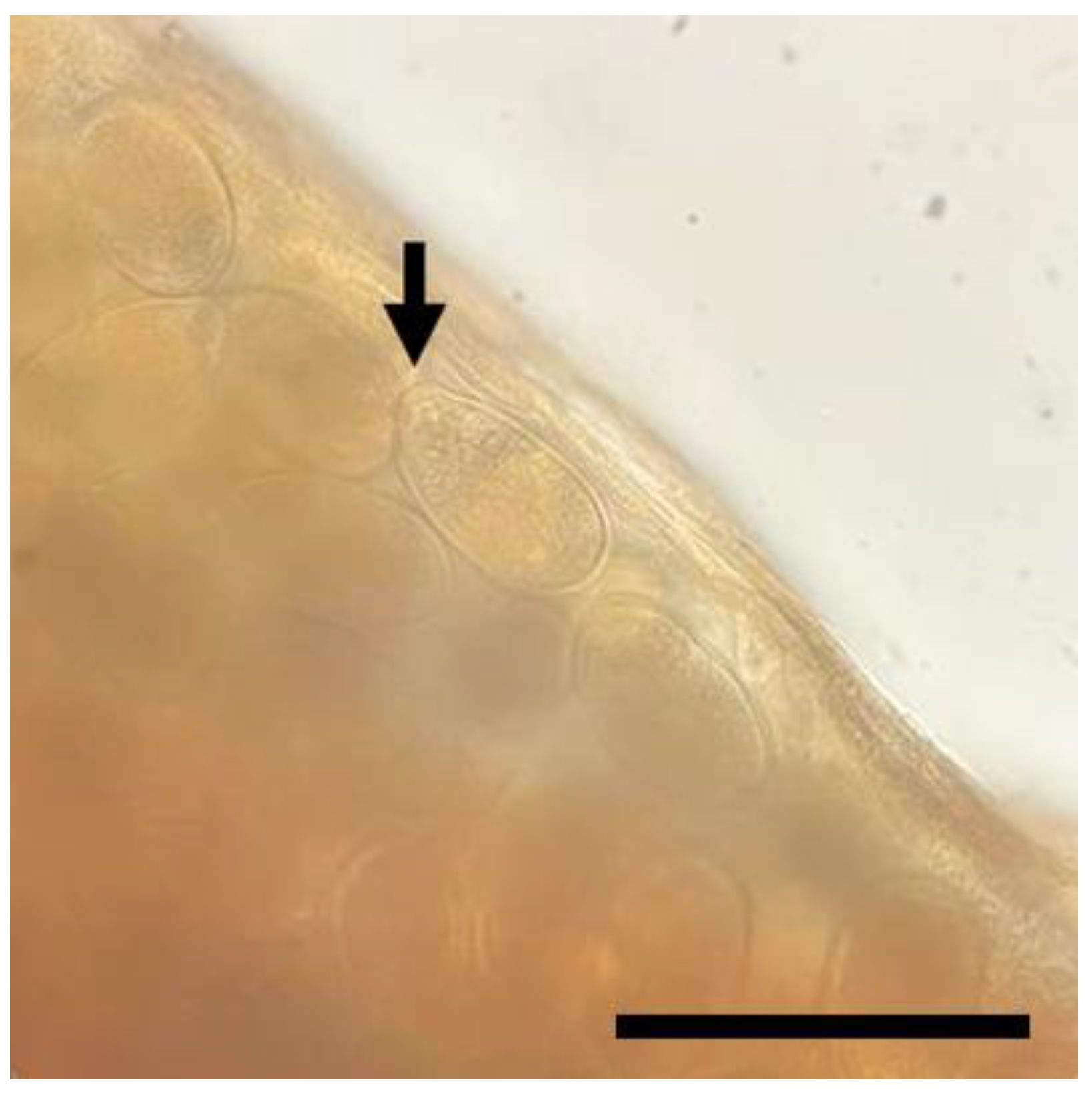
The burrowing owls with respiratory clinical signs went through a multimodal diagnostic approach, which included a full physical examination under general anesthesia with isoflurane, when possible, according to the clinical status of each individual. Blood sampling was performed for an in-house complete blood count and general biochemistry panel, the latter through a VETSCAN VS chemistry analyzer (Avian/Reptilian Profile Plus). Both total leukocyte and erythrocyte counts were manually completed, in which a 5 µL blood-filled pipette was inserted into a Natt-Herricks-TIC (Bioanalytic GmbH, Umkirch/Freiburg, Germany) 1:200 stain solution vial, and counting was performed with a Neubauer chamber. Evaluation of blood smears was performed after Diff-Quik staining. Hemoglobin levels were obtained through a hemoglobin analyzer HemoCue (HemoCue AB, Ängelholm, Sweden) and hematocrit after centrifugation of microhematocrit tubes (Centurion Scientific Ltd.—Pro-Vet, West Sussex, UK) at 12,000 rpm for 5 min. Radiographic studies included ventrodorsal and left lateral views with portable radiographic equipment (GIERTH HF300, GIERTH X-RAY International GmbH, Riesa, Germany). For in-house fecal wet mounts, one drop of new methylene blue was added immediately before microscopic observation.
The adult nematode parasites were sent to the Faculty of Veterinary Medicine at the University of Lisbon for analysis conducted by the Parasitology and Parasitic Diseases Service. The parasites were subjected to morphological identification and measurement and underwent the following procedures: (i) examination under a stereomicroscope for initial observation; (ii) mounting on slides with Hoyer’s medium for subsequent examination using a compound microscope.
The identification of parasite subgenera employed the Syngaminae classification system as proposed by Lichtenfels [15]. For the identification of the parasite species, the authors consulted the review of Cyathostoma sp. nematodes by Kanarek et al. [8]. Detailed observations and measurements encompassed body length, body width, depth of buccal capsule, spicule length in males, and egg dimensions.

3. Results

A substantial proportion, specifically 45% (5/11), of the population of burrowing owls developed clinical signs of respiratory origin. These clinical signs included dyspnea (5/5), tachypnea (5/5), crackles (3/5), and sneezes (1/5). Other clinical findings included compression of the abdominal air sacs’ radiographic silhouette (3/5), mild leukocytosis (2/5), eosinophilia (1/5), and subcutaneous emphysema (1/5) (Figure 1). Additionally, observation of the direct fecal smears revealed the presence of ovoid unioperculate eggs (Figure 2).
The postmortem examination of one individual who presented hyperacute dyspnea showed severe signs of pulmonary congestion. Adult nematodes were found in the parenchyma of the left lung apex. Notwithstanding the signs of air sacculitis, there were no parasites found in the trachea or air sacs. The male parasites exhibited a total length of 1.2 cm, while the females measured 2.3 cm. Notably, the morphological configuration of the dorsal ray of the copulatory bursa, which does not extend beyond the end (Figure 3), was identified as being under the subgenus Cyathostoma (Hovorkonema) (Turemuratov, 1963).
The ensuing measurements are all expressed in micrometers (μm). The dimensions of the male buccal capsule (width × depth) were 255 × 255 (Figure 4), while the female’s buccal capsule measured 350 × 210 (Figure 5). The male spicule length measured 435 (Figure 6), and the eggs exhibited dimensions of 85–92.5 in length and 50 in width, with an ovoid form and a single operculum (Figure 7). Notably, given the prevailing attributes, including the smaller spicules and the host being part of the parasite’s type group (birds of prey), the specimens were identified as C. (Hovorkonema) americana [16].

4. Discussion

Animal welfare assessment is crucial in modern zoological institutions in order to assure the highest possible standards of welfare for animals under professional care. This assessment is based on the Five Domains model, which includes nutrition, environment, health, behavior, and mental state [19,20,21]. The negative effects of parasitic infections in zoos are numerous and may extensively affect the health dominion (i.e., the absence of diseases, the absence of injuries, and the absence of pain induced by management procedures). The parasitic consequences may vary from the development of secondary infections or nutritional deficiencies to death in the case of severe parasitosis. Moreover, the systematic impact of parasitism may include reproduction impairment, which may be decisive not only for the success of a specific animal collection but also for conservation purposes. Lastly, in a zoological context, close contact between animals and humans is possible, hence the higher risk of the dispersion and spread of parasitic zoonoses [22,23].
The overall management of parasitic infections in zoos may present itself as a challenge, not only from a diagnostic and treatment perspective but also from an environmental point of view. The original population of burrowing owls under professional care at Zoomarine was captive-bred in other European institutions and arrived at Zoomarine around six years before the onset of the respiratory parasitic infection. Since their arrival, they inhabited a mixed-species outdoor walkthrough enclosure, which included other bird species (giant wood rail (Aramides ypecaha), cattle egret (Bubulcus ibis), striated heron (Butorides striata), reg-legged seriema (Cariama cristata), scarlet ibis (Eudocimus ruber), guira cuckoo (Guira guira), black-crowned night heron (Nycticorax nycticorax), southern wigeon (Mareca sibilatrix), ocellated turkey (Meleagris ocellata), roseate spoonbill (Platalea ajaja), glossy ibis (Plegadis falcinellus), and green aracari (Pteroglossus viridis)) and reptiles (green iguana (Iguana iguana) and red-eared slider (Trachemys scrypta scrypta)). This type of natural-looking outdoor habitat is a creative outlet for physical activity and mental stimulation, providing several environmental enrichment opportunities covering both generic (choice, control, variety, and complexity) and specific needs [21,24]. However, one of the logistical challenges of this type of enclosure involves the difficult access to the animals, so the group of owls in Zoomarine was trained to enter a crate, allowing easy access for medical reasons whenever needed. This played a key part, as these owls were under a preventative health program, which included but was not limited to fecal sampling for coprological examination (wet mount, Gram stain, Diff-Quik stain, and flotation). However, even under a comprehensive medical protocol, the untimely and low sensitivity of certain diagnostic tools may delay treatment and worsen the overall prognosis of a certain parasitic infection [25,26]. Considering this diagnosis challenge along with the characteristics of the mixed-species outdoor enclosure and the possibility of nearby access for wild fauna and pathogens cross-transmission, all birds were under a deworming protocol every six months, which consisted of fenbendazole (50 mg/kg, per os (PO), once), ivermectin (0.2 mg/kg, PO, once, 15 days after fenbendazole) and toltrazuril (10 mg/kg, PO, three administrations, every other day (EOD), 7 days after ivermectin).
Even considering the regular anti-parasitic protocol, respiratory parasitosis was included as a differential diagnosis after the development of respiratory signs in 45% of the owl population. Both the clinical signs and findings from the physical examination and complementary exams fall inside the non-specific signs of respiratory disease in birds [27,28]. Considering the complexity of the avian respiratory tract, disease processes can be located in different anatomic regions, though the distinction between clinical signs is not clear, as many of them (e.g., dyspnea, breathing with an extended neck, tachypnea, respiratory noises, and a change in pitch or voice) may refer to both upper and lower respiratory tract disease. However, upper airway disease does not usually present with severe respiratory distress [27,28]. Due to its specific anatomic characteristics (e.g., air sacs), the avian respiratory system constitutes a substantial target for infection by a myriad of infectious agents [27]. Previous authors reported unspecific clinical signs of infections by Cyathostoma sp., such as depression and sudden death, along with syndromes and lesions, such as pneumonia, bronchitis, and air sacculitis, in birds of prey [10,11,13,29].
Hematology abnormalities may include leukocytosis, whereas serum biochemistries are not generally of particular interest in respiratory/parasitic diseases [27]. Indeed, only two individuals, one of them with eosinophilia, showed a slight increase in their total leukocyte counts. Although eosinophilia is commonly associated with mammals with parasitic diseases, this relation in avian species is not straightforward, so more complementary studies are needed on this subject [30]. A physical examination confirmed one case of severe and generalized subcutaneous emphysema, with the typical crackling and air-filled distention, with the common causes being trauma or lung/air sac inflammation associated with parasites or other infectious agents [31,32,33].
The treatment protocol was adapted according to each individual’s clinical condition. The description of the specific clinical approach falls beyond the scope of this work. It is important to note, however, the use of a broad-spectrum protocol that included fenbendazole (50 mg/kg, PO, once a day (SID), 3 to 5 days), in conjunction in some cases with ivermectin (0.2 mg/kg, PO, once, 5 to 10 days after the therapeutic protocol with fenbendazole). Metaphylaxis measures through the same deworming protocol may have prevented the development of a clinical parasitosis in the rest of the owl population. Only one of the five individuals that presented with clinical signs died after a hyperacute clinical respiratory presentation. The literature includes several reports of mortality in birds of prey infected with Cyathostoma sp. Fatal parasitic pneumonia was reported in three injured wild owls in Southern Ontario along with four juvenile A. cunicularia bred in captivity [10]. A survey in Canada of 394 specimens of Falconiformes and Strigiformes also described fatal infections [13]. Finally, a case of fatal epicarditis associated with Cyathostoma species in a hen harrier (Circus cyaneus) was also reported [14].
Up until now, the identification of Cyathostoma (Hovorkonema) nematodes collected from the respiratory tracts of Accipitriformes, Falconiformes, and Strigiformes in Europe and North America has been highly inconsistent among authors [8]. These inconsistencies arose due to several shifts in the taxonomic placement of Cyathostoma species, which stem from different interpretations of morphological characteristics, various revisions, and questionable synonymities [34,35,36,37]. Furthermore, these discrepancies have been compounded by different authors utilizing distinct classification systems. For instance, Hartwich amalgamated three C. (Hovorkonema) species—C. (Hovorkonema) americana, C. (Hovorkonema) bronchialis, and C. (Hovorkonema) variegatum sensu stricto—into C. (Hovorkonema) variegatum [37]. This conflation resulted in several identifications of C. (Hovorkonema) variegatum in birds of prey that corresponded to the C. (Hovorkonema) americana species [8].
C. (Hovorkonema) americana, which is a typical parasite of the trachea and air sacs of raptors, C. (Hovorkonema) bronchialis, typically found in Anseriformes and also detected in Casuariiformes, and C. (Hovorkonema) variegatum, a typical parasite of the trachea of cranes and storks, constitute the three species within this subgenus. According to Kanarek’s comprehensive morphological and molecular examination of Cyathostoma nematodes parasitizing the respiratory tracts of birds of prey in Europe and North America, only one species of C. (Hovorkonema) in birds of prey is confirmed to exist: C. (Hovorkonema) americana [8]. However, the possible but rare incidental occurrence of other C. (Hovorkonema) species, typical for different avian host groups, is acknowledged.
With respect to our samples and focusing on the characteristics highlighted as the most reliable for Cyathostoma sp. identification, the subgenus C. (Hovorkonema) is defined by spicules measuring 0.45–0.8 mm, while Cyathostoma (Cyathostoma) (Blanchard, 1849) has measurements ranging from 0.08 to 0.4 mm [38]. Our specimens fall between these ranges (0.435 mm). However, the absence of thorn-like projections on the copulatory bursa aligns with classification under the C. (Hovorkonema) subgenus.
Two of the three species of this subgenera can be clearly distinguished based on spicular length. C. (Hovorkonema) variegatum boasts notably longer spicules (0.58–0.77 mm) [34,39], compared to C. (Hovorkonema) americana (0.47–0.511 mm) [16,40]. The spicules length of C. (Hovorkonema) bronchialis (0.51–0.7 mm) is closely aligned with that of C. (Hovorkonema) variegatum [16,41,42,43]. Although this matter is still in need of further research, it suggests the potential synonymy of C. (Hovorkonema) bronchialis with C. (Hovorkonema) variegatum, as theorized by Kanarek et al. [8].
Considering these observed spicule lengths and given the close similarity of C. (Hovorkonema) americana’s spicules to our exemplars, along with the fact that it is the parasite of our designated host type (birds of prey), the sole plausible identification is C. (Hovorkonema) americana. Furthermore, the total lengths of the male and female parasites also fall within the species’ described measurements.
While C. (Hovorkonema) variegatum in typical hosts exclusively occurs in the trachea, C. (Hovorkonema) americana in birds of prey was mainly recorded in air sacs and less frequently in the trachea [8,10,11,34,39,40]. There are reports on the presence of Cyathostoma nematodes in the trachea, bronchi, and air sacs of birds of prey [12,36]. However, there is no specific mention of these parasites being found in the lung parenchyma, least of all exclusively in this site, like in the present case [8].
Hunter et al. reported the presence of a Cyathostoma species parasite in an A. cunicularia specimen; however, the species of the parasite was not specified [10]. To the best of our knowledge, the current report is the first description of Cyathostoma (Hovorkonema) americana in A. cunicularia. The morphological differences and lack of previous reports do not rule out, but rather suggest, that these parasites could be a new species, with C. (Hovorkonema) americana being the closest match among the described species. Further research is needed to confirm this hypothesis, including the molecular characterization of these parasites, comparing the results with the molecular analyses available in the literature [8,11].
Although there are several studies available on parasitism in wild animals, more specifically wild birds, the possibility of free-ranging avifauna acting as a source of infection for captive specimens is a rarely explored subject [25,44]. It has been established, however, that exposure to wild avifauna may result in the parasitism of captive birds, depending if certain conditions for parasitic development are suitable such as parasite–host specificity, parasite life cycle, host resistance, husbandry-related factors, and environmental factors [25,44]. The host specificity of parasites is variable. While some parasite species are only found in a limited number of host species (otherwise known as highly host-specific), other parasites can affect multiple host species (considered to be host generalists, with low host specificity) [44].
There was a potential origin of the parasitic infection in the A. cunicularia population, namely, a group of more than 50 wild cattle egrets (B. ibis) that used to gather on top of the enclosure net, attracted by individuals of the same species that were part of the zoological collection and in that same enclosure. This gathering followed years of a particularly high birth rate of the captive B. ibis population and an increase in the total number of individuals kept in the enclosure. The wild B. ibis built nests and stayed above the enclosure throughout the day, and several nestlings and fledglings were seen throughout this period. Moreover, an estimated count of these wild B. ibis, not immediately above the enclosure but in the adjacent trees, included more than 600 animals.
Cattle egrets are free-range migratory birds in the family Ardeidae (order Pelecaniformes) with widespread distribution. Inclusively, they are the only member of the genus Bubulcus found in the tropics, subtropics, and warm temperate zones [45,46]. They are frequently found in association with cattle, with whom they establish a symbiotic relationship. This species has an extremely large range and does not approach the thresholds for vulnerable conservation status under the range size criterion—cattle egrets are classified as Least Concern under the IUCN conservation status [45,46].
Even though metaphylaxis measures were adopted, and all cleaning and disinfection protocols for the owls’ underground burrows were revised, the incidence of reinfection was high. The complete clinical resolution of the parasitic infection of the population of burrowing owls was only accomplished when all the resident cattle egrets were transferred to other zoological institutions, and their wild counterparts immediately stopped congregating above the enclosure. This probable focus of infection cannot, however, be confirmed since a coprological analysis of these wild individuals was not performed. Cattle egrets are not yet known to be definitive hosts of C. (Hovorkonema) americana, but several nematodes were reported in these migratory birds, including from the Syngamidae family [46]. In this study, 40 free-ranging specimens of B. ibis were captured and examined for endoparasites and associated ectoparasites. The gastrointestinal content of each specimen was examined for adults and eggs of different helminths and protozoan cysts. The observed endoparasites included Ascaridia galli (with the highest prevalence, 35%), Heterakis spp. (17.5%), Capillaria spp. (12.5%), Trichostrongylus tenuis (12.5%), Fascioloides magna (10%), and Syngamus trachea (7.5%). This study also showed that there was a significantly higher risk of co-infection by the different types of parasites and that the ubiquitous presence of B. ibis may play an important in parasite dispersal in a global perspective. Furthermore, cattle egrets feed daily on a variety of insects, crustaceans, fish, lizards, frogs, spiders, moths, rodents, and earthworms, making it a favorable host through the ingestion of intermediate and paratenic hosts [46,47].
Interestingly, Syngamidae nematodes, including Cyathostoma sp., have been reported in free-ranging birds from across Europe, yet, there is only one available occurrence of Syngamidae in wild avifauna in Portugal, namely, the genus Syngamus. This study was developed in a wildlife rehabilitation center in Castelo Branco, Portugal, from January 2016 to May 2017, concerning the examination of the fecal samples of a total of 65 birds, in which birds of prey were the most representative group (77% of the samples) [11,34,37,39,40,43,48].
Generalist parasites that tolerate host physiology differences can infect a large variety of avian species [44]. In the present case of monoxenous parasites, owls may have become directly infected by eating soil or other materials contaminated with the feces of wild avifauna. Moreover, since burrowing owls are territorial and tend to stay in the same area, in or close to their underground burrows, there might have been a higher exposure to the feces of the wild B. ibis population. The possibility of paratenic hosts (e.g., earthworms) facilitating the chance of infection cannot be excluded, since the owls’ habitat included areas of earthy soil. Finally, reinfection is possible through the ingestion of food/substrates contaminated with their own feces.
Parasites seldom lead to subclinical effects in wild avifauna, thus contributing to the dispersion and transmission to birds under human care inhabiting enclosures that allow close proximity to wild birds or paratenic hosts [25,44]. This report highlights the need to consider external sources of excretion and parasitic contamination as part of the management of parasitic infections when the type of zoological enclosures allows the possibility of opportunistic parasitism in susceptible avian collections. More studies are needed demonstrating cases of parasite transmission in a zoological context and describing the species of parasites affecting both captive and free-ranging birds. More studies on zoo–wildlife cross-transmission would benefit the adaptation of specific environment control measures (e.g., architectural designing; the removal of nearby roosts; paratenic host–control programs), as well as enhance effective preventative medical and husbandry protocols.

Author Contributions

Conceptualization, G.N.M. and L.M.d.C.; methodology, G.N.M., J.T.C. and M.L.; validation, G.N.M., J.T.C. and M.P.; formal analysis, G.N.M. and J.T.C.; investigation, G.N.M., J.T.C., M.P. and M.L.; writing—original draft preparation, G.N.M., J.T.C., M.P. and C.F.; writing—review and editing, G.N.M., J.T.C., M.P., M.L., C.F., N.U. and L.M.d.C.; supervision, L.M.d.C.; project administration, G.N.M.; funding acquisition, J.T.C. and L.M.d.C. All authors have read and agreed to the published version of the manuscript.

Funding

The research developed by J.T.C., M.P., and L.M.d.C. is supported by national funds through FCT—Fundação para a Ciência e Tecnologia, under the scope of projects UIDB/00276/2020 (CIISA-FMV-ULisboa) and LA/P/0059/2020 (AL4AnimalS). J.T.C. holds a scientific initiation grant (ref. UIDB/00276/2020) and a master grant, MSC22Jul-04, both provided by CIISA-FMV-ULisboa.

Institutional Review Board Statement

Ethical review and approval were waived for this study, due to the absence of interference with the normal animal health program designed by the zoo’s veterinarians. This study followed the daily activity of the institution and its normal animal management, in strict collaboration with the zoo owners and assistant veterinarians. No interferences were made during the regular health management of all collections, since all animals were dewormed, and fecal samples were mostly collected from soil after natural excretion by the animals.

Informed Consent Statement

Not applicable.

Data Availability Statement

The authors confirm that the data supporting the findings of this study are available within the article.

Acknowledgments

The authors would like to thank the veterinary nurses and all the bird trainers of Zoomarine for their fundamental work and dedication throughout this clinical situation.

Conflicts of Interest

The authors declare no conflict of interest. The funders had no role in the design of the study; in the collection, analyses, or interpretation of data; in the writing of the manuscript; or in the decision to publish the results.

References

  1. The IUCN Red List of Threatened Species Home Page. BirdLife International 2016, Athene cunicularia. Available online: https://www.iucnredlist.org/species/pdf/93227732 (accessed on 6 July 2023).
  2. Rosenberg, K.V.; Kennedy, J.A.; Dettmers, R.; Ford, R.P.; Reynolds, D.; Alexander, J.D.; Beardmore, C.J.; Blancher, P.J.; Bogart, R.E.; Butcher, G.S.; et al. Partners in Flight Landbird Conservation Plan: 2016 Revision for Canada and Continental United States; Bogart, R.E., Ed.; Partners in Flight. 2016, pp. 1–105. Available online: https://partnersinflight.org/resources/the-plan/ (accessed on 27 November 2023).
  3. Brandes, S. Athene cunicularia, Animal Diversity Web. Available online: http://animaldiversity.org/accounts/Athene_cunicularia/ (accessed on 12 July 2023).
  4. Haug, E.; Oliphant, L. Movements, activity patterns, and habitat use of Burrowing Owls in Saskatchewan. J. Wildl. Manag. 1990, 54, 27–35. [Google Scholar] [CrossRef]
  5. Sarno, R.J.; Nixon, P.A.; Mealey, B.K.; Concoby, R.E.; Mrykalo, R.J.; Grigione, M.M. Suitability of translocation sites for Florida burrowing owls: Prey availability and diet. Southeast. Nat. 2012, 11, 755–764. [Google Scholar] [CrossRef]
  6. Trulio, L.; Higgins, P. The diet of western burrowing owls in an urban landscape. West. N. Am. Nat. 2012, 73, 348–356. [Google Scholar] [CrossRef]
  7. BBL—Longevity Records of North American Birds. Available online: http://genomics.senescence.info/species/biblio.php?id=427 (accessed on 23 October 2014).
  8. Kanarek, G.; Zaleśny, G.; Sitko, J.; Blanco, A.I. Taxonomic status of Cyathostoma nematodes (Nematoda: Syngaminae) parasitizing respiratory tracts of birds of prey and owls in Europe and North America: How many species are there? Helminthologia 2016, 53, 47–54. [Google Scholar] [CrossRef]
  9. Fernando, M.A.; Barta, J.R. Tracheal Worms. In Parasitic Diseases of Wild Birds, 1st ed.; Atkinson, C.T., Thomas, N.J., Hunter, D.B., Eds.; John Wiley & Sons, Ltd.: Ames, IA, USA, 2008; pp. 343–354. ISBN 978-0-813-82081-1. [Google Scholar]
  10. Hunter, D.B.; McKeever, K.; Bartlett, C. Cyathostoma Infections in Screech Owls, Saw-whet Owls, and Burrowing Owls in Southern Ontario. In Raptor Biomedicine, 1st ed.; Redig, P.T., Cooper, J.E., Remple, J.D., Hunder, D.B., Eds.; University of Minnesota Press: Minneapolis, MN, USA, 1993; pp. 54–56. ISBN 978-0816622191. [Google Scholar]
  11. Krone, O.; Friedrich, D.; Honisch, M. Specific status and pathogenicity of syngamid nematodes in bird species (Ciconiformes, Falconiformes, Gruiformes) from Germany. J. Helminthol. 2007, 81, 67–73. [Google Scholar] [CrossRef]
  12. Kanarek, G.; Horne, E.C.; Zaleśny, G. Cyathostoma (Cyathostoma) phenisci Baudet, 1937 (Nematoda: Syngamidae), a parasite of respiratory tract of African penguin Spheniscus demersus: Morphological and molecular characterization with some ecological and veterinary notes. Parasitol. Int. 2013, 62, 416–422. [Google Scholar] [CrossRef] [PubMed]
  13. Lavoie, M.; Mikaelian, I.; Sterner, M.; Villeneuve, A.; Fitzgerald, G.; McLaughlin, J.; Lair, S.; Martineau, D. Respiratory Nematodiases in Raptors in Quebec. J. Wildl. Dis. 1999, 35, 375–380. [Google Scholar] [CrossRef]
  14. Vaughan-Higgins, R.; Murphy, S.; Carter, I.; Pocknell, A.; Harris, E.; Sainsbury, A.W. Fatal epicarditis in a hen harrier (Circus cyaneus) a red-listed bird of high conservation concern in Britain associated with Cyathostoma species and Escherichia coli infection. Vet. Rec. 2013, 173, 477. [Google Scholar] [CrossRef]
  15. Lichenfels, J.R. Keys to the genera of the superfamily Strongyloidea. In CIH Keys to the Nematode Parasites of Vertebrates; Anderson, R.C., Chabaud, A.G., Willmott, S., Eds.; Commonwealth Institute of Helminthology: Farnham Royal, UK, 1980; Volume 7, 41p. [Google Scholar]
  16. Chapin, E.A. Review of the nematode genera Syngamus Sieb. and Cyathostoma E. Blanch. J. Agric. Res. 1925, 30, 557–570. [Google Scholar]
  17. Borgsteede, F.H.; Okulewicz, A.; Zoun, P.M.; Okulewicz, J. The helminth fauna of birds of prey (Accipitriformes, Falconiformes and Strigiformes) in the Netherlands. Acta Parasitol. 2003, 48, 200–207. [Google Scholar]
  18. Stiko, J.; Okulewicz, A. Checklist of the Nematodes in Birds in the Czech Republic and the Slovak Republic; Comenius Museum: Přerov, Czech Republic, 2010; 104p, ISBN 9788090371392. [Google Scholar]
  19. Kells, N.J. Review: The Five Domains model and promoting positive welfare in pigs. Animals 2022, 16, 100378. [Google Scholar] [CrossRef] [PubMed]
  20. Mellor, D.J.; Beausoleil, N.J.; Littlewood, K.E.; McLean, A.N.; McGreevy, P.D.; Jones, B.; Wilkins, C. The 2020 Five Domains Model: Including Human–Animal Interactions in Assessments of Animal Welfare. Animals 2020, 10, 1870. [Google Scholar] [CrossRef] [PubMed]
  21. Tallo-Parra, O.; Salas, M.; Manteca, X. Zoo Animal Welfare Assessment: Where Do We Stand? Animals 2023, 13, 1966. [Google Scholar] [CrossRef] [PubMed]
  22. Panayotova-Pencheva, M.S. Parasites in Captive Animals: A Review of Studies in Some European Zoos. Zool. Gart. 2013, 82, 60–71. [Google Scholar] [CrossRef]
  23. Papini, R.; Girivetto, M.; Marangi, M.; Mancianti, F.; Giangaspero, A. Endoparasite Infections in Pet and Zoo Birds in Italy. Sci. World J. 2012, 2012, 253127. [Google Scholar] [CrossRef] [PubMed]
  24. Mellen, J.; MacPhee, M.S. Philosophy of Environmental Enrichment: Past, Present, and Future. Zoo Biol. 2001, 20, 211–226. [Google Scholar] [CrossRef]
  25. Carrera-Játiva, P.D.; Morgan, E.R.; Barrows, M.; Wronski, T. Gastrointestinal parasites in captive and free-ranging birds and potential cross-transmission in a zoo environment. J. Zoo Wildl. Med. 2018, 49, 116–128. [Google Scholar] [CrossRef]
  26. Carvalho, A.F.; Barnabe, A.S.; Federsoni, I.P.; Ferraz, R.R.; Marco, R.M.; Garcia, I.P. Efficacy of parasitological diagnosis methods in wild animals kept in captivity. Arq. Do Inst. Biol. 2017, 84, e0502016. [Google Scholar] [CrossRef]
  27. Crosta, L. Respiratory Diseases of Parrots: Anatomy, Physiology, Diagnosis and Treatment. Vet. Clin. Exot. Anim. Pract. 2021, 24, 397–418. [Google Scholar] [CrossRef]
  28. Forbes, N.A. Raptors: Parasitic disease. In BSAVA Manual of Raptors, Pigeons and Passerine Birds, 1st ed.; Chitty, J., Lierz, M., Eds.; British Small Animal Veterinary Association: Gloucester, UK, 2008; Chapter 19; pp. 205–206. ISBN 978-1-905319-04-6. [Google Scholar]
  29. Lierz, M.; Schuster, R.; Ehrlein, J.; Gobel, T. Findings of Hovorkonema variegatum in northern goshawk (Accipiter gentilis). Kleintierpraxis 1998, 43, 43–46. [Google Scholar]
  30. Mitchell, E.; Johns, J. Avian Hematology and Related Disorders. Vet. Clin. Exot. Anim. Pract. 2008, 11, 501–522. [Google Scholar] [CrossRef] [PubMed]
  31. Bowles, H.L.; Odberg, E.; Harrison, G.J.; Kottwitz, J.J. Surgical resolution of soft tissue disorders. In Clinical Avian Medicine, 2nd ed.; Harrison, G.J., Lightfoot, T.L., Eds.; Spix Publishing, Inc.: Palm Beach, FL, USA, 2006; Volume 2, p. 794. ISBN 00-9754994-0-8. [Google Scholar]
  32. Doneley, B.; Harrison, G.J.; Lightfoot, T.L. Maximizing Information from the Physical Examination. In Clinical Avian Medicine, 2nd ed.; Harrison, G.J., Lightfoot, T.L., Eds.; Spix Publishing, Inc.: Palm Beach, FL, USA, 2006; Volume 1, p. 169. ISBN 00-9754994-0-8. [Google Scholar]
  33. Lee, S.-Y.; Kim, H.-J.; Kim, J.-W. Deflation Treatment for Subcutaneous Emphysema in a Goffin Cockatoo (Cacatua goffini). J. Vet. Clin. 2011, 28, 519–521. [Google Scholar]
  34. Vogel, H. About biology and morphology of Cyathostoma variegatum (Creplin, 1849) [Syngamus variegatum]. Z. Infekt.-Skrankh 1928, 34, 97–117. [Google Scholar]
  35. Ali, M.M. A review and revision of the subfamily Cyathostominae Nicoll, 1927 (Nematoda, Syngamidae). Acta Parasitol. Pol. 1970, 17, 237–246. [Google Scholar]
  36. Ryzhikov, K.M. The biology of a nematode Cyathostoma bronchialis (Syngamidae: Strongylata). Helminthologia 1980, 17, 241–244. [Google Scholar]
  37. Hartwich, G.; Nematoda, I.I. Strongylida: Strongyloidea und Ancylostomatoidea. In The Wildlife of Germany; Gustav Fisher Verlag: Jena, Germany; Frankfurt, Germany, 1994; p. 157. [Google Scholar]
  38. Kanarek, G. The occurrence of Cyathostoma (Cyathostoma) microspiculum (Skrjabin, 1913) (Nematoda: Syngamidae) in the great cormorant [Phalacrocorax carbo (L., 1758)] in north-eastern Poland. J. Helminthol. 2009, 83, 391–398. [Google Scholar] [CrossRef] [PubMed]
  39. Okulewicz, A. Cyathostoma variegatum (Creplin, 1849) Chapin, 1925 and Cyathostoma lari Blanchard, 1849 (Nematoda, Syngamidae) in Poland. Wiad. Parazytol. 1984, 30, 53–56. [Google Scholar]
  40. Borgsteede, F.H.; Okulewicz, A. Justification of the species Cyathostoma (Hovorkonema) americana. Helminthologia 2001, 38, 115–121. [Google Scholar]
  41. Griffiths, H.J.; Leary, R.M.; Fenstermacher, R. A new record for gapeworm (Cyathostoma bronchialis) infection of domestic geese in North America. Am. J. Vet. Res. 1954, 15, 298–299. [Google Scholar]
  42. Ryžikov, K.M.; Zavadil, R. On the identity of species of the genus Cyathostoma collected from ostrich. Acta Univ. Agric. Silvic. Brno 1958, 2, 125–132. [Google Scholar]
  43. Hernandez-Rodriguez, S.; Calerto-Carretero, R.; Becerra-Martel, C.; Dominguez de Tena, M.; Moreno-Montañez, T.; Martinez-Gomez, F.; Barasona-Mata, J. Cyathostoma bronchialis (Mühling, 1884) Chapin, 1925 (Nematoda: Syngamidae) in Anser anser, first record in Spain. Rev. Iber. Parasitol. 1975, 35, 367–371. [Google Scholar]
  44. Carrera-Játiva, P.D.; Morgan, E.R.; Barrows, M.; Jiménez-Uzcátegui, G.; Roosevelt, J.; Tituaña, A. Free-ranging avifauna as a source of generalist parasites for captive birds in zoological settings: An overview of parasite records and potential for cross-transmission. J. Adv. Vet. Anim. Res. 2020, 7, 482–500. [Google Scholar] [CrossRef] [PubMed]
  45. The IUCN Red List of Threatened Species Home Page. BirdLife International 2016, Bubulcus ibis. Available online: https://www.iucnredlist.org/fr/species/22697109/155477521 (accessed on 27 November 2023).
  46. Omonona, A.O.; Ademola, I.O.; Ogunrotimi, M.O. Parasites of cattle egrets (Bubulcus ibis) and the associated haematological and biochemical changes. J. Environ. Ext 2014, 12, 31–40. [Google Scholar]
  47. Adediran, O.A.; Jubril, A.J.; Uwalaka, E.C.; Jarikre, T.A. Parasites, haematology and morphological changes in tissues of a migratory Bubulcus ibis. J. Zool. 2015, 4, 26–31. [Google Scholar] [CrossRef]
  48. Pires, H.; Matos, A.; Brida, T.; Lopes, F.; Alho, A.M.; Madeira de Carvalho, L.M. Parasitology survey in wild birds kept for rehabilitation in central eastern Portugal. In Proceedings of the10th International Symposium on Wild Fauna, Vila Real, Portugal, 21–23 September 2017; p. 92. [Google Scholar]
Figure 1. Ventrodorsal radiographic image of an Athena cunicularia specimen, depicting a general subcutaneous emphysema.
Jzbg 04 00056 g001
Figure 2. Direct fecal smear showing ovoid unioperculate eggs (new methylene blue, ×100). Bar: 500 μm.
Jzbg 04 00056 g002
Figure 3. Posterior end of male parasite. Note that no ray of the copulatory bursa extends beyond the end. Bar: 1 mm.
Jzbg 04 00056 g003
Figure 4. Anterior end of male parasite. Bar: 500 μm.
Jzbg 04 00056 g004
Figure 5. Anterior end of female parasite. Bar: 500 μm.
Jzbg 04 00056 g005
Figure 6. Posterior end of male parasite with copulatory bursa. Note the spicule end (arrow). Bar: 250 μm.
Jzbg 04 00056 g006
Figure 7. Detail of parasite eggs inside a female specimen. Note the operculum (arrow). Bar: 120 μm.
Jzbg 04 00056 g007
Disclaimer/Publisher’s Note: The statements, opinions and data contained in all publications are solely those of the individual author(s) and contributor(s) and not of MDPI and/or the editor(s). MDPI and/or the editor(s) disclaim responsibility for any injury to people or property resulting from any ideas, methods, instructions or products referred to in the content.

