Systematic Review

Chemical Elements in Hair and Their Association with Autism Spectrum Disorder: A Comprehensive Systematic Review

by
Katarzyna Chojnacka
1,* and
Marcin Mikulewicz
2
1
Department of Advanced Material Technologies, Faculty of Chemistry, Wroclaw University of Science and Technology, Smoluchowskiego 25, 50-372 Wroclaw, Poland
2
Department of Dentofacial Orthopaedics and Orthodontics, Division of Facial Abnormalities, Medical University of Wroclaw, Krakowska 26, 50-425 Wroclaw, Poland
*
Author to whom correspondence should be addressed.
Pollutants 2023, 3(4), 587-602; https://doi.org/10.3390/pollutants3040038
Submission received: 8 October 2023 / Revised: 7 November 2023 / Accepted: 30 November 2023 / Published: 5 December 2023

Abstract

:
Background: Autism Spectrum Disorder (ASD) is a complex neurodevelopmental condition with increasing prevalence. This review aims to systematically investigate the relationship between the levels of toxic and trace elements in hair and the occurrence of ASD. Methods: Original articles reporting trace element levels in hair were included. A comprehensive search was conducted in databases such as Medline (via PubMed), Scopus, Web of Science, and Google Scholar, covering publications from 1 January 2000, to 2 January 2023. Keywords including “hair element analysis”, “trace elements”, and “autism”, were utilized in the search. Results: A direct comparison of the data was not possible due to the variety of methodologies observed in the available studies, including variations in sample sizes and analytical procedures. Conclusions: Exposure to toxic elements, notably Mercury (Hg) and Lead (Pb), which act as neurotoxicants, was found to be associated with the pathogenesis of ASD. Furthermore, a significant correlation was identified between the Zinc (Zn) to Copper (Cu) ratio and ASD.

Graphical Abstract

1. Introduction

Autism spectrum disorder (ASD)
The scientific literature shows that environmental exposure to toxic elements can have an impact on the occurrence of neurodevelopmental diseases, including (ASD)—the symptoms of which include intellectual disabilities, social interaction deficits, repetitive behavioral patterns, and cognitive impairment [1]. The pathogenesis of ASD involves both environmental and genetic factors that alter physiological processes during development. Environmental toxins undoubtedly influence neurological development. Neurotoxic metals such as Cd, Pb, Hg, Ni, and Mn have been implicated in ASD [2].
The global prevalence of autism has risen over the last decade, underscoring the urgency of this research. Therefore, intensive research is ongoing to identify the factors that contribute to the occurrence of ASD. Factors related to environmental pollution and genetics are mentioned [3]. ASD is associated with inflammatory neuronal damage. The causes of neuronal injury include various external conditions that lead to developmental disorders. The most common environmental factors are listed as follows: lead, methyl-mercury, polychlorinated biphenyls, polybrominated diphenyl ethers, organophosphate pesticides, organochlorine pesticides, endocrine disruptors, polycyclic aromatic hydrocarbons, automotive fumes, and perfluorinated compounds. However, the greatest role is assigned to mercury and its compounds. Kern et al., 2016 [4] reviewed 91 studies published between 1999 and 2016. As many as 74% of these studies proved a close relationship between mercury exposure and the occurrence of ASD [4].
Based on epidemiological studies, the following developmental neurotoxicants are identified as contributing to the appearance of ASD pathology: lead, methylmercury, polychlorinated biphenyls, arsenic, and toluene. The greatest role is assigned to mercury compounds, especially methylmercury. However, the sources of mercury exposure raise discussion: methylmercury (found in fish), ethylmercury (thiomersal in vaccines), and mercury vapors (dental amalgams) [4]. Research has shown that areas with high levels of lead and mercury emissions in the air are related to more cases of autism. There is a correlation between the incidence of autism in children and their exposure to mercury from the environment or seafood. [5]. The literature shows that autism is related to exposure to toxic elements. Hair mineral analysis can help investigate the mechanisms of ASD by identifying the state of chemical elements in autistic children. This could inform potential mitigation measures and detoxification protocols.
To assess elemental status, researchers have employed various biological matrices such as blood, urine, and serum. However, this review focuses on hair mineral analysis (HMA) as a diagnostic tool for ASD [6,7].

1.1. Biomarkers of Exposure in Research and ASD

1.1.1. Blood

Blood is not a recommended biomarker as the blood half-life of mercury is several weeks. Therefore, chronic exposure biomarkers are used [5].

1.1.2. Teeth

The content of Hg, Pb, and Zn in the teeth was assessed. It was shown that the Hg level was 2 times higher in the teeth of autistic children (a statistically significant difference). The Pb content was 30% higher, but it was not a statistically significant difference. The content of Zn was similar. This suggests that prenatal mercury exposure is the confirmation that mercury is a neurodevelopmental toxin responsible for the development of autism [8].

1.1.3. Hair

Hair mineral analysis (HMA) serves as a useful tool in the assessment of the effect of chemical element status on disorders. Human hair contains a level of trace elements that are about 10 times higher. Therefore, their detectability in biological material is greater. The content of elements in human hair is determined by various factors: exogenous (air and water pollution) [9] and endogenous (metabolic pathways) [10,11,12]. The usefulness of hair mineral analysis is related to its non-invasiveness and ease of sampling [13]. However, there are interpretation problems related to the influence of individual factors on the content of elements in hair.
In the case of elemental analysis of children’s hair, the problem is even more complex, as this group is more susceptible to contact with trace elements [14], and has a low detoxification and excretion capacity and a high absorption rate [10]. Compared to other mediums, hair has a slow growth rate and a broad accumulation window [6]. The methodology of human hair is to determine the standardized baseline of the content of elements in hair, taking into account the population of healthy and unexposed people [15]. In this way, reference ranges are established. As a rule, gender is the criterion for dividing into groups. Additional criteria are lifestyle, diet, hair thickness, determination, and how fast it grows [16].
Studies using hair as an exposure marker rely on the assessment of exposure to specific chemicals with a known dose-effect relationship. Baseline values are established for specific population groups, and populations at risk are identified, taking into account the influence of specific factors: toxicological or nutritional risk. This also has an impact on legislative action. Hair is a non-invasive matrix, easy to store and transport, and its main advantage is a wide time window for exposure evaluation [17,18]. Although hair mineral analysis is an attractive biomarker of exposure to toxic elements, there are more questions than answers about the validity of this method in the diagnostics of elemental status [19]. There are still controversies regarding the use of hair mineral analysis as a diagnostic method. It is related to the multitude of factors that affect the reference values. It is imperative to follow an appropriate hair washing procedure before analysis into more elements related to the outer surface of the hair [20].
The HMA should reflect internal exposure, as it integrates all exposure sources and absorption routes. Testing exposure to elements should take into account individual variability and its kinetics, in particular absorption, distribution, metabolism, and excretion [21,22]. The level of trace elements in the hair reflects the average state of the body, assuming that hair grows at a rate of 1 cm per month. The analyte content in this matrix reflects the condition of the body and provides information on historical exposure to water and food by dermal exposure. HMA is considered a good screening tool to assess the state of the human body with respect to toxic elements, because hair incorporates elements into its structure during growth [23]. Unlike other tissues, hair grows outside of the skin and eliminates toxic elements from metabolic processes [24]. It is crucial to adhere to a standardized hair washing procedure prior to analysis to ensure accurate assessment of trace element compounds associated with the outer surface of the hair, including but not limited to, zinc (Zn), copper (Cu), lead (Pb), selenium (Se), chromium (Cr), and aluminum (Al). Hair washing procedures can significantly impact trace element concentrations in hair samples, affecting the accuracy and reliability of hair mineral analysis (HMA). Various studies show that the levels of specific trace elements can be altered by washing, in both laboratory and personal hygiene settings.

1.2. Examples of Using HMA

Many studies confirm that HMA is useful in assessing human exposure to toxic elements. Many examples confirm the relation between the content of elements in hair. For example, mineral hair analysis was used to assess the exposure of the population to arsenic from groundwater contaminated with arsenic and other toxic elements [25]. Such monitoring studies are combined with population surveys to identify additional factors that influence hair mineral composition, such as gender, dietary and environmental exposure, diseases, or medications [26].
Hair mineral analysis is used for the assessment of workplace exposure. For example, the usefulness of HMA is useful in evaluation of the exposure to Fe, Zn, Mn and P in welders and steelworkers for assembly personnel [27,28].
The objective of this review is to systematically examine the literature concerning the relationship between the levels of toxic and trace elements in hair and the occurrence of ASD in the determination of the status of the human organism. The aim of this work was to systematically review the literature on the relation between the levels of elements in hair (toxic elements and trace elements) on the appearance of autism spectrum disorder.
A comprehensive analysis of hair samples and introducing new variables into the study of ASD and toxic elements adds a novel dimension to this paper, and to the existing literature, enhancing the understanding of the intricate relationships between toxic and trace elements and ASD. This distinguishes our work from previous studies, which primarily investigated blood, urine, and red blood cell samples. The exclusive analysis of hair samples in our review allows for the exploration of long-term exposure and accumulation of these elements, providing insights into their role in the pathogenesis of ASD and their potential protective or detrimental effects. The significance of microelements, particularly the Zn/Cu ratio, and their protective role in ASD, aspects not thoroughly explored in previous studies, was underlined. This study introduces a unique approach to understanding the relationship between toxic and trace element levels and the incidence of ASD, focusing exclusively on hair samples.
In contrast, the work by Ding M et al. (2023) [29] encompasses a variety of biological matrices and focuses specifically on heavy metals such as cadmium, lead, arsenic, and mercury. Ding M et al. (2023) [29] provide a quantitative synthesis of data from included studies, potentially offering more conclusive evidence on the associations between specific heavy metals and child autistic disorder. The study by Amir Shiani et al. (2023) [30] shows that individuals with ASD have significantly elevated levels of lead, mercury, and cadmium in their blood compared to healthy controls. This emphasizes the importance of controlling exposure to these elements in mothers before and during pregnancy and in infants. Eleonor Blaurock-Busch et al. (2012) [31] describes assessing the levels of toxic metals and essential elements in hair samples of children with autism, establishing significant correlations between elevated levels of specific metals and various autism symptoms, and concluding that heavy metals play a role in the development of ASD. The systematic review by Amadi et al. (2022) [32] reports data from six case-control studies. The authors concluded that elevated levels of toxic metals are associated with ASD. Also, the need for therapeutic strategies to reduce the body burden of these metals in affected children was underlined.
This review focused on original English articles measuring trace element levels in hair from select databases between 2000 and 2023. Studies were identified using specific search terms like ‘hair element analysis’ and ‘autism’. Systematic reviews, case reports, and other reviews were excluded.

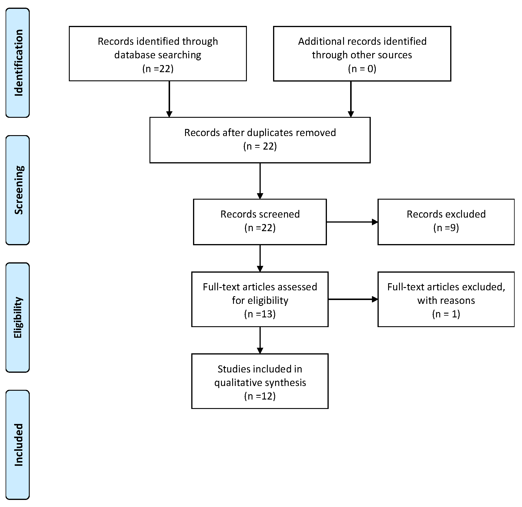
2. Materials and Methods

Search Strategy and Study Selection: This review included original articles that met the following inclusion criteria: studies measuring levels in hair and providing sufficient data, including the total number of subjects in both the investigated and control groups, and reporting mean or median and standard deviation (SD) of trace element levels in hair; articles written in English. Eligible studies were identified by reviewing titles and abstracts, and full texts were obtained for further assessment. References in the retrieved studies were also reviewed. Databases screened included Medline (via PubMed), Scopus, Web of Science, and Google Scholar, covering literature from 1 January 2000 to 2 January 2023. The search strategy combined MeSH heading words with free text words, focusing on ‘hair element analysis’, ‘trace elements’, and ‘autism’. Figure 1 presents the Prisma diagram. Exclusion criteria included systematic reviews, case reports, and reviews.

3. Results

The search revealed 12 original papers meeting the inclusion criteria, reporting on the relationship between the levels of elements in hair and ASD in children. Table 1 outlines the main objectives, materials, methodology, and conclusions of the included studies, while Table 2 and Table 3 present the results regarding essential trace and toxic elements. The included studies employed varied methodologies, sampling methods, analytical methods, and experimental group sizes, and reported results as mean, median, or percentile range, complicating the comparison of absolute values.
Table 1 offers an exhaustive synthesis of diverse studies included in the systematic review, each probing into varied facets of trace elements in children diagnosed with ASD. The majority of the studies leveraged Inductively Coupled Plasma Mass Spectrometry (ICP-MS) as the analytical technique to assess the levels of trace elements primarily in hair and occasionally in serum and nails.
Each study is characterized by a distinct objective, from the evaluation of trace element and mineral levels [33] to the exploration of associations between metal-based hair levels and specific characteristics of ASD [34]. The demographic groups studied are diverse, encompassing different patient counts, age brackets, and gender distributions, facilitating a comprehensive understanding of ASD across varied populations.
Another research identified specific microbial indicators, such as increased abundance of nine genera, and established a predictive model with an 84.00% accuracy, shedding light on the potential role of metal metabolism and gut microecology in the pathogenesis of ASD [35].
The analytical techniques deployed are pivotal for the precise quantification of trace element levels. ICP-MS is predominantly used due to its high sensitivity and precision, with one study [33] utilizing Atomic Absorption Spectroscopy (AAS). The conclusions derived are multifarious, reflecting the intricate nature of ASD. For example, study [33] posits that elements like Mg, Mn, and Zn could influence the development of ADHD (Attention Deficit Hyperactivity Disorder) and ASD, while study [36] uncovers a negative correlation between Hg levels in hair and catatonia in ASD.
The outcomes of these studies are disparate, mirroring the complex and multifaceted nature of ASD. Some studies, such as [34,35,36,37,38,39], did not discern significant discrepancies in the content of certain elements between groups, whereas others, like identified elevated concentrations of specific elements in individuals with ASD compared to controls. Study [40] underscores the potential implication of zinc and magnesium deficiency in the pathogenesis of autism, highlighting the critical role of trace elements in understanding ASD.
The varied findings elucidate the complex interplay between trace elements and ASD, indicating potential directions for future research and intervention methodologies. The diversity in methodologies and conclusions accentuates the imperative for standardized methodologies and further explorations to fully comprehend the implications of trace elements in ASD.
Table 2 and Table 3 meticulously contrasts the levels of essential trace elements across the subjects of the reviewed studies, depicting a wide array of values and units, including median and mean, to represent the concentration of each element. The elements analyzed comprise Chromium (Cr), Cobalt (Co), Iron (Fe), Manganese (Mn), Zinc (Zn), Copper (Cu), and Selenium (Se).
Table 2 and Table 3 also incorporates studies that have employed varied statistical values and units to represent the concentrations of trace elements, emphasizing the necessity for cautious interpretation and potential further standardization in reporting to enable more precise cross-study comparisons. The absence of values for certain elements in some studies, like [35,40,41,42,43,44], denotes the selective focus of each study on particular trace elements, possibly attributed to the primary objectives of the study or resource availability.
Table 4 and Table 5 provides a detailed comparative analysis of metal concentrations, including Lithium (Li), Beryllium (Be), Aluminum (Al), Nickel (Ni), Arsenic (As), Molybdenum (Mo), Cadmium (Cd), Mercury (Hg), Uranium (U), and Lead (Pb) across various studies. It serves as a crucial resource in understanding the diverse range of metal concentrations found in the subjects of the reviewed studies.
The tables reveal significant differences in the levels of these trace elements among various studies, highlighting the variety in methodologies and populations examined.
Some studies, such as [33,37,40], have not reported values for many metals, indicating the specific focus or limitations of each study in analyzing certain metals. This selective reporting underscores the importance of considering the objectives and scope of each study when interpreting the results.
Table 4 and Table 5 also reveals that some studies have reported concentrations for specific functioning levels of autism, as seen in [43], where concentrations of Cadmium (Cd) and Mercury (Hg) are reported for low, medium, and high functioning autism (LFA, MFA, HFA). This differentiation provides a more nuanced understanding of metal concentrations in relation to the severity of autism spectrum disorder.
Table 1. Summary of Systematic Review Studies Investigating Trace Element Levels in Individuals with ASD.
Ref.Main ObjectiveNo of Patients/Sex/AgeAnalytical Technique/UnitMain Conclusions
[33]Evaluation of hair trace element and mineral levels in boys with ADHD, ASD, as well as ADHD with ASD.M 52 (ASD)
CTR 52
Age * 5.18 ± 1.00
ICP-MSThe results obtained suggest that elements, such as Mg, Mn, and Zn may have an impact on the development of ADHD and ASD.
[34]Associations between metal-based hair levels and essential elements and some specific characteristics of ASD.ASD 48
M 34
F 14
Age: 6.5 ± 3.8
ICP-MSThe results obtained show the relationship of metallomic analysis with the essential characteristics of ASD in order to identify potential environmental risk factors at the individual level.
Investigation of the trace element and gut microbiota profiles of Chinese autistic children.ASD 78
M 56
F 22
58 CTR
Age: 4.96 ± 1.01
ICP-MSThe results obtained in Chinese children with ASD indicate significant changes in the profile of micronutrients and intestinal microbiota.
[36]Investigation of the association between catatonia in ASD and the levels of trace elements in hair and serum.ASD 30
ASD + CAT 30
M 30
CTR 30
ICP-MSThe results showed (regression) that Hg levels in hair were negatively associated with catatonia in ASD in raw and corrected models.
[37]Evaluation of the levels of essential trace elements in hair and serum in children with autism spectrum disorder.ASD 70
M40
F30
CTR 70
Age: 6.4 ± 2.9
Two groups:
(2–5)
(6–10)
ICP-DRC-MSTo develop a personal diet for patients with ASD, it is recommended to evaluate several bioindication matrices to critically assess the status of trace elements in patients.
[38]Analysis of hair trace elements content in children with communication disorder and autism spectrum disorder.ASD 33 **
M 33
CTR 33
Age: 5.0 ± 1.8
Two groups:
(3–4)
(5–8)
ICP-MSOn the basis of the obtained results, it can be hypothesized that children suffering from ASD are characterized by a deeper change in metal handling and excretion.
[39]Investigation of hair trace elements content in children with ASD.ASD 74
CTR 74
AGE: two groups:
(2–4)
(5–9)
ICP-MSThere was no significant difference between the groups in the content of mercury (Hg), zinc (Zn), and copper (Cu) in the hair.
Children with ASD are characterized by lower values of not only essential, but also toxic trace elements in their hair.
[40]Determination of the concentrations of trace elements.ASD 1967
M 1553
F 414
Age: 0–15
ICP-MSZinc and magnesium deficiency may play an important role in the pathogenesis of autism.
[41]Examination of possible environmental risk factors and sources of exposure to mercury in children with autism.ASD 25
M 22
F 3
CTR 25
Age: 3–9
ICP-MSThe baseline levels of arsenic, cadmium, and cerium in the urine did not reflect statistically significant differences of these elements in the the mean levels in hair.
[42]Investigation of the levels of both toxic and nontoxic
essential metals in the hair of autistic children.
ASD 44
M 37
F 7
CTR 61
Age: 9.00 ± 4.05
ICP-MSThe results of the current study (as well as the previous one) assessing the concentration of elements in the hair-when it comes to Hg-are not meaningful.
[43]Assessment of the levels of trace elements in hair and nail samples of autistic children.ASD 45
LFA/MFA/HFA ***
15 each
CTR 50
M:F = 4:1
Age: 4–12
AASZn showed a significantly different level in hair and nails, Cu, Pb, and Hg-increased concentration, and Mg and Se decreased concentration (hair, nails) in people with autism vs. control.
[44]Concentration of toxic metals in the hair of children with autism.ASD 40
M 40
40 CTR
Age: 4–7
ICP-MSLead, mercury and uranium-higher content in the hair of children with autism.
* in the ASD group (it concerns all presented studies); ** selected group from the study; *** LFA: low functioning autism, MFA: medium functioning autism, HFA: high functioning autism; CTR—control; F—females; M—males.
Table 2. Comparative Analysis of Essential Trace Elements Across Reviewed Studies.
Ref.Statistical Value/UnitCrCoFe
[33]Median/(μg/g)0.125 (0.092–0.184)0.008
(0.005–0.01)
13.11 (9.81–15.68)
[34]Median/(μg/g)1.151 (0.714–1.714)0.025
(0.025–0.025)
-
[35]Median/(mg/kg)--19.00
[36]Median/(μg/g)0.110 (0.085–0.150)0.008
(0.006–0.011)
12.37 (9.744–14.85)
[37]Median/(μg/g)-0.0066 (0.0048–0.0104) *11.7 (9.1–18.5) *
[38]Median/(μg/g)-0.0111 (0.0086–0.0157)0.0068 (0.0048–0.0104)16.0 (11.5–22.9)10.5 (8.8–15.7)
[39]Median/(μg/g)0.110 (0.082–0.156) *0.008
(0.006–0.011) *
11.7 (9.6–18.0) *
[40]**12% **2.0% **17% **
[41]Mean + SD/(μg/g)0.09 ± 0.060.04 ± 0.0510.89 ± 4.28
[42]Median/(μg/g)0.200.017.60
[43]Mean + SD/(μg/g)LFA
MFA
HFA
--
[44]Median/(μg/g)---
* in the ASD group (it concerns all presented studies); ** selected group from the study.
Table 3. Comparative Analysis of Essential Trace Elements Across Reviewed Studies.
Ref.Statistical Value/UnitMnZnCuSe
[33]Median/(μg/g)0.224 (0.161–0.404)122.3 (86.6–152.9)10.11 (8.46–11.99)0.422 (0.35–0.484)
[34]Median/(μg/g)0.144 (0.058–0.322)136 (107–168)9.226 (7.939–12.07)0.511 (0.465–0.610)
[35]Median/(mg/kg)-78.008.30-
[36]Median/(μg/g)0.254 (0.194–0.325)121.59 (77.17–156.1)9.604 (8.454–11.02)0.341 (0.261–0.388)
[37]Median/(μg/g)0.2233 (0.1384–0.3220) *122 (77–169) *10.9 (9.7–13.3) *0.4061 (0.3427–0.4598) *
[38]Median/(μg/g)0.326 (0.195–0.469)0.241 (0.174–0.340)77.0 (63.8–141.8)153.2 (117.3–202.0)10.6 (8.8–12.5)8.7 (8.2–11.1)0.364 (0.317–0.407)0.406 (0.253–0.437)
[39]Median/(μg/g)0.230 (0.170–0.327) *-10.2 (8.7–12.5) *0.365 (0.315–0.424) *
[40]**4% **29.7% **4% **-
[41]Mean + SD/(μg/g)0.38 ± 0.24101.042 ± 52.021.94 ± 21.70.80 ± 0.25
[42]Median/(μg/g)0.20149.0010.200.90
[43]Mean + SD/(μg/g)-130.46 ± 15.65
172.81 ± 20.73
171.92 ± 20.63
36.62 ± 4.39
23.16 ± 2.77
12.35 ± 1.48
0.57 ± 0.06
1.98 ± 0.23
2.55 ± 0.30
[44]Median/(μg/g)----
* in the ASD group (it concerns all presented studies); ** selected group from the study.
Table 4. Comparative Analysis of Metal Concentrations Across Reviewed Studies.
Ref.LiBeAlNiAs
[33]0.023
(0.017–0.034)
----
[34]0.025
(0.025–0.025)
0.025
(0.025–0.025)
7.738 (5.797–12.79)0.158 (0.067–0.365)0.082
(0.030–0.165)
[35]----0.21
[36]----0.033 (0.023–0.039)
[37]-----
[38]0.0165 (0.0119–0.0470)0.0202 (0.0115–0.0290)0.0005 (0.0003–0.0007)0.0006 (0.0003–0.0008)8.8 (7.4–19.1)9.2 (5.3–11.8)-0.0391 (0.0284–0.0591)0.0301 (0.0250–0.0408)
[39]0.020
(0.012–0.033)
0.0004 (0.0001–0.0010)8.0 (5.3–11.7)-0.034
(0.021–0.044)
[40]-----
[41]0.002 ± 0.0040.0001 ± 0.00038.89 ±6.160.55 ± 0.830.20 ± 0.26
[42]0.008 11.650.200.03
[43]LFA
MFA
HFA
----
[44]-0.05 (0.01, 0.10)61.0 (59.0, 70.0)-0.13 (0.12, 0.18)
Table 5. Comparative Analysis of Metal Concentrations Across Reviewed Studies.
Ref.MoCdHgUPb
[33]-----
[34]0.117 (0.051–0.179)0.021
(0.010–0.035)
0.338 (0.121–1.255)0.047 (0.026–0.086)0.542 (0.316–1.690)
[35]-0.040.41-2.0
[36]-0.022 (0.015–0.030)0.229 (0.072–0.393)--
[37]-----
[38]-0.0277 (0.0224–0.0617)0.0222 (0.0149–0.0297)0.116 (0.066–0.203)0.097 (0.049–0.168)-0.717 (0.547–1.273)0.463 (0.210–0.612)
[39]-0.023
(0.014–0.035)
0.127 (0.049–0.250)-0.506 (0.330–0.673)
[40]-----
[41]0.08 ± 0.10.23 ± 0.270.47 ± 0.420.02 ± 0.010.01 ± 0.02
[42]0.040.010.500.031.30
[43]--3.09 ± 0.37
1.10 ± 0.13
0.65 ± 0.07
17.97 ± 2.15
3.24 ± 0.38
2.04 ± 0.24
[44]-0.14 (0.12, 0.16)4.50 (4.10, 4.90)0.42 (0.40, 0.50)6.75 (5.70, 7.00)

4. Discussion

The World Health Organization (WHO) reports indicate a rising prevalence of ASD among children. Currently, the diagnosis rate stands at 1 in 160 children. The incidence of ASD has increased significantly in the last 10 years. It is postulated that this increase is associated with environmental pollution, stemming from urbanization and industrialization [45]. Further, the rapid advancements in technology and the proliferation of chemical industries have led to the release of numerous pollutants and toxicants into the environment, potentially escalating the risk of ASD.
Studies investigating the status of trace elements and their correlation with ASD employ a range of exposure biomarkers, including hair mineral analysis, blood, urine, and teeth compartments. Autism is also conceptualized as a manifestation of mercury (Hg) poisoning. Certain micronutrients, like copper (Cu), are implicated in the development of ASD. The imbalance of essential micronutrients can disrupt the physiological equilibrium, potentially leading to neurodevelopmental disorders. The role of nutritional status and dietary habits in ASD development also warrants further exploration. Conversely, zinc (Zn) is recognized for its protective role against neurodevelopmental issues, participating in detoxification and antioxidation processes, involving, for example, metallothioneins [38].
Given the rising incidence of autism, exposure to environmental factors, specifically toxic substances, is hypothesized to play a role in the pathogenesis of ASD (Table 6). Table 6, substantiated by references [5,37,44,45,46,47,48] meticulously elucidates the intricate relationships between various toxic metals, their chemical forms, and the mechanisms through which they contribute to ASD. This table provides a comprehensive synthesis of the multifaceted impacts of these metals on individuals diagnosed with ASD, offering insights into the complex interplay between environmental exposure and neurodevelopmental disorders. Children are more susceptible to exposure to environmental toxins due to their higher absorption rates and lower detoxification capacities compared to adults. Mercury (Hg) is considered to have the most significant impact on ASD owing to its neurotoxic properties. Additionally, the cumulative exposure to multiple toxic elements may have synergistic effects, exacerbating the neurotoxic impact and complicating the understanding of individual element contributions to ASD. Thiomersal, an organic compound of mercury used as a preservative in vaccines such as Measles-Mumps-Rubella and as an antiseptic and antifungal agent, has historically been suspected to contribute to a substantial number of autism cases. Metabolic issues with toxic elements in children with autism are thought to be associated with oxidative stress, diminished methylation and transsulfuration potential, and mitochondrial dysfunction. Additionally, elevated levels of porphyrins are found in the urine of children with autism, indicative of the body’s mercury load [42].
An interesting interpretation is the link between ASD and biomarkers using metallomics (biometal studies) in combination with genomics and proteomics. A meta-analysis has demonstrated a correlation between ASD and elements such as copper (Cu), iron (Fe), and zinc (Zn). However, it is crucial to consider the heterogeneity in study designs, populations, and methodologies when interpreting these findings, as they can significantly influence the observed associations. The results of studies in patients with ASD showed higher levels of Cu that were associated with oxidative damage, Fe deficiency causing anemia, behavioral and emotional problems, lower Zn content, which is associated with autoimmune infection, hyperactivity, and mental retardation. Studies have shown that too high of a Cu administration causes Zn deficiency and is associated with synaptic dysfunction. The optimal value was suggested at the level of 0.66–0.81 [46].
Scientific literature underscores the pivotal role of glutathione in the pathogenesis of ASD, highlighting its importance in maintaining cellular redox balance. In individuals with ASD, there are observed lower levels of reduced glutathione and higher levels of oxidized glutathione. This imbalance in glutathione levels underscores the critical need for interventions aimed at restoring antioxidant defenses, potentially mitigating the progression of ASD. This results in the malfunction of glutathione-related enzymes in the blood and brain. Exposure to toxic metals is reported to inhibit synthesis and increase glutathione excretion. Low levels of reduced glutathione cause oxidative stress, which is an important factor in the inflammation of the nervous system. In turn, the effect of low levels of reduced glutathione causes a lower ability to eliminate toxic metals from the body, the consequence of which can be ASD [21].
The underlying causes of the pathophysiological processes leading to autism are often challenging to pinpoint with certainty. Both genetic and environmental influences have been suggested. For instance, the impact of reduced levels of iodine and lithium in a mother’s hair, and subsequently in the child’s, has been recognized. There was a correlation between the severity of autism and the content of toxic elements such as lead (Pb) or mercury (Hg) in hair [43].
The etiology of ASD has been linked to toxic metals such as arsenic (As), aluminum (Al), lead (Pb), and mercury (Hg). The pathogenetic mechanisms underlying ASD remain incompletely understood. Ongoing research endeavors are crucial to unraveling the multifaceted pathogenetic mechanisms and to develop effective therapeutic interventions for ASD. Furthermore, the development of advanced diagnostic tools and biomarkers is imperative for early detection and intervention, which can significantly improve the prognosis and quality of life for individuals with ASD. Nerve inflammation in various areas of the brain, an increased cytokine inflammatory profile, and abnormal expression of the kappa B factor are indicated. Due to the increasing level of environmental pollution, the impact of exposure to toxic metals on the appearance of neurodevelopmental disorders is of particular importance [47]. Thus, implementing stringent environmental regulations and monitoring is paramount to mitigating the exposure to toxic elements and reduce the incidence of neurodevelopmental disorders.
Fiore et al. demonstrated a statistically significant positive correlation between the levels of lead (Pb), aluminum (Al), arsenic (As), and cadmium (Cd) in hair and the severity of ASD symptoms, including repetitive and restricted behaviors and communication deficits. The content of Pb, Mo, and Mn in the hair was antagonistic with respect to the cognitive level measured as IQ. In turn, a low zinc content was associated with the severity of ASD symptoms [48]. Conclusively, the multifactorial etiology of ASD necessitates a multidisciplinary approach, integrating environmental, genetic, and nutritional perspectives to formulate comprehensive prevention and management strategies.

Microelements and ASD

Many studies conclusively demonstrate that nutritional deficiencies can play a role in the pathogenesis of ASD (Table 7). The coexistence of ASD and iron (Fe) deficiency in patients has been particularly noted [49]. Further exploration into the implications of iron deficiency in ASD patients is crucial, as it may offer insights into potential therapeutic interventions and preventative measures. Understanding the underlying mechanisms linking iron deficiency to ASD can pave the way for the development of targeted nutritional strategies aimed at mitigating the risks associated with iron deficiency in ASD patients. In recent years, many studies have been published that compare the content of trace elements in hair (Cu, Zn, Mg, and Se). Analyzed criteria encompass age and sex, alongside a control group of healthy children. Should an increase in the level of toxic elements be detected, the implementation of a detoxification program, utilizing chelators like EDTA, can aid in purging the organism of toxic metals [43]. Such detoxification programs need to be approached with caution and should be administered under strict medical supervision to avoid any adverse effects. It is theorized that the imbalance between excitatory and inhibitory synaptic functions could be a causative factor of ASD, with trace elements playing a significant role [50]. Further research is imperative to substantiate this theory and to explore the potential therapeutic implications of modulating trace element levels in ASD management.
A study involving 40 boys, both autistic and healthy, and the content of Sb, As, U, Be, Hg, Cd, Al, Be and Pb was found. It has been proven that the hair of children with autism had significantly higher levels of Pb, Hg, and U. The relationship between the content of nutritional and toxic elements did not differ between autistic and healthy children [44].
In a study with 104 boys, 52 of whom had ASD, hair analyses showed reduced levels of Co, Mg, Mn, and V in ASD patients [51]. Furthermore, factor analysis connected ASD with decreased contents of multiple elements in hair, with Zn levels being 20% lower. Regression models highlighted a link between lower Zn and Mg levels in hair and heightened neurodevelopmental disorders [52]. Notably, findings also suggested potential overlaps in elemental content in hair between ASD and ADHD cases, hinting that supplementation with Mg, Mn, and Zn could be beneficial, though this warrants further clinical trials for validation.
Skalny et al. studied 148 children, half with ASD, categorized by age and gender. The ASD group predominantly exhibited reduced Cr, I, and V levels in hair but an increase in Se [39]. On the whole, children diagnosed with ASD showcased a decline in various essential and toxic elements in their hair.
A separate study involving 99 children with ASD revealed that younger ASD patients manifested more pronounced elemental discrepancies in their hair compared to older ones, with a notable decline in elements like Cu, As, Be, Cd, and I, and an elevation in Al, Fe, and Se [38].
Tinkov et al.’s research on 60 boys with ASD, 30 of whom had catatonia, found that the catatonic group had three times the Hg content in their hair compared to the non-catatonic ASD group [52]. There were also significant variances in multiple elements between the ASD group and controls.
Yasuda and Tsutsui, in their expansive study of 1967 children, determined that those with ASD typically had reduced levels of Zn and Mg but elevated toxic elements such as Al, Cd, Pb, Hg, and As [52]. Intriguingly, a deficiency in Zinc has been specifically associated with the onset of autism. The research underscored the potential of early metallomic screening as an essential tool in ASD diagnosis and prevention [52].

5. Conclusions

Empirical research corroborates the detrimental influences of toxic elements such as Arsenic (As), Lead (Pb), and Mercury (Hg) on neural development. A multitude of studies draw parallels between the severity of autism manifestations and exposure to these toxic elements, however, a definitive consensus is yet to be reached. Studies into the mineral concentrations in individuals with ASD are grounded in the premise that hair acts as a pathway for the expulsion of toxic elements from the human body. A prevailing agreement within the scientific community suggests that individuals with ASD display significant irregularities in metal metabolism and the expulsion of toxic metals. Current literature validates the association between ASD prevalence and heightened levels of toxic metals in children’s hair. There is a notable association between reduced zinc levels in hair and the onset of ASD. The mechanisms causing mineral imbalances in those diagnosed with ASD are yet to be fully understood. Continued advancements in the field of clinical metallomics pertaining to specific ASD symptoms are crucial. This will aid in the identification of environmental risk elements, especially during early developmental stages. The significant roles of Magnesium (Mg), Manganese (Mn), and Zinc (Zn) as protective agents against ASD are emphasized. The divergence in conclusions across different studies is potentially a reflection of the complex interactions between genetic predispositions and environmental influences in ASD etiology. Almost all research accentuates alterations in the concentration of both essential and non-essential/toxic elements in the hair of children with neurodevelopmental disorders such as ASD.
Future perspective studies should cover:
-
Future research can center on the development and validation of standardized methods to ensure consistency and comparability across studies,
-
Since alterations in both essential and non-essential/toxic elements have been noted in children with ASD, a comparative analysis studying the impact and significance of these alterations could be of immense value,
-
Genetic sequencing coupled with environmental exposure data could be employed to unravel these interactions.
Yet, the field is marked by a lack of concise, quantitative, and comparable studies due to the absence of standardized protocols for Hair Mineral Analysis (HMA) testing.

Author Contributions

Conceptualization, M.M. and K.C.; Data curation, M.M. and K.C.; Formal analysis, K.C. and M.M.; Investigation, M.M. and K.C.; Methodology, M.M. and K.C.; Writing—original draft, K.C. and M.M.; Writing—review & editing, K.C. and M.M. All authors have read and agreed to the published version of the manuscript.

Funding

This research received no external funding.

Data Availability Statement

Data are contained within the article.

Conflicts of Interest

The authors declare no conflict of interest.

Abbreviations

ADHDAttention Deficit Hyperactivity Disorder
ASDAutism Spectrum Disorder
HMAHair Mineral Analysis

References

  1. Virolainen, S.; Hussien, W.; Dalibalta, S. Autism spectrum disorder in the United Arab Emirates: Potential environmental links. Rev. Environ. Health 2020, 35, 359–369. [Google Scholar] [CrossRef] [PubMed]
  2. Ijomone, O.M.; Olung, N.F.; Akingbade, G.T.; Okoh, C.O.; Aschner, M. Environmental influence on neurodevelopmental disorders: Potential association of heavy metal exposure and autism. J. Trace Elements Med. Biol. 2020, 62, 126638. [Google Scholar] [CrossRef] [PubMed]
  3. Currenti, S.A. Understanding and Determining the Etiology of Autism. Cell. Mol. Neurobiol. 2010, 30, 161–171. [Google Scholar] [CrossRef] [PubMed]
  4. Kern, J.K.; Geier, D.A.; Sykes, L.K.; Haley, B.E.; Geier, M.R. The relationship between mercury and autism: A comprehensive review and discussion. J. Trace Elements Med. Biol. 2016, 37, 8–24. [Google Scholar] [CrossRef] [PubMed]
  5. Modabbernia, A.; Velthorst, E.; Reichenberg, A. Environmental risk factors for autism: An evidence-based review of systematic reviews and meta-analyses. Mol. Autism 2017, 8, 13. [Google Scholar] [CrossRef] [PubMed]
  6. Semenova, Y.; Zhunussov, Y.; Pivina, L.; Abisheva, A.; Tinkov, A.; Belikhina, T.; Skalny, A.; Zhanaspayev, M.; Bulegenov, T.; Glushkova, N.; et al. Trace element biomonitoring in hair and blood of occupationally unexposed population residing in polluted areas of East Kazakhstan and Pavlodar regions. J. Trace Elements Med. Biol. 2019, 56, 31–37. [Google Scholar] [CrossRef] [PubMed]
  7. Savinov, S.S.; Sharypova, R.M.; Drobyshev, A.I. Determination of the Trace Element Composition of Human Nails. J. Anal. Chem. 2020, 75, 409–415. [Google Scholar] [CrossRef]
  8. Adams, J.B.; Holloway, C.E.; George, F.; Quig, D. Analyses of Toxic Metals and Essential Minerals in the Hair of Arizona Children with Autism and Associated Conditions, and Their Mothers. Biol. Trace Element Res. 2006, 110, 193–210. [Google Scholar] [CrossRef]
  9. Watts, C.; Sun, J.; Jones, P.D.; Peng, H.; Giesy, J.P. Monthly variations of unregulated brominated disinfection by-products in chlorinated Water are correlated with total Bromine. Eco-Environ. Health 2022, 1, 147–155. [Google Scholar] [CrossRef]
  10. Esplugas, R.; Mari, M.; Marquès, M.; Schuhmacher, M.; Domingo, J.L.; Nadal, M. Biomonitoring of trace elements in hair of schoolchildren living near a hazardous waste incinerator—A 20 years follow-up. Toxics 2019, 7, 52. [Google Scholar] [CrossRef]
  11. de Oliveira, L.M.; Das, S.; da Silva, E.B.; Gao, P.; Gress, J.; Liu, Y.; Ma, L.Q. Metal concentrations in traditional and herbal teas and their potential risks to human health. Sci. Total. Environ. 2018, 633, 649–657. [Google Scholar] [CrossRef] [PubMed]
  12. Yu, M.; Teitelbaum, S.L.; Dolios, G.; Dang, L.-H.T.; Tu, P.; Wolff, M.S.; Petrick, L.M. Molecular Gatekeeper Discovery: Workflow for Linking Multiple Exposure Biomarkers to Metabolomics. Environ. Sci. Technol. 2021, 56, 6162–6171. [Google Scholar] [CrossRef] [PubMed]
  13. Cole, S.A. Individual and collective identification in contemporary forensics. Biosocieties 2020, 15, 350–375. [Google Scholar] [CrossRef]
  14. Alekseenko, S.I.; Skalny, A.V.; Karpischenko, S.A.; Tinkov, A.A. Serum, Whole Blood, Hair, and Mucosal Essential Trace Element and Mineral Levels in Children with Verified Chronic Rhinosinusitis Undergoing Functional Endoscopic Sinus Surgery. Biol. Trace Element Res. 2020, 199, 2112–2120. [Google Scholar] [CrossRef] [PubMed]
  15. Çelik, B.; Nalçacıoğlu, H.; Karakükçü, Ç.; Aslaner, H.; Şahiner, Ü.M. Assessment of Hair Zinc in the School Children in Kayseri, Turkey. Biol. Trace Element Res. 2020, 196, 343–348. [Google Scholar] [CrossRef] [PubMed]
  16. Sazakli, E.; Leotsinidis, M. Hair biomonitoring and health status of a general population exposed to Nickel. J. Trace Elements Med. Biol. 2017, 43, 161–168. [Google Scholar] [CrossRef]
  17. Shin, W.-J.; Jung, M.; Ryu, J.-S.; Hwang, J.; Lee, K.-S. Revisited digestion methods for trace element analysis in human hair. J. Anal. Sci. Technol. 2020, 11, 1. [Google Scholar] [CrossRef]
  18. Zhang, S.; Chu, Y.; Ma, S.; Chen, F.; Zhang, D.; Hu, Z.; Zhang, Z.; Jin, H.; Guo, L. Highly accurate determination of Zn and Cu in human hair by ultrasound-assisted alkali dissolution combined with laser-induced breakdown spectroscopy. Microchem. J. 2020, 157, 105018. [Google Scholar] [CrossRef]
  19. Bocato, M.Z.; Ximenez, J.P.B.; Hoffmann, C.; Barbosa, F. An overview of the current progress, challenges, and prospects of human biomonitoring and exposome studies. J. Toxicol. Environ. Health B Crit. Rev. 2019, 22, 131–156. [Google Scholar] [CrossRef]
  20. Peña-Fernández, A.; del Carmen Lobo-Bedmar, M.; González-Muñoz, M.J. Effects of sex on the levels of metals and metalloids in the hair of a group of healthy Spanish adolescents (13 to 16 years old). Environ. Sci. Pollut. Res. 2017, 24, 23666–23678. [Google Scholar] [CrossRef]
  21. Bjørklund, G.; Tinkov, A.A.; Hosnedlová, B.; Kizek, R.; Ajsuvakova, O.P.; Chirumbolo, S.; Skalnaya, M.G.; Peana, M.; Dadar, M.; El-Ansary, A.; et al. The role of glutathione redox imbalance in autism spectrum disorder: A review. Free. Radic. Biol. Med. 2020, 160, 149–162. [Google Scholar] [CrossRef] [PubMed]
  22. Li, P.; Guo, S.; Zhao, J.; Gao, Y.; Li, Y.-F. Human Biological Monitoring of Mercury through Hair Samples in China. Bull. Environ. Contam. Toxicol. 2019, 102, 701–707. [Google Scholar] [CrossRef] [PubMed]
  23. Protano, C.; Astolfi, M.L.; Marconi, E.; Antonucci, A.; Canepari, S.; Piamonti, D.; Brunori, M.; Vitali, M. Occupational Exposure Assessment of Major and Trace Elements in Human Scalp Hair Among a Group of Eritrean Workers. Biol. Trace Element Res. 2019, 197, 89–100. [Google Scholar] [CrossRef] [PubMed]
  24. Noreen, F.; Sajjad, A.; Mahmood, K.; Anwar, M.; Zahra, M.; Waseem, A. Human Biomonitoring of Trace Elements in Scalp Hair from Healthy Population of Pakistan. Biol. Trace Element Res. 2020, 196, 37–46. [Google Scholar] [CrossRef]
  25. Wei, B.; Yu, J.; Wang, J.; Li, H.; Yang, L.; Kong, C. Trace Metals in the Urine and Hair of a Population in an Endemic Arsenism Area. Biol. Trace Element Res. 2018, 182, 209–216. [Google Scholar] [CrossRef]
  26. Wongsasuluk, P.; Chotpantarat, S.; Siriwong, W.; Robson, M. Using hair and fingernails in binary logistic regression for bio-monitoring of heavy metals/metalloid in groundwater in intensively agricultural areas, Thailand. Environ. Res. 2018, 162, 106–118. [Google Scholar] [CrossRef] [PubMed]
  27. Vinnikov, D.; Semizhon, S.; Rybina, T.; Zaitsev, V.; Pleshkova, A.; Rybina, A. Occupational exposure to metals and other elements in the tractor production. PLoS ONE 2018, 13, e0208932. [Google Scholar] [CrossRef]
  28. Mauriello, M.C.; Sbordone, C.; Montuori, P.; Alfano, R.; Triassi, M.; Iavicoli, I.; Manno, M. Biomonitoring of toxic metals in incinerator workers: A systematic review. Toxicol. Lett. 2017, 272, 8–28. [Google Scholar] [CrossRef]
  29. Ding, M.; Shi, S.; Qie, S.; Li, J.; Xi, X. Association between heavy metals exposure (cadmium, lead, arsenic, mercury) and child autistic disorder: A systematic review and meta-analysis. Front. Pediatr. 2023, 11, 1169733. [Google Scholar] [CrossRef]
  30. Shiani, A.; Sharafi, K.; Omer, A.K.; Kiani, A.; Karamimatin, B.; Massahi, T.; Ebrahimzadeh, G. A systematic literature review on the association between exposures to toxic elements and an autism spectrum disorder. Sci. Total Environ. 2023, 857, 159246. [Google Scholar] [CrossRef]
  31. Blaurock-Busch, E.; Dessoki, H.H.; Rabah, T. Toxic Metals and Essential Elements in Hair and Severity of Symptoms among Children with Autism. Maedica 2012, 7, 38–48. [Google Scholar] [PubMed]
  32. Amadi, C.N.; Orish, C.N.; Frazzoli, C.; Orisakwe, O.E. Association of autism with toxic metals: A systematic review of case-control studies. Pharmacol. Biochem. Behav. 2022, 212, 173313. [Google Scholar] [CrossRef] [PubMed]
  33. Skalny, A.V.; Mazaletskaya, A.L.; Ajsuvakova, O.P.; Bjørklund, G.; Skalnaya, M.G.; Notova, S.V.; Chernova, L.N.; Skalny, A.A.; Burtseva, T.I.; Tinkov, A.A. Hair trace element concentrations in autism spectrum disorder (ASD) and attention deficit/hyperactivity disorder (ADHD). J. Trace Elements Med. Biol. 2020, 61, 126539. [Google Scholar] [CrossRef]
  34. Fiore, M.; Barone, R.; Copat, C.; Grasso, A.; Cristaldi, A.; Rizzo, R.; Ferrante, M. Metal and essential element levels in hair and association with autism severity. J. Trace Elements Med. Biol. 2020, 57, 126409. [Google Scholar] [CrossRef]
  35. Zhai, Q.; Cen, S.; Jiang, J.; Zhao, J.; Zhang, H.; Chen, W. Disturbance of trace element and gut microbiota profiles as indicators of autism spectrum disorder: A pilot study of Chinese children. Environ. Res. 2019, 171, 501–509. [Google Scholar] [CrossRef] [PubMed]
  36. Tinkov, A.A.; Skalnaya, M.G.; Simashkova, N.V.; Klyushnik, T.P.; Skalnaya, A.A.; Bjørklund, G.; Notova, S.V.; Kiyaeva, E.V.; Skalny, A.V. Association between catatonia and levels of hair and serum trace elements and minerals in autism spectrum disorder. Biomed. Pharmacother. 2019, 109, 174–180. [Google Scholar] [CrossRef]
  37. Skalny, A.V.; Simashkova, N.V.; Skalnaya, A.A.; Klyushnik, T.P.; Bjørklund, G.; Skalnaya, M.G.; Tinkov, A.A. Assessment of gender and age effects on serum and hair trace element levels in children with autism spectrum disorder. Metab. Brain Dis. 2017, 32, 1675–1684. [Google Scholar] [CrossRef]
  38. Skalny, A.V.; Simashkova, N.V.; Klyushnik, T.P.; Grabeklis, A.R.; Radysh, I.V.; Skalnaya, M.G.; Tinkov, A.A. Analysis of Hair Trace Elements in Children with Autism Spectrum Disorders and Communication Disorders. Biol. Trace Element Res. 2016, 177, 215–223. [Google Scholar] [CrossRef]
  39. Skalny, A.V.; Simashkova, N.V.; Klyushnik, T.P.; Grabeklis, A.R.; Bjørklund, G.; Skalnaya, M.G.; Nikonorov, A.A.; Tinkov, A.A. Hair toxic and essential trace elements in children with autism spectrum disorder. Metab. Brain Dis. 2017, 32, 195–202. [Google Scholar] [CrossRef]
  40. Yasuda, H.; Yoshida, K.; Yasuda, Y.; Tsutsui, T. Infantile zinc deficiency: Association with autism spectrum disorders. Sci. Rep. 2011, 1, 129. [Google Scholar] [CrossRef]
  41. Blaurock-Busch, E.; Amin, O.R.; Rabah, T. Heavy metals and trace elements in hair and urine of a sample of arab children with autistic spectrum disorder. Maedica 2011, 4, 247–257. [Google Scholar] [CrossRef]
  42. De Palma, G.; Catalani, S.; Franco, A.; Brighenti, M.; Apostoli, P. Lack of correlation between metallic elements analyzed in hair by ICP-MS and autism. J. Autism Dev. Disord. 2012, 42, 342–353. [Google Scholar] [CrossRef] [PubMed]
  43. Lakshmi Priya, M.D.; Geetha, A. Level of trace elements (copper, zinc, magnesium and selenium) and toxic elements (lead and mercury) in the hair and nail of children with autism. Biol. Trace Element Res. 2011, 142, 148–158. [Google Scholar] [CrossRef] [PubMed]
  44. Fido, A.; Al-Saad, S. Toxic trace elements in the hair of children with autism. Autism 2005, 9, 290–298. [Google Scholar] [CrossRef] [PubMed]
  45. Nabgha-e-Amen; Eqani, S.A.M.A.S.; Khuram, F.; Alamdar, A.; Tahir, A.; Shah, S.T.A.; Nasir, A.; Javed, S.; Bibi, N.; Hussain, A.; et al. Environmental exposure pathway analysis of trace elements and autism risk in Pakistani children population. Sci. Total. Environ. 2020, 712, 136471. [Google Scholar] [CrossRef] [PubMed]
  46. Scassellati, C.; Bonvicini, C.; Benussi, L.; Ghidoni, R.; Squitti, R. Neurodevelopmental disorders: Metallomics studies for the identification of potential biomarkers associated to diagnosis and treatment. J. Trace Elements Med. Biol. 2020, 60, 126499. [Google Scholar] [CrossRef] [PubMed]
  47. Bjørklund, G.; Skalny, A.V.; Rahman, M.; Dadar, M.; Yassa, H.A.; Aaseth, J.; Chirumbolo, S.; Skalnaya, M.G.; Tinkov, A.A. Toxic metal(loid)-based pollutants and their possible role in autism spectrum disorder. Environ. Res. 2018, 166, 234–250. [Google Scholar] [CrossRef]
  48. Petrova, M.V.; Ourgaud, M.; Boavida, J.R.; Dufour, A.; Onrubia, J.A.T.; Lozingot, A.; Heimbürger-Boavida, L.-E. Human mercury exposure levels and fish consumption at the French Riviera. Chemosphere 2020, 258, 127232. [Google Scholar] [CrossRef]
  49. Tseng, P.-T.; Cheng, Y.-S.; Chen, Y.-W.; Stubbs, B.; Whiteley, P.; Carvalho, A.F.; Li, D.-J.; Chen, T.-Y.; Tang, C.-H.; Chu, C.-S.; et al. Peripheral iron levels in children with autism spectrum disorders vs controls: A systematic review and meta-analysis. Nutr. Res. 2018, 50, 44–52. [Google Scholar] [CrossRef]
  50. Saghazadeh, A.; Ahangari, N.; Hendi, K.; Saleh, F.; Rezaei, N. Status of essential elements in autism spectrum disorder: Systematic review and meta-analysis. Rev. Neurosci. 2017, 28, 783–809. [Google Scholar] [CrossRef]
  51. Skalny, A.V.; Mazaletskaya, A.L.; Ajsuvakova, O.P.; Bjørklund, G.; Skalnaya, M.G.; Chao, J.C.-J.; Chernova, L.N.; Shakieva, R.A.; Kopylov, P.Y.; Skalny, A.A.; et al. Serum zinc, copper, zinc-to-copper ratio, and other essential elements and minerals in children with attention deficit/hyperactivity disorder (ADHD). J. Trace Elements Med. Biol. 2020, 58, 126445. [Google Scholar] [CrossRef] [PubMed]
  52. Tinkov, A.A.; Ajsuvakova, O.P.; Skalny, A.V. A Case-Control Study of Essential and Toxic Trace Elements and Minerals in Hair of 0–4-Year-Old Children with Cerebral Palsy. Biol. Trace Element Res. 2020, 195, 399–408. [Google Scholar] [CrossRef] [PubMed]
Figure 1. PRISMA flow diagram.
Pollutants 03 00038 g001
Table 6. Toxic Metals, Their Chemical Forms, and Mechanisms Contributing to ASD [5,37,44].
MetalChemical FormMechanism of ASD Contribution
Hg (Mercury)-Higher levels of antineuronal antibodies; neurological, motor, immune, and sensory dysfunctions.
Exposure can occur through fish contaminated with methylmercury or through fungicides used as grain preservatives in bread.
Children with ASD exhibit higher levels of Hg in primary teeth and blood.
Hg induces metallothionein dysfunction, related to Zn deficit.
Mercury ions (Hg2+)Nephrotoxic and causes damage to muscle tissue.
Methylmercury (CH3Hg+)Most toxic form, can cross the blood-brain barrier due to its lipophilic nature, binding to neurons and causing high neurotoxicity.
Main sources for humans include fish, bacteria, and algae, leading to the biotransformation of elemental Hg to methylmercury.
As (Arsenic)-Alters brain morphology and causes Mcl-1 depression in the cerebral cortex.
Induces gliosis degeneration and up-regulates Bax and Bak expression.
Impairs neurite growth through suppression of AMPK kinase activation and inhibits the Wnt/β-catenin signaling pathway.
Pb (Lead)-Induces neuroinflammation and autoimmunity, stimulating the synthesis of anti-ribosomal P antibodies.
Exposure to Pb from leaded gasoline, used in the past, is another theorized pathogenesis of autism.
Al (Aluminum)-Interacts with glycolytic enzymes and inhibits cellular energy synthesis.
Intensifies neurotoxic effects through Al3+ ion by oxygen-based ligands and activates microglia producing IL-6, TNF-α, iNOS, NOS-2, neuroinflammatory PICs, and ROS.
U (Uranium)-Uranium from coal combustion and phosphate fertilizers can contribute to autism.
Elevated levels of non-radioactive isotope of uranium have been found in the hair of autistic children compared to controls.
Table 7. Essential Microelements, Their Chemical Forms, and Their Protective Roles in ASD [37,51].
MetalMechanism of ASD Contribution
ZnZinc is crucial for the scaffolding of ProSAP/Shank proteins, linked to excitatory synapses. Imbalances, either excess or deficiency, are associated with epileptogenesis, depression, and ASD, respectively, confirming the role of abnormal zinc levels in brain dysfunctions. Elevated levels of copper, being antagonistic to zinc, lead to synaptic dysfunction [50]. Zn is integral to the active site of 300 enzymes.
MgMagnesium ions are pivotal for the synthesis of the key neurotransmitter gamma-aminobutyric acid (GABA), thus playing a crucial role in ASD as they regulate GABA activity [50].
FeIron is imperative for proper brain functioning, playing a key role in gene expression and myelination. Disruption in iron homeostasis is observed in various neurodegenerative diseases. Iron deficiency can lead to conditions such as depression and anxiety, influencing social and emotional behavior and contributing to the development of ASD [50].
Zn/CuResearch indicates that the Zn/Cu ratio and zinc content are significantly lower in children with ASD compared to healthy children, underlining the significant role of the Zn to Cu ratio in ASD [37].
Other elementsNo associations with ASD were found in the case of the content of Cr, I, Se, in hair [37].
Disclaimer/Publisher’s Note: The statements, opinions and data contained in all publications are solely those of the individual author(s) and contributor(s) and not of MDPI and/or the editor(s). MDPI and/or the editor(s) disclaim responsibility for any injury to people or property resulting from any ideas, methods, instructions or products referred to in the content.

