
Study of the Influence of Process Parameters on the Morphology of ZnO Nanostructures †


Abstract
:1. Introduction
2. Experimental Detail
2.1. Synthesis of ZnO Nanostructures
2.2. Characterization
3. Results and Discussion
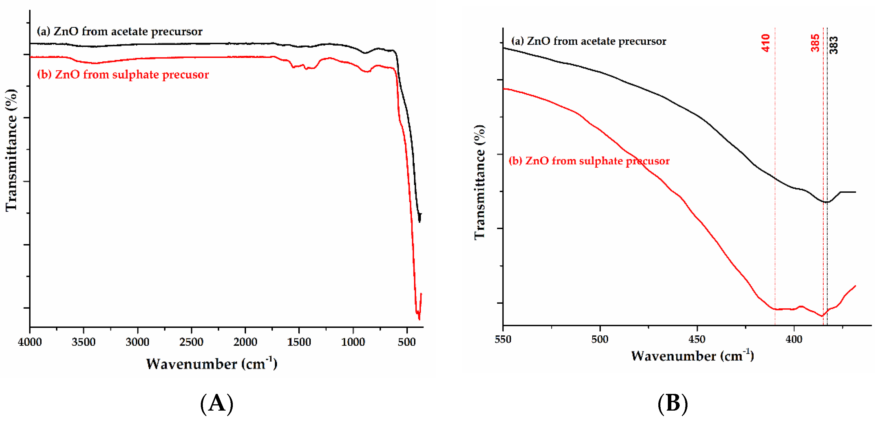
3.1. FTIR Analysis
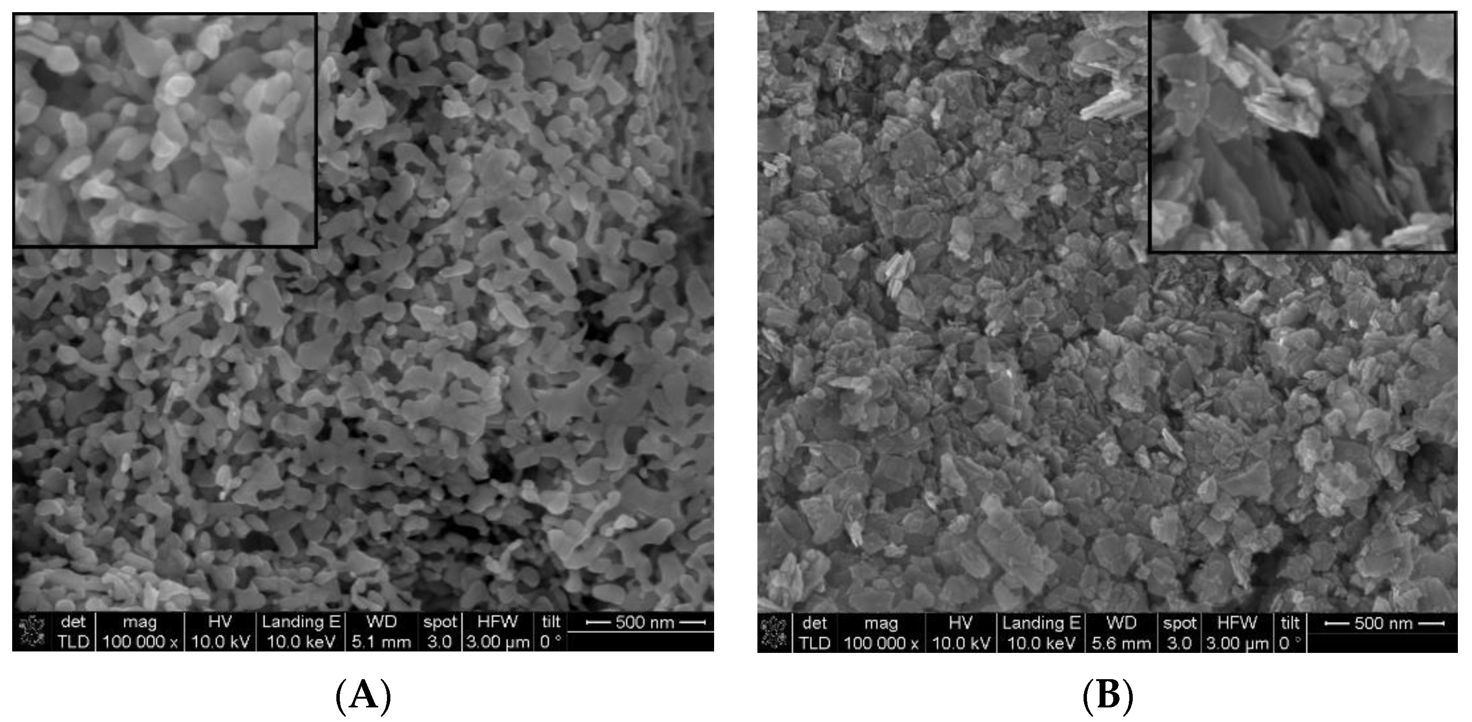
3.2. SEM Analysis
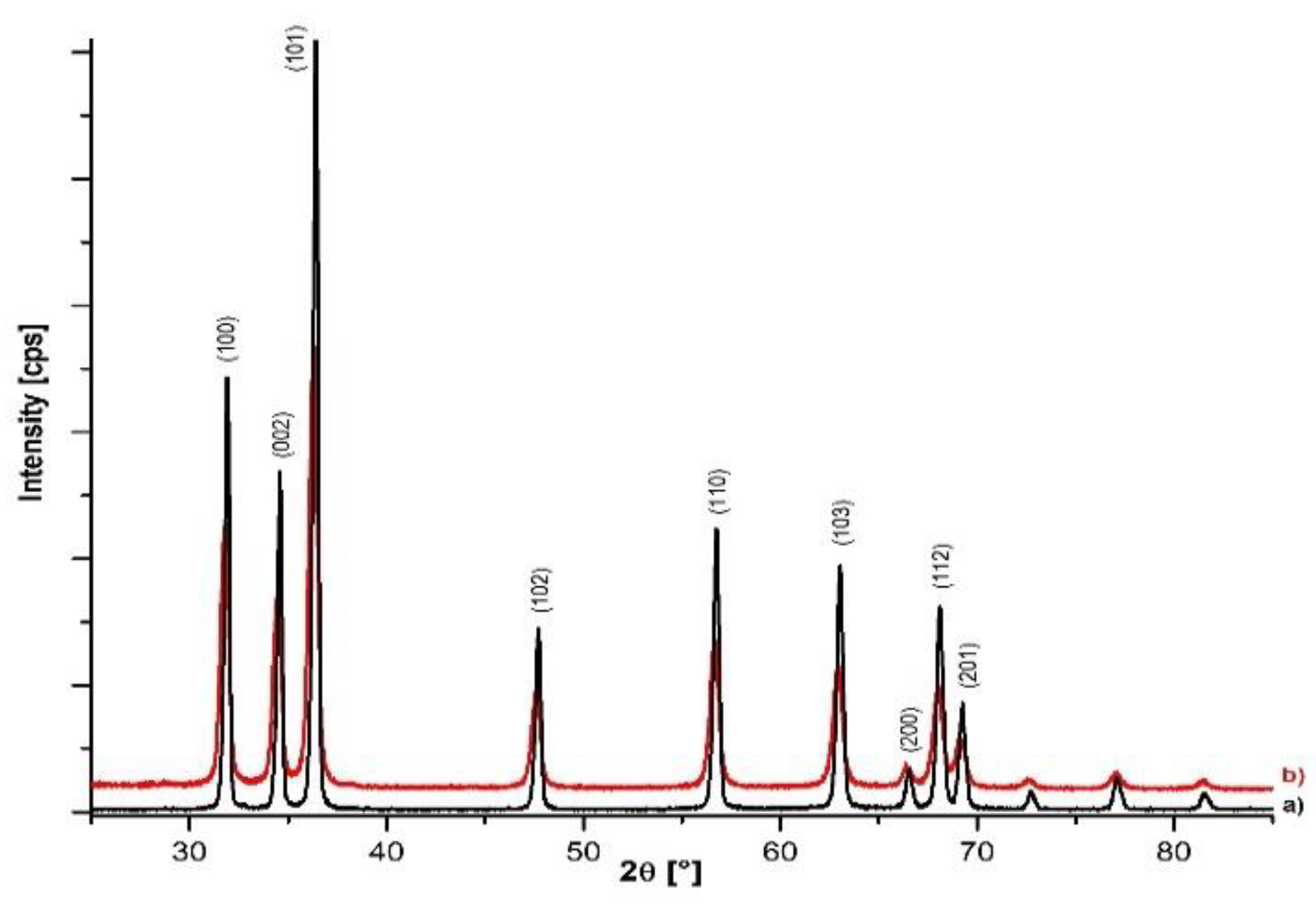
3.3. XRD Analysis
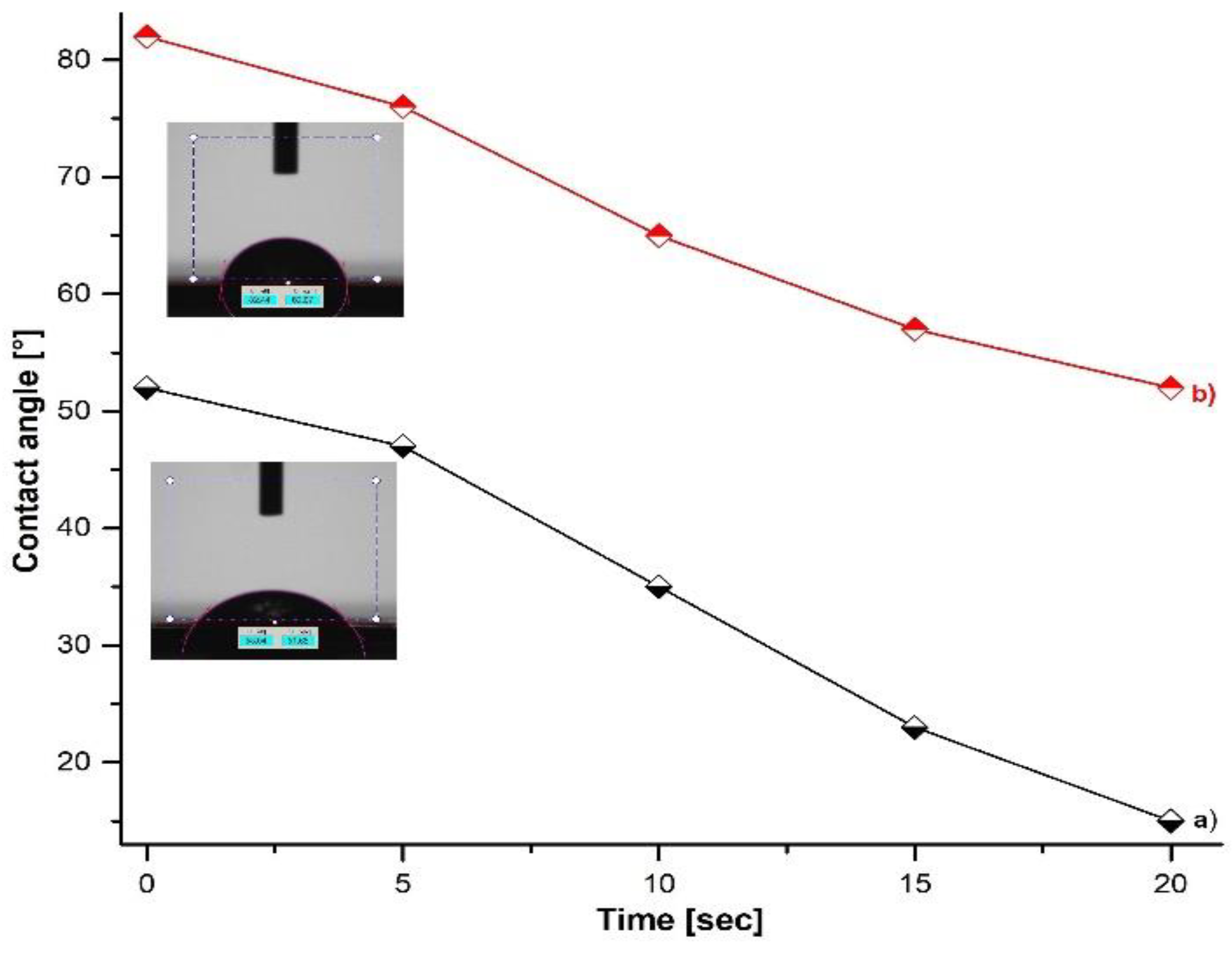
3.4. Wetting Capacity (Contact Angle)
4. Conclusions
Author Contributions
Funding
Institutional Review Board Statement
Informed Consent Statement
Data Availability Statement
Acknowledgments
Conflicts of Interest
References
- Hedlund Orbeck, J.K.; Hamers, R.J. Surface properties and interactions of transition metal oxide nanoparticles: A perspective on sustainability. J. Vac. Sci. Technol. 2020, 38, 031001. [Google Scholar] [CrossRef]
- Borysiewicz, M.A. ZnO as a Functional Material, a Review. Crystal 2019, 9, 505. [Google Scholar] [CrossRef]
- Matei, A.; Tucureanu, V.; Dumitrescu, L. Aspects regarding synthesis and applications of ZnO nanomaterials. Bull. Transilv. Univ. Brasov. Ser. I Eng. Sci. 2014, 7, 45–52. [Google Scholar]
- Raha, S.; Ahmaruzzaman, M. ZnO nanostructured materials and their potential applications: Progress, challenges and perspectives. Nanoscale Adv. 2022, 4, 1868–1925. [Google Scholar] [CrossRef] [PubMed]
- Jiang, J.; Pi, J.; Cai, J. The advancing of zinc oxide nanoparticles for biomedical applications. Bioinorg. Chem. Appl. 2018, 2018, 1062562. [Google Scholar] [CrossRef] [PubMed]
- Matei, A.; Dumitrescu, L.; Cernica, I.; Tucureanu, V.; Mihalache, I.; Bita, B.; Danila, M.; Manciulea, I. Study of the influence of capping agents on the structural and optical properties of ZnO nanostructures. J. Optoelectron. Adv. Mater. 2015, 17, 952–957. [Google Scholar]
- Mousavi, S.M.; Behbudi, G.; Gholami, A.; Hashemi, S.A.; Nejad, Z.M.; Bahrani, S.; Chiang, W.H.; Wei, L.C.; Omidifar, N. Shape-controlled synthesis of zinc nanostructures mediating macromolecules for biomedical applications. Biomater. Res. 2022, 26, 4. [Google Scholar] [CrossRef] [PubMed]
- Gatou, M.A.; Lagopati, N.; Vagena, I.A.; Gazouli, M.; Pavlatou, E.A. ZnO Nanoparticles from Different Precursors and Their Photocatalytic Potential for Biomedical Use. Nanomaterials 2023, 13, 122. [Google Scholar] [CrossRef] [PubMed]
- Shaba, E.Y.; Jacob, J.O.; Tijani, J.O.; Suleiman, M.A.T. Critical review of synthesis parameters affecting the properties of zinc oxide nanoparticle and its application in wastewater treatment. Appl. Water Sci. 2021, 11, 48. [Google Scholar] [CrossRef]
- Sambath, K.; Saroja, M.; Rajendran, K.; Muthukumarasamy, N. Morphology controlled synthesis of ZnO nanostructures by varying pH. J. Mater. Sci. Mater. Electron. 2012, 23, 431–436. [Google Scholar] [CrossRef]
- Rathnasekara, R.; Hari, P. Impedance spectroscopy of nanostructured ZnO morphologies. J. Mater. Res. 2021, 36, 1937–1947. [Google Scholar] [CrossRef]
- Matei, A.; Tucureanu, V.; Popescu, C.M.; Romanitan, C.; Mihalache, I. Influence of Cu dopant on the morpho-structural and optical properties ZnO nanoparticles. Ceram. Int. 2019, 45, 10826–10833. [Google Scholar] [CrossRef]


| Precurors | Lattice Parameters | Strain [%] | Average Crystallite Size [nm] | ||
|---|---|---|---|---|---|
| A = b (A) | c (A) | c/a Ratio | |||
| Zn(CH3COO)2 | 3.2512 | 5.2084 | 1.6019 | 0.11 | 16.2 |
| ZnSO4 | 3.2482 | 5.2028 | 1.6017 | 0.02 | 31.6 |
Disclaimer/Publisher’s Note: The statements, opinions and data contained in all publications are solely those of the individual author(s) and contributor(s) and not of MDPI and/or the editor(s). MDPI and/or the editor(s) disclaim responsibility for any injury to people or property resulting from any ideas, methods, instructions or products referred to in the content. |
© 2023 by the authors. Licensee MDPI, Basel, Switzerland. This article is an open access article distributed under the terms and conditions of the Creative Commons Attribution (CC BY) license (https://creativecommons.org/licenses/by/4.0/).
Share and Cite
Matei, A.; Brincoveanu, O.; Romanitan, C.; Tucureanu, V. Study of the Influence of Process Parameters on the Morphology of ZnO Nanostructures. Mater. Proc. 2023, 14, 9. https://doi.org/10.3390/IOCN2023-14455
Matei A, Brincoveanu O, Romanitan C, Tucureanu V. Study of the Influence of Process Parameters on the Morphology of ZnO Nanostructures. Materials Proceedings. 2023; 14(1):9. https://doi.org/10.3390/IOCN2023-14455
Chicago/Turabian StyleMatei, Alina, Oana Brincoveanu, Cosmin Romanitan, and Vasilica Tucureanu. 2023. "Study of the Influence of Process Parameters on the Morphology of ZnO Nanostructures" Materials Proceedings 14, no. 1: 9. https://doi.org/10.3390/IOCN2023-14455