Proceeding Paper

Synthesis and Characterization of Nanocomposites Based on Carbon Materials and Transitional Oxides †

National Institute for Research and Development in Microtechnologies, IMT-Bucharest, 126A Erou Iancu Nicolae Street, 077190 Bucharest, Romania
*
Authors to whom correspondence should be addressed.
Presented at the 4th International Online Conference on Nanomaterials, 5–19 May 2023; Available online: https://iocn2023.sciforum.net/.
Mater. Proc. 2023, 14(1), 8; https://doi.org/10.3390/IOCN2023-14453
Published: 5 May 2023
(This article belongs to the Proceedings of The 4th International Online Conference on Nanomaterials)

Abstract

:
Nanostructured materials in the biomedical field are commonly used for diagnostics, drug delivery, therapy activation and monitoring therapeutic responses in real-time, thus maximizing the therapeutic benefits simultaneously with a minimally invasive effect and low toxicity. Electrochemical analysis and, implicitly, the development of materials for biosensors have become of vital importance for the monitoring of biomolecules. The conductivity of nanocomposites is usually determined by characteristics related to the concentration, size and dispersion of the nanoparticles. Generally, graphene’s high surface energy and strong interactions moderate its uniform compatibility with different media. In the present paper, we propose the synthesis of yttrium oxide nanoparticles for developing nanocomposites based on transition oxides and carbon materials for electrochemical applications. The precipitation method was used to obtain nanostructured Y2O3. The Hummer method was used for the synthesis of graphene material. After the activation step of the Y2O3 surface, the ex-situ method was chosen to obtain the nanocomposites, allowing the insertion of oxide nanoparticles into the sheets of carbon materials. The developed materials were studied from a structural point of view using Raman and FTIR spectroscopy. The surface morphology, particle size and distribution of nanoparticles in the carbon material were studied using a field emission scanning electron microscope. The goniometric studies followed the wetting and percolation capacity of the nanocomposite.

1. Introduction

Nanotechnology has gained fundamental importance in the development of biomedical systems due to the fact that nanostructured materials present unique properties and dimensions appropriate to many biological systems; their use is expected to bring revolutionary changes in medical diagnosis and therapy with minimal damage. For the detection of biomolecules, although there are many analysis techniques, the development of fast, cheap and accessible detection methods is desired. In this regard, electrochemistry has registered many advances as a result of the development of materials that have allowed for the improvement of reproducibility, the reduction of detection limits and the ease in the detection of the analyte of interest. In recent decades, nanomaterial composite systems have attracted significant interest as they combine different types of materials into a single multifunctional nanostructure, which exhibits the properties of its component modules. Nanostructured composites based on carbon materials and transitional metals have become a topical area with multiple applications. Nanomaterial-based sensors offer a new class of fast, reliable, selective, etc., detectors at a low cost. Additionally, carbon materials have begun to be used more and more in the development of electrodes for various applications due to their unique structure and extraordinary electronic properties. Carbon materials (CNT, RGO, etc.) have a large surface area, significant mechanical strength, high electrical conductivity and efficient electrocatalytic activity. It is known that the attachment of inorganic nanoparticles to graphene can inhibit aggregation and significantly improve the persuasive effect on the electrochemical properties by attaching them to the surface of the carbon material [1,2]. Yttrium oxide nanoparticles (Y2O3) are mechanically, chemically and thermally stable, have a narrow bandgap that allows electron transfer and provide excellent electrochemical sensitivity [3,4]. However, the literature presents little data regarding the realization of microsensors based on Y2O3. Studies have been found regarding the realization of sensors based on a PVA-Y2O3:Eu composite designed for the non-enzymatic detection of H2O2 in biological environments. By using the GO-Y2O3 or CNT-Y2O3 type composites, studies were conducted for detecting acetaminophen, and with the RGO-Y2O3:Cr3+ nanocomposite for detecting dopamine [1,4,5].
In the present paper, we propose the synthesis of Y2O3 and RGO nanoparticles for the development of nanocomposites based on transition oxides and carbon materials for integration in electrochemical applications. The materials are characterized from a structural and morphological point of view using FTIR spectrometry, Raman spectrometry and SEM. The goniometric studies followed the wetting and percolation capacity of the nanocomposite. The knowledge obtained from the study provides a better understanding of the synthesis steps of high-purity nanomaterials to develop electrochemical sensors for detection in biomedical applications.

2. Experimental

2.1. Synthesis of Carbon Material

Reduced graphene oxide (RGO) was prepared using Hummer’s method by oxidizing graphite with sulfuric acid (H2SO4), sodium nitrate (NaNO3) and potassium permanganate (KMnO4) [6,7], followed by neutralization with hydrogen peroxide (H2O2) and purification in the presence of hydrochloric acid (HCl). The reduction of graphene oxide was carried out with hydrazine in the presence of N-methyl-2-pyrrolidone (C5H9NO). Lastly, steps of decanting, washing with ethanol and redispersion were repeated for the purification of RGO.

2.2. Synthesis of Oxide Nanoparticles

To obtain the oxide precursor, a mixture of acetyl acetone (C5H8O2) and dimethyl sulfoxide (C2H6OS) was added over the yttrium nitrate solution (Y(NO3)3·6H2O). Acetyl acetone acts as a chelating agent. Stirring and heating are started under reflux conditions, and ethylene glycol (C₂H₆O₂) and CTAB (C19H42BrN) surfactant are added. Ethylene glycol (C₂H₆O₂) acts as a stabilizing agent, allowing the binding of the chelating agent through polyesterification reactions, which favors the formation of the gel. During the process, it was found that the hydrolysis and condensation steps are more achievable by keeping the pH constant at the value of 4–5, while it was undesirable to increase the pH, as, in this case, flocculation and the formation of an unwanted precipitate may occur. Nitric acid (HNO3) was used to adjust the pH. After 3 h, reflux conditions were removed, stirring and heating were stopped and it was left to mature for polymerization and gel formation. The final thermal treatment occurs at 900 °C for 6 h.

2.3. Synthesis of Nanocomposite

In the first part of the synthesis process of the Y2O3-RGO composite, the oxide is dispersed in hydrogen peroxide to form OH groups on the surface; then it is subjected to repeated steps of centrifugation-decantation-washing-redispersion in ethanol. RGO is added to the previous solution. To achieve a good insertion of the oxide nanoparticles between the RGO sheets, ultrasonication is used for 4 h at 45 kHz.

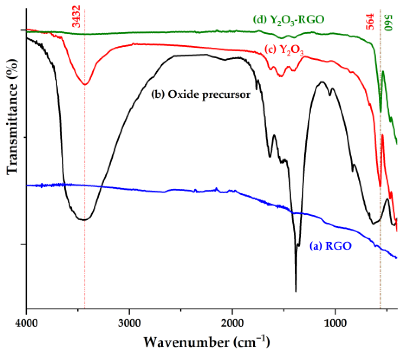
3. Results and Discussion

The physicochemical properties and applicable capacity of Y2O3 nanoparticles in the biomedical field are strongly influenced by the morphology and structure of the particles. Figure 1 shows the comparative ATR-FTIR spectra taken for the oxide precursor, Y2O3 nanoparticles after the surface activation stage, RGO and the Y2O3-RGO composite. The spectrum of the oxide is characterized by absorption bands that can be associated with Y-O bonds from Y2O3 (564 cm−1) and OH bonds anchored to the oxide surface (3432 cm−1). In the spectra of the composites, the existence of the characteristic peak of Y-O bonds can be observed, but with a very slight shift of it towards lower wavenumbers (560 cm−1) compared to the oxide nanoparticles and the disappearance of the OH band, as a result of the interference with the carbon material. In IR spectroscopy, the RGO does not show any specific absorption band, which is the reason why the presence of RGO in composites was studied using Raman spectroscopy [5,8,9,10,11].
Figure 2 shows the Raman spectra for the RGO and the Y2O3-RGO nanocomposite. The Raman spectrum plotted for RGO is characterized by two bands centered at 1581 cm−1 (the maximum assigned to a G band that confirms the graphitic nature of the filler) and at 1345 cm−1 (the D band that can be associated with the sp2 carbon out-of-plane vibration from graphene). In the spectra of the Y2O3-RGO composite, the existence of similar peaks can be observed, but with a slight shift, broadening of the bands and an increase in the intensity of the peaks as a result of the insertion of the oxide between the graphene sheets [5,8,10].
The SEM image in Figure 3a for the Y2O3 sample highlights the formation of agglomerated particles (without defects on the surface), for which an average particle size of 30–40 nm was found. In Figure 3b, the SEM image for the Y2O3-RGO composite can be seen, confirming the formation of the nanocomposite, a close interaction between the main components of the composite where the RGO nanosheets are well interposed with oxide nanoparticles.
The wetting capacity of a film is determined by the chemical composition and morphology of the surface and is reflected in the value of the contact angle when the water interacts with the surface of the composite film. Figure 4 shows the values of water contact angles obtained for a Y2O3-RGO composite film, measured in the interval 0–30 s after the water drop comes into contact with the composite film. A hydrophilic character was found for the composite, which can be attributed to the insertion of Y2O3 particles between the RGO sheets and their interaction with them. The percolation capacity of the composites is supported by the decrease of the contact angle with increasing time [6].

4. Conclusions

Y2O3 nanoparticles were obtained by the sol–gel method, RGO using the Hummer method, and the Y2O3-RGO nanocomposite was obtained by ex-situ process. The FTIR spectrum confirms the formation of Y2O3, and in the case of the composite, the existence of the characteristic Y-O peak is observed, but with a slight shift of it toward lower wave numbers compared to the raw material, as a result of the interference with the carbon material. The Raman spectrum plotted for RGO is characterized by two peaks that show the graphitic nature of the carbon material (band G) and the sp2 carbon from graphene (band D). In the spectra of the Y2O3-RGO composite, the existence of these peaks is confirmed, with a slight shift due to the insertion of the oxide between the RGO sheets. The SEM images confirm the nanostructuring of Y2O3 particles, and for the Y2O3-RGO composite, it shows the uniform dispersion of the fillers, the insertion of the Y2O3 nanoparticles into the RGO sheets and the close interaction between the components of the composite. The contact angle is lower than 90°, confirming the obtaining of a hydrophilic composite with good percolation capacity.

Author Contributions

V.Ţ. conceived, planned and carried out the experiments for Y2O3 and composite synthesis and FTIR characterization; C.A.O. carried out the experiments for RGO synthesis; O.B. performed SEM characterization; C.P. carried out the Raman characterization; A.M. performed the wettability studies; V.Ţ. wrote the manuscript with support from A.M. All authors have read and agreed to the published version of the manuscript.

Funding

This research was funded by Core Program within the National Research Development and In-novation Plan 2022–2027, carried out with the support of MCID, project no. 2307 (µNanoEl).

Institutional Review Board Statement

Not applicable.

Informed Consent Statement

Not applicable.

Data Availability Statement

Not applicable.

Acknowledgments

This work was supported by the Core Program within the National Research Development and Innovation Plan 2022–2027 and was carried out with the support of MCID, project no. 2307 (µNanoEl).

Conflicts of Interest

The authors declare no conflict of interest.

References

  1. Shashikumara, J.K.; Kalaburgi, B.; Kumara Swamy, B.E.; Nagabhushana, H.; Sharma, S.C.; Lalitha, P. Effect of RGO-Y2O3 and RGO-Y2O3:Cr3+ nanocomposite sensor for dopamine. Sci. Rep. 2021, 11, 9372. [Google Scholar] [CrossRef] [PubMed]
  2. Loh, K.P.; Bao, Q.; Ang, P.K.; Yang, J. The chemistry of graphene. J. Mater. Chem. 2010, 20, 2277. [Google Scholar] [CrossRef]
  3. Kumar, S.; Panwar, S.; Kumar, S.; Augustine, S.; Malhotra, B.D. Biofunctionalized Nanostructured Yttria Modified Non-Invasive Impedometric Biosensor for Ecient Detection of Oral Cancer. Nanomaterials 2019, 9, 1190. [Google Scholar] [CrossRef] [PubMed]
  4. Rajakumar, G.; Mao, L.; Bao, T.; Wen, W.; Wang, S.; Gomathi, T.; Gnanasundaram, N.; Rebezov, M.; Shariati, M.A.; Chung, I.M.; et al. Yttrium Oxide Nanoparticle Synthesis: An Overview of Methods of Preparation and Biomedical Applications. Appl. Sci. 2021, 11, 2172. [Google Scholar] [CrossRef]
  5. Sánchez, C.M.; Montiel-González, F.; Rodríguez-González, V. Electrochemical sensing of acetaminophen using a practical carbon paste electrode modified with a graphene oxide-Y2O3 nanocomposite. J. Taiwan Inst. Chem. Eng. 2019, 96, 382–389. [Google Scholar] [CrossRef]
  6. Tucureanu, V.; Obreja, C.A.; Craciun, G.; Romanițan, C.; Mihailescu, C.M.; Stan, D.; Matei, A. Preparation and evaluation of nanocomposites based on transitional oxides and carbon materials for electrochemical applications. Ceram. Int. 2022, 48, 27201–27212. [Google Scholar] [CrossRef]
  7. Obreja, A.C.; Cristea, D.; Gavrila, R.; Schiopu, V.; Dinescu, A.; Danila, M.; Comanescu, F. Functionalized graphene/poly 3-hexyl thiophene based nanocomposites. In Proceedings of the CAS 2011 Proceedings, Sinaia, Romania, 17–19 October 2011; pp. 27–30. [Google Scholar] [CrossRef]
  8. Saravanan, T.; Anandan, P.; Azhagurajan, M.; Arivanandhan, M.; Pazhanivel, K.; Hayakawa, Y.; Jayavel, R. Synthesis and characterization of Y2O3-reduced graphene oxide nanocomposites for photocatalytic applications. Mater. Res. Express 2016, 3, 075502. [Google Scholar] [CrossRef]
  9. Munawar, T.; Mukhtar, F.; Nadeem, M.S.; Mahmood, K.; Hussain, A.; Ali, A.; Arshad, M.I.; Ajaz un-Nabi, M.; Iqbal, F. Structural, optical, electrical, and morphological studies of rGO anchored direct dual-Z-scheme ZnO-Sm2O3–Y2O3 heterostructured nanocomposite: An efficient photocatalyst under sunlight. Solid State Sci. 2020, 106, 106307. [Google Scholar] [CrossRef]
  10. Mandal, G.; Choudhary, R.B. rGO–Y2O3 intercalated PANI matrix (PANI–rGO–Y2O3) based polymeric nanohybrid material as electron transport layer for OLED application. Res. Chem. Intermed. 2019, 45, 3755. [Google Scholar] [CrossRef]
  11. Tucureanu, V.; Matei, A.; Avram, A.M. FTIR Spectroscopy for Carbon Family Study. Crit. Rev. Anal. Chem. 2016, 46, 502–520. [Google Scholar] [CrossRef] [PubMed]
Figure 1. ATR-FTIR spectra for samples of (a) RGO, (b) oxide precursor, (c) Y2O3 after surface activation and (d) Y2O3-RGO composite.
Materproc 14 00008 g001
Figure 2. Raman spectra for the sample of (a) RGO and (b) Y2O3-RGO nanocomposite.
Materproc 14 00008 g002
Figure 3. SEM images for (a) Y2O3 nanoparticles and (b) Y2O3–RGO composite.
Materproc 14 00008 g003
Figure 4. The variation of the contact angle depending on the time elapsed since the contact of the water droplet with the Y2O3-RGO nanocomposite.
Materproc 14 00008 g004
Disclaimer/Publisher’s Note: The statements, opinions and data contained in all publications are solely those of the individual author(s) and contributor(s) and not of MDPI and/or the editor(s). MDPI and/or the editor(s) disclaim responsibility for any injury to people or property resulting from any ideas, methods, instructions or products referred to in the content.

