Proceeding Paper

Study of Phosphine Tellurides as Precursors in the Synthesis of HgTe CQDs for IR Applications †

by
Alaa Alddin Mardini
*,
Ivan Alekseevich Shuklov
and
Vladimir Fedorovich Razumov
Moscow Institute of Physics and Technology, National Research University, 141701 Dolgoprudny, Russia
*
Author to whom correspondence should be addressed.
Presented at the 4th International Online Conference on Nanomaterials, 5–19 May 2023; Available online: https://iocn2023.sciforum.net.
Mater. Proc. 2023, 14(1), 40; https://doi.org/10.3390/IOCN2023-14512
Published: 5 May 2023
(This article belongs to the Proceedings of The 4th International Online Conference on Nanomaterials)

Abstract

:
Here, we report the study of several phosphine tellurides in the preparation of HgTe CQDs. The efficient synthesis and characterization of HgTe CQDs were developed for the first time by applying Tris(dimethylamino)phosphine telluride in THF (TDMAPTe/THF) and mercury chloride (HgCl2) precursors. The preparation was conducted through an organometallic hot-injection route. Transmission electron microscopy (TEM), SAED analysis, X-ray photoelectron spectroscopy, UV–Vis–NIR spectroscopy, Fourier-transform infrared spectroscopy (FT-IR), and Raman spectroscopy were employed in the characterization of obtained nanocrystals. The comparison between TDMAPTe/THF and TOPTe precursors revealed that the former had a higher chemical reactivity. The band edge peak of the nanocrystals prepared using TDMAPTe/THF was at 1297 nm after a reaction time of 15 min with narrow size distribution (FWHM~214 nm). The first absorption peak red-shifted to ~1336 nm after 60 min and no significant red-shift was observed far ahead.

1. Introduction

Mercury telluride colloidal quantum dots are considered an ideal nanomaterial for infrared applications [1]. The nearly zero band gap energy of bulk mercury telluride and the quantum confinement effect enabled us to prepare HgTe CQDs with absorption that covers all infrared windows [2]. The importance of infrared technology was observed in several applications, such as medical imaging, infrared lasers, sensors, and detectors [2,3,4]. Tri-octylphosphine telluride (TOPTe) is the most common precursor used in the synthesis of HgTe colloidal quantum dots (CQDs) [5]. The weak stability of TOPTe towards dissociation compared with TOPS and TOPSe has motivated scientists to explore new tellurium precursors [6]. Tris(dimethylamino)phosphine telluride (Me2N)3PTe is a novel tellurium precursor that was used in the synthesis of CdTe nanocrystals [6]. In this study, three new tellurium precursors, (Me2N)3PTe, methyldiphenylphosphine telluride Me(Ph)2PTe, and dimethylphenylphosphine telluride (Me)2PhPTe, were examined in the synthesis of HgTe CQDs. Only TDMAPTe/THF succeeded. The efficiencies of the prepared precursors were studied based on the comparison of their chemical reactivity with TOPTe. The positions of the first excitonic peaks and the mean size distributions of the obtained HgTe nanocrystals from both precursors were investigated as well.

2. Materials and Methods

2.1. Materials

Te (Sigma-Aldrich, St. Louis, MO, USA, 99.999%, pieces), Trioctylphosphine (ACROS, Pasadena, CA, USA, technical grade 90%), Tris(dimethylamino)phosphine (Sigma-Aldrich, 97%), Tetrahydrofuran (Sigma-Aldrich, ≥99.9%, anhydrous), Mercury (II) chloride (Honeywell/Fluka, Charlotte, NC, USA, ≥99.5%), 1-Dodecanethiol (Sigma-Aldrich, ≥98%), Tetrachloroethylene (Sigma-Aldrich, HPLC grade ≥99.9%), Methanol (Chimmed, Moscow, Russia, HPLC grade), and Acetonitrile (MACRON, Paris, France, HPLC grade ≥99.8%) were utilized in this study; all chemicals were used as received. Oleylamine (ACROS, approximate C18 content 80–90%) was purified under vacuum at 70 °C for two hours to remove water and absorbed CO2.

2.2. Methods

2.2.1. Preparation of Tellurium Precursors

A 1 M solution of Te dissolved in TOP was prepared by solvating 0.635 g (5 mmol) of Te powder in 5 mL (11 mmol) of TOP (Mw = 370.64 g/mol, d = 0.831 g/cm3) at 90 °C. A yellow solution of TOPTe (2:1) was obtained [1,4,7,8]. In order to prepare a 0.5 M solution of Te solvated in TDMAP/THF, 0.3175 g (2.5 mmol) of Te powder and 0.7 mL (3.75 mmol) of tris(dimethylamino)phosphine TDMAP (Mw = 163.20 g/mol, d = 0.898 g/cm3) were added to 4.3 mL of tetrahydrofuran (THF) into a Schlenk tube at 50 °C. A light yellow solution of TDMAPTe (1.5:1) in THF was synthesized.

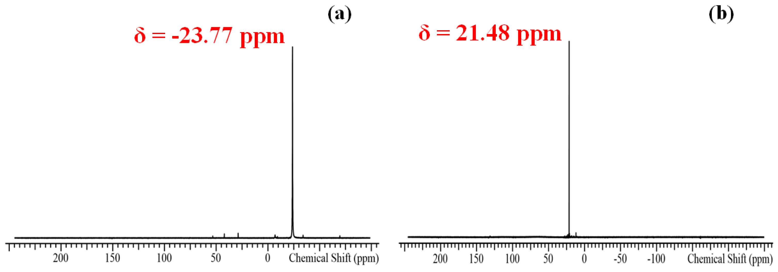
2.2.2. Characterization of Tellurium Precursors

The characterization of TDMAPTe and TOPTe was performed using 31P NMR spectroscopy. In the case of TOPTe, one signal at δ = −23.77 ppm (solvent, C6D6) was observed, as shown in Figure 1a. This indicated that the precursor solution consisted of a mixture of TOPTe and TOP. The ability to detect the pattern of pure TOPTe in the presence of unreacted TOP was not available [5]. A signal for TOPO at δ = 43.8 ppm (solvent, C6D6) was not observed [9]. For TDMAPTe, one pattern was shown at δ = 21.48 ppm (solvent, C6D6), agreeing with the formation of TDMAPTe/TDMAP mixture in THF. No signals for pure TDMAPTe at δ = 58.8 ppm (solvent, C6H6) or pure TDMAP at δ = 123 ppm (solvent, C6H6) were detected [6,10], as shown in Figure 1b.

2.2.3. Synthesis of Mercury Telluride Colloidal Quantum Dots

HgTe colloidal quantum dots (QDs) were prepared using the procedure mentioned elsewhere [1,4,7,8,11,12], with a slight modification. The samples were synthesized using the hot-injection route at 60 °C. The synthesis involved oleylamine solutions of mercury and tellurium precursors: 0.2 mmol of HgCl2 dissolved in 6 mL of oleylamine at 100 °C for 1 h (solution A); 0.2 mmol of tellurium precursor resolved in 2 mL of oleylamine at 100 °C for 1 h (solution B). When the solutions cooled to 60 °C, reagent B was rapidly inserted by syringe into solution A under Ar flow. At a specific reaction time, a quenching solution consisting of 16 mL tetrachloroethylene (TCE), 3.1 mL 1-dodecanethiol (DDT), and 1.5 mL Tri-n-octylphosphine (TOP) was added to the previous solution and cooled to room temperature using an ice bath.
The purification procedure was conducted as follows: 8 mL of methanol was added to 12 mL of the quantum dot solution. The mixture was centrifuged to isolate the precipitate. The obtained precipitate was re-dispersed in 4 mL of tetrachloroethylene (TCE) and 4 mL of acetonitrile was added. The centrifugate product was dried under Ar flow, re-dispersed, filtered using a 0.22 µm hydrophobic polytetrafluoroethylene syringe filter, and diluted to 2 mL using tetrachloroethylene (TCE).

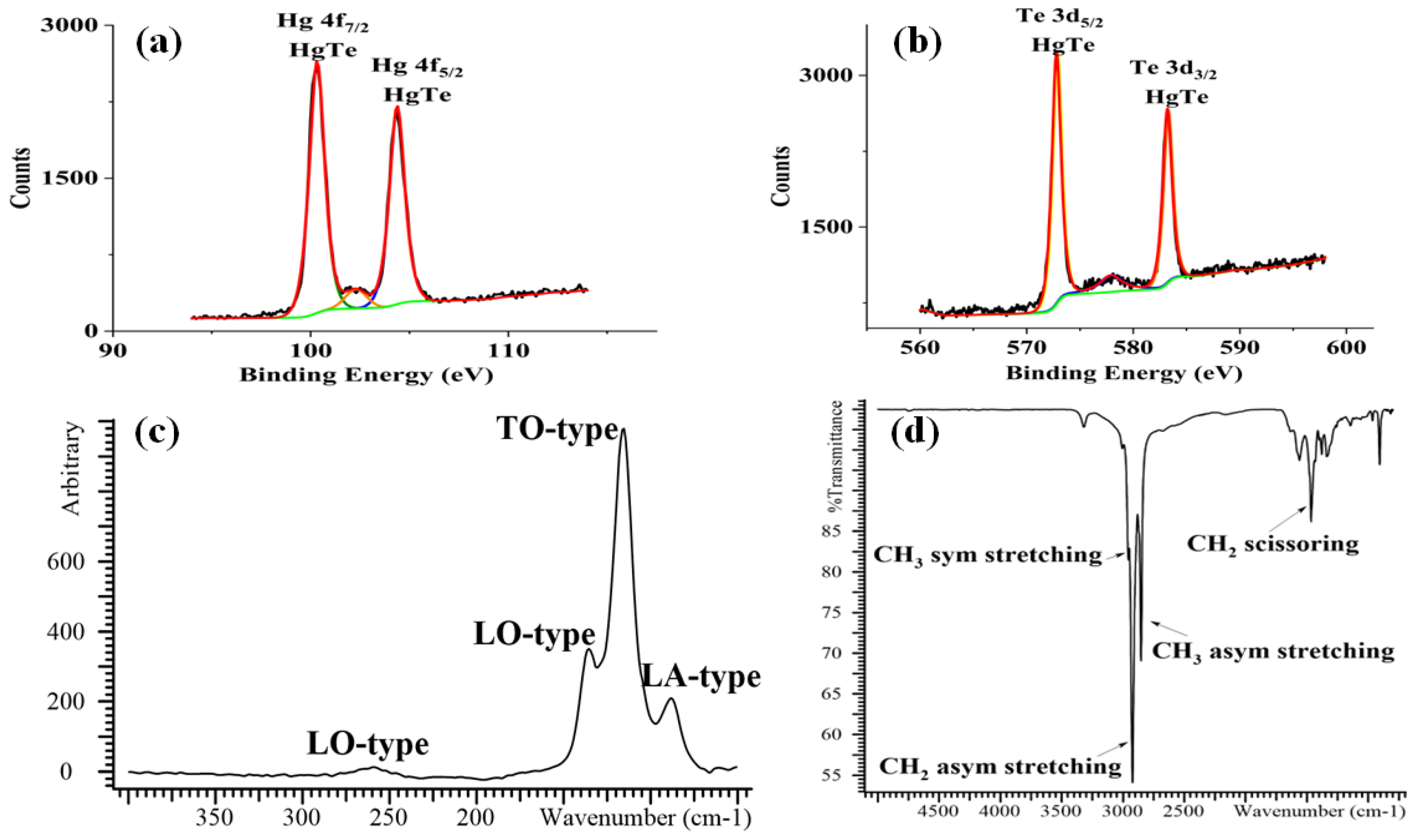
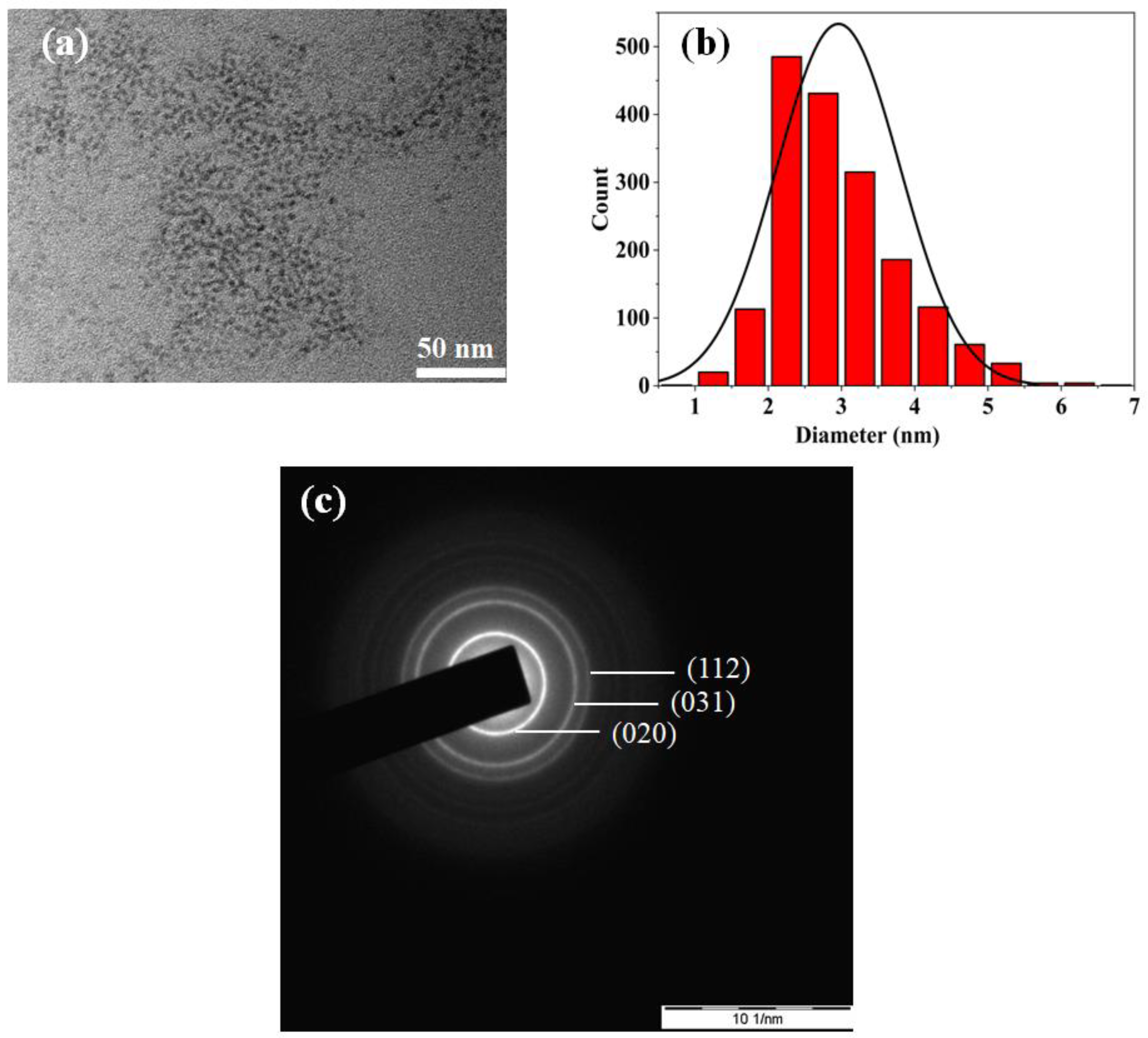
2.2.4. Characterization of HgTe CQDs

Characterization of the obtained HgTe NCs was performed using UV–Vis–NIR spectroscopy, Fourier-transform infrared spectroscopy (FT-IR), Raman spectroscopy, X-ray photoelectron spectroscopy (XPS), transmission electron microscopy (TEM) and selected area electron diffraction (SAED). The quantum confinement effect was observed in the N-IR spectra, Figure 2a,b, of prepared NCs through the red-shifting of the first excitonic peak to a longer wavelength when size increased [4,13,14]. In addition, infrared spectra of HgTe CQDs in TCE demonstrated that the chemical reactivity of TDMAPTe/THF is higher than that for TOPTe/TOP. The first excitonic peak of the synthesized quantum dots was ~1297 nm after 15 min of reaction time with narrow size distribution (FWHM~214 nm). A red-shifting occurred to ~1336 nm after 60 min, although no considerable shift was shown later (Table 1).
HgTe CQDs are materials sensitive to oxygen in the air; therefore, X-ray photoelectron spectroscopy (XPS) was used to determine the chemical structure of the prepared nanocrystals. The XPS spectrum, Figure 3a,b, revealed two spectral lines of 4f7/2 and 4f5/2 for Hg in HgTe at binding energies of 100 and 104 eV. Two other peaks of 3d5/2 and 3d3/2 for Te in HgTe at binding energies of 573 and 583 eV were observed, respectively [2,12]. No signals attributed to the heavy oxidation of Te−2 to Te+4, such as HgTeO3 or TeO2 (paratellurite), were shown. Raman spectroscopy was also used in order to identify the obtained nanocrystals. A thin film of mercury telluride CQDs on a glass substrate was prepared by employing a drop-cast route. The measurement was between 50 and 500 cm−1. The observed phonon lines were as follows: a phonon of longitudinal acoustic (LA) at 88.31 cm−1; a phonon of transverse optic (TO) at 116 cm−1; two phonons of longitudinal optic (LO) at 136 cm−1 and at 259 cm−1 as a weak signal (Figure 3c). This result agreed well with the findings in reference [7]. The shell was investigated using Fourier-transform infrared spectroscopy (FT-IR). The measured FT-IR spectrum, the 5000–1000 cm−1 region in Figure 3d, revealed C–H stretching of methylene and methyl groups at 2955 cm−1, 2922 cm−1, and 2852 cm−1. Methylene groups exhibited bending at 1467 cm−1. No band of free thiol at 2595 cm−1 was observed [15].
The TEM image revealed that the synthesized HgTe CQDs are sphere-shaped with a mean size distribution of ~3 nm, as shown in Figure 4a,b. Analysis of selected-area electron diffraction (SAED) showed that the prepared HgTe nanocrystals had a tetragonal crystal structure with lattice planes of (020), (031), and (112) that matched well with Miller indices of HgTe (COD No. 96-152-3885) [16].

3. Conclusions

The preparation of mercury telluride colloidal quantum dots using novel tellurium precursors was presented. Among the three phosphine–telluride precursors that were tested, only TDMAPTe/THF prospered. The higher chemical reactivity of TDMAPTe/THF compared with TOPTe enabled us to obtain small sizes of HgTe CQDs in the same empirical conditions. The first excitonic peak of the NCs prepared using TDMAPTe/THF was red-shifted by 50 nm in the N-IR window, as opposed to 416 nm in the SW-IR window for TOPTe between reaction times of 15 and 90 min and a temperature of 60 °C.

Author Contributions

A.A.M.: investigation; writing—review and editing. I.A.S.: methodology; investigation; writing—review and editing. V.F.R.: supervision; writing—review and editing. All authors have read and agreed to the published version of the manuscript.

Funding

This research was funded by the Ministry of Science and Education of Russian Federation (Agreement No. 075-03-2022-107/10 at 03.11.2022, Project No. FSMG-2022-0034).

Institutional Review Board Statement

Not applicable.

Informed Consent Statement

Not applicable.

Data Availability Statement

Not applicable.

Acknowledgments

Laboratory of Photonics of Quantum Nanostructures-Moscow Institute of Physics and Technology (State University) acknowledges funding from the Ministry of Science and Education of Russian Federation (Agreement No. 075-03-2022-107/10 at 03.11.2022, Project No. FSMG-2022-0034).

Conflicts of Interest

The authors declare no conflict of interest.

References

  1. Keuleyan, S.E.; Guyot-Sionnest, P.; Delerue, C.; Allan, G. Mercury Telluride Colloidal Quantum Dots: Electronic Structure, Size-Dependent Spectra, and Photocurrent Detection up to 12 μm. ACS Nano 2014, 8, 8676–8682. [Google Scholar] [CrossRef] [PubMed]
  2. Kim, S.; Kim, T.; Im, S.H.; Seok, S.I.; Kim, K.W.; Kim, S.; Kim, S.-W. Bandgap engineered monodisperse and stable mercury telluride quantum dots and their application for near-infrared photodetection. J. Mater. Chem. 2011, 21, 15232–15236. [Google Scholar] [CrossRef]
  3. Kagan, C.R.; Lifshitz, E.; Sargent, E.H.; Talapin, D.V. Building devices from colloidal quantum dots. Science 2016, 353, aac5523. [Google Scholar] [CrossRef] [PubMed]
  4. Keuleyan, S.; Kohler, J.; Guyot-Sionnest, P. Photoluminescence of Mid-Infrared HgTe Colloidal Quantum Dots. J. Phys. Chem. C 2014, 118, 2749–2753. [Google Scholar] [CrossRef]
  5. Shuklov, I.A.; Mikhel, I.S.; Nevidimov, A.V.; Birin, K.P.; Dubrovina, N.V.; Lizunova, A.A.; Razumov, V.F. Mechanistic Insights into the Synthesis of Telluride Colloidal Quantum Dots with Trioctylphosphine-Tellurium. ChemistrySelect 2020, 5, 11896–11900. [Google Scholar] [CrossRef]
  6. Sun, H.; Wang, F.; Buhro, W.E. Tellurium Precursor for Nanocrystal Synthesis: Tris(dimethylamino)phosphine Telluride. ACS Nano 2018, 12, 12393–12400. [Google Scholar] [CrossRef] [PubMed]
  7. Goubet, N.; Jagtap, A.; Livache, C.; Martinez, B.; Portales, H.; Xu, X.Z.; Lobo, R.; Dubertret, B.; Lhuillier, E. Terahertz HgTe Nanocrystals: Beyond Confinement. J. Am. Chem. Soc. 2018, 140, 5033–5036. [Google Scholar] [CrossRef] [PubMed]
  8. Livache, C.; Martinez, B.; Goubet, N.; Greboval, C.; Qu, J.; Chu, A.; Royer, S.; Ithurria, S.; Silly, M.G.; Dubertret, B.; et al. A colloidal quantum dot infrared photodetector and its use for intraband detection. Nat. Commun. 2019, 10, 2125. [Google Scholar] [CrossRef] [PubMed]
  9. Chauzov, V.A.; Kostina, L.P. Alkylation In Situ of Arylphosphines Formed during Thermolysis of Hydrophosphoryl Compounds. J. Gen. Chem. USSR 1991, 61, 2181–2186. [Google Scholar]
  10. Du Mont, W.W.; Kroth, H.J. Zur Reaktion von Organophosphien mit Chalkogenen und Halogenen Rasche Ubertragung von Tellurund Jod Zwischen Phosphinen. J. Organomet. Chem. 1976, 113, C35–C37. [Google Scholar] [CrossRef]
  11. Chu, A.; Martinez, B.; Ferre, S.; Noguier, V.; Greboval, C.; Livache, C.; Qu, J.; Prado, Y.; Casaretto, N.; Goubet, N.; et al. HgTe Nanocrystals for SWIR Detection and Their Integration up to the Focal Plane Array. ACS Appl. Mater. Interfaces 2019, 11, 33116–33123. [Google Scholar] [CrossRef] [PubMed]
  12. Shen, G.; Chen, M.; Guyot-Sionnest, P. Synthesis of Nonaggregating HgTe Colloidal Quantum Dots and the Emergence of Air-Stable n-Doping. J. Phys. Chem. Lett. 2017, 8, 2224–2228. [Google Scholar] [CrossRef] [PubMed]
  13. Brichkin, S.B.; Razumov, V.F. Colloidal quantum dots: Synthesis, properties and applications. Russ. Chem. Rev. 2016, 85, 1297–1312. [Google Scholar] [CrossRef]
  14. Im, S.H.; Kim, H.J.; Kim, S.W.; Kim, S.W.; Seok, S.I. Efficient HgTe colloidal quantum dot-sensitized near-infrared photovoltaic cells. Nanoscale 2012, 4, 1581–1584. [Google Scholar] [CrossRef] [PubMed]
  15. Nyquist, R.A. Interpreting Infrared, Raman, and Nuclear Magnetic Resonance Spectra; Academic Press: San Diego, CA, USA, 2001; Volume 2, pp. 65–83. [Google Scholar]
  16. Grazulis, S.; Chateigner, D.; Downs, R.T.; Yokochi, A.F.T.; Quiros, M.; Lutterotti, L.; Manakova, E.; Butkus, J.; Moeck, P.; Le Bail, A. Crystallography Open Database—An open-access collection of crystal structures. J. Appl. Crystallogr. 2009, 42, 726–729. [Google Scholar] [CrossRef] [PubMed]
Figure 1. 31P spectra of (a) 1 M TOPTe/TOP and (b) 0.5 M TDMAPTe/THF precursors in C6D6.
Materproc 14 00040 g001
Figure 2. N-IR spectra of HgTe CQDs prepared using (a) 1 M TOPTe/TOP and (b) 0.5 M TDMAPTe/THF precursors.
Materproc 14 00040 g002
Figure 3. XPS spectra of (a) mercury and (b) tellurium, Raman (c), and FTIR (d) spectra of HgTe CQDs obtained using 0.5 M TDMAPTe/THF precursors.
Materproc 14 00040 g003
Figure 4. (a) TEM image, (b) size distribution histogram, and (c) SAED patterns and the corresponding Miller indices of HgTe CQDs synthesized using 0.5 M TDMAPTe/THF precursors.
Materproc 14 00040 g004
Table 1. Investigation of the chemical reactivity of tellurium precursors.
Precursor15 minFWHM60 minFWHM90 minFWHM
TDMAPTe/THF129721413342411342253
TOPTe/TOP184033520523752256425
Disclaimer/Publisher’s Note: The statements, opinions and data contained in all publications are solely those of the individual author(s) and contributor(s) and not of MDPI and/or the editor(s). MDPI and/or the editor(s) disclaim responsibility for any injury to people or property resulting from any ideas, methods, instructions or products referred to in the content.

