Proceeding Paper

Characterization of Spinal Cord Stimulation Electrode for Chronic Implant in Animal Models †

1
Federal Institute for Education, Science and Technology of Rio Grande do Norte (IFRN), Ceará-Mirim 59580-000, Brazil
2
Edmond and Lily Safra International Institute of Neuroscience (ELS-IIN), Macaíba 59280-000, Brazil
3
Department of Materials Engineering, Federal University of Rio Grande do Norte (UFRN), Natal 59072-970, Brazil
4
School of Medicine, State University of Rio Grande do Norte (UERN), Mossoró 59607-360, Brazil
5
Anita Garibaldi Center for Education and Research in Health, Santos Dumont Institute (ISD), Macaíba 59280-000, Brazil
6
Neuron—Neurosurgical Team, Natal Hospital Center, Natal 59020-505, Brazil
*
Author to whom correspondence should be addressed.
Presented at the 3rd International Electronic Conference on Biosensors, 8–21 May 2023; Available online: https://iecb2023.sciforum.net
These authors contributed equally to this work.
Eng. Proc. 2023, 35(1), 34; https://doi.org/10.3390/IECB2023-14579
Published: 8 May 2023
(This article belongs to the Proceedings of The 3rd International Electronic Conference on Biosensors)

Abstract

:
Spinal cord electrical (SCS) stimulation alleviates motor deficits in rodent and primate models of Parkinson’s disease due to a suppression of synchronous corticostriatal low-frequency oscillation. Limited epidural space requires resistant biocompatible microelectrodes to deliver efficiently electrical currents through a metal–cellular interface. Platinum (Pt) microelectrodes may lead to material degradation and topography modification under prolonged electrical stimulation. Thus, microstimulation performance over time can deteriorate and affect the functional recovery produced by SCS. To investigate electrodes commonly implanted in the epidural space of rats, Pt microelectrodes immersed in physiological saline underwent 48 h of electrical stimulation (100 Hz; 1.0, 1.3, and 1.6 mA). A wettability test was performed to characterize the interaction of the contact angle before and after stimulation, and it was found that there was an increase in this angle after the stimulation. An electrical impedance test showed that electrochemical interactions caused an increase in impedance after the stimulation. A roughness analysis also showed an increase in roughness after stimulation. Pt electrodes under chronic electric stimulation are susceptible to degradation, and further studies can improve electrode stability and efficacy as new sensor technologies become available.

1. Introduction

Spinal cord electrical stimulation (SCS) was first used in 1967 to inhibit severe diffuse pain in the chest and abdomen [1,2]. Since then, SCS has emerged as a potential neuromodulation method for pain relief and motor disorders. More recently, SCS in mice [3] and marmoset models of Parkinson’s disease reduced akinesia [4], a significant step toward alternative therapies for PD, since subthalamic nucleus (STN) deep brain stimulation (DBS) eligibility is limited to 1.6 to 4.5% of PD patients [5].
Recent findings support the neurophysiological mechanism of SCS on PD motor symptoms, such as desynchronizing corticostriatal oscillations to improve the control of movements [3,4].
Despite the lack of a complete neurophysiological mechanism describing the effects of SCS on akinesia, other aspects, such as electrode geometry, stimulation parameters, and spinal cord stimulation level have been deemed critical to understanding different outcomes [1,6].
One of the most critical issues concerning implantable devices is biocompatibility. Biocompatible materials are used to reduce the intensity and time duration of the inflammatory processes. However, the implanted material interacts with physical, biological, and chemical agents over time and may change its composition, surface, biocompatibility, and electrochemical properties. Platinum is extensively used as a biocompatible metal for mechanical and electrical applications. Nevertheless, when platinum is used at thin thicknesses, its structure may be disrupted after several cycles of stimuli [7]. Another critical factor is impedance, since a low value of electrochemical impedance between the electrode and the electrolyte (tissue) is a vital and necessary condition to reduce tissue damage during stimulation. The reduction of these lesions contributes directly to an increase in the longevity of the electrode [8,9,10].
A well-characterized and fully functional electrode leads to an optimized electrode–tissue interface coupling. The electrode characterization contributes to chronic SCS studies for motor symptoms in PD [3] since this technique still needs a standardized method for its therapeutic consistency. This characterization supports the robustness of electrode behavior throughout chronic use and as a preliminary approach to providing safety and effectiveness for its use in animal models.
Here, we present a characterization with wettability, impedance, and roughness tests on electrodes that underwent electric current stimulation in vitro. The wettability test characterizes the interaction between the electrode and the electrolyte, impedance testing indicates the resistance to flow current between the electrode and the tissue, and roughness testing evaluates the texture of the surface that interfaces with tissue. These tests can assist in predicting the behavior of the submitted electrode in vivo.

2. Materials and Methods

2.1. Manufacture of Electrodes

Platinum foil (99.9% purity) was used to make two rectangular electrical contacts measuring 1.0 × 0.8 mm (25 µm thick). These contacts were micro-welded to stainless steel wires of ø25.4 µm with welding alloy (PbSn). NuSil MED2-4420 (in a ratio of 1:1) medical silicone was used to insulate the welding surface of the pairs of platinum contacts, resulting in a 100 µm total thick paddle electrode.
The dimensions of the platinum contacts were based on the spinal cord anatomy of rats at the high thoracic level, accordingly to previous successful results [3,4,11]. The rectangular contacts were placed side by side and spaced by 0.4 mm (center–center distance is 1.2 mm), and their longer dimension runs longitudinally to the spinal cord. This electrode geometry intends to deliver an electrical current to stimulate dorsal column fibers and to avoid side effects resulting from the activation of the dorsal root fiber.

2.2. Wettability Test

Wettability tests are performed as an indicator of biocompatibility, and they consist of a technique to measure the surface energy of a material sample. This energy is given as a function of the interaction between the surface and a liquid with a known surface energy, such as water, for example. This interaction can be numerically associated with the measurement of the contact angle that exists between the liquid droplet and the material’s surface [12]. If a material has a low wettability, it means it exhibits hydrophobicity, and thus it would not recruit cells in a biological environment once there is no water adhered to it in first place [13]. However, considering stimulation electrodes inserted in a fluid, the goal is to invoke a minimal inflammatory response and produce an optimal interface between the solid and liquid surfaces to facilitate the transfer of charge [14]. Thus, the characterization of the wettability of platinum electrodes is necessary to verify their ability to conduct charges through fluid, where it is desired that it be hydrophilic.
Our method was based on a wettability test technique shown in Mittal [15]. Sessile drop measurement was performed with an adapted apparatus coupled to an automatic injector system (Robot Stereotaxic, NEUROSTAR) to precisely deliver pure water droplets of 0.5 µL using a Hamilton syringe over the platinum contacts and silicone paddle (substrate) (Figure 1a).
Images were captured within 5 s after the droplet met the surface of the substrate using a digital camera (AxioCam MRc, Zeiss) attached to a microscope, which was tilted to obtain images of the droplet according to previous procedures shown in Mittal [15]. The temperature of the room was maintained at 17 C in order to avoid rapid water evaporation, and contact angles were computed offline using AutoCAD Autodesk software (Figure 1b).

2.3. Electrical Impedance Measurement

An impedance meter (SIGGI II, Falk Minow) was used at a frequency of 100 Hz (same frequency of stimulation). The electrodes that were to be measured were dipped into saline solution (0.9%) with a reference wired to its metallic beaker (circuited with the saline), and the system was properly grounded.
Stabilization was observed before the measurement was taken after a period of becoming accustomed to the electrolyte in use. The electrode is meant to have an electrical potential, but if it reaches a value of 300 mV, the electrode is considered faulty. Measurements occurred when the electrical potential was lower than 1 mV (following specifications by the impedance meter’s manufacturer). For each electrode, three measurements were taken before and after the stimulation cycles for the retroactive calculation of their average.

2.4. In Vitro Electrical Stimulation

An STG 4004 stimulator (Multichannel Systems) was used to generate a bipolar square wave signal at 100 Hz with 1.6 mA and a 500 µs pulse width to continuously stimulate the electrodes dipped in 0.9% saline solution for 48 h. Over 1.7 × 10⁷ pulses were generated, which is at least 2 times more than a previous study with SCS in PD [11].

2.5. Stimulation Characterization

The Shannon [16] limit was used to characterize the electrical stimulation performed by the electrode. Based on past neural stimulation results in cats, Shannon made a chart with upper limits for safe electrical stimulation. Shannon proposed that the maximum stimulation limit was k = 1.5. However, because this value is restrictive, a value of 1.75 was considered by the literature, as it was slightly below the appearance of tissue damage [17]. The values obtained without damage by McCreery [18] can be obtained from:
L o g ( D ) = K L o g ( D )
where D is the charge density in μ Coulombs/cm 2 /phase and Q is the charge in μ Coulombs/phase. D = Q / A , where A is the electrode surface area in cm 2 . The formula can be expressed as
L o g ( Q A ) = K L o g ( Q )
From Equation (2), K calculations were made for the stimulation analyzed in vitro. The surface contact area of the electrode was A = 0.008 cm², whereas the charge was Q = 0.8 µC, where Q is the product of 1.6 mA and 500 µs. The influence of other K values on the change in the surface of the electrodes was carried out to analyze the roughness before and after the in vitro electrostimulation. Electrostimulations were performed with loads of 1.6 mA, 1.3 mA, and 1.0 mA, respectively, representing K = 1.90, K = 1.75, and K = 1.50.

2.6. Roughness Analysis

The roughness of the electrodes was measured through analysis by an atomic force microscope (AFM). Three pairs of electrodes were characterized at room temperature (25 °C) using 11 N/m spring cantilevers with a 10 nm tip radius of curvature, a 10:1 aspect ratio, and a 150 kHz resonance frequency. For all measurements, two measurements were performed per sample to be scanned, and data were collected. The surface scan was performed perpendicular to the cantilever axis and with a scan area of 5 × 5 μ m. The measure of the roughness used to compare the samples was the mean squared deviation (Rq), which was provided by the equipment.

3. Results and Discussion

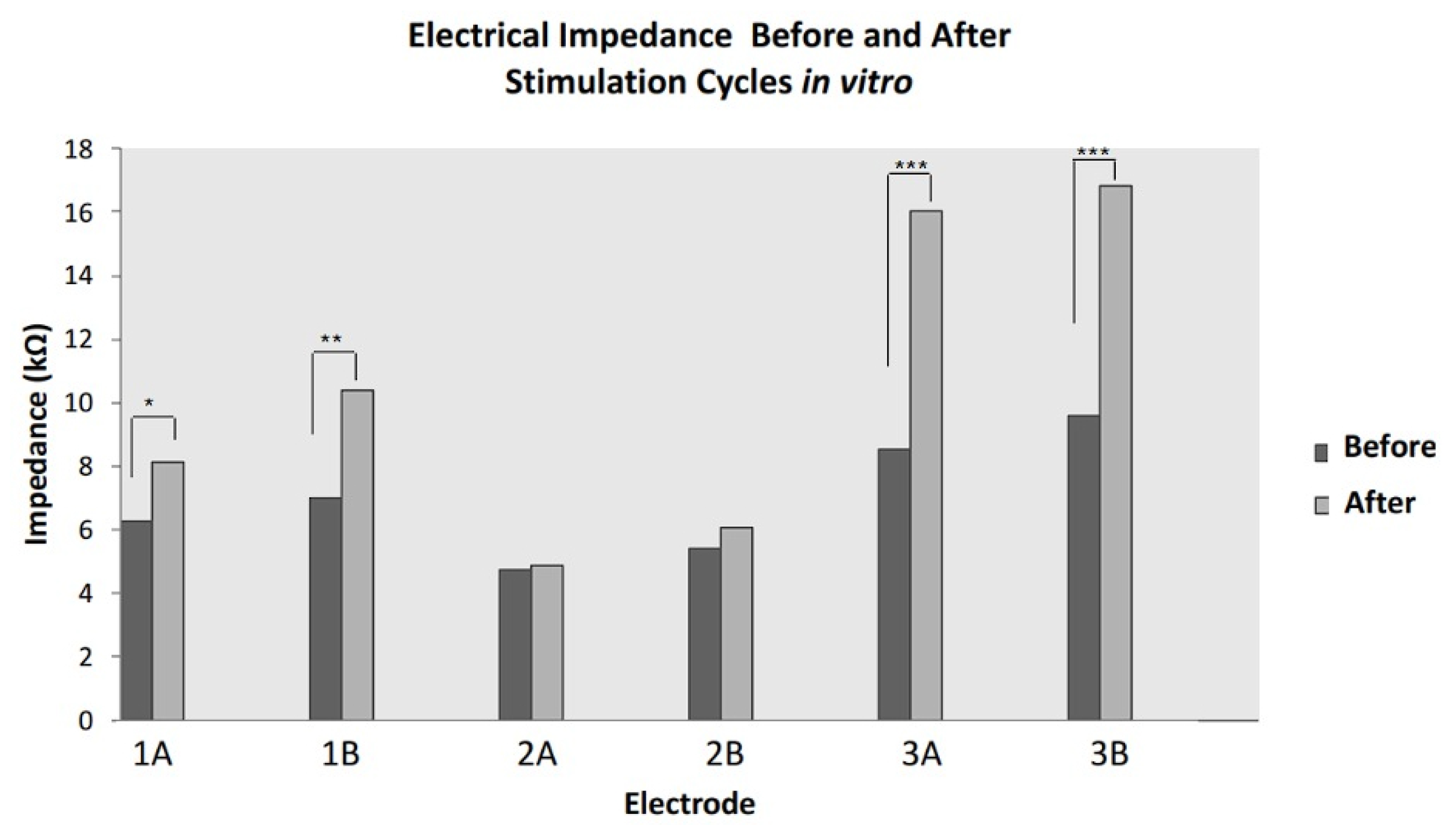
3.1. Electrode and Impedance Characterization

Three pairs of electrodes were manufactured, tested, and stimulated for an uninterrupted 48 h. The averages of each set of electrode measurements taken before and after the electrical stimulation cycles are presented graphically in Figure 2. Overall, the electrical impedance showed an increase of 7.8% (from 5.1 kΩ to 5.5 kΩ after stimulation), and the two pairs (1 and 3) presented a significant increase in impedance value (p ≤ 0.05).
The impedance results were higher after stimulation than before. This was due to the oxidation of the platinum at values of k > 1.75, which resulted in superficial changes in the electrode interface, thus increasing its contact angle. This increase in the contact angle generated a different solid–liquid interaction and increased the impedance. Nevertheless, the average increase was within the experimental measure’s acceptable range of 10%; thus, the electrodes were considered robust in their electrical behavior while experiencing electrical stimulation of over more than 1.7 × 10 7 pulses (2.4 times more than used in Yadav et al. [11]). It is important, however, to highlight that different results are expected if other frequencies are used, as capacitive polarization in an electrode–electrolyte interaction is frequency-dependent, according to Fricke’s law [19].
The characterization of the stimulation resulted in k = 1.90, which is above the limit of k = 1.75 proposed by Shannon as safe and incapable of generating damage to the tissue where it is implanted [16]. The increase in the impedance of the electrodes after the stimulation tests can be attributed to the value of k = 1.90. The combination of the platinum used for electrical stimulation with k > 1.75 and an environment with available oxygen results in surface oxidation. This oxidation can generate an increase in impedance due to superficial changes to the electrode, as well as a release of platinum oxide, which in large quantities, can cause damage to the tissue where it was implanted [14].

3.2. Wettability Test and Roughness

The wettability test showed average contact angles of 106º and 120º for the platinum contacts before and after stimulation and 130º for the silicone pad. These values showed that the electrodes had increased hydrophobic properties, which is not desirable for stimulation electrodes. The value of K = 1.90 was obtained according to Equation (2). This characterization is necessary to predict whether the electrode will cause tissue damage through electrical stimulation. With this result, we enter the level of tissue damage and platinum degradation.
The contact angle was measured before and after stimulation, and it showed an increase after stimulation compared to the measurement before stimulation. This change occurs due to the oxidation of platinum, which, in turn, occurs due to changes in the electrode surface, thus increasing its roughness. This results particularly from stimulation with values of K > 1.75. Correlating roughness and wettability is not a simple association. Traditionally, the theories of Wenzer and Cassie-Baxter explain the moistening of rough interfaces. According to Wenzer’s theory, a smooth surface, which is hydrophobic, will increase its contact angle if roughness is increased. This statement is also valid for a smooth hydrophilic surface; its contact angle will be reduced if roughness is increased [20]. Cassie-Baxter’s theory considers gas bubbles that are trapped in the solid and liquid phases [21]. Thus, with the increase in the contact angle due to the degradation of the platinum, there was an increase in the impedance due to the greater hydrophobicity of the electrode. Figure 3 shows an increase in roughness after stimulation, which thus increased the contact angle.
This study examined three different electrostimulation loads to assess their effect on the surface roughness of the electrodes. Loads of 1.6 mA, 1.3 mA, and 1.0 mA were chosen, corresponding to K values of 1.90, 1.75, and 1.50, respectively, based on the Shannon parameter [16]. Surface changes before and after in vitro electrostimulation were compared using the difference in the current, and mean square deviation was used to quantify roughness in both conditions. The results showed an increase in roughness of up to 371% (Table 1 compared to the initial condition, with the greatest increase being observed at the highest load of 1.6 mA, corresponding to K = 1.90. The increase in roughness was found to be directly linked to the increase in the applied load during in vitro electrostimulation. However, it is noteworthy that the coefficient was above the safety threshold proposed by Shannon, indicating the potential risk of damage to the tissue where the electrode is implanted. The electrostimulation loads of 1.3 mA and 1.0 mA, corresponding to K values of 1.75 and 1.50, respectively, were within the safety range for electrostimulation but still resulted in an increase in roughness of 212.3% and 181.5%, respectively. The oxidation and modification of the platinum electrode’s surface was also observed even within the safe electrostimulation parameters. This oxidation can lead to the formation of platinum oxide, which, in large amounts, can cause tissue damage [14].

4. Conclusions

In conclusion, the characterization of invasive electrodes is crucial to ensure their safe and effective use in chronic experimentation. Consistent with the existing literature, our study showed that the electrostimulation of platinum electrodes results in increased roughness, even at lower loads within safe electrostimulation parameters. The modification of these electrode’s roughness can form platinum oxide, which may cause tissue damage in large amounts. Therefore, it is crucial to consider the electrostimulation parameters used in medical devices considering the potential risks of tissue damage caused by roughness modification in platinum electrodes. Finally, the potential of SCS to reduce the invasiveness of several therapies for neurological disorders is advancing. Furthermore, this characterization in animal models supports further human studies in neuromodulation.

Author Contributions

Conceptualization, L.C., E.M. and H.S.; methodology, L.C., G.F., R.M., H.D., I.D. and E.M.; formal analysis, L.C., G.F. and E.M.; investigation, L.C. and G.F.; resources, L.C., E.M. and H.S.; writing—original draft preparation, L.C., G.F. and E.M.; writing—review and editing, L.C., G.F., E.M. and H.S.; visualization, L.C., G.F. and I.D.; supervision, E.M. and H.S. All authors have read and agreed to the published version of the manuscript.

Funding

This study was funded by the Brazilian Ministry of Education (MEC), CNPq (313381/2017-8), CAPES (scholarship to G.F. and L.C.) and Santos Dumont Institute.

Institutional Review Board Statement

Not applicable.

Informed Consent Statement

Not applicable.

Data Availability Statement

The data that support the findings of this study are available on request from the corresponding author, E.M.

Acknowledgments

This work was supported by the Brazilian research agency CAPES and the Santos Dumont Institute (ISD).

Conflicts of Interest

The authors declare no conflict of interest.

References

  1. De Andrade, E.M.; Ghilardi, M.G.; Cury, R.G.; Barbosa, E.R.; Fuentes, R.; Teixeira, M.J.; Fonoff, E.T. Spinal cord stimulation for Parkinson’s disease: A systematic review. Neurosurg. Rev. 2016, 39, 27–35. [Google Scholar] [CrossRef] [PubMed]
  2. Shealy, C.N.; Mortimer, J.T.; Reswick, J.B. Electrical inhibition of pain by stimulation of the dorsal columns: Preliminary clinical report. Anesth. Analg. 1967, 46, 489–491. [Google Scholar] [CrossRef] [PubMed]
  3. Fuentes, R.; Petersson, P.; Siesser, W.B.; Caron, M.G.; Nicolelis, M.A. Spinal cord stimulation restores locomotion in animal models of Parkinson’s disease. Science 2009, 323, 1578–1582. [Google Scholar] [CrossRef] [PubMed]
  4. Santana, M.B.; Halje, P.; Simplício, H.; Richter, U.; Freire, M.A.M.; Petersson, P.; Fuentes, R.; Nicolelis, M.A. Spinal cord stimulation alleviates motor deficits in a primate model of Parkinson disease. Neuron 2014, 84, 716–722. [Google Scholar] [CrossRef] [PubMed]
  5. Morgante, L.; Morgante, F.; Moro, E.; Epifanio, A.; Girlanda, P.; Ragonese, P.; Antonini, A.; Barone, P.; Bonuccelli, U.; Contarino, M.F.; et al. How many parkinsonian patients are suitable candidates for deep brain stimulation of subthalamic nucleus? Results of a questionnaire. Park. Relat. Disord. 2007, 13, 528–531. [Google Scholar] [CrossRef] [PubMed]
  6. Nicolelis, M.A.; Thevathasan, W.; Fuentes, R.; Petersson, P.; Brown, P. Spinal cord stimulation failed to relieve akinesia or restore locomotion in Parkinson disease. Neurology 2010, 75, 1484–1485. [Google Scholar] [CrossRef] [PubMed]
  7. Ordonez, J.S.; Rudmann, L.; Cvancara, P.; Bentler, C.; Stieglitz, T. Mechanical deformation of thin film platinum under electrical stimulation. In Proceedings of the 2015 37th Annual International Conference of the IEEE Engineering in Medicine and Biology Society (EMBC), Milan, Italy, 25–29 August 2015; pp. 1045–1048. [Google Scholar]
  8. Howlader, M.; Doyle, T.; Mohtashami, S.; Kish, J. Charge transfer and stability of implantable electrodes on flexible substrate. Sensors Actuators B Chem. 2013, 178, 132–139. [Google Scholar] [CrossRef]
  9. Wang, K.; Liu, C.C.; Durand, D.M. Flexible nerve stimulation electrode with iridium oxide sputtered on liquid crystal polymer. IEEE Trans. Biomed. Eng. 2009, 56, 6–14. [Google Scholar] [CrossRef] [PubMed]
  10. Mohtashami, S. Electrochemical Properties of Flexible Electrodes for Implanted Neuromuscular Excitation Applications. Ph.D. Thesis, McMaster University, Hamilton, ON, Canada, 2011. [Google Scholar]
  11. Yadav, A.P.; Fuentes, R.; Zhang, H.; Vinholo, T.; Wang, C.H.; Freire, M.A.M.; Nicolelis, M.A. Chronic spinal cord electrical stimulation protects against 6-hydroxydopamine lesions. Sci. Rep. 2014, 4, 3839. [Google Scholar] [CrossRef] [PubMed]
  12. Ziauddin, M.; Montaron, B.; Hussain, H.; Habashy, T.; Seleznev, N.; Signer, C.; Abdallah, W. Fundamentals of wettability. Schlumberger Oilfield Rev. 2007, 19, 40–67. [Google Scholar]
  13. Lotfi, M.; Nejib, M.; Naceur, M. Advances in Biomaterials Science and Biomedical Applications; Pignatello, R., Ed.; InTech: Rijeka, Croatia, 2013; Volume 2. [Google Scholar]
  14. Kumsa, D.W.; Montague, F.W.; Hudak, E.M.; Mortimer, J.T. Electron transfer processes occurring on platinum neural stimulating electrodes: Pulsing experiments for cathodic-first/charge-balanced/biphasic pulses for 0.566 ≤ k ≥ 2.3 in oxygenated and deoxygenated sulfuric acid. J. Neural Eng. 2016, 13, 056001. [Google Scholar] [CrossRef] [PubMed]
  15. Mittal, K.L. Contact Angle, Wettability and Adhesion; CRC Press: Boca Raton, FL, USA, 2003; Volume 3. [Google Scholar]
  16. Shannon, R.V. A model of safe levels for electrical stimulation. IEEE Trans. Biomed. Eng. 1992, 39, 424–426. [Google Scholar] [CrossRef] [PubMed]
  17. Merrill, D.R.; Bikson, M.; Jefferys, J.G. Electrical stimulation of excitable tissue: Design of efficacious and safe protocols. J. Neurosci. Methods 2005, 141, 171–198. [Google Scholar] [CrossRef] [PubMed]
  18. McCreery, D.B.; Agnew, W.F.; Yuen, T.G.; Bullara, L. Charge density and charge per phase as cofactors in neural injury induced by electrical stimulation. IEEE Trans. Biomed. Eng. 1990, 37, 996–1001. [Google Scholar] [CrossRef] [PubMed]
  19. Fricke, H. XXXIII. The theory of electrolytic polarization. Lond. Edinb. Dublin Philos. Mag. J. Sci. 1932, 14, 310–318. [Google Scholar] [CrossRef]
  20. Belaud, V.; Valette, S.; Stremsdoerfer, G.; Bigerelle, M.; Benayoun, S. Wettability versus roughness: Multi-scales approach. Tribol. Int. 2015, 82, 343–349. [Google Scholar] [CrossRef]
  21. Cassie, A.; Baxter, S. Wettability of porous surfaces. Trans. Faraday Soc. 1944, 40, 546–551. [Google Scholar] [CrossRef]
Figure 1. (a) Schematic representation for the setup of wettability test. (b) Example of contact angle measurement by AutoCAD geometry drawing tools.
Engproc 35 00034 g001
Figure 2. Electrical impedance measurements of three pairs of electrodes. Contacts were numbered 1 to 3; A and B means left and right, respectively. Dark bars represent measurements before stimulation, and light bars represent measurements after stimulation. * p < 0.05, ** p < 0.02 and *** p < 0.001.
Engproc 35 00034 g002
Figure 3. (a) Electrode before and after stimulation with a load of 1.0 mA (K = 1.50). (b) Electrode before and after stimulation with a load of 1.3 mA (K = 1.75). (c) Electrode before and after stimulation with a load of 1.6 mA (K = 1.90).
Engproc 35 00034 g003
Table 1. Roughness of platinum electrodes before and after electrostimulation.
CurrentRoughness before
Stimulation (Rq)
Roughness after
Stimulation (Rq)
Percentage
Change
1.0 mA32.08 ± 5.6190.32 ± 31.76181.5%
1.3 mA37.47 ± 11.79115.73 ± 30.35212.3%
1.6 mA18.48 ± 4.7386.89 ± 32.86371.2%
Disclaimer/Publisher’s Note: The statements, opinions and data contained in all publications are solely those of the individual author(s) and contributor(s) and not of MDPI and/or the editor(s). MDPI and/or the editor(s) disclaim responsibility for any injury to people or property resulting from any ideas, methods, instructions or products referred to in the content.

