1. Introduction
Zinc oxide (ZnO) is an intrinsic binary n-type semiconductor material that belongs to class II-VI, has a bandgap of about 3.37 eV, and an excitonic binding energy of 60 meV. This semiconductor is considered a good host network for doping elements, such as transition metals (MTs), which have an ionic radius comparable to Zn
2+ and are highly soluble [
1].
Doping with transition metals promotes the insertion of discrete energy levels in the bandgap, allowing the occurrence of lower energy electronic transitions and tuning the spectral region profiles associated with them. Doping also improves the mobility of charge carriers (electrons and holes), the appearance of donor and acceptor defects, conductivity as well as different optical and magnetic properties. These characteristics open up the possibilities of application of pure and doped ZnO in several fields of nanotechnology, such as optoelectronic devices, LEDs [
1], diluted magnetic semiconductors (DMS), photonics [
1], and in nanomedicine [
2]. Therefore, this material has been attracting the attention of the scientific community in recent decades due to its potential applications.
These materials have been mainly prepared by wet methods such as sol-gel, solvothermal, hydrothermal, and coprecipitation [
1,
2]. Many of them use organic solvents and high temperatures. Since the properties of interest are generally related to the morphology and size of the nanoparticles, which can be modulated from synthetic parameters [
3], the preparation of ZnO nanocrystals, with dimensions lower than 100 nm, in water is still a challenge. In this context, we aimed to develop an alternative synthetic procedure to obtain ZnO nanocrystals in water. For this, we used the colloidal synthesis methodology, followed by a heat treatment. Thus, this manuscript presents the preparation of pure ZnO and ZnO:Co NPs via colloidal chemistry in an aqueous medium using mercaptosuccinic acid (MSA) as a surface-stabilizing agent.
2. Materials and Methods
Colloidal synthesis of pure and Co-doped ZnO was carried out using zinc chloride (ZnCl
2) or zinc nitrate hexahydrate [Zn(NO
3)
2·6H
2O] as a zinc source, cobalt chloride hexahydrate (CoCl
2·6H
2O) or cobalt nitrate hexahydrate [Co(NO
3)
2·6H
2O)] as cobalt source, mercaptosuccinic acid (MSA) as the stabilizing agent, and sodium hydroxide (NaOH) as the precipitant agent for pH control. The molar ratio between Zn and Co was utilized according to the expression, Zn
1−xCo
xO (
x = 0.05, 0.075, 0.10) [
4], and the molar ratio between Zn:MSA was 1:4 [
5].
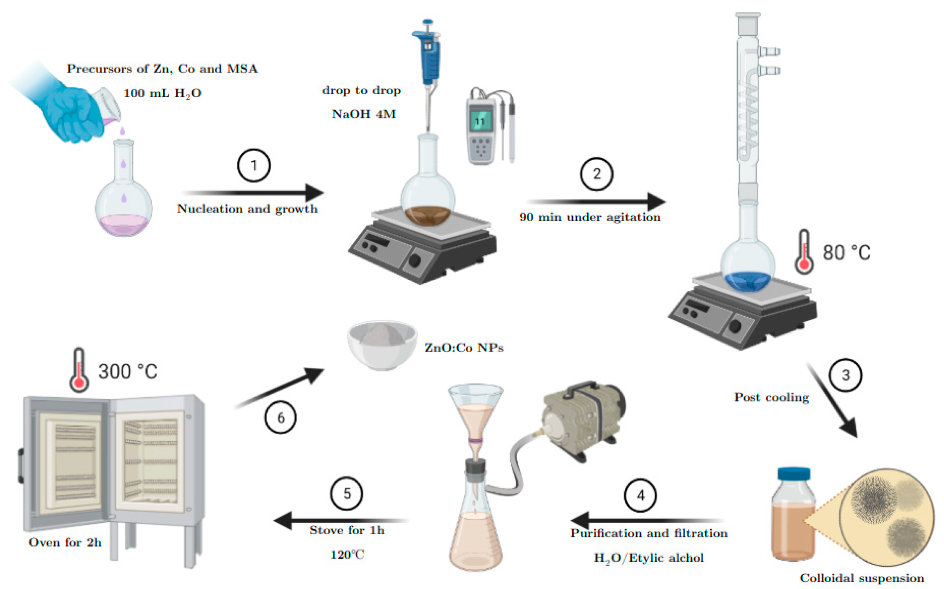
Initially, 0.004 mol of zinc precursor and 0.016 mol of MSA were added to a two-neck synthesis flask and solubilized in 50 mL of double-distilled water. For the system containing the dopant, the same procedure was followed with the addition of the cobalt precursor according to the Zn:Co molar ratio. Then, under stirring, the pH of the solution was adjusted to 11 with a 4 mol·L−1 solution of NaOH. Subsequently, the sample was heated and stirred for 90 min at a temperature of 80 °C.
Thus, the colloidal suspensions obtained were frozen to aid the destabilization of the colloidal system. Then, after thawing, isopropyl alcohol was added to the suspensions to force precipitation. The precipitate was washed with ethyl alcohol and distilled water, filtered using a vacuum pump and a sintered plate filter, dried in an oven for 1 h at 120 °C, and calcined in a muffle furnace for 2 h at 300 °C. A schematic representation of the synthetic procedure is shown in
Figure 1. Finally, all samples obtained were analyzed with an X-ray Diffractometer SmartLab of the brand Rigaku.
3. Results and Discussion
X-ray Diffraction (XRD) patterns of Zn
1−xCo
xO (
x = 0.05, 0.075, 0.10) NPs synthesized using MSA as a stabilizing agent and chloride salts as the precursors of Zn and Co are shown in
Figure 2a. The XRD profiles are well-matched with standard data (ICSD-13950), corresponding to the structure of hexagonal wurtzite, similar to ZnS. For the Zn
0.95Co
0.05O, an uncommon peak was observed around 45° that can be attributed to some secondary phase, such as Co clusters or cobalt oxides, which present characteristic peaks between 40 and 47° [
6], or other impurities of the samples. In order to study the effect of Co doping, a careful analysis of the position of XRD peaks was carried out, especially the prominent (100), (002), and (101) peaks that are shifted toward a lower 2
θ, as shown in
Figure 2b.
In Co-doped ZnO a decrease in the diffraction peak intensities with increasing the Co concentration was also observed. This can indicate that the dopant Co
2+ ions (ionic radius: 0.58 Å) entered in the inner lattice of Zn
2+ ions (ionic radius: 60 Å) [
7] more efficiently with the increase in doping amount. Since the ionic radius of Co
2+ is close to that of Zn
2+, the changes in the FWHM are in accordance with the crystallite size, according to the Debye−Scherrer’s Equation.
Evidently, the average crystal size decreases for Co-doped ZnO NPs with the increase in dopant concentration (
Table 1). The reduction in crystal size is mainly due to the distortion in the host ZnO lattice by the foreign impurities of Co
2+ that decreased the nucleation and subsequent growth rate of the ZnO NPs [
4]. Furthermore, the surface-stabilizing agent (MSA) also has an important role in the nucleation process because of the possibility of a higher control in the size of the particles retarding the growth process.
Figure 3a shows the structural analysis of Co-doped ZnO NPs synthesized using nitrate salts as the precursors for Zn and Co. The diffractogram shows that all peaks are in accordance with the standard data, also corresponding with the hexagonal wurtzite structure. Differently from
Figure 2a, there were no observed peaks of a secondary phase.
Table 2 illustrates the same trend of the Co-doped ZnO NPs with chloride precursors, in which the increase in the concentration of doping (Co) in synthesis leads to a decrease in the average size crystal and an increase in the microstrain in the crystal lattice.
Another parameter to analyze is the microstrain suffered by the ZnO network due to the insertion of Co
2+ ions (
Table 1 and
Table 2). The substitution of Co
2+ into an interstitial position would affect the concentration of interstitial Zn, oxygen, and Zn vacancies. The observation of small changes in 2
θ values of diffraction peaks and peak broadening (
Figure 2b and
Figure 3b) is due to the increase in microstrain [
8], and the line broadening may be due to the size and microstrain [
9] of the nanoparticles. The slight change observed in the diffraction peaks indicates that the incorporation of Co
2+ ions into the ZnO lattice causes minimal changes in the crystal lattice. Nevertheless, when increasing the dopant concentration, the microstrain also increases, which indicates that more Co was incorporated into the ZnO host network, causing major compaction of the crystal lattice due to the replacement of Zn by Co.
In terms of the counterion used, stoichiometrically, we report a molar ratio of 1:2 Zn:counterion for M2+:Cl− and M2+:NO3−, where M = Zn or Co. A slower precipitation was observed for the system containing nitrate than chloride as the counterion. One of the explanations for this may be related to the high colloidal stability of the system containing nitrate as a counterion due to the morphological aspects of the nanoparticles obtained or that nitrate ions may interact with surface ligands (MSA) via hydrogen bonding, thus increasing the negative charge concentration around the ligand and consequently the colloidal stability of the suspension.
The availability of these ions for the processes of NP nucleation and growth is one of the factors determining the nanoparticles average size. Comparing the enthalpy and Gibbs energy of the formation of ions in aqueous media under normal temperature and pressure conditions, we found that nitrate has a more exothermic enthalpy than chloride, −207.4 kJ/mol and −167.2 kJ/mol, respectively, and the Gibbs energy corresponds with the exergonic processes, −131.2 kJ/mol to Cl
− and −111.3 kJ/mol to NO
3− [
10].
Therefore, it is plausible that the nitrate interacts for a longer time with the constituent species of the colloidal suspension, favoring morphological changes and ligand–counterion interactions that are reflect in the smaller average nanoparticles sizes observed based on nitrate salts.
4. Conclusions
Zn1−xCoxO NPs were successfully prepared by a colloidal aqueous synthesis method utilizing MSA as a surface-stabilizing agent. The hexagonal (wurtzite) crystalline structure was identified for doped ZnO through XRD analysis and confirmed the probable incorporation of Co2+ ions into the ZnO matrix. Using nitrate salt precursors, the existence of CoO (Co cluster) peaks was no observed. However, the presence of a probable CoO phase for Zn0.95Co0.05O NPs with chloride salts as precursors was observed. The average crystal size for Co-doped ZnO NPs showed that with the increase in Co concentration the size of the crystals decreased while the microstrain of the host ZnO crystal lattice increased due to the probable substitution of Zn by Co in the network.
As prospective work, additional measurements by transmission (TEM) and scanning electron microscopy (SEM), X-ray photoluminescence spectroscopy (XPS), and active surface measurements (BET) will be carried out for better characterization in terms of compositional analysis, size distribution and morphology of the nanoparticles to evaluate their future application potential.
In addition, more established methods in the literature, such as synthesis via coprecipitation, will be used as a comparative base to evaluate the most promising properties via colloidal synthesis, whose presence of the surface-stabilizing agent has a crucial impact on the nucleation and growth processes, resulting in smaller nanoparticles.
Finally, it is also important to highlight that the synthetic procedure developed in this work is practical, reproducible, and environmentally friendly, allowing the preparation of doped ZnO nanocrystals in an aqueous medium.


 ,
, 


