Proceeding Paper

In Vitro Response of Dental Stem Cells on Decellularized Extracellular Matrix-Derived Hydrogels †

by
Alexandra B. dos Santos
1,2,
Cláudia L. da Silva
1,2 and
Marta S. Carvalho
1,2,*
1
Department of Bioengineering and iBB—Institute of Bioengineering and Biosciences, Instituto Superior Técnico, Universidade de Lisboa,1049-001 Lisboa, Portugal
2
Associate Laboratory i4HB—Institute for Health and Bioeconomy, Instituto Superior Técnico, Universidade de Lisboa, 1049-001 Lisboa, Portugal
*
Author to whom correspondence should be addressed.
Presented at the 3rd International Electronic Conference on Applied Sciences, 1–15 December 2022; Available online: https://asec2022.sciforum.net/.
Eng. Proc. 2023, 31(1), 38; https://doi.org/10.3390/ASEC2022-13832
Published: 9 December 2022
(This article belongs to the Proceedings of The 3rd International Electronic Conference on Applied Sciences)

Abstract

:
Periodontitis is an infectious inflammatory disease that damages the tissues supporting the tooth. Hydrogels are suitable candidates for periodontal regeneration due to their capacity to interact with soft and hard tissues and to conform to the 3D defect through minimal invasion procedures. Cell-derived decellularized extracellular matrix (dECM) can recreate cellular niches and model cellular function. In this work, collagen hydrogels were developed by incorporating lyophilized cell-derived dECM and their effects on the proliferation and osteogenic differentiation of dental stem cells were evaluated. Overall, our results confirmed the beneficial effect of dECM-derived hydrogels in proliferation and osteogenic differentiation of dental stem cells.

1. Introduction

Periodontal disease can be defined as an acquired disorder of the tissues surrounding and supporting the teeth, such as cementum, alveolar bone and periodontal ligament, forming the complex structure called the periodontium [1]. Being caused by bacterial inflammation and plaque accumulation, clinical treatments of periodontitis focus on cause-related, non-surgical and conservative approaches, such as plaque removal and local inflammation control [2]. Although these therapies minimize symptoms and prevent further disease progression, they are not able to restore all the lost tissues [3]. Hence, tissue engineering approaches have been explored for periodontal regeneration, such as the development of hydrogels. Hydrogels are attractive candidates for periodontal applications due to their capacity to interact with both soft and hard tissues, as well as to conform to the 3D defect, requiring less invasive procedures. Hydrogels are highly hydrophilic polymeric networks with the ability to simulate the natural microenvironment of cells and can be composed of natural or synthetic polymers [4]. More specifically, collagen type I hydrogels are very attractive for tissue engineering applications, given collagen is the most abundant protein present in the extracellular matrix (ECM) [5].
In vivo, cells grow in a complex and bioactive microenvironment—the ECM [6]. Hence, to properly mimic the native cell niche, ECM-derived hydrogels might be a good alternative to synthetic polymers. Aiming to develop ECM-derived hydrogels, decellularization techniques can be used to obtain the native ECM from tissues or cultured cells. In both cases, decellularization techniques are necessary to remove all the cellular content while preserving the ECM structure and composition. In particular, cell-derived ECM can better recreate a specific cell niche, being easily obtained from autologous cells, and thus eliminating pathogen content that can be present in the tissue-derived ECM [7]. In our study, we evaluated the effect of decellularized ECM (dECM)-derived hydrogels in proliferation and osteogenic differentiation of dental stem cells, such as periodontal ligament stem cells (PDLSC) for periodontal regeneration.

2. Materials and Methods

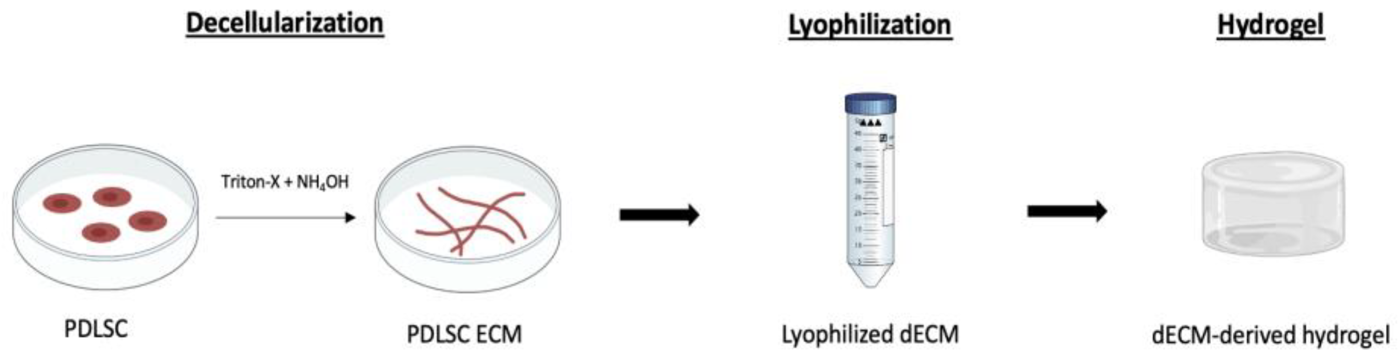
2.1. Cell-Derived ECM Production

PDLSC were seeded in tissue culture polystyrene wells at 3000 cells/cm2 and expanded in culture medium composed of low-glucose Dulbecco’s Modified Eagle Medium (DMEM, Gibco, New York, NY, USA) supplemented with 10% fetal bovine serum (FBS, Gibco) and 1% antibiotic-antimycotic (A/A, Gibco) for 10–12 days, with complete medium renewal every 3–4 days. After reaching confluency, the medium was discarded and cells were washed in Dulbecco’s phosphate-buffered saline (PBS). ECM isolation was performed using a solution composed of 20 mM ammonium hydroxide (NH4OH, Sigma-Aldrich) and 0.5% Triton X-100 (Sigma-Aldrich, St. Louis, MO, USA) in PBS, according to previously reported methods [7,8,9,10]. The solution was directly added to the wells and incubated for 5 min at 37 °C. ECM was gently washed three times with distilled water and cell-derived ECM was detached from the plates using a cell scraper. Finally, the solution was collected and freeze-dried.

2.2. ECM-Derived Hydrogels Production

Collagen hydrogels were prepared with and without lyophilized dECM incorporation. Rat tail type I collagen was used at 2.5 mg/mL and mixed with 1/10 volume of 0.1 M NaOH (Sigma-Aldrich), 1/9 volume of 10× PBS and the remnant volume of 1× PBS. PDLSC were characterized by flow cytometry and differentiation potential assays, according to previously reported methods [7]. PDLSC were added to the collagen mixture (1 × 10⁶ cells per hydrogel). To prevent gelation, the temperature of the mixture was maintained at 2–10 °C. The mixture was then transferred to a 24 well-plate and incubated at 37 °C for 2 h. After incubation, the collagen hydrogels with embedded PDLSC were hydrated in expansion medium (DMEM + 10% FBS + 1% A/A) at 37 °C.

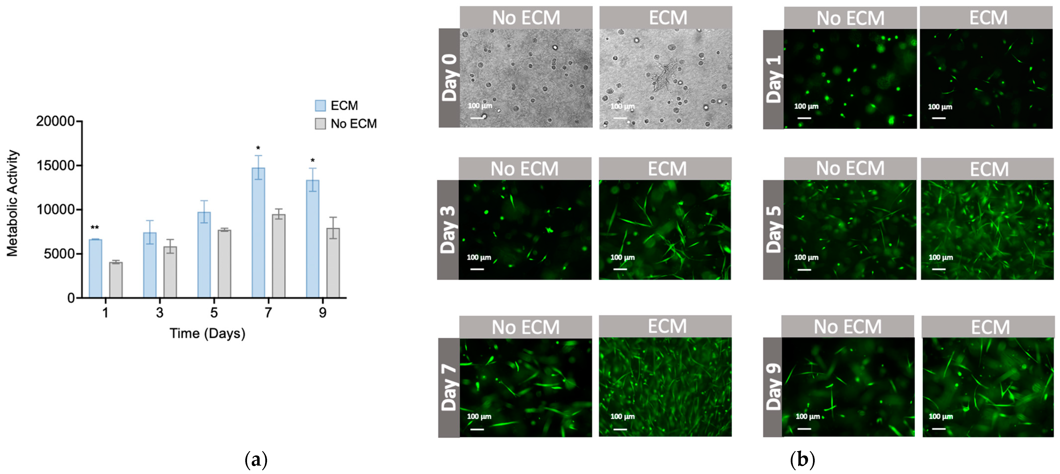
2.3. Cell Viability and Metabolic Activity

Cell viability was assessed by washing the cells in PBS, followed by an incubation in 1 µM acetoxymethyl (AM) calcein solution (Sigma-Aldrich) for 30 min. Cells were then imaged by fluorescence microscope (Olympus IX51 Inverted Microscope; Olympus America Inc., Center Valley, PA, USA) and recorded by an attached digital camera. The metabolic activity of the embedded PDLSC was evaluated using AlamarBlue® cell viability reagent (Molecular Probes, Eugene, OR, USA) after days 1, 3, 5, 7 and 9 of cell culture. After incubation for 3 h at 37 °C, fluorescence intensity was measured in a plate reader at an excitation/emission wavelength of 560/590 nm.

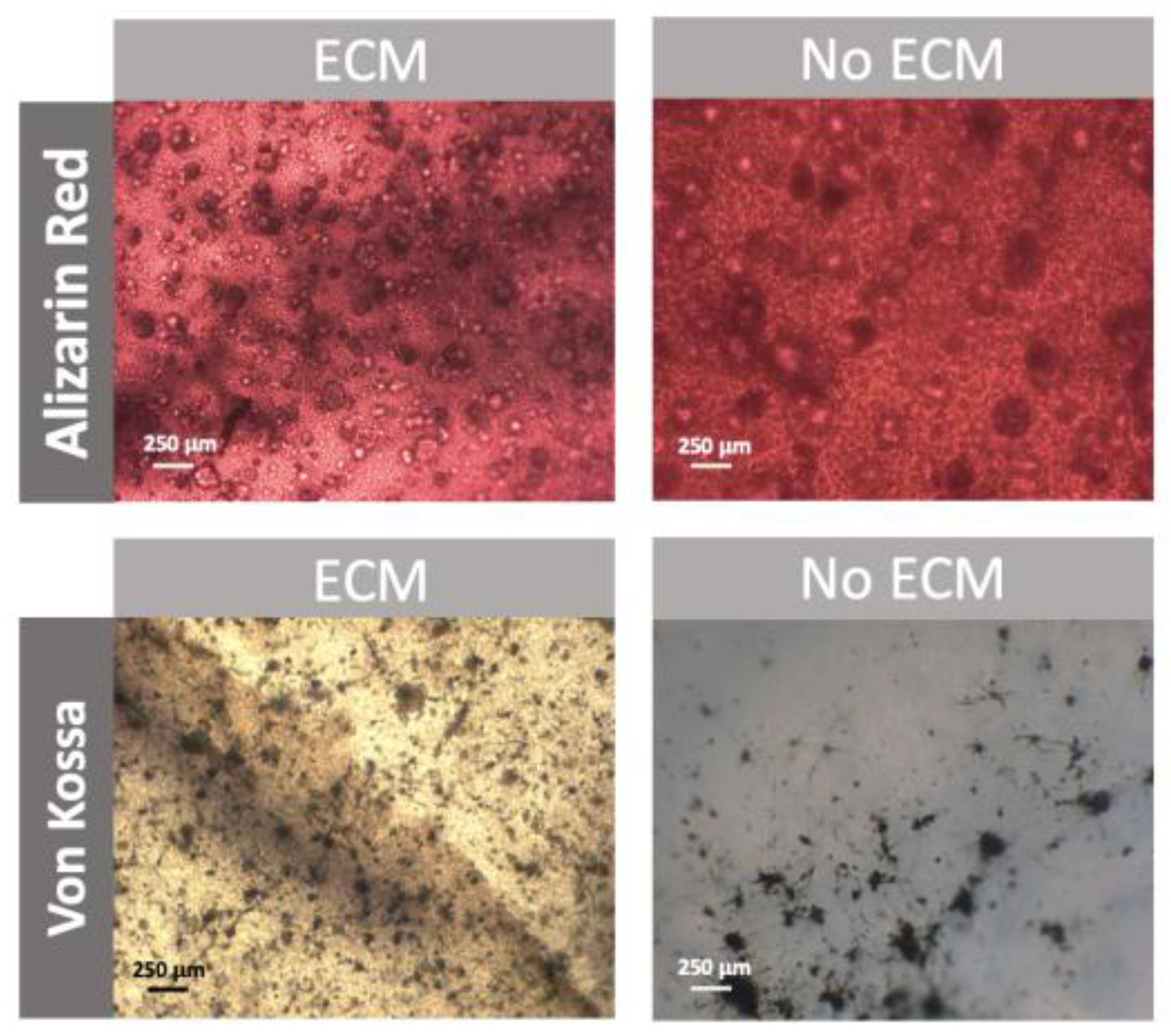
2.4. Osteogenic Differentiation

After 9 days of cell culture, osteogenic medium composed of DMEM, 10% FBS, 1% A/A, 10 mM β-glycerophosphate (Sigma-Aldrich), 10 nM dexamethasone (Sigma-Aldrich), and 50 μg/mL ascorbic acid (Sigma-Aldrich), was added to the hydrogels. Osteogenic differentiation was qualitatively confirmed through von Kossa (VK) and Alizarin Red (AR) stainings after 21 days of osteogenic differentiation.

3. Results

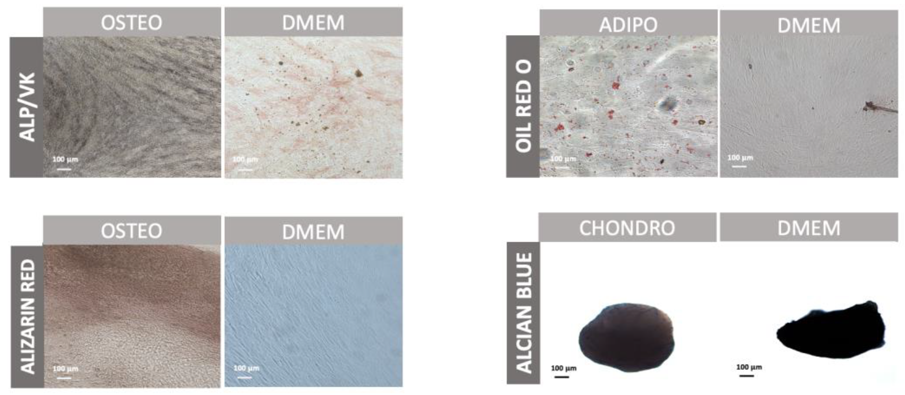
3.1. Characterization of Periodontal Ligament Stem Cells

PDLSC were cultured in expansion medium (DMEM + 10% FBS + 1% A/A) and immunophenotypic profile was assessed by flow cytometry (Figure 1). Results demonstrated positive expression of mesenchymal stem cell (MSC)-associated markers CD73 (94.0% ± 4.30%), CD90 (94.7% ± 0.23%), and CD105 (71.8% ± 0.39%), as well as negative expression of hematopoietic markers, such as CD34 and CD45. In addition, it was also shown that CD146, a marker previously used to identify PDLSC, was slightly expressed by PDLSC (24% ± 15%).
To understand the differentiation potential of PDLSC, cells were differentiated into adipogenic, osteogenic and chondrogenic lineages (Figure 2). Adipogenic differentiation resulted in the accumulation of lipid vacuoles (stained in red). As for chondrogenic differentiation, the deposition of glycosaminoglycans was observed (stained in blue). Finally, osteogenic differentiation was also confirmed by VK and AR stainings, presenting a great deposition of mineral deposits (stained in black for VK staining and in red for AR staining).

3.2. Characterization of Decellularized ECM Derived from PDLSC

dECM was obtained from PDLSC according to the first step of Figure 3.
Immunocytochemistry analysis confirmed the presence of the most common ECM proteins present in the PDL, such as collagen type I, laminin, asporin, fibronectin, osteopontin and osteocalcin (Figure 4). DAPI staining also confirmed the successful decellularization, showing the absence of well-defined nuclei after decellularization. This indicates that cellular nuclei were successfully disrupted and most of the cellular material was removed.

3.3. Effect of ECM-Derived Hydrogels in PDLSC

PDLSC-derived dECM was obtained and incorporated in collagen hydrogels as depicted in Figure 3. The metabolic activity of PDLSC embedded in collagen gels with and without dECM particles increased over time (Figure 5a). Furthermore, cells cultured in the ECM-derived hydrogels presented a statistically significant level of higher metabolic activity than cells embedded in collagen hydrogels without dECM. Calcein AM cell staining confirmed the viability of PDLSC cultured on ECM-derived hydrogels (Figure 5b).
To explore the effect of ECM-derived hydrogels on the osteogenic differentiation of PDLSC, we qualitatively assessed calcium deposition through osteogenic stainings (Figure 6). Alizarin Red staining confirmed the osteogenic differentiation of PDLSC in both hydrogels, while von Kossa staining showed an increased deposition of minerals when cells were cultured on ECM-derived hydrogels compared to hydrogels without ECM.

4. Discussion

In our study, we evaluated the effect of ECM-derived hydrogels in the proliferation and osteogenic differentiation of PDLSC for developing a new approach for periodontal regeneration. We hypothesized that dECM could stimulate both proliferation and differentiation of dental stem cells, such as PDLSC, when incorporated into collagen hydrogels.
Firstly, aiming to characterize PDLSC, immunophenotypic analysis was performed by flow cytometry. PDLSC showed expression of MSC-surface markers, such as CD73, CD90, and CD105, which is in accordance with previous studies [11,12,13]. In addition to these typical MSC-surface markers, PDLSC also expressed CD146. CD146 is a pericyte-associated marker and has been used to identify PDLSC populations [14,15]. Additionally, and likewise similarly to the existing literature [12], HSC markers such as CD34 and CD45 were absent. Apart from the positive expression of MSC-related surface markers, the MSC phenotype was also confirmed through the successful differentiation of PDLSC in three distinct lineages: adipogenic, chondrogenic and osteogenic. To understand if PDLSC-derived ECM was composed of most common ECM proteins, immunocytochemistry analysis before and after decellularization was performed. The results showed that after decellularization, the remnant material stained positive for important ECM proteins, such as asporin, collagen type I, fibronectin and laminin, suggesting that decellularization treatment did not compromise the ECM composition [7].
After PDLSC-dECM lyophilization, ECM particles were incorporated into collagen hydrogels to evaluate the effect of dECM on PDLSC proliferation. Our results showed increased metabolic activity of PDLSC in dECM-collagen hydrogels over time, when compared to hydrogels without ECM, indicating increased proliferation. Although no studies report the use of dECM in collagen hydrogels for periodontal regeneration, ECM was already shown to positively influence MSC proliferation when cultured onto polycaprolactone scaffolds and electrospun fibers [16,17], as well as in dECM coatings [16]. In addition, osteogenic differentiation was also confirmed through osteogenic stainings. Our results demonstrated that PDLSC cultured on ECM-derived hydrogels were producing more mineral deposits compared to cells cultured on hydrogels without ECM, presenting a positive effect of dECM on osteogenic differentiation of PDLSC. Further studies will focus on quantitative analysis of osteogenic/periodontal differentiation of PDLSC cultured on ECM-derived hydrogels, such as calcium content quantification and gene expression analysis.

5. Conclusions

Hydrogels have been extensively explored in a variety of biomedical applications, such as drug delivery systems, scaffolds for 3D cell culture, and wound healing and tissue engineering. Although it has been demonstrated that cell-derived dECM can be used to deliver proteins and growth factors to achieve tissue regeneration, dECM hydrogels for periodontal regeneration have not been explored yet. Here, we addressed for the first time the effect of ECM-derived hydrogels in PDLSC proliferation and osteogenic differentiation. Our results suggest that the novel dECM technology might represent a potential approach for periodontal regeneration, enhancing cell proliferation and osteogenic differentiation.

Author Contributions

Conceptualization, A.B.d.S., C.L.d.S. and M.S.C.; investigation, A.B.d.S. and M.S.C.; writing—original draft preparation, A.B.d.S. and M.S.C.; writing—review and editing, A.B.d.S., C.L.d.S. and M.S.C.; supervision, C.L.d.S. and M.S.C.; funding acquisition, C.L.d.S. and M.S.C. All authors have read and agreed to the published version of the manuscript.

Funding

This research was financed by national funds from FCT-Fundação para a Ciência e a Tecnologia, I.P., in the scope of the project UIDB/04565/2020 and UIDP/04565/2020 of the Research Unit Institute for Bioengineering and Biosciences—iBB and the project LA/P/0140/2020 of the Associate Laboratory Institute for Health and Bioeconomy—i4HB. Funding was also provided from the FCT project PTDC/BTM-BTM/3538/2020—DentalBioMatrix.

Institutional Review Board Statement

Not applicable.

Informed Consent Statement

Informed consent was obtained from all subjects involved in the study.

Data Availability Statement

The authors declare that the data generated in the current study are available within the article or from the corresponding author upon reasonable request.

Conflicts of Interest

The authors declare no conflict of interest.

References

  1. Pihlstrom, B.L.; Michalowicz, B.S.; Johnson, N.W. Periodontal diseases. Lancet 2005, 366, 1809–1820. [Google Scholar] [CrossRef] [PubMed] [Green Version]
  2. Woo, H.N.; Cho, Y.J.; Tarafder, S.; Lee, C.H. The recent advances in scaffolds for integrated periodontal regeneration. Bioact. Mater. 2021, 6, 3328–3342. [Google Scholar] [CrossRef] [PubMed]
  3. Liang, Y.; Luan, X.; Liu, X. Recent advances in periodontal regeneration: A biomaterial perspective. Bioact. Mater. 2020, 5, 297–308. [Google Scholar] [CrossRef] [PubMed]
  4. Zhang, W.; Du, A.; Liu, S.; Lv, M.; Chen, S. Research progress in decellularized extracellular matrix-derived hydrogels. Regen 2021, 18, 88–96. [Google Scholar] [CrossRef] [PubMed]
  5. Antoine, E.E.; Vlachos, P.P.; Rylander, M.N. Review of collagen i hydrogels for bioengineered tissue microenvironments: Characterization of mechanics, structure, and transport. Tissue Eng. Part B Rev. 2014, 20, 683–696. [Google Scholar] [CrossRef] [PubMed] [Green Version]
  6. Frantz, C.; Stewart, K.M.; Weaver, V.M. The extracellular matrix at a glance. J. Cell Sci. 2010, 123, 4195–4200. [Google Scholar] [CrossRef] [PubMed] [Green Version]
  7. Carvalho, M.S.; Silva, J.C.; Cabral, J.M.S.; da Silva, C.L.; Vashishth, D. Cultured cell-derived extracellular matrices to enhance the osteogenic differentiation and angiogenic properties of human mesenchymal stem/stromal cells. J. Tissue Eng. Regen. Med. 2019, 13, 1544–1558. [Google Scholar] [CrossRef] [PubMed]
  8. Yang, Y.; Lin, H.; Shen, H.; Wang, B.; Lei, G.; Tuan, R.S. Mesenchymal stem cell-derived extracellular matrix enhances chondrogenic phenotype of and cartilage formation by encapsulated chondrocytes in vitro and in vivo. Acta Biomater. 2018, 69, 71–82. [Google Scholar] [CrossRef] [PubMed]
  9. Gong, T.; Heng, B.C.; Xu, J.; Zhu, S.; Yuan, C.; Lo, E.C.M.; Zhang, C. Decellularized extracellular matrix of human umbilical vein endothelial cells promotes endothelial differentiation of stem cells from exfoliated deciduous teeth. J. Biomed. Mater. Res. A 2017, 105, 1083–1093. [Google Scholar] [CrossRef] [PubMed]
  10. Silva, J.C.; Carvalho, M.S.; Han, X.; Xia, K.; Mikael, P.E.; Cabral, J.M.S.; Ferreira, F.C.; Linhardt, R.J. Compositional and structural analysis of glycosaminoglycans in cell-derived extracellular matrices. Glycoconj J. 2019, 36, 141–154. [Google Scholar] [CrossRef] [PubMed]
  11. Iwata, T.; Yamato, M.; Zhang, Z.; Mukobata, S.; Washio, K.; Ando, T.; Feijen, J.; Okano, T.; Ishikawa, I. Validation of human periodontal ligament-derived cells as a reliable source for cytotherapeutic use. J. Clin. Periodontol. 2010, 37, 1088–1099. [Google Scholar] [CrossRef] [PubMed]
  12. Nagatomo, K.; Komaki, M.; Sekiya, I.; Sakaguchi, Y.; Noguchi, K.; Oda, S.; Muneta, T.; Ishikawa, I. Stem cell properties of human periodontal ligament cells. J. Periodontal. Res. 2006, 41, 303–310. [Google Scholar] [CrossRef] [PubMed]
  13. Banavar, S.R.; Rawal, S.Y.; Paterson, I.C.; Singh, G.; Davamani, F.; Khoo, S.P.; Tan, E.L. Establishing a technique for isolation and characterization of human periodontal ligament derived mesenchymal stem cells. Saudi Dent. J. 2021, 33, 693–701. [Google Scholar] [CrossRef] [PubMed]
  14. Saito, Y.; Watanabe, E.; Mayahara, K.; Watanabe, N.; Morokuma, M.; Isokawa, K.; Shimizu, N.; Honda, M.J. CD146 Marker for PDL CD146/MCAM Surface Marker for Identifying Human Periodontal Ligament-derived Mesenchymal Stem Cells. J. Hard Tissue Biol 2013, 22, 115–128. [Google Scholar] [CrossRef] [Green Version]
  15. Seo, B.M.; Miura, M.; Gronthos, S.; Bartold, P.M.; Batouli, S.; Brahim, J.; Young, M.; Robey, P.G.; Wang, C.Y.; Shi, S. Investigation of multipotent postnatal stem cells from human periodontal ligament. Lancet 2004, 364, 149–155. [Google Scholar] [CrossRef] [PubMed]
  16. Silva, J.C.; Carvalho, M.S.; Udangawa, R.N.; Moura, C.S.; Cabral, J.M.; Da Silva, C.L.; Ferreira, F.C.; Vashishth, D.; Linhardt, R.J. Extracellular matrix decorated polycaprolactone scaffolds for improved mesenchymal stem/stromal cell osteogenesis towards a patient-tailored bone tissue engineering approach. J. Biomed. Mater. Res. B Appl. Biomater. 2020, 108, 2153–2166. [Google Scholar] [CrossRef] [PubMed]
  17. Carvalho, M.S.; Silva, J.C.; Udangawa, R.N.; Cabral, J.M.; Ferreira, F.C.; da Silva, C.L.; Linhardt, R.J.; Vashishth, D. Co-culture cell-derived extracellular matrix loaded electrospun microfibrous scaffolds for bone tissue engineering. Mater. Sci. Eng. C 2019, 99, 479–490. [Google Scholar] [CrossRef] [PubMed]
Figure 1. Immunophenotypic analysis of PDLSC cultured in expansion conditions (DMEM + 10% FBS + 1% A/A) by flow cytometry. Data are expressed as mean ± SD (n = 2).
Engproc 31 00038 g001
Figure 2. In vitro multilineage differentiation of PDLSC. Alkaline phosphatase (ALP) and von Kossa (VK) stainings show ALP activity in red and mineralized extracellular matrix deposits in black. Alizarin red stains the calcium deposited in the extracellular matrix in red. Adipogenic differentiation was detected by Oil red O staining showing the lipid vacuoles in red. Alcian blue stains proteoglycans in blue. As controls, stainings were also performed in PDLSC cultured under expansion conditions (DMEM). Scale bar, 100 μm.
Engproc 31 00038 g002
Figure 3. Schematic representation of the decellularization process of PDLSC and its incorporation in collagen hydrogels.
Engproc 31 00038 g003
Figure 4. Comparison of immunofluorescent staining images for PDLSC before and after decellularization for the expression of laminin (LAM, red), osteopontin (OPN, red), osteocalcin (OC, red), asporin (ASP, red), collagen type I (COL I, red), and fibronectin (FIB, green). Nuclei were stained with DAPI (blue). Scale bar, 50 μm.
Engproc 31 00038 g004
Figure 5. PDLSC proliferation in collagen hydrogels enriched with dECM particles (ECM) and collagen hydrogels without dECM (No ECM). (a) Metabolic activity of PDLSC after 1, 3, 5, 7 and 9 days of culture; (b) representative images of viable cells at different timepoints (calcein AM cell viability assay). Data is expressed as Mean ± SD; ** p < 0.01, * p < 0.05. Scale bar, 100 μm.
Engproc 31 00038 g005
Figure 6. In vitro osteogenic differentiation of PDLSC embedded in collagen hydrogels enriched with dECM particles (ECM), and collagen hydrogels (No ECM). Von Kossa stains mineralized extracellular matrix deposits in black. Alizarin red stains the calcium deposited in the extracellular matrix in red. Scale bar, 250 μm.
Engproc 31 00038 g006
Disclaimer/Publisher’s Note: The statements, opinions and data contained in all publications are solely those of the individual author(s) and contributor(s) and not of MDPI and/or the editor(s). MDPI and/or the editor(s) disclaim responsibility for any injury to people or property resulting from any ideas, methods, instructions or products referred to in the content.

