Proceeding Paper

Biological Docking and BSA Binding Studies of 1,4-Disubstituted Piperdine Containing 1,2,4-Triazoles: Comparative Synthesis Leveraging Microwave-Assisted and Conventional Protocols †

1
Department of Chemistry, University of Sahiwal, Sahiwal 57000, Punjab, Pakistan
2
Department of Chemistry, Government College University, Lahore 54000, Punjab, Pakistan
3
Department of Chemistry, Universidad Técnica Federico Santa Maria Av. Santa Maria 6400, Vitacura, Santiago 766025, Chile
4
Department of Chemistry, University of Lahore, Lahore 54000, Punjab, Pakistan
5
Faculty of Pharmacy, Universiti Teknologi MARA Cawangan Selangor Kampus Puncak Alam, Bandar Puncak Alam 42300, Selangor, Malaysia
6
Atta-ur-Rahman Institute for Natural Products Discovery (AuRIns), Universiti Teknologi MARA Cawangan Selangor Kampus Puncak Alam, Bandar Puncak Alam 42300, Selangor, Malaysia
*
Authors to whom correspondence should be addressed.
Presented at the 27th International Electronic Conference on Synthetic Organic Chemistry (ECSOC-27), 15–30 November 2023; Available online: https://ecsoc-27.sciforum.net/.
Chem. Proc. 2023, 14(1), 106; https://doi.org/10.3390/ecsoc-27-16281
Published: 15 November 2023

Abstract

:
A biologically effective study regarding the synthesis of a library of hybrids based on a triazole ring, propanamide and azinane was performed in this study. The targeted hybrids, 9a–9l, were synthesized through a multistep protocol followed by two methodologies, that is, conventional and microwave-assisted ones. Initially, compound 3 was synthesized via the room-temperature stirring of 4-methoxybenzenesulfonyl chloride (1) and ethyl ester (2). Resulting carboxylate was converted into carbohydrazide 4, which was refluxed with phenyl isothiocyanate and KOH to synthesize product 5. A library of amides, 8a–8l, was stirred at room temperature with compound 5 to avail the targeted library of hybrids 9a–9l. The designed hybrids were screened for their antioxidant, urease, AChE and BChE inhibition potential. All the compounds were found to be active with variable potential. The best antioxidant agent was compound 9c with an IC50 value of 45.2 ± 0.15. The compound 9e (63.27 ± 1.21) was the best AChE inhibitor; 9g (20.2 ± 0.21) and 9k (19.2 ± 0.09) were the best anti-urease agents; and 9d (15.5 ± 0.39) and 9e (15.9 ± 0.67) were the best BChE inhibitors. The computational and BSA binding studies of the selected synthesized compounds against urease, BChE and AChE enzymes were carried out to elaborate the strong and weak enzyme-inhibition potential through the binding forces of the synthesized compounds with the different enzymatic sites that are responsible for their activity.

1. Introduction

The microwave-assisted synthesis of heterocyclic compounds has become very effective. It has been frequently utilized by synthetic chemists around the world because of its efficiency in term of large-scale reactions in minimal time, its environment-friendly behavior and its high yield [1]. During the last few years, microwave-assisted synthesis has been utilized to agreat extent on the basis of its improved yield, region-selectivity and chemo-selectivity [2,3].
Nitrogenous heterocyclic systems based on privileged patterns drawn from azinane and 1,2,4-triazole possess an extreme level of biological applications like natural products. These molecules could be a step toward the drug discovery route [4]. Triazoles and their derived compounds have become very attractive and attained the attention of the world because of their important usage in biology, agriculture, material science and pharmaceuticals [5]. The application of triazole-based compounds in these different fields could be justified by their high thermal stability, heteroatomic character, hydrogen bonding and dipole moment properties [6,7].
Azole-based compounds, especially triazoles, are the part of broad range of drugs utilized to cure different diseases. Triazoles are a part of anastrozole, letrozole and vorozole being used as anticancer agents; voriconazole being used as an antifungal agent; and fluconazole and itraconazole being used as antimycotic agents [8,9]. Another part of the main core of the designed drugs, the piperidine ring, possesses a wide range of biological applications like anticancer, antimicrobial, antimycobacterial and antihypertensive ones [10,11].
Prompted by our previously designed projects, observations and literature survey, we designed an array of compoundswith 1,2,4-triazole, piperidine and various substituted aromatic functionalities submerged in a single unit to enhance their biological potential. Likewise, the microwave-assisted technique was employed to synthesize the designed products and compared to the conventional technique. It was observed that microwave-assisted method was better and more efficient compared to the conventional one with respect to time and the yield of the synthesized compounds.

2. Experimental Section

The services of a local supplier were availed to access all the chemicals utilized during the current research. Chromatographic techniques were used to assess the progress of the reaction, while spectroscopic approaches were used to analyze and confirm the structures of all the synthesized compounds.

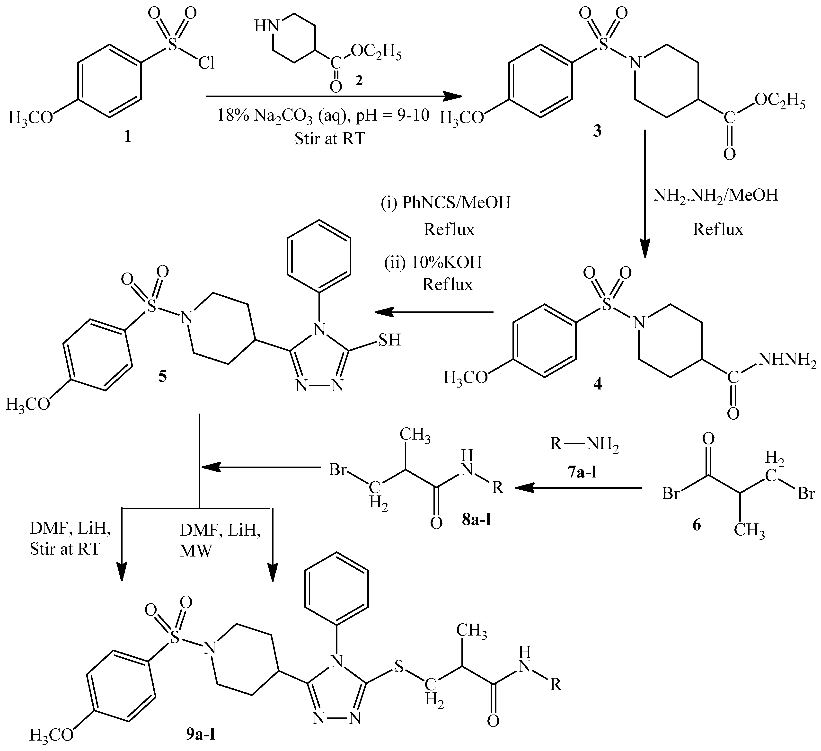
2.1. Synthesis of Ethyl 1-[(4-Methoxyphenyl)sulfonyl]-4-piperidincarboxylate (3)

Compound 3 was prepared by stirring 4-methoxybenzenesulfonyl chloride (1; 0.04 mol) with ethyl isonipecotate (2; 0.04 mol) for 14 h. During the whole process, a 18% Na2CO3 solution was used at regular intervals to adjust the pH at 9–10. TLC confirmed the completion of the reaction. By adding cold distilled water, the precipitates of target compound 3 were obtained.

2.2. Synthesis of 1-(4-Methoxyphenylsulfonyl)piperidin-4-carbohydrazide (4)

The corresponding carbohydrazide 4 of product 3 was synthesized by a 5-h reflux reaction with hydrazine hydrate. Thin-layer chromatography was employed to monitor the reaction. After the reaction completion, the surplus solvent was evaporated, and crystals of the pure target product 4 were availed.

2.3. Synthesis of 5-{1-[(4-Methoxyphenyl)sulfonyl]-4-piperidinyl}-4-methyl-4H-1,2,4-triazole-3-thiol (5)

Compound 4 and phenyl isothiocyanate were mixed in equimolar quantities (0.04 mol) in ethanol as a solvent and refluxed for 1 h. The uncyclized product was separated after confirmation through TLC. It was subjected to cyclization via reflux reaction in the presence of equimolar 10% KOH. TLC was used to monitor the reaction completion. After the reaction completion, dilute HCl was added on continuous stirring to adjust the pH to 4–5. The resulting product was washed and dried at ambient temperature.

2.4. Synthesis of N-(Substituted)-2-bromopropanamides (8a8l)

Aralkyl/aryl amines (7a7l; 0.02 mol) were reacted with equimolar quantity of 2-bromopropionyl bromide (6) in the presence of distilled water. By the addition of a 18% Na2CO3 solution, the pH was maintained at 9–10. Propanamides were obtained in the precipitate form and were available for further utilization after washing and drying.

2.5. General Procedure for the Synthesis of N-(Substituted)-2-[(5-{1-[(4-methoxyphenyl) sulfonyl]-4-piperidinyl}-4-methyl-4H-1,2,4-triazol-3-yl)sulfanyl] Propanamide (9a9l)

Conventional synthesis: Compound 5 (0.0005 mol) was stirred with LiH for 15 min in the presence of DMF. Equimolar amounts of synthesized propanamides (8a8l) were added and stirred at room temperature for 8–16 h. The reaction progress was monitored using TLC. At the reaction completion, the title compounds were precipitated on addition of cold distilled water. These precipitates were filtered, washed, and dried for further analysis.
Microwave assisted synthesis: Compound 5 (0.0005 mol) was stirred in the presence of LiH for 15 min in the presence of DMF. Equimolar quantities of synthesized propanamides (8a8l) were added and stirred in microwave for 30–60 s. The reaction progress was monitored through TLC. On completion of reaction, desired compounds were precipitated by adding cold distilled water. These precipitates were filtered, washed, and dried for further analysis.

2.5.1. N-(2,5-Dimethylphenyl)-2-[(5-{1-[(4-methoxyphenyl)sulfonyl]-4-piperidinyl}-4-phenyl-4H-1,2,4-triazol-3-yl)sulfanyl]propanamide (9a)

Light pink amorphous solid; yield: 88%; M.P: 203.5 °C; Molecular formula: C31H35N5O4S2; Molecular mass: 605.77 g/mol; IR (KBr, wave number, cm−1): 2800 (Ar C-H), 1680 (C=O), 1600 (C=N), 1500 (Ar C=C), 1350 (CH3), 1300 (S=O), 1240 (C-O-C), 710 (C-H); 1H-NMR (CDCl3, 600 MHz, δ(ppm)): 9.51 (s, 1H, NH), 7.88 (s, 1H, H-6′′′′), 7.69 (d, J = 8.2 Hz, 2H, H-2′, H-6′), 7.58–7.54 (m, 3H, H-3″, H-4″, H-5″), 7.21 (d, J = 6.9 Hz, 2H, H-2″, H-6″), 7.06 (d, J = 7.7 Hz, 1H, H-3′′′′), 6.99 (d, J = 8.1 Hz, 2H, H-3′, H-5′), 6.88 (d, J = 7.3 Hz, 1H, H-4′′′′) 4.66 (q, J = 7.3 Hz, 1H, H-2‴), 3.88 (s, 3H, H-1‴), 3.76–3.65 (m, 2H, He-2, He-6), 2.53–2.51 (m, 1H, H-4), 2.40–2.36 (m, 2H, Ha-2, Ha-6), 2.32 (s, 3H, H-4‴), 2.26 (s, 3H, H-5‴), 2.07–1.81 (m, 4H, He-3, He-5, Ha-3, Ha-5), 1.61 (d, J = 7.8 Hz, 3H, H-3‴); 13C-NMR (CDCl3, 150 MHz, δ (ppm)): 168.00 (C-4′), 162.98 (C-5′′′′), 157.74 (C-3′′′′), 153.12 (C-6′′′′), 136.18 (C-1′′′′), 132.42 (C-1″), 130.68 (C-6′′′′), 130.38 (C-3″, C-5″), 131.20 (C-2′′′′), 126.74 (C-2′, C-6′), 129.01 (C-1′), 127.76 (C-5′′′′), 126.99 (C-2″, C-6″), 125.73 (C-4″), 125.28 (C-3′′′′),122.63 (C-4′′′′), 114.20 (C-3′,C-5′), 55.59 (C-1‴), 45.30 (C-2, C-6), 42.85 (C-2‴), 31.75 (C-4), 29.46 (C-3, C-5), 21.16 (C-4‴), 17.74 (C-5‴), 16.31 (C-3‴).

2.5.2. N-(2,6-Dimethylphenyl)-2-[(5-{1-[(4-methoxyphenyl)sulfonyl]-4-piperidinyl}-4-phenyl-4H-1,2,4-triazol-3-yl)sulfanyl]propanamide (9b)

Off white amorphous solid; yield: 87%; M.P: 176.5 °C; Molecular formula: C31H35N5O4S2; Molecular mass: 605.77 g/mol; IR (KBr, wave number, cm−1): 2830 (Ar C-H), 1700 (C=O), 1620 (C=N), 1500 (Ar C=C), 1350 (CH3), 1280 (S=O), 1140 (C-O-C), 730 (C-H); 1H-NMR (CDCl3, 600 MHz, δ(ppm)): 9.31 (s, 1H, NH), 7.68 (d, J = 7.8 Hz, 2H, H-2′, H-6′), 7.58–7.56 (m, 3H, H-3″, H-4″, H-5″), 7.20–7.19 (m, 2H, H-2″, H-6″), 7.10–7.04 (m, 3H, H-3′′′′, H-4′′′′, H-5′′′′), 6.98 (d, J = 7.8 Hz, 2H, H-3′, H-5′), 4.68 (q, J = 7.3 Hz, 1H, H-2‴), 3.86 (s, 3H, H-1‴), 3.74–3.68 (m, 2H, He-2, He-6), 2.56–2.52 (m, 1H, H-4), 2.41–2.36 (m, 2H, Ha-2, Ha-6), 2.173 (s, 6H, H-4‴, H-5‴), 2.04–1.95 (m, 2H, He-3, He-5), 1.90–1.83 (m, 2H, Ha-3, Ha-5), 1.61 (d, J = 8.5 Hz, 3H, H-3‴); 13C-NMR (CDCl3, 150 MHz, δ (ppm)): 169.55 (C-4′), 162.99 (C-5′′′′), 157.67 (C-3′′′′), 152.72 (C-6′′′′), 135.29 (C-1′′′′), 133.95 (C-1″), 132.48 (C-1′), 130.74 (C-2′′′′, C-6′′′′), 130.74 (C-3″, C-5″), 129.75 (C-2′, C-6′), 128.05 (C-3′′′′, C-5′′′′), 127.80 (C-4″), 127.07 (C-4′′′′), 126.97 (C-2″, C-6″), 114.21 (C-3′, C-5′), 55.59 (C-1‴), 45.37 (C-2, C-6), 42.49 (C-2‴), 31.83 (C-4), 29.37 (C-3, C-5), 18.11 (C-3‴, C-4‴), 16.54 (C-3‴).

2.5.3. N-(3,5-Dimethylphenyl)-2-[(5-{1-[(4-methoxyphenyl)sulfonyl]-4-piperidinyl}-4-phenyl-4H-1,2,4-triazol-3-yl)sulfanyl]propanamide (9c)

White amorphous solid; yield: 85%; M.P: 98.0 °C; Molecular formula: C31H35N5O4S2; Molecular mass: 605.77 g/mol; IR (KBr, wave number, cm−1): 2840 (Ar C-H), 1700 (C=O), 1550 (C=N), 1500 (Ar C=C), 1320 (CH3), 1250 (S=O), 1150 (C-O-C), 720 (C-H); 1H-NMR (CDCl3, 600 MHz, δ(ppm)): 9.93 (s, 1H, NH), 7.69 (d, J = 7.6 Hz, 2H, H-2′, H-6′), 7.57–7.54 (m, 5H, H-3″, H-4″, H-5″, H-2′′′′, H-6′′′′), 7.21 (d, J = 7.1 Hz, 2H, H-2″, H-6″), 6.99 (d, J = 7.9 Hz, 2H, H-3′, H-5′), 6.75 (s, 1H, H-4′′′′), 4.51 (q, J = 7.0 Hz, 1H, H-2‴), 3.88 (s, 3H, H-1‴), 3.80–3.75 (m, 2H, He-2, He-6), 2.52–2.48 (m, 1H, H-4), 2.31 (s, 6H, H-4‴, H-5‴), 2.29–2.27 (m, 2H, Ha-2, Ha-6), 2.08–1.99 (m, 2H, He-3, He-5), 1.91–1.82 (m, 2H, Ha-3, Ha-5), 1.57 (d, J = 7.2 Hz, 3H, H-3‴); 13C-NMR (CDCl3, 150 MHz, δ (ppm): 169.09 (C-4′), 162.99 (C-5′′′′), 157.64 (C-3′′′′), 152.84 (C-6′′′′), 138.60 (C-1′′′′), 138.19 (C-1″), 132.46 (C-1′), 130.69 (C-2′′′′, C-6′′′′), 130.37 (C-3″, C-5″), 129.77 (C-2′, C-6′), 127.80 (C-3′′′′, C-5′′′′), 127.00 (C-4″), 125.77 (C-4′′′′), 117.40 (C-2″, C-6″), 114.20 (C-3′, C-5′), 55.59 (C-1‴), 45.55 (C-2, C-6), 43.32 (C-2‴), 32.10 (C-4), 29.50 (C-3, C-5), 21.36 (C-4‴, C-5‴), 16.24 (C-3‴).

2.5.4. 2-[(5-{1-[(4-Methoxyphenyl)sulfonyl]-4-piperidinyl}-4-phenyl-4H-1,2,4-triazol-3-yl)sulfanyl]-N-phenylpropanamide (9d)

White amorphous solid; yield: 89%; M.P: 123.0 °C; Molecular formula: C29H21N5O4S2; Molecular mass: 577.71 g/mol; IR (KBr, wave number, cm−1): 2800 (Ar C-H), 1700 (C=O), 1630 (C=N), 1500 (Ar C=C), 1310 (CH3), 1250 (S=O), 1130 (C-O-C), 750 (C-H); 1H-NMR (CDCl3, 600 MHz, δ (ppm)): 10.19 (s, 1H, NH), 7.69 (d, J = 7.3 Hz, 2H, H-2′, H-6′), 7.65 (d, J = 7.8 Hz, 2H, H-2′′′′, H-6′′′′), 7.58–7.54 (m, 3H, H-3″, H-4″, H-5″), 7.33 (t, J = 7.2 Hz, 2H, H-3′′′′, H-5′′′′), 7.21 (d, J = 7.1 Hz, 2H, H-2″, H-6″), 7.11 (t, J = 7.4 Hz, H-4′′′′), 6.99 (d, J = 7.3 Hz, 2H, H-3′, H-5′), 4.51 (q, J = 7.2 Hz, 1H, H-2‴), 3.88 (s, 3H, H-1‴), 3.79–3.73 (m, 2H, He-2, He-6), 2.53–2.49 (m, 1H, H-4), 2.35–2.29 (m, 2H, Ha-2, Ha-6), 2.08–1.97 (m, 2H, He-3, He-5), 1.91–1.82 (m, 2H, Ha-3, Ha-5), 1.58 (d, J = 7.1 Hz, 3H, H-3‴); 13C-NMR (CDCl3, 150 MHz, δ (ppm)): 169.13 (C-4′), 163.00 (C-5′′′′), 157.68 (C-3′′′′), 152.85 (C-6′′′′), 138.43 (C-1′′′′), 132.43 (C-1″), 130.72 (C-1′), 130.38 (C-3″, C-5″), 129.78 (C-2′, C-6′), 128.88 (C-2′′′′,C-6′′′′), 127.80 (C-4″), 127.00 (C-4′′′′), 123.98 (C-3′′′′, C-5′′′′), 119.65 (C-2″, C-6″), 114.21 (C-3′, C-5′), 55.60 (C-1‴), 45.51 (C-2, C-6), 43.32 (C-2‴), 32.02 (C-4), 29.45 (C-3, C-5), 16.21 (C-3‴).

2.5.5. 2-[(5-{1-[(4-Methoxyphenyl)sulfonyl]-4-piperidinyl}-4-phenyl-4H-1,2,4-triazol-3-yl)sulfanyl]-N-(2-methylphenyl)propanamide (9e)

Off white amorphous solid; yield: 85%; M.P: 172.8 °C; Molecular formula: C30H33N5O4S2; Molecular mass: 591.74 g/mol; IR (KBr, wave number, cm−1): 2830 (Ar C-H), 1700 (C=O), 1630 (C=N), 1500 (Ar C=C), 1325 (CH3), 1220 (S=O), 1110 (C-O-C), 740 (C-H); 1H-NMR (CDCl3, 600 MHz, δ (ppm)): 9.59 (s, 1H, NH), 8.04 (d, J = 7.9 Hz, 2H, H-6′′′′), 7.69 (d, J = 7.4 Hz, 2H, H-2′, H-6′), 7.59–7.54 (m, 3H, H-3″, H-4″, H-5″), 7.21–7.18 (m, 4H, H-2″, H-6″, H-3′′′′, H-5′′′′), 6.99 (d, J = 7.4 Hz, 2H, H-3′, H-5′), 4.66 (q, J = 7.2 Hz, 1H, H-2‴), 3.88 (s, 3H, H-1‴), 3.76–3.65 (m, 2H, He-2, He-6), 2.55–2.51 (m, 1H, H-4), 2.41–2.34 (m, 2H, Ha-2, Ha-6), 2.31 (s, 3H, H-4‴), 2.08–2.04 (m, 2H, He-3, He-5), 1.95–1.88 (m, 2H, Ha-3, Ha-5), 1.57 (d, J = 7.1 Hz, 3H, H-3‴); 13C-NMR (CDCl3, 150 MHz, δ (ppm)): 169.46 (C-4′), 162.98 (C-5′′′′), 157.77 (C-3′′′′), 152.72 (C-6′′′′), 136.45 (C-1′′′′), 132.41 (C-1″), 130.70 (C-1′), 130.43 (C-6′′′′), 130.39 (C-3″, C-5″), 126.77 (C-2′, C-6′), 128.87 (C-2′′′′), 128.80 (C-4″), 126.98 (C-5′′′′), 126.46 (C-3′′′′), 124.53 (C-4′′′′), 122.02 (C-2″, C-6″), 114.20 (C-3′, C-5′), 55.59 (C-1‴), 45.41 (C-2, C-6), 42.88 (C-2‴), 31.76 (C-4), 29.46 (C-3, C-5), 18.22 ((C-4‴), 21.47 (C-3‴).

2.5.6. 2-[(5-{1-[(4-Methoxyphenyl)sulfonyl]-4-piperidinyl}-4-phenyl-4H-1,2,4-triazol-3-yl)sulfanyl]-N-(3-methylphenyl)propanamide (9f)

Off white amorphous solid; yield: 85%; M.P: 97.5 °C; Molecular formula: C30H33N5O4S2; Molecular mass: 591.74 g/mol; IR (KBr, wave number, cm−1): 2850 (Ar C-H), 1700 (C=O), 1600 (C=N), 1500 (Ar C=C), 1320 (CH3), 1250 (S=O), 1100 (C-O-C), 750 (C-H); 1H-NMR (CDCl3, 600 MHz, δ (ppm)): 10.06 (s, 1H, NH), 7.69 (d, J = 7.2 Hz, 2H, H-2′, H-6′), 7.58–7.54 (m, 3H, H-3″, H-4″, H-5″), 7.50 (s, 1H, H-2′′′′), 7.43 (d, J = 8.0 Hz, H-6′′′′), 7.22–7.20 (m, 3H, H-2″, H-6″, H-5′′′′), 6.99 (d, J = 7.4 Hz, 2H, H-3′, H-5′), 6.93 (d, J = 7.4 Hz, 1H, H-4′′′′), 4.51 (q, J = 7.2 Hz, 1H, H-2‴), 3.88 (s, 3H, H-1‴), 3.79–3.74 (m, 2H, He-2, He-6), 2.52–2.48 (m, 1H, H-4), 2.36 (s, 3H, H-4‴), 2.35–2.28 (m, 2H, Ha-2, Ha-6), 2.08–2.01 (m, 2H, He-3, He-5), 1.91–1.82 (m, 2H, Ha-3, Ha-5), 1.57 (d, J = 7.2 Hz, 3H, H-3‴); 13C-NMR (CDCl3, 150 MHz, δ (ppm)): 169.11 (C-4′), 162.99 (C-5′′′′), 157.66 (C-3′′′′), 152.84 (C-6′′′′), 138.81 (C-1′′′′), 138.31 (C-1″), 132.45 (C-1′), 130.70 (C-6′′′′), 130.37 (C-3″, C-5″), 129.77 (C-2′, C-6′), 129.44 (C-2′′′′), 128.71 (C-2″, C-6″), 127.80 (C-3′′′′), 126.99 (C-4″), 124.81 (C-5′′′′), 116.77 (C-4′′′′), 114.20 (C-3′, C-5′), 55.60 (C-1‴), 45.54 (C-2, C-6), 43.32 (C-2‴), 32.06 (C-4), 29.47 (C-3, C-5), 21.48 (C-4‴), 16.22 (C-3‴).

2.5.7. N-(2-Ethylphenyl)-2-[(5-{1-[(4-methoxyphenyl)sulfonyl]-4-piperidinyl}-4-phenyl-4H-1,2,4-triazol-3-yl)sulfanyl]propanamide (9g)

White amorphous solid; yield: 89%; M.P: 175.6 °C; Molecular formula: C31H37N5O4S2; Molecular mass: 607.78 g/mol; IR (KBr, wave number, cm−1): 2830 (Ar C-H), 1690 (C=O), 1610 (C=N), 1550 (Ar C=C), 1325 (CH3), 1250 (S=O), 1140 (C-O-C), 700 (C-H); 1H-NMR (CDCl3, 600 MHz, δ (ppm)): 9.53 (s, 1H, NH), 7.93 (d, J = 7.9 Hz, 1H, H-6′′′′), 7.68 (d, J = 7.2 Hz, 2H, H-2′, H-6′), 7.58–7.55 (m, 3H, H-3″, H-4″, H-5″), 7.21–7.19 (m, 4H, H-2″, H-6″, H-3′′′′, H-5′′′′), 7.11 (t, J = 7.4 Hz, 1H, H-4′′′′), 6.98 (d, J = 7.3 Hz, 2H, H-3′, H-5′), 4.66 (q, J = 7.2 Hz, 1H, H-2‴), 3.88 (s, 3H, H-1‴), 3.76–3.66 (m, 2H, He-2, He-6), 2.65–2.60 (m, 2H, H-4‴), 2.56–2.52 (m, 1H, H-4), 2.40–2.35 (m, 2H, Ha-2, Ha-6), 2.08–2.04 (m, 2H, He-3, He-5), 1.96–1.89 (m, 2H, Ha-3, Ha-5), 1.56 (d, J = 7.7 Hz, 3H, H-3‴), 1.14 (t, J = 7.5 Hz, 3H, H-5″); 13C-NMR (CDCl3, 150 MHz, δ (ppm)): 166.66 (C-4′), 162.99 (C-5′′′′), 157.76 (C-3′′′′), 152.80 (C-6′′′′), 135.51 (C-1′′′′), 135.22 (C-1″), 132.44 (C-1′), 130.71 (C-6′′′′), 130.41 (C-3″, C-5″), 129.76 (C-2′, C-6′), 128.68 (C-4″), 127.81 (C-2′′′′), 126.95 (C-2″, C-6″), 126.35 (C-5′′′′), 125.07 (C-3′′′′), 123.18 (C-4′′′′), 114.21 (C-3′, C-5′), 55.59 (C-1‴), 45.40 (C-2, C-6), 42.78 (C-2‴), 31.76 (C-4), 29.48 (C-3, C-5), 24.55 (C-3‴), 16.41 (C-5‴), 14.20 (C-4‴).

2.5.8. N-(4-Ethoxyphenyl)-2-[(5-{1-[(4-methoxyphenyl)sulfonyl]-4-piperidinyl}-4-phenyl-4H-1,2,4-triazol-3-yl)sulfanyl]propanamide (9h)

Light brown amorphous solid; yield: 87%; M.P: 166.0 °C; Molecular formula: C31H35N5O5S2; Molecular mass: 621.77 g/mol; IR (KBr, wave number, cm−1): 2830 (Ar C-H), 1700 (C=O), 1610 (C=N), 1500 (Ar C=C), 1310 (CH3), 1230 (S=O), 1150 (C-O-C), 750 (C-H); 1H-NMR (CDCl3, 600 MHz, δ (ppm)): 9.99 (s, 1H, NH), 7.69 (d, J = 7.5 Hz, 2H, H-2′, H-6′), 7.59–7.53 (m, 5H, H-3″, H-4″, H-5″, H-2′′′′, H-6′′′′), 7.21 (d, J = 7.8 Hz, 2H, H-2″, H-6″), 6.99 (d, J = 7.6 Hz, 2H, H-3′, H-5′), 6.86 (d, J = 7.6 Hz, 2H, H-3′′′′, H-5′′′′), 4.49 (q, J = 7.2 Hz, 1H, H-2‴), 4.03 (q, J = 6.9 Hz, 2H, H-4‴), 3.88 (s, 3H, H-1‴), 3.78–3.73 (m, 2H, He-2, He-6), 2.52–2.48 (m, 1H, H-4), 2.35–2.28 (m, 2H, Ha-2, Ha-6), 2.07–1.98 (m, 2H, He-3, He-5), 1.90–1.82 (m, 2H, Ha-3, Ha- 5), 1.57 (d, J = 7.2 Hz, 3H, H-3‴), 1.41 (t, J = 6.9 Hz, 3H, H-5‴); 13C-NMR (CDCl3, 150 MHz, δ (ppm)): 168.78 (C-4′), 162.99 (C-5′′′′), 157.64 (C-3′′′′), 155.49 (C-4′′′′), 152.85 (C-6′′′′), 132.46 (C-1′′′′), 131.57 (C-1″), 130.69 (C-1′), 130.36 (C-3″, C-5″), 129.77 (C-2′, C-6′), 127.79 (C-4″), 127.00 (C-2′′′′, C-6′′′′), 121.12 (C-2″, C-6″), 114.70 (C-3′′′′, C-5′′′′), 114.21 (C-3′, C-5′), 63.71 (C-4‴), 55.60 (C-1‴), 45.52 (C-2, C-6), 43.33 (C-2‴), 32.02 (C-4), 29.45 (C-3, C-5), 16.31 (C-3‴), 14.85 (C-5‴).

2.5.9. N-(2-Ethyl-6-methylphenyl)-2-[(5-{1-[(4-methoxyphenyl)sulfonyl]-4-piperidinyl}-4-phenyl-4H-1,2,4-triazol-3-yl)sulfanyl]propanamide (9i)

White amorphous solid; yield: 90%; M.P: 162.0 °C; Molecular formula: C32H37N5O4S2; Molecular mass: 619.79 g/mol; IR (KBr, wave number, cm−1) 2840 (Ar C-H), 1670 (C=O), 1610 (C=N), 1500 (Ar C=C), 1340 (CH3), 1250 (S=O), 1140 (C-O-C), 750 (C-H); 1H-NMR (CDCl3, 600 MHz, δ (ppm)): 9.14 (s, 1H, NH), 7.68 (d, J = 7.6 Hz, 2H, H-2′, H-6′), 7.59–7.57 (m, 3H, H-3″, H-4″, H-5″), 7.21 (d, J = 6.0 Hz, 2H, H-2″, H-6″), 7.14 (t, J = 7.6 Hz, 1H, H-4′′′′),7.08–7.07 (m, 2H, H-3′′′′, H-6′′′′), 6.98 (d, J = 7.6 Hz, 2H, H-3′, H-5′), 4.69 (q, J = 7.2 Hz, 1H, H-2‴), 3.86 (s, 3H, H-1‴), 3.74–3.68 (m, 2H, He-2, He-6), 2.57–2.47 (m, 3H, H-4, Ha-2, Ha-6), 2.38 (q, J = 10.9 Hz, 2H, H-4‴), 2.14 (s, 3H, H-6‴), 2.04–1.82 (m, 4H, He-3, He-5, Ha-3, Ha-5),1.62 (3H, H-3 merged with HDO‴), 1.08 (t, J = 7.2 Hz, 3H, H-5‴); 13C-NMR (CDCl3, 600 MHz, δ (ppm)): 169.80 (C-4′), 162.99 (C-5′′′′), 157.64 (C-3′′′′), 152.75 (C-6′′′′), 141.10 (C-1′′′′), 135.79 (C-1″), 133.30 (C-1′), 132.49 (C-6′′′′), 130.74 (C-4″), 130.46 (C-3″, C-5″), 129.75 (C-2′, C-6′), 128.11 (C-5′′′′), 127.81 (C-2′′′′), 127.41 (C-3′′′′), 126.93 (C-2″, C-6″), 126.30 (C-4′′′′), 114.21 (C-3′, C-5′), 55.58 (C-1‴), 45.36 (C-2, C-6), 42.34 (C-2‴), 31.82 (C-4), 29.37 (C-3, C-5), 24.82 (C-4‴), 18.18 (C-6‴), 16.54 (C-3‴), 14.64 (C-5‴).

2.5.10. N-Cyclohexyl-2-[(5-{1-[(4-methoxyphenyl)sulfonyl]-4-piperidinyl}-4-phenyl-4H-1,2,4-triazol-3-yl)sulfanyl]propanamide (9j)

Off white amorphous solid; yield: 85%; M.P: 135.0 °C; Molecular formula: C29H37N5O4S2; Molecular mass: 583.76 g/mol; IR (KBr, wave number, cm−1): 2820 (Ar C-H), 1700 (C=O), 1620 (C=N), 1500 (Ar C=C), 1310 (CH3), 1250 (S=O), 1150 (C-O-C), 750 (C-H); 1H-NMR (CDCl3, 600 MHz, δ (ppm)): 7.69 (d, J = 7.7 Hz, 2H, H-2′, H-6′), 7.56–7.53 (m, 3H, H-3″, H-4″, H-5″), 7.19 (d, J = 7.1 Hz, 2H, H-2″, H-6″), 6.99 (d, J = 7.6 Hz, 2H, H-3′, H-5′), 4.30 (q, J = 7.2 Hz, 1H, H-2‴), 3.88 (s, 3H, H-1‴), 3.77–3.71 (m, 2H, He-2, He-6), 2.55–2.47 (m, 1H, H-4), 2.37–2.30 (m, 2H, Ha-2, Ha-6), 1.89–1.84 (m, 4H, He-3, He-5, Ha-3, Ha-5), 1.72–1.70 (m, 1H, H-1′′′′), 1.49 (d, J = 7.2 Hz, 3H, H-3‴), 1.36–1.17 (m, 10H, H-2′′′′, H-3′′′′, H-4′′′′, H-5′′′′, H-6′′′′); 13C-NMR (CDCl3, 600 MHz, δ (ppm)): 170.12 (C-4′), 163.00 (C-5′′′′), 157.46 (C-3′′′′), 152.16 (C-6′′′′), 133.51 (C-1″), 130.54 (C-1′), 130.28 (C-3″, C-5″), 129.76 (C-2′, C-6′), 127.08 (C-4″), 124.98 (C-2″, C-6″), 114.20 (C-3′, C-5′), 55.59 (C-1‴), 48.00 (C-1′′′′), 45.49 (C-2, C-6), 43.34 (C-2‴), 32.60 (C-4), 31.99 (C-2′′′′, C-6′′′′), 29.52 (C-3, C-5), 25.53 (C-3′′′′, C-5′′′′), 24.45 (C-4′′′′), 16.67 (C-3‴).

2.5.11. Methyl 2-({2-[(5-{1-[(4-Methoxyphenyl)sulfonyl]-4-piperidinyl}-4-phenyl-4H-1,2,4-triazol-3-yl)sulfanyl]propanoyl}amino)benzoate (9k)

Off white amorphous solid; yield: 85%; M.P: 130.0 °C; Molecular formula: C32H35N5O6S2; Molecular mass: 649.78 g/mol; IR (KBr, wave number, cm−1): 2810 (Ar C-H), 1680 (C=O), 1600 (C=N), 1510 (Ar C=C), 1300 (CH3), 1230 (S=O), 1140 (C-O-C), 710 (C-H); 1H-NMR (CDCl3, 600 MHz, δ (ppm)): 7.66 (d, J = 8.2 Hz, 2H, H-2′, H-6′), 7.52–7.46 (m, 3H, H-3″, H-4″, H-5″), 7.26–7.25 (m, 2H, H-2″, H-6″), 7.14–6.98 (m, 5H, H-2′′′′, H-3′′′′, H-4′′′′, H-5′′′′, H-6′′′′), 6.95 (d, J = 7.9 Hz, 2H, H-3′, H-5′), 4.68 (q, J = 7.0 Hz, 2H, H-2‴), 3.88 (s, 3H, H-1‴), 3.73–3.66 (m, 2H, He-2, He-6), 2.45–2.43 (m, 5H, H-5‴, Ha-2, Ha-6), 2.43–2.41 (m, 1H, H-4), 1.99–1.82 (m, 4H, He-3, He-5, Ha-3, Ha-5), 1.79–1.78 (m, 3H, H-3‴); 13C-NMR (CDCl3, 600 MHz, δ (ppm)): 168.22 (C-2′′′′), 166.01 (C-4′), 162.91 (C-5′′′′), 161.01 (C-7′′′′), 154.93 (C-3′′′′), 152.38 (C-6′′′′), 132.74 (C-1′′′′), 131.56 (C-1″), 131.11 (C-6′′′′), 130.70 (C-3″, C-5″), 130.21 (C-1′), 129.75 (C-2′, C-6′), 129.25 (C-4″), 128.61 (C-3′′′′), 128.01 (C-5′′′′), 127.60 (C-4′′′′), 127.47 (C-2″, C-6″), 114.20 (C-3′,C-5′), 55.60 (C-4‴), 55.57 (C-1‴), 47.71 (C-2‴), 45.33 (C-2, C-6), 31.72 (C-4), 29.20 (C-3, C-5), 14.01 (C-5‴), 16.12 (C-3‴).

2.5.12. N-(3,4-Dimethylphenyl)-2-[(5-{1-[(4-methoxyphenyl)sulfonyl]-4-piperidinyl}-4-phenyl-4H-1,2,4-triazol-3-yl)sulfanyl]propanamide (9l)

Light pink amorphous solid; M.P: 109.5 °C; Molecular formula: C31H35N5O4S2; Molecular mass: 605.77 g/mol; IR (KBr, wave number, cm−1): 2850 (Ar C-H), 1700 (C=O), 1600 (C=N), 1500 (Ar C=C), 1375 (CH3), 1250 (S=O), 1150 (C-O-C), 750 (C-H); 1H-NMR (CDCl3, 600 MHz, δ(ppm)): 9.64 (s, 1H, NH), 7.69 (d, J = 8.8 Hz, 2H, H-2′, H-6′), 7.64 (d, J = 7.9 Hz, 1H, H-6′′′′), 7.60–7.55 (m, 3H, H-3″, H-4″, H-5″), 7.23 (dd, J = 7.7 Hz, 2H, H-2″, H-6″), 7.12 (t, J = 7.7 Hz, 1H, H-5′′′′), 7.00 (d, J = 7.5 Hz, 1H, H-4′′′′), 6.99 (d, J = 8.8 Hz, 2H, H-3′, H-5′), 4.60 (q, J = 7.0 Hz, 2H, H-2‴), 3.87 (s, 3H, H-1‴), 3.76–3.74 (m, 2H, He-2, He-6), 2.53–2.49 (m, 1H, H-4), 2.34–2.30 (m, 5H, Ha-2, Ha-6, H-4‴), 2.20 (s, 3H, H-5‴), 2.03–2.01 (m, 2H, He-3, He-5), 1.87–1.87 (m, 2H, Ha-3, Ha-5), 1.59 (d, J = 6.1 Hz, 3H, H-3‴); 13C-NMR (CDCl3, 150 MHz, δ (ppm)): 167.01 (C-5′′′′), 163.00 (C-4′), 158.07 (C-3′′′′), 152.69 (C-6′′′′), 137.31 (C-1′′′′), 135.78 (C-1″), 132.43 (C-2′′′′), 130.75 (C-6′′′′), 130.43 (C-3″, C-5″), 129.75 (C-2″, C-6″), 128.98 (C-4″), 128.00 (C-3′′′′), 127.02 (C-5′′′′), 126.96 (C-2′, C-6′), 125.69 (C-1′), 121.27 (C-4′′′′), 114.20 (C-3′, C-5′), 55.57 (C-1‴), 45.43 (C-2, C-6), 42.67 (C-2‴), 31.96 (C-4), 29.44 (C-3, C-5), 20.60 (C-4‴), 13.83 (C-5‴), 16.65 (C-3‴).

2.6. Antioxidant Activity by DPPH Method

The antioxidant activity of all the synthesized compounds was determined using the previously reported method [12], with a few changes. DPPH (90 µL) and the synthesized compounds (10 µL) were incubated at 35 °C for 30 min. A Synergy HT BioTek® USA microplate reader was used to measure absorbance at 517 nm. The decrease in absorbance indicated DPPH scavenging activity. The given formula was used to quantify the percentage of scavenging activity, and the IC50 values were calculated using EZ-Fit Enzyme Kinetics Software (Perrella Scientific Inc., Amherst, MA, USA software).
Percent scavenging activity = 100 − (Abs of test compound/Abs of control) × 100

2.7. Butyryl Cholinesterase Inhibition Assay

BChE inhibitory activity was investigated using the previously reported approach [13,14], with minor modifications. BChE is responsible for neuromuscular junctions and brain synapses. A mixture of Na2HPO4, butyryl cholinesterase enzyme and synthesized compounds was prepared with absorbance measurement at 405 nm before and after incubation. The IC50 values were calculated using the EZ-Fit Enzyme Kinetics software (Perrella Scientific Inc., Amherst, MA, USA).

2.8. Urease Inhibition Assay

The urease inhibition potential of all synthesized compounds was determined using the published method, with a few subtle changes [15]. A mixture of phosphate buffer, sample solution and enzyme solution was produced. The absorbance was measured at 625 nm before and after incubation. The IC50 values were calculated using the EZ-Fit Enzyme Kinetics software (Perrella Scientific Inc., Amherst, MA, USA).

2.9. Statistical Analysis

All calculations were conducted in triplicate to collect the statistical data and the statistical analysis was carried out using Microsoft Excel 2010. Results were provided as mean ± SEM. The IC50 values were determined using the EZ-Fit Enzyme Kinetics Software (Perrella Scientific Inc., Amherst, MA, USA).

2.10. Molecular Docking Studies

To study the potential binding relationship with urease and BChE, we performed molecular docking studies on synthesized compounds. Urease and BChE protein structures were retrieved from the Protein Data Bank with PDB IDs 4h9m and 4BDS, respectively, with resolutions of 1.5 Å and 2.1 Å [16,17]. ChemDraw Professional 15.0 was used to derive the structure of the synthesized products [18]. The Ligand Preparation tool, which is included in Discovery Studio 4.0, is then used to turn the 3D structures into minimized versions. Prior to molecular docking investigations, the Discovery Studio Client program was utilized to prepare the target protein and ligand structures [19]. For docking calculations, FRED 3.2.0 was utilized [20]. FRED needs a set of input conformers for each substance. Each ligand’s conformers were synthesized using OMEGA 3.0.0 [21]. We used OMEGA’s default parameters to generate conformers. The active site of urease was defined to include all residues within 10 Å of nickel atoms, as well as non-standard residues (KCX and CME) in X-ray structures. For BChE, the active site was selected around the co-crystal ligand, tacrine. FRED default parameters were utilized to set the high dock resolution. The docking process was optimized by re-docking the co-crystal ligand into the active site. FRED created ten poses for each ligand, and the pose with the lowest chemguass4 was chosen for additional investigation. The binding interactions between the best docked postures were visualized using Discovery Studio client v16.1.0 [19].

3. Results and Discussion

Heterocyclic compounds merged into single unit bearing amide functionalities were synthesized to acquire bioactive molecules. The protocol for the synthesized hybrids is given in Scheme 1 and the varying groups are listed in Table 1. All the synthesized hybrids of 1,2,4-triazole were characterized through spectroscopic techniques to justify their structures. Their successful synthesis included different analytical techniques like TLC, filtration, extraction, crystallization and re-crystallization. Comparative synthetic study through conventional and microwave assisted methods was one of the main objectives of the current studies. Using comparative synthetic strategies, we have found that the microwave assisted technique is more suitable and efficient with respect to time and yield (Table 2). The whole library of synthesized hybrids was subjected to biological screening against different enzyme and securitization of the most active synthesized members. The factors responsible for better biological activity were analyzed through docking studies.

3.1. Chemistry

Compound 9a was selected for an in-depth structural analysis. It was obtained as a pale pink amorphous solid. Protons of aromatic ring with sulfonyl group were justified by two doublet peaks appearing at 7.69 (d, J = 8.2 Hz, 2H, H-2′, H-6′) and 6.99 (d, J = 8.1 Hz, 2H, H-3′, H-5′) respectively. The aromatic ring attached with nitrogen of triazole ring have been justified by the following two signals appeared at 7.58–7.54 (m, 3H, H-3″, H-4″, H-5″), 7.21 (d, J = 6.9 Hz, 2H, H-2″, H-6″). Similarly the third aromatic ring of amide was justified by one singlet and two doublet peaks as 7.88 (s, 1H, H-6′′′′), 7.06 (d, J = 7.7 Hz, 1H, H-3′′′′) and 6.88 (d, J = 7.3 Hz, 1H, H-4′′′′) respectively. The nine protons of piperidine ring were justified by the following signals 3.76–3.65 (m, 2H, He-2, He-6), 2.53–2.51 (m, 1H, H-4), 2.40–2.36 (m, 2H, Ha-2, Ha-6) and 2.07–1.81 (m, 4H, He-3, He-5, Ha-3, Ha-5). Three protons of methoxy group attached with aromatic ring of sulfonyl moiety was justified by a singlet signal appearing at δ 3.88 while the –CH group attached with heteroatom was justified by quartet signal as 4.66 (q, J = 7.3 Hz, 1H, H-2′′′). Two substituted methyl groups attached at position 2 & 5 were justified by two singlet peaks appearing at 2.32 (s, 3H, H-4′′′) and 2.26 (s, 3H, H-5′′′) respectively. The carbon skeleton of the concerned compound was explained on the basis of 13C-NMR spectral data. All the quaternary carbons of aromatic rings and two quaternary carbons of triazole ring were justified by the following peaks appearing at 168.00 (C-4′), 129.01 (C-1′), 132.42 (C-1″), 136.18 (C-1′′′′), 131.20 (C-2′′′′), 127.76 (C-5′′′′), 162.98 (C-5′′′′) and 157.74 (C-3′′′′). The carbons of aromatic ring having sulfonyl moiety were justified by two peaks at 126.74 (C-2′, C-6′) and 114.20 (C-3′, C-5′). The aromatic carbons of the ring attached to the triazole ring were justified by 130.38 (C-3″, C-5″) and 126.99 (C-2′″, C-6″) respectively. The carbon of methoxy group was justified by the peak 55.59 (C-1′′′) while the carbons at S-substitution and methyl groups attached to amidic aromatic ring were justified by the following peaks as 42.85 (C-2′′′), 16.31 (C-3′′′), 21.16 (C-4′′′) and 17.74 (C-5′′′). The detailed spectroscopic studies made us able to define the structure of concerned compound 9a with name of N-(2,5-dimethylphenyl)-2-[(5-{1-[(4-methoxyphenyl)sulfonyl]-4-piperidinyl}-4-phenyl-4H-1,2,4-triazol-3-yl)sulfanyl]propanamide.

3.2. Acetyl Cholinesterase Inhibition Potential

The ability of the given drugs to inhibit the AChE enzyme was assessed (Table 3). All of the compounds 9a9l have been found to have various levels of activity. With IC50 values of 63.45 ± 1.21 in comparison to the standard used, compound 9g exhibited the best inhibition potential among the synthesized compounds. Even though the synthesized compounds have slightly lower potential than expected, they nevertheless have the strongest potential to block AChE’s activity. The modification at the aromatic ring connected to the amidic functionality may be a root cause of varied potential of the synthesized compounds.

3.3. Antioxidant Activity Studies

The whole array of designed hybrids was screened for antioxidant potential (Table 3). Every member of the current synthesis was found very active for their antioxidant potential with variable range. The following members, 9b9d and 9k and 9l were found the most active among the synthesized compounds. The compound 9c showed the highest potential among the synthesized compounds and also comparable to that of BHA used as reference standard. The highest antioxidant potential possessed by 9c might be due to the presence of two methyl groups at meta position of phenyl ring attached to nitrogen of amide functionality.

3.4. Urease Inhibition Studies

Urease inhibition potential of every compound was tested and very outstanding results were availed as shown in Table 3. The list of compounds showed variable potential including low, comparable and better potential than that of the reference standard, thiourea. Compounds, 9b, 9c and 9g9i have a little low activity as compared to the reference, while compound 9a presented comparable potential with IC50 value of 21.1 ± 0.11 as compared to that of reference with IC50 value of 21.3 ± 0.24. The urease inhibition potential of compounds, 9g and 9k was even better than the standard utilized here with IC50 values of 20.2 ± 0.21 and 19.2 ± 0.09 respectively. The better potential presented by these two compounds might be due the ortho substitution of aromatic ring of amidic functionality by bulky groups.

3.5. Butyryl Cholinesterase (BChE) Studies

The inhibition potential of the presented compounds 9a9l against BChE enzyme was evaluated (Table 3). All the compounds were found active with variable range. Two compounds 9d and 9e showed the highest potential among the synthesized compounds with IC50 values of 15.5 ± 0.39 and 15.9 ± 0.67 respectively as compared to the reference standard, eserine. Although the potential of synthesized compounds is little less than the standard yet they could inhibit the action of BChE at the highest rank. The structural variation include un-substituted or small methyl substituted phenyl ring attached to amidic functionality.

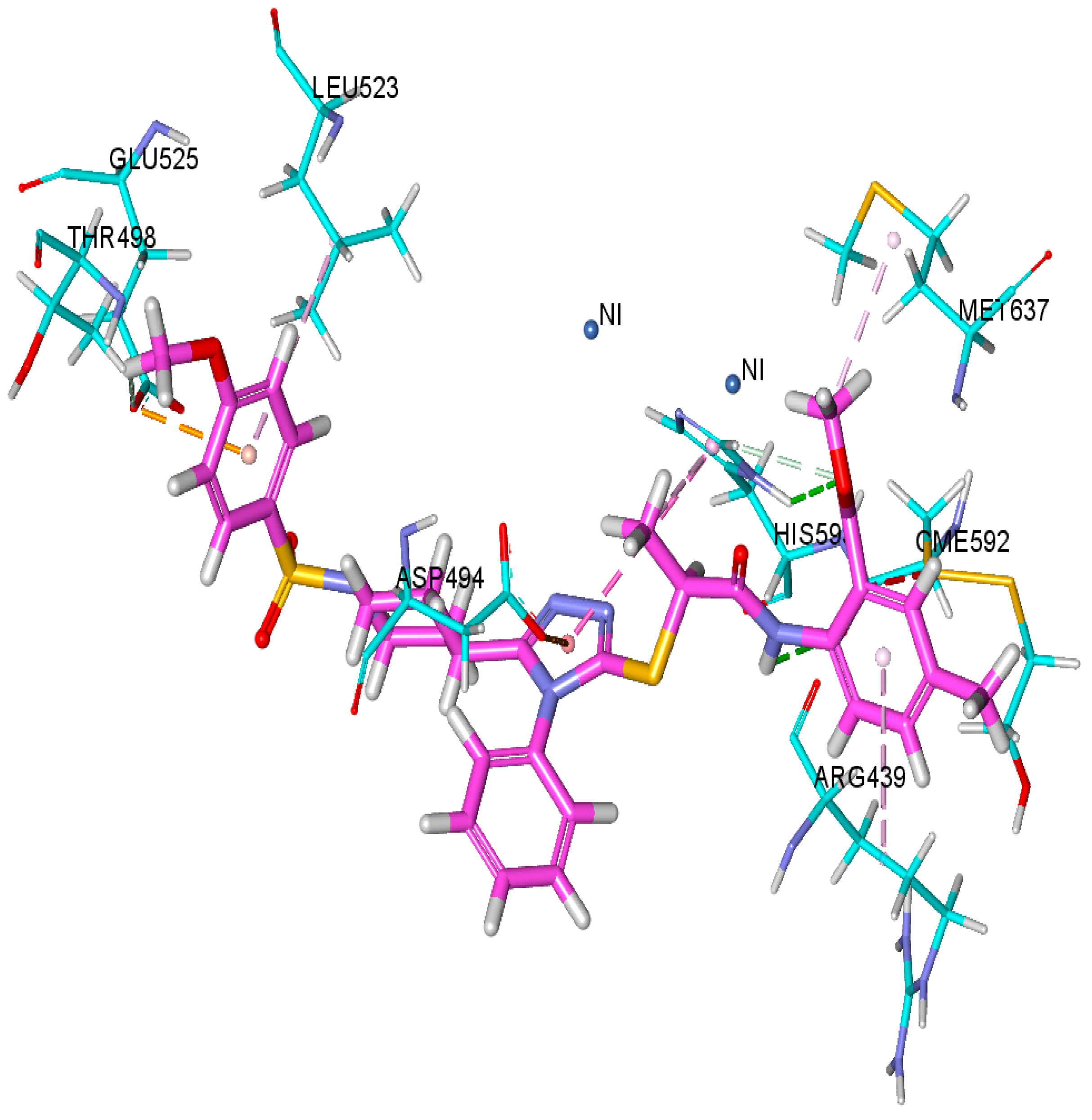
3.6. Docking Studies

3.6.1. Docking Studies against Urease and BChE Enzymes

Molecular docking calculations are broadly used for investigation of the binding affinities of ligands with target structures. We also performed molecular docking studies of all compounds 9a9l, Table 4, to investigate the putative binding orientation of these compounds within the active site of urease and BChE. Binding affinity of the synthesized compounds against urease and BChE were evaluated based on chemguass4 score implemented in FRED docking software (3.2.0) as shown in Table 4. FRED uses multi-conformer docking procedure which separately creates a set of low-energy conformers, and then does rigid docking for each conformer.
The best docked pose based on the lowest chemguass4 was selected for further deciphering the biding interactions. Detail binding interaction of the most potent compound 9k and compound 9d against urease and BChE, respectively are shown in Figure 1 and Figure 2. Compound 9k was forming a great network of different hydrogen bonding, hydrophobic and electrostatic interactions with various amino acid residues within the active site of urease. Two hydrogen bonds were formed by amino acid residue His593 and CME592 as shown in green dotted line in Figure 1. Two pi-anion electrostatic interactions were formed with Asp494 and Glu525 while only one pi-pi T-shaped interaction between triazole moiety of compound 9k and amino acid residue His593 as shown by pink dotted line. Additionally, Met637, His593, Leu523 and Arg439 presented hydrophobic interactions with side chains of compound 9k.
Compound 9d was the most potent compound against urease, so it was selected to investigate the detailed binding interaction with the target structure. Figure 2 shows the binding interactions of compound 9d with different amino acid residue of urease. One of oxygen atom attached to sulfur was making hydrogen bond with amino acid residue of Asn289 as shown in green dotted line. Ala277 and Ser287 along with Gly283 were also forming carbon hydrogen bond as shown in light green color in Figure 2. In addition to hydrogen bonding network, hydrophobic interactions were also formed by Phe329, Trp332, Ala277 and Ala328.

3.6.2. Docking Studies against AChE Enzyme

To identify the binding orientation of compound 9l with the protein that forms the active site of the AChE enzyme, a molecular docking analysis of the relevant molecule was performed. Out of all the synthesized compounds, ligand 9l demonstrated the best ability to inhibit the AChE enzyme. In order to dock ligand 9l or the active site of the AChE enzyme, SybylX-1.3 (module Surflex-Dock) wasutilized (Table 5, Figure 3). By re-docking the docked compounds into the active cite of AChE, the legitimacy of the chemical was verified. In order to verify the docking methodology, donepezil was removed from the co-crystal complex 4EY7In and then docked again in the same binding pocket. Experimentally, the docking capabilities and donepezil’s AChE binding behavior were compared. This proves the accuracy of our docking method. Additionally, it was demonstrated that the inhibitor exhibits a similar binding mechanism to that of the experimentally discovered 4EY7 complex. According to the visual analysis of docked ligands, compound 9l, an inhibitor, deeply enters the CAS and PAS of the AChE enzyme. The ligand has created a hydrogen bonding interaction with the backbone of Glu292, Ser203, Phe295, Arg296, and Phe338 in the active site of AChE. It is possible that the methyl groups substituted at the aromatic ring linked with the amidic group created the hydrophobic contact necessary for its optimal activity in the CAS area. C score, PMF score, and Polar score are experimental findings that are 6.84, 15.098, and 0.09, respectively. This proves the accuracy of our docking method. Numerous amino acids, including Glu 292, Ser 203, Phe 295, Arg 296 and Phe 338, interacted with inhibitor drugs to increase their effectiveness. The variation in ligand acceptor interactions could be the cause of possible inhibitory behavior of compound 9l.

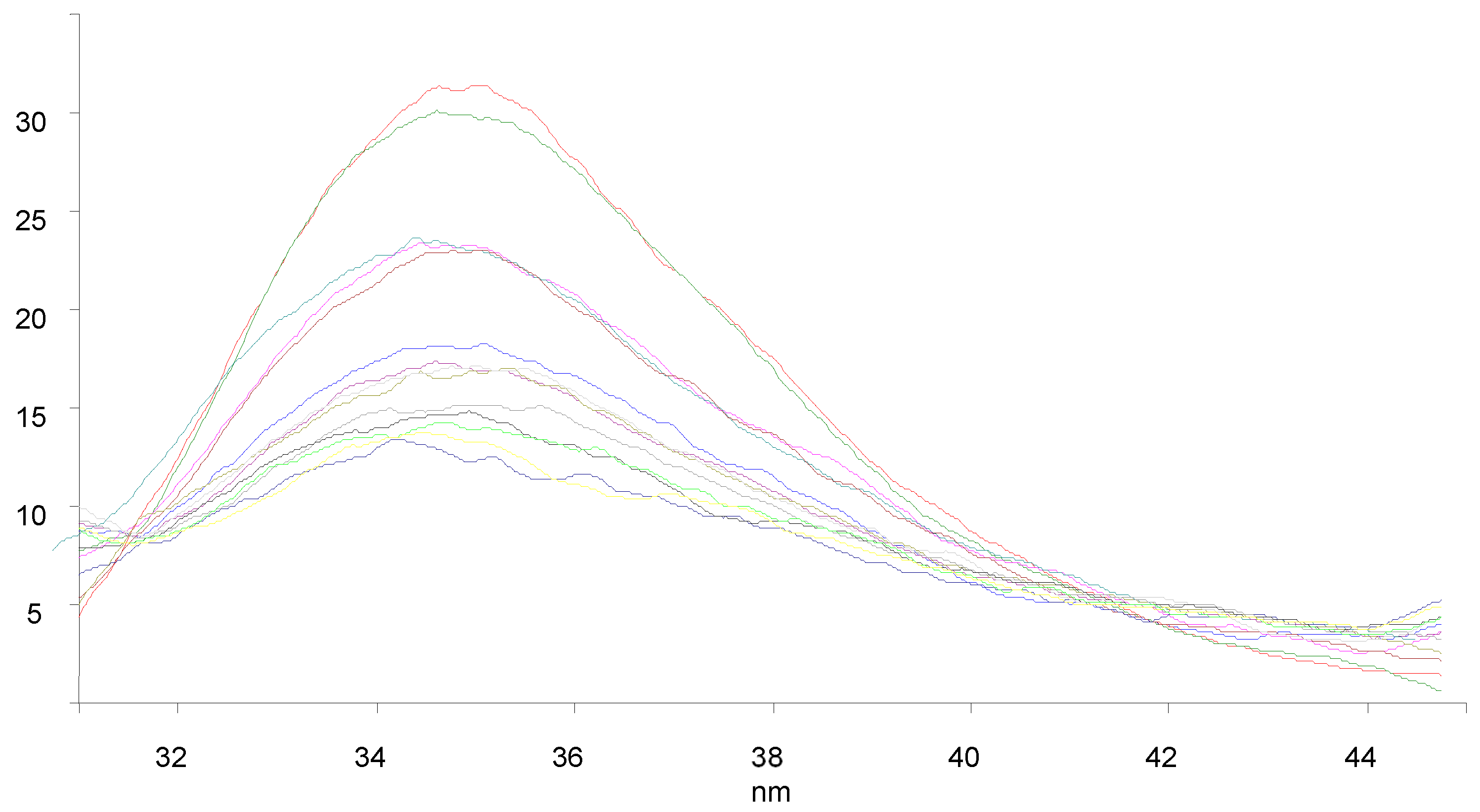
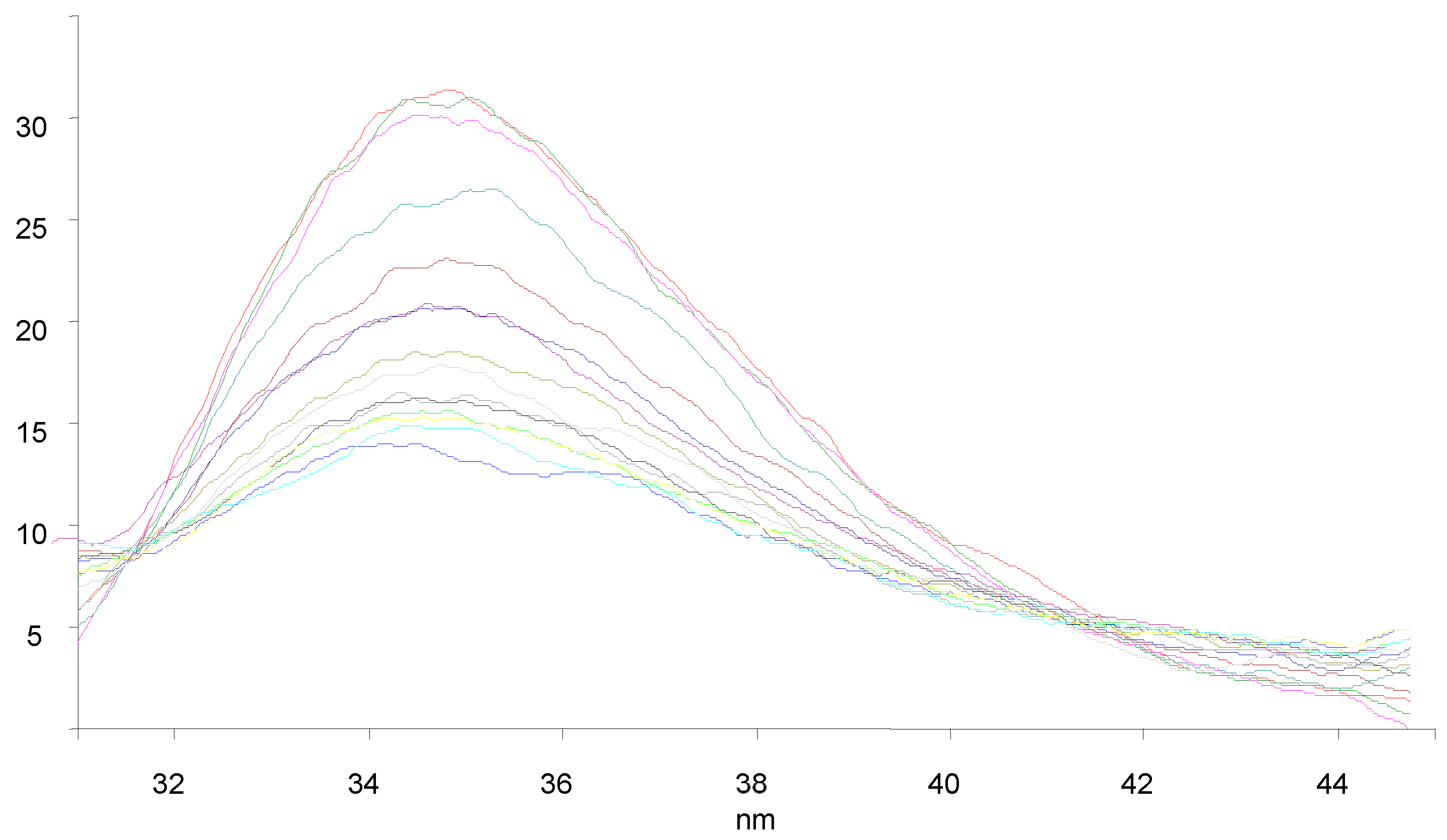
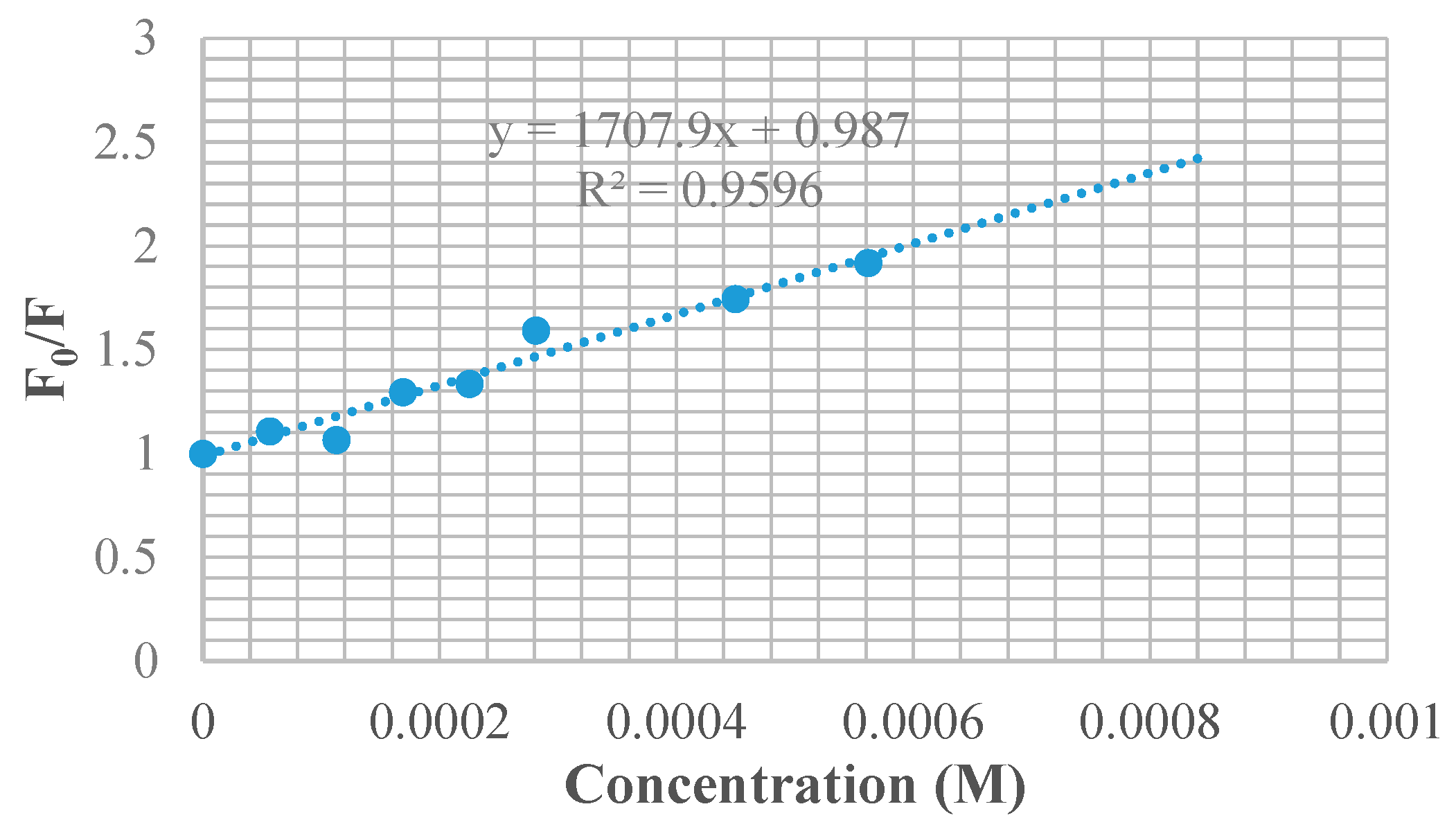
3.7. BSA Binding Studies

The quenching of BSA with the synthesized molecules appears to follow a static process rather than a dynamic one, as the predicted kq was greater than the maximum scattering collision quenching rate constant (2 × 10¹⁰ M−1 s−1) in dynamic quenching. The apparent constant (Ka) and the total number of binding sites (n) can be determined when tiny molecules bind individually to a set of analogous sites on a macromolecule using the double reciprocal plot. The double reciprocal charts of synthesized molecules are displayed (Figure 4, Figure 5, Figure 6, Figure 7, Figure 8 and Figure 9). The intercept and slope of the linear plot, respectively, are used to determine the values of Ka and n. Among the studied compound, 9a showed the least bonding constant values while 9f showed the highest bonding constant value with BSA as given in Table 6. So this binding pattern will justify that strongest bonding with ‘lead’ does not radially release the drug and the least bonding will not carry the drug in proper way towards the target as 9a and 9f respectively. The compound, 9c showing intermediate bonding capabilities with BSA will perform in a better way.

4. Conclusions

Current research was conducted to synthesize a library of hybrids of 1,2,4-triazole having azinane and propanamides as key components and to explore their versatile biological potential. Synthesis of target molecules was conducted for comparison of conventional and microwave assisted methods. The highest yield with high purity in minimum time was obtained through microwave assisted technique. The whole library of designed hybrids was characterized by spectroscopic techniques. The biological studies included the assessment of antioxidant activity, urease, AChE and BChE inhibition potential followed by docking and BSA binding analysis. All the hybrids were found active with variable potential against mentioned enzymes and antioxidant activity. Compounds, 9g and 9k were the most active member of the series and proved to be the best urease inhibitors. After further biological exploration, these compounds might be the best anti-urease drugs in the market in future to serve the humanity.

Author Contributions

Methodology, J.I. and A.U.R.; Investigation, J.I., N.A.V., A.K. and M.Y.; Data curation, J.I., A.U.R. and S.A.A.S.; Supervision, J.I., A.U.R. and S.A.A.S.; Formal analysis, N.A.V., A.K. and M.Y.; Writing – Original Draft, N.A.V.; Conceptualization, A.U.R.; Validation, A.U.R. and S.A.A.S.; Resources, A.U.R. and S.A.A.S.; Visualization, A.U.R., S.R. and S.A.A.S.; Project administration, A.U.R.; Software, A.K., M.Y. and S.A.A.S.; Writing – Review & Editing, S.R. All authors have read and agreed to the published version of the manuscript.

Funding

This research received no external funding.

Institutional Review Board Statement

Not applicable.

Informed Consent Statement

Not applicable.

Data Availability Statement

The data presented in this study are available on request from the corresponding author.

Acknowledgments

Authors are thankful to Higher Education Commission (HEC) of Pakistan.

Conflicts of Interest

The authors declare no conflict of interest.

References

  1. Jhuang, H.S.; Liu, Y.W.; Reddy, D.M.; Tzeng, Y.Z.; Lin, W.Y.; Lee, C.F. Microwave assisted synthesis of thioesters from aldehydes and thiols in water. J. Chin. Chem. Soc. 2018, 65, 24–27. [Google Scholar] [CrossRef]
  2. Lidström, P.; Tierney, J.; Watheyb, B.; Westmana, J. Microwave assisted organic synthesis—A review. Tetrahedron 2001, 57, 9225–9283. [Google Scholar] [CrossRef]
  3. Lee, J.; Hong, M.; Jung, Y.; Cho, E.J.; Rhee, H. Synthesis of 1,3,5-trisubstituted-1,2,4-triazoles by microwave-assisted N-acylation of amide derivatives and the consecutive reaction with hydrazine hydrochlorides. Tetrahedron 2012, 68, 2045–2051. [Google Scholar] [CrossRef]
  4. Sebeka, A.A.H.; Osman, A.M.A.; ElSayed, I.E.T.; ElBahanasawy, M.; Tantawy, M.A. Synthesis and antiproliferative activity of novel neocryptolepine-hydrazides hybrids. J. Appl. Pharm. Sci. 2017, 7, 9–15. [Google Scholar]
  5. Potewar, T.M.; Petrova, K.T.; Barros, M.T. Efficient microwave assisted synthesis of novel 1,2,3-triazole–sucrose derivatives by cycloaddition reaction of sucrose azides and terminal alkynes. Carbohydr. Res. 2013, 379, 60–67. [Google Scholar] [CrossRef] [PubMed]
  6. Sangshetti, J.N.; Chabukswar, A.R.; Shinde, D.B. Microwave assisted one pot synthesis of some novel 2,5-disubstituted1,3,4-oxadiazoles as antifungal agents. Bioorg. Med. Chem. Lett. 2011, 21, 444–448. [Google Scholar] [CrossRef] [PubMed]
  7. Medvedeva, A.S.; Demina, M.M.; Kon, T.V.; Nguyen, T.L.H.; Afonin, A.V.; Ushakov, I.A. Microwave assisted solvent-and catalyst-free three-component synthesis of NH-1,2,3-triazoloimines. Tetrahedron 2017, 73, 3979–3985. [Google Scholar] [CrossRef]
  8. Kharb, R.; Sharma, P.C.; Yar, M.S. Pharmacological significance of triazole scaffold. J. Enzyme Inhib. Med. Chem. 2011, 26, 1–21. [Google Scholar] [CrossRef] [PubMed]
  9. Mishra, P.; Upmanyu, N.; Mehta, A. In vitro anti-microbial activity of some 5-aryl-4-(substituted amino)-3-mercapto-1,2,4-triazoles. Indian J. Microbiol. 2006, 46, 401–404. [Google Scholar]
  10. Sahu, S.K.; Dubey, B.K.; Tripathi, A.C.; Koshy, M.; Saraf, S.K. Piperidin-4-one: The potential pharmacophore. Mini Rev. Med. Chem. 2013, 13, 565–583. [Google Scholar] [CrossRef] [PubMed]
  11. Gopalakrishnan, M.; Thanusu, J.; Kanagarajan, V.; Govindaraju, R. Unexpected formation of 3-chloro-1-hydroxy-2,6-diarylpiperidin-4-ones: Synthesis, antibacterial and antifungal activities. J. Enzyme Inhib. Med. Chem. 2009, 24, 52–58. [Google Scholar] [CrossRef] [PubMed]
  12. Koleva, I.I.; VanBeek, T.A.; Linssen, J.P.H.; Groot, A.D.; Evstatieva, L.N. Screening of plant extracts for antioxidant activity: A comparative study on three testing methods. Phytochem. Anal. 2002, 13, 8–17. [Google Scholar] [CrossRef] [PubMed]
  13. Cygler, M.; Schrag, J.D.; Sussman, J.L.; Harel, M.; Silman, I.; Gentry, M.K.; Doctor, B.P. Relationship between sequence conservation and three-dimensional structure in a large family of esterases, lipases, and related proteins. Protein Sci. 1993, 2, 366–382. [Google Scholar] [CrossRef] [PubMed]
  14. Tougu, V. Acetyl cholinesterase: Mechanism of catalysis and inhibition. Curr. Med. Chem. 2001, 1, 155–170. [Google Scholar]
  15. Mobley, H.L.; Cortesia, M.J.; Rosenthal, L.E.; Jones, B.D. Characterization of urease from Campylobacterpylori. J. Clin. Microbiol. 1988, 26, 831–836. [Google Scholar] [CrossRef] [PubMed]
  16. Balasubramanian, A.; Ponnuraj, K. Crystal structure of the first plant urease from jackbean: 83 years of journey from itsfirst crystal to molecular structure. J. Mol. Biol. 2010, 400, 274–283. [Google Scholar] [CrossRef] [PubMed]
  17. Nachon, F.; Carletti, E.; Ronco, C.; Trovaslet, M.; Nicolet, Y.; Jean, L.; Renard, P.Y. Crystal structures of human cholinesterases in complex with huprine Wandtacrine: Elements of specificity for anti-Alzheimer’s drugs targeting acetyl-and butyryl-cholinesterase. Biochem. J. 2013, 453, 393–399. [Google Scholar] [CrossRef] [PubMed]
  18. Elmer, P. ChemBioDraw Professional, version (15.0.0.106); Cambridge Soft Waltham: Waltham, MA, USA, 2017.
  19. BIOVIA Discovery Studio Client, v16.1.0.15350; Dassault Systemes: SanDiego, CA, USA, 2015.
  20. FRED, version 3.0.0; Open Eye Scientific Software: SantaFe, NM, USA, 2013. Available online: www.eyesopen.com(accessed on 12 April 2023).
  21. OMEGA, version 2.4.6; Open Eye Scientific Software: SantaFe, NM, USA, 2013. Available online: www.eyesopen.com(accessed on 12 April 2023).
Scheme 1. General scheme for the synthesis library of hybrids of 1,2,4-triazole having piperidine and propanamides.
Chemproc 14 00106 sch001
Figure 1. Binding orientation of most potent Compound 9k within the active site if Urease (4H9M). Hydrogen bonding are shown in green line while other Hydrophobic interactions are shown in pink dotted line, yellow color show sulfur bond, while pink color show pi bonding.
Chemproc 14 00106 g001
Figure 2. Binding orientation of most potent Compound 9d within the active site if Urease (4BDS). Hydrogen bonding is shown in green line while other Hydrophobic interactions are shown in pink dotted line, yellow color show sulfur bond, while pink color show pi bonding.
Chemproc 14 00106 g002
Figure 3. Molecular docking generated poses for selected AChE inhibitor (9l) (A) Compound 9l bonded with in the active site of AChE enzyme (B) binding mode of 9l in AChE ligand binding site (C) 2D-ligand-protein interaction diagram was generated for the best poses obtained with compound 9l against AChE enzyme.
Chemproc 14 00106 g003
Figure 4. Fluorescence graph of BSA in the presence of 9a at different concentrations shown by different colors in graph.
Chemproc 14 00106 g004
Figure 5. Fluorescence graph of BSA in the presence of 9c at different concentrations shown by different colors in graph.
Chemproc 14 00106 g005
Figure 6. Fluorescence graph of BSA in the presence of 9e at different concentrations shown by different colors in graph.
Chemproc 14 00106 g006
Figure 7. Stern-Volmer plots of 9a.
Chemproc 14 00106 g007
Figure 8. Stern-Volmer plots of 9c.
Chemproc 14 00106 g008
Figure 9. Stern-Volmer plots of 9f.
Chemproc 14 00106 g009
Table 1. Different N-substituted aryl/phenyl/alkyl groups.
CompoundRCompoundR
9aChemproc 14 00106 i0019gChemproc 14 00106 i002
9bChemproc 14 00106 i0039hChemproc 14 00106 i004
9cChemproc 14 00106 i0059iChemproc 14 00106 i006
9dChemproc 14 00106 i0079jChemproc 14 00106 i008
9eChemproc 14 00106 i0099kChemproc 14 00106 i010
9fChemproc 14 00106 i0119lChemproc 14 00106 i012
Table 2. Comparison of conventional and microwave assisted methods.
CompoundsReaction TimeReaction Yield (%)
Conventional (hours)Microwave (sec)ConventionalMicrowave
9a10326294
9b13365892
9c14436693
9d11336188
9e8597086
9f11396789
9g12407267
9h14485492
9i16476890
9j13527688
9k9384985
9l8495882
Table 3. Anti-oxidant and enzyme inhibition studies.
CompoundsIC50 Values
AChEAntioxidantUrease InhibitionBChE
9a270.83 ± 1.2165.2 ± 0.1521.1 ± 0.1166.1 ± 0.62
9b269.25 ± 1.1352.1 ± 0.2636.2 ± 0.2179.2 ± 0.22
9c134.63 ± 1.1245.2 ± 0.1539.4 ± 0.4577.7 ± 0.36
9d215.91 ± 1.1349.5 ± 0.21>50015.5 ± 0.39
9e63.27 ± 1.2172.1 ± 0.69>50015.9 ± 0.67
9f169.83 ± 1.1277.7 ± 0.29>50019.2 ± 0.67
9g63.45 ± 1.2175.5 ± 0.3220.2 ± 0.2129.2 ± 0.26
9h87.32 ± 1.1885.5 ± 0.5545.5 ± 0.5226.1 ± 0.14
9i182.73 ± 1.1582.2 ± 0.5649.2 ± 0.2639.5 ± 0.21
9j-55.5 ± 0.2055.2 ± 0.1179.8 ± 0.29
9k133.91 ± 1.1252.1 ± 0.0919.2 ± 0.0972.1 ± 0.35
9l402.83 ± 1.1247.7 ± 0.29-29.2 ± 0.37
BHA 44.2 ± 0.41--
Thiourea -21.3 ± 0.24-
Eserine0.19 ± 0.05--7.8 ± 0.05
Table 4. Chemgauss4 scores of all synthesized compounds against Urease and BChE.
CompoundChemGauss4 Score
against Urease
ChemGauss4 Score
against BChE
9a−6.57−9.80
9b−6.06−9.92
9c−7.49−8.98
9d−6.23−8.82
9e−6.56−9.47
9f−4.66−8.94
9g−4.17−8.95
9h−5.37−8.21
9i−6.17−9.32
9j−4.27−9.02
9k−6.75−8.68
9l−4.21−9.12
Table 5. Surflex score of docked ligands compound 9l for AChE enzyme.
Docking Complex = AChE- 9l
C
Score a
Crash Score bPolar Score cD Score dPMF Score eG
Score f
Chem
Score g
Amino Acid Interaction
6.84−3.890.09−215.07415.098−345.68−32.964Glu292, Ser203, Phe295, Arg296, Phe338
Note: a a consensus scoring which uses multiple types of scoring functions to rank the affinity of ligands, b revealing the inappropriate penetration into the binding site, c region of the ligand, d for chRe and van der Waals interactions between the protein and the ligand, e indicating the Helmholtz free energies of interactions for protein-ligand atom pairs (Potential of Mean Force, PMF), f showing hydrogen bonding, complex (ligand-protein), and internal (ligand-ligand) energies, g points for hydrogen bonding, lipophilic contact, and rotational entropy, along with an intercept term.
Table 6. Stern–Volmer quenching constants, binding constant and number of binding site for compounds.
CompoundsKSV × 101 (M−1)kq × 1011 (M−1 s−1)Ka (M−1)n
9a1.2381.23861.20.58
9c1.8471.8472.33 × 1031.03
9f1.7081.7084.11 × 1031.11
Disclaimer/Publisher’s Note: The statements, opinions and data contained in all publications are solely those of the individual author(s) and contributor(s) and not of MDPI and/or the editor(s). MDPI and/or the editor(s) disclaim responsibility for any injury to people or property resulting from any ideas, methods, instructions or products referred to in the content.

