Article

Patients’ Perspective on Barriers to Utilization of a Diabetic Retinopathy Screening Service

by
Bismark Owusu-Afriyie
1,2,*,†,
Theresa Gende
1,2,†,
Martin Tapilas
1,
Nicholas Zimbare
1 and
Jeffrey Kewande
1
1
Faculty of Medicine and Health Sciences, Divine Word University, Madang 511, Papua New Guinea
2
The Fred Hollows Foundation NZ, Auckland 1010, New Zealand
*
Author to whom correspondence should be addressed.
These authors contributed equally to this work.
Diabetology 2023, 4(3), 393-405; https://doi.org/10.3390/diabetology4030033
Submission received: 30 July 2023 / Revised: 25 August 2023 / Accepted: 5 September 2023 / Published: 11 September 2023
(This article belongs to the Special Issue Feature Papers in Diabetology 2023)

Abstract

:
This study was conducted to determine the barriers to the utilization of diabetic retinopathy (DR) screening in Papua New Guinea (PNG). A list of patients booked for DR screening at Madang Provincial Hospital Eye Clinic (MPHEC) between January 2017 and December 2021 who had not been screened was retrieved, and the patients were invited to participate in the study. The data were collected using a structured questionnaire, and IBM Statistical Package for Social Sciences version 26 was used for the analysis. p < 0.05 was considered statistically significant. One hundred and twenty-nine patients (37.4%) did not attend DR screening for the period under study. The study response rate was 80.6%. The mean ± SD age of the respondents was 51.5 ± 10.9 years. The majority of the study respondents were female (62.5%), people living in rural settings (53.8%), and farmers (22.1%). Time constraints, poor knowledge about DR, and long waiting periods at the DR screening center were the main barriers to the uptake of DR screening. Compared to respondents in urban communities, those in rural settings were significantly concerned about cost (p < 0.001), travel distance to the MPHEC (p < 0.001), and poor information about DR screening (p = 0.002). More than half of the respondents (63.5%) had discontinued using pharmacotherapy for DM. There is a high rate of nonadherence to diabetes (DM) and DR treatment in PNG. There is a need for public health campaigns about DM and strategic DR screening at the community level in PNG and similar countries.

1. Introduction

Diabetic retinopathy (DR) is the most common complication of diabetes (DM) [1,2,3], and its main risk factors are disease duration, poor glycemic control, and hypertension [1]. The disease remains a clinical problem [4], and it is the leading cause of working age and adult-onset blindness in spite of improvements in diabetes care [2,5]. Thus, it is a great concern for people with diabetes and healthcare providers. Several years of research into the pathophysiology and management of DR have improved the understanding of the disease process [6,7,8]. In order to prevent sight loss from DR, all persons with diabetes are encouraged to undergo a comprehensive eye examination for the early detection and treatment of DR, herein referred to as DR screening.
DR is categorized into two main forms based on the clinical features of the disease, namely non-proliferative DR (NPDR) and proliferative DR (PDR) [9]. Cotton wool spots, retinal hemorrhages, retinal exudates, and microaneurysms are often the hallmark of NPDR. PDR is distinguished from NPDR by the presence of new blood vessels in the retina and/or iris (neovascularization). These new blood vessels are fragile and often cause further complications, such as vitreous hemorrhage. PDR, together with diabetic macular oedema, are the main causes of vision-threatening DR [10].
The low level of DR screening in Papua New Guinea (PNG), coupled with the growing prevalence of diabetes, is worrisome. Burnett et al. indicated in their study that more than three-quarters of the patients with known diabetes in the National Capital District of PNG never had an eye examination [11]. They further discovered that nearly half of those with diabetes had developed retinopathy and/or maculopathy [11]. While this low level of DR screening may be attributed to a lack of a national DR screening program in PNG, it is worth exploring the barriers to the utilization of DR screening in settings in the country where the service is available.
In 2007, the Madang Provincial Hospital Eye Clinic (MPHEC) implemented systematic DR screening for all people with diabetes to identify and treat sight-threatening retinopathy using the “Pacific Diabetes Retinal Screening, Grading and Management Guidelines” [12]. In this guideline, people with DM but no DR are expected to undertake annual retinal screening, while those with DR are scheduled for periodic screenings, depending on the clinical features and severity of the condition [12]. The MPHEC is one of the three centers in the country that offer DR screening, such as fundus photography and laser treatment. Ophthalmic clinicians and ophthalmologists in the country are trained to detect and refer DR cases to either the MPHEC or the other two centers at Port Moresby for further assessment and management. DR screening at the MPHEC is at no cost to the patients.
A recent finding indicated that 50% of all ophthalmic patients aged ≥30 years who visited the MPHEC in the first half of 2021 were either pre-diabetic or diabetic [13]; hence, it was paramount that they undergo DR screening. Despite the availability of the free retinal screening service at the facility, the uptake of the service has been very low, at an average of two patients per month for the years 2017 to 2021. It was therefore necessary to identify the reasons for the nonadherence to DR screening among patients who had records at the MPHEC.
This study explored the views of patients who had not yet attended DR screening or missed review appointments over a 5-year period. Our findings have the potential to direct policymakers to develop strategies to enhance the quality and uptake of DM and DR services in PNG and other similar countries.

2. Materials and Methods

2.1. Study Setting

The study was designed and conducted by using clinical records from the MPHEC. It was one of the three DR screening centers in PNG at the time of this study. The facility routinely screens patients aged ≥30 years for DM at no cost and, in addition, provides free DR services for walk-in and referred patients with diabetes (DM). Therefore, our study included patients from across the country, not just Madang Province. At the MPHEC and the other eye clinics, patients are first examined by an ophthalmologist or ophthalmic clinician before a recommendation is given for DR screening.

2.2. Study Design and Sampling Techniques

Purposive sampling was used in this descriptive cross-sectional study, as only DM and DR patients’ records at the MPHEC were selected for the study.

2.3. Inclusion and Exclusion Criteria

The records of DM patients and the DR referral list of the MPHEC from January 2017 to December 2021 were reviewed, and those who had not yet undertaken DR screening or missed follow-up retinal screenings were selected. These patients were contacted via the phone, and standardized information about the identity of the researchers, the purpose of the study, the estimated time to complete the questionnaire, voluntary participation, privacy, confidentiality, and anonymity of the data to be collected was given to the patients before inviting them to participate in the study. The study excluded 20 patients who could not be reached, 4 patients who did not consent, 1 deceased patient, and records before January 2017 and after December 2021.

2.4. Data Collection Procedure

A structured questionnaire was designed based on similar studies [14,15,16,17,18] and used for this study (see Supplementary Material). It comprised four parts: the first part determined the respondents’ sociodemographic data, the second portion evaluated the barriers directly related to the patients, the third aspect investigated service-related challenges, and the final set of questions inquired if the patients were on any diabetes treatment. The responses were rated on a 10-point scale, where 1 meant that it was not a barrier at all and 10 meant that it was a very strong barrier. Respondents were also given the opportunity to provide any further comments. Data collection was done during phone calls and in-person interactions when possible, and the respondents’ responses were recorded by researchers M.T., J.K., and N.Z. Data was collected from June to October 2022.

2.5. Data Management and Analysis

The data were analyzed using IBM Statistical Package for Social Sciences (SPSS) version 26. Frequencies and percentages were used to summarize the categorical variables, while the continuous variables were summarized using means (±standard deviation) and medians (interquartile range). The Wilcoxon rank-sum test and Kruskal–Wallis test were used to determine associations, and Bonferroni correction was done for multiple comparisons. Statistical significance was established at p < 0.05.

3. Results

3.1. Sociodemographic Features of the Respondents

A total of 345 patients were listed in the DR screening and referral registers for the period under study, out of which 129 patients (37.4%) failed to undertake their first DR screenings or missed follow-up visits. Of this number, 104 patients participated in the study, giving a response rate of 80.6%. There were more female respondents (62.5%) than male. The age of the study respondents ranged from 24 to 75 years, with a mean of 51.5 ± 10.9 years. The majority of the respondents (53.8%) were from rural settings, and more than three-quarters of them were residents of Madang Province. Farming (22.1%) was the most common primary occupation among the respondents, and people with tertiary education (42.3%) were the highest respondents. The demographic characteristics of the study respondents are detailed in Table 1.

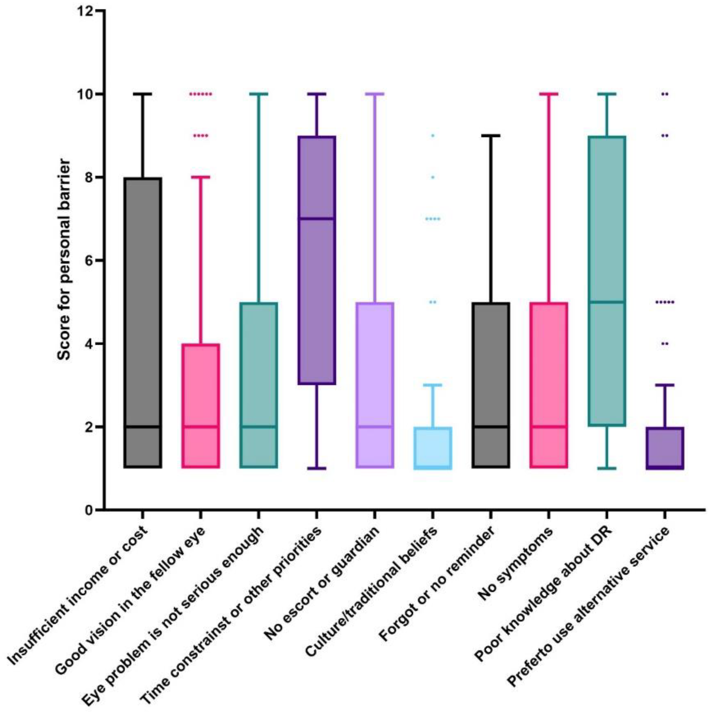
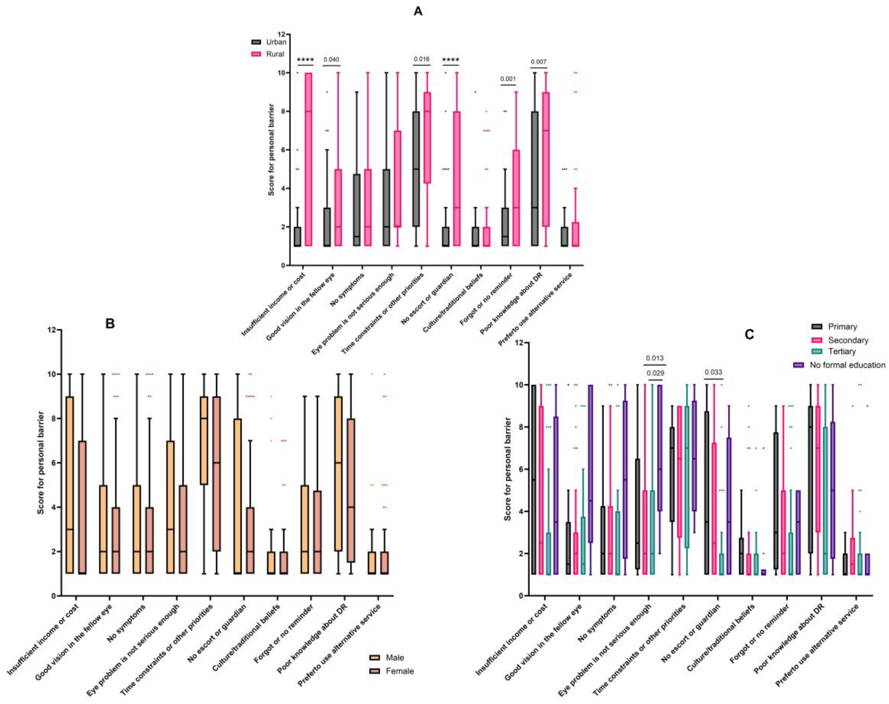
3.2. Personal Barriers

The majority of respondents indicated that time constraints (86.5%) was a barrier to their uptake of DR screenings. In addition, more than half of the respondents considered poor knowledge about DR, cost, good vision in the fellow eye, their eye problem not being a serious issue, the need for a guardian, no reminders about screening appointments, and the asymptomatic nature of their conditions as their personal reasons for not attending DR screenings. The details are shown in Table 2.
Among all the respondents, the most important personal barriers to DR screenings were time constraints (median (IQR) = 7/10 (9/10–3/10)) and poor knowledge about DR (median (IQR) = 5/10 (9/10–2/10)). Overall, culture and traditional beliefs and the use of an alternative treatment did not appear to be hindrances to DR screening services among the respondents (each median (IQR) = 1/10 (2/10–1/10)). The details are shown in Figure 1.
Respondents from rural settings more often indicated that insufficient income or cost (median (IQR) = 8/10 (10/10–1/10)), having other priorities that demanded their time (median (IQR) = 8/10 (9/10–4.3/10)), and poor knowledge about DR (median (IQR) = 7/10 (9/10–2/10)) were the main personal challenges to DR screenings (Figure 2A). Similarly, the most important personal barriers to DR screening among respondents in urban communities were time constraints (median (IQR) = 5/10 (8/10–2/10)) and poor knowledge about DR (median (IQR) = 3/10 (8/10–1/10)). The details are shown in Figure 2A. Respondents from rural settings reported cost (p < 0.001), having good vision in the fellow eye (p = 0.040), time constraints (p = 0.016), poor knowledge about DR (p = 0.007), the need for a guardian (p < 0.001), and reminders (p = 0.001) as significant barriers compared to respondents from urban settings (Figure 2A).
There was no statistically significant difference in the responses from males and females (Figure 2B). From Figure 2C, respondents without formal education significantly considered their eye problems as not being serious enough compared to respondents with secondary (p = 0.013) and tertiary educations (p = 0.029).

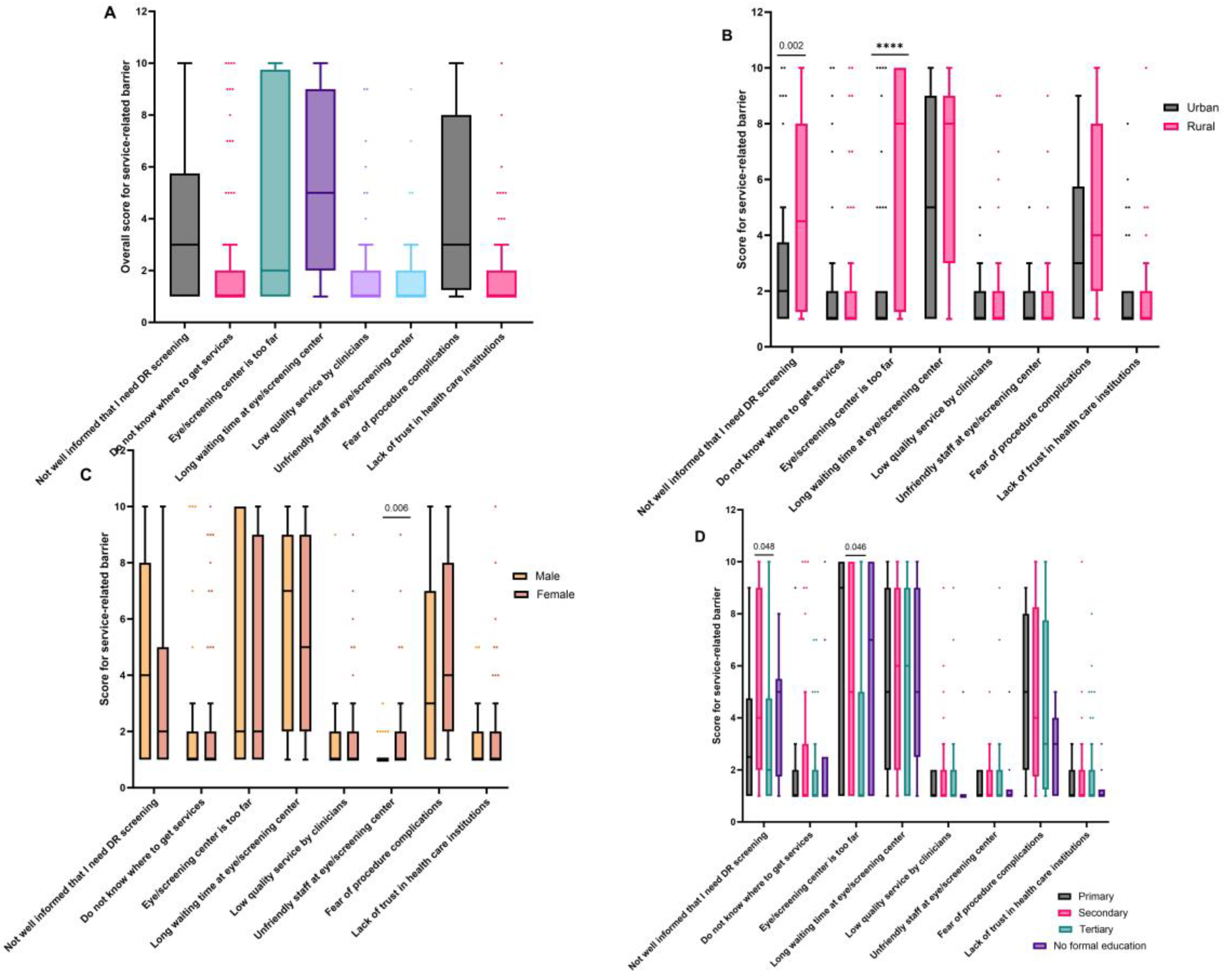
3.3. Service-Related Barriers

At the service level, the majority of the respondents were concerned about long waiting times at the eye screening center (77.9%), fear of procedure complications (75.0%), not being well informed about DR screening (65.4%), and the distant location of the screening center (55.8%) (see Table 2). The main service-related barriers to the uptake of DR screening among all the respondents were the long waiting periods at the eye center (median (IQR) = 5/10 (9/10–2/10)), not being well informed about DR screening (median (IQR) = 3/10 (5.8/10–1/10)), and a fear of procedure complications (median (IQR) = 3/10(3/10–1.3/10)). Further details are shown in Figure 3A.
Respondents from rural communities were more concerned about the long waiting times at the screening center (median (IQR) = 8/10 (9/10–3/10)) and proximity (median (IQR) = 8/10(10/10–1.25/10)). Respondents from urban settings also showed increased concern about the long waiting periods at the screening center (median (IQR) = 5/10 (9/10–1/10)). Barriers such as not being well informed that the respondents needed DR screening and the long-distance location of the screening center were significantly reported by respondents in rural settings compared to their urban counterparts (p = 0.002 and <0.001, respectively). These are highlighted in Figure 3B. Similar responses were reported by respondents with secondary and tertiary educations (p = 0.048 and 00.46, respectively; Figure 3D). The responses from male and female respondents were comparable (Figure 3C); however, the female respondents were significantly concerned about unfriendly staff at the screening center (p = 0.006).

4. Discussion

There is a rise in the global prevalence of DM, and current projections indicate that 1.3 billion people will have DM by the year 2050 [19]. This will have significant implications for the eye health sector due to the effect and complications of DM on the eyes. Patients with DM may not be aware of its damaging effects on their eyes until the onset of visual symptoms; by which time, any sight loss is irreversible. However, sight loss caused by complications of DM can be prevented by timely and effective interventions, such as regular DR screening and early treatment [15]. An effective screening and management program requires that patients and their caregivers are willing to actively adhere to treatment protocols. This study aimed to understand the barriers influencing DR screening nonattendance at one of the three DR screening centers in PNG and propose measures to address these challenges.
The World Health Organization recently recommended that countries should monitor the proportion of people with DM attending DR screening appointments, preferably by using data from health facilities [20]. DR screening is at no cost to patients who visit the MPHEC, but a substantial proportion of DM patients (37.4%) did not attend DR screening during the period under review. Suboptimal adherence to DR screening is not unique to this setting and other developing countries. In Australia, retinal screening coverage among people with DM remains a challenge in spite of Medicare benefits that include non-mydriatic fundus photography due to other barriers, such as time constraints, the cost of retinal cameras, and a lack of expertise in delivering DR services [16]. It is well established that DR is the most significant cause of visual impairment and blindness among adults and the working age population [21,22]. In this study, the average age of the respondents was 51.5 ± 10.9 years, similar to a study in Saudi Arabia that reported an average age of 54.0 years [14]. All these data suggest that both developed and developing economies are at risk of losing a productive workforce if measures are not put in place to strengthen the adherence to DR screening programs.
Irrespective of the sociodemographic characteristics of the respondents, poor knowledge about DR, time constraints, and long waiting periods at the eye center were the main influencers associated with nonadherence to DR screening. A recent study reported that the majority of ophthalmic patients in Madang Province depend on health facilities for information about DM and DR [23]. Taken together with the outcomes of the current study, the two studies support a need for public education on DM and its complications in PNG and similar settings. An effective public health education about the complications of DM, effects of DR, and the benefits of timely and regular DR screening among persons with DM could reduce the burden of sight-threatening DR in PNG. This is especially important among respondents in rural communities and those with less than a secondary level of education.
High proportions of the patients who failed to attend DR screening in this study were female (62.5%), people living in rural communities (53.8%), and farmers (22.1%). Cost (such as for transport, accommodations, and food) was a major barrier to DR screening among respondents living in rural settings. Respondents from rural settings were also more often concerned about the distant location of the DR screening center from their homes. Socioeconomic deprivation is a major risk for nonattendance of DR screenings even in developed countries such as the United States of America, the United Kingdom, and Saudi Arabia [15]. Xiao et al. suggested that DR outreach screenings in rural communities in China are more reliable in reaching women, older populations, and less-educated individuals compared to passive case detection in hospitals and referral centers [24]. Therefore, a mobile DR screening program is an effective strategy that could be adopted in PNG to meet the population demands and enhance the uptake of DR services.
With the advancements in technology and teleophthalmology and the easy access to smartphones, stakeholders of eye care in PNG could consider implementing smartphone-based and/or portable handheld fundus imaging in rural and resource-constrained communities. Although this strategy may not replace conventional fundus photography at present, reports indicate that it is cost effective and has high accuracy at detecting sight-threatening DR [25,26,27,28]. This can increase the accessibility to DR screening services and minimize the long waiting periods at the screening center. In addition, the government and non-governmental organizations in PNG could consider subsidizing the cost of conventional retinal cameras to make them readily available in primary and secondary health care facilities and plan towards a national DR screening program. These proposed measures would require that clinicians in these facilities are well trained to use the fundus cameras for effective DR screening, a greater investment into the eye health workforce, and integration with the diabetes sector.
In many resource-constrained settings, clinicians rely on microvascular changes observed via ophthalmoscopy and/or retinal photography to diagnose, grade, and monitor the treatment of DR. However, the available data have shown that DR is not only a vascular disease but also shows neurodegenerative dysfunction [9]. Neural changes may occur before microvascular changes and visual defects are detected in the clinic. For example, in their quest to find biomarkers for the early diagnosis of DR, Harrison et al. found that multifocal electroretinogram was a powerful tool to predict the development of DR among persons with DM but no retinopathy [29]. Therefore, it is important that health care service providers and clinicians explore diagnostic techniques that examine both neural and vascular changes for the efficient and accurate diagnosis of DM and DR.
At present, binocular indirect ophthalmoscopy and fundus photography are the tests performed to detect DR among patients reporting to the MPHEC. Evidence suggests that other techniques such as electroretinogram [29,30], optical coherence tomography (OCT), and OCT angiography [10,30,31,32,33,34] provide additional key features and information for the early and improved diagnosis of DM and DR, as well as monitoring the treatments. These retinal imaging tools could be explored by DR screening centers such as the MPHEC, albeit this would require the training of clinicians and financial investments in the technologies, as previously noted above.
The study further investigated whether the study respondents were still taking their DM medications. Unexpectedly, two-thirds of them (63.5%) were not on any medication; out of which, 32 (30.8%) had no reason for refusing treatment, while 21 (20.2%) reported that they were managing their condition with diet instead of drugs. Despite the fact that there is no complete cure for DM at the time of this study, patient-centered care [35,36,37] and adherence to pharmacotherapy and lifestyle changes are extremely valuable in reducing hyperglycemia and, hence, complications of DM, including DR [36,38,39,40]. This study remarks the need for a future study to identify the barriers and influencers of nonadherence to DM treatment in PNG to reduce the burden of DM and its complications in the country. In addition, since blood glucose levels fluctuate [41], we recommend that healthcare facilities that provide DM and DR screening services such as the MPHEC could include the measurement of glycated hemoglobin (HbA1c) in their tests. HbA1c measures the blood glucose history over the previous two to three months and therefore provides a better understanding of a glycemic control compared to only a fasting or random blood glucose measurement [42,43]. Hence, it is a better predictor of DM and its complications.
The current evidence provides insight into DR screening services in PNG, but the findings are focused only on patients who were identified by or referred to the MPHEC during the 5-year period. Further study is necessary to detect the barriers to DM and DR care across communities in PNG and at the national level and implement appropriate interventions.

5. Conclusions

Ophthalmic clinicians and ophthalmologists in PNG are trained to examine the retina and refer patients for appropriate treatment based on the Pacific Diabetes Retinal Screening, Grading and Management Guidelines, yet a large proportion of persons with DM remain unscreened. This study identified time constraints, cost, poor knowledge about DR, long waiting times, and long travel distance to the DR screening center as the main barriers to the uptake of DR screening services. Several enabling strategies have been proposed in this study to increase the access to and adherence to DR screening at the MPHEC and across PNG.

Supplementary Materials

The following supporting information can be downloaded at https://www.mdpi.com/article/10.3390/diabetology4030033/s1: File S1: the questionnaire.

Author Contributions

Conceptualization, B.O.-A., T.G., M.T., N.Z. and J.K.; Data curation, B.O.-A., T.G., M.T., N.Z. and J.K.; Formal analysis, B.O.-A., T.G., M.T., N.Z. and J.K.; Methodology, B.O.-A., T.G., M.T., N.Z. and J.K.; Resources, B.O.-A., T.G., M.T., N.Z. and J.K.; Writing—original draft, B.O.-A., T.G., M.T., N.Z. and J.K.; Writing—review and editing, B.O.-A., T.G., M.T., N.Z. and J.K. All authors have read and agreed to the published version of the manuscript.

Funding

This research received no external funding, and the APC was funded by The Fred Hollows Foundation, NZ.

Institutional Review Board Statement

The study was conducted according to the guidelines of the Declaration of Helsinki and approved by The Faculty of Medicine and Health Sciences Research Committee of Divine Word University (approval number FRC/MHS/58-22 on 10 June 2022). Permission was granted by the management of the MPHEC in order to access the patients’ records and contact details.

Informed Consent Statement

Informed consent was obtained from all subjects involved in the study. No personally identifiable information was collected as part of the study, and participation was voluntary and confidential.

Data Availability Statement

The data presented in this study are available upon request from the corresponding author. The data are not publicly available due to the use of a questionnaire for data collection.

Acknowledgments

We are grateful to the management of the MPHEC for granting us access to the DR screening lists and Carmilla Yawi for helping us to retrieve the lists. We thank The Fred Hollows Foundation, NZ for funding the APC.

Conflicts of Interest

The authors declare no conflict of interest.

References

  1. Simó-Servat, O.; Hernández, C.; Simó, R. Diabetic Retinopathy in the Context of Patients with Diabetes. Ophthalmic Res. 2019, 62, 211–217. [Google Scholar] [CrossRef]
  2. Jenkins, A.J.; Joglekar, M.V.; Hardikar, A.A.; Keech, A.C.; O’Neal, D.N.; Januszewski, A.S. Biomarkers in diabetic retinopathy. Rev. Diabet. Stud. 2015, 12, 159–195. [Google Scholar] [CrossRef] [PubMed]
  3. Wang, W.; Lo, A.C.Y. Diabetic retinopathy: Pathophysiology and treatments. Int. J. Mol. Sci. 2018, 19, 1816. [Google Scholar] [CrossRef]
  4. Hammes, H.P. Diabetic retinopathy: Hyperglycaemia, oxidative stress and beyond. Diabetologia 2018, 61, 29–38. [Google Scholar] [CrossRef]
  5. Kang, Q.; Yang, C. Oxidative stress and diabetic retinopathy: Molecular mechanisms, pathogenetic role and therapeutic implications. Redox Biol. 2020, 37, 101799. [Google Scholar] [CrossRef] [PubMed]
  6. Heng, L.Z.; Comyn, O.; Peto, T.; Tadros, C.; Ng, E.; Sivaprasad, S.; Hykin, P.G. Diabetic retinopathy: Pathogenesis, clinical grading, management and future developments. Diabet. Med. 2013, 30, 640–650. [Google Scholar] [CrossRef]
  7. Rodríguez, M.L.; Pérez, S.; Mena-Mollá, S.; Desco, M.C.; Ortega, Á.L. Oxidative Stress and Microvascular Alterations in Diabetic Retinopathy: Future Therapies. Oxid. Med. Cell. Longev. 2019, 2019, 4940825. [Google Scholar] [CrossRef]
  8. Al Ghamdi, A.H. Clinical Predictors of Diabetic Retinopathy Progression; A Systematic Review. Curr. Diabetes Rev. 2019, 16, 242–247. [Google Scholar] [CrossRef]
  9. Chen, X.D.; Gardner, T.W. A critical review: Psychophysical assessments of diabetic retinopathy. Surv. Ophthalmol. 2021, 66, 213–230. [Google Scholar] [CrossRef]
  10. Jampol, L.M.; Glassman, A.R.; Sun, J. Evaluation and Care of Patients with Diabetic Retinopathy. N. Engl. J. Med. 2020, 382, 1629–1637. [Google Scholar] [CrossRef]
  11. Burnett, A.; Lee, L.; D’Esposito, F.; Wabulembo, G.; Cama, A.; Guldan, G.; Nelisse, M.; Koim, S.P.; Keys, D.; Poffley, A.J.; et al. Rapid assessment of avoidable blindness and diabetic retinopathy in people aged 50 years and older in the National Capital District of Papua New Guinea. Br. J. Ophthalmol. 2019, 103, 743–747. [Google Scholar] [CrossRef] [PubMed]
  12. Pacific Eye Institute. Diabetes Retinal Screening, Grading and Management Guidelines for Use in Pacific Island Nations, 2010; Diabetes Work Group, Fred Hollows Found Pacific Eye Institute: Auckland, New Zealand, 2010; pp. 1–37. Available online: https://www.worlddiabetesfoundation.org/sites/default/files/WDF08-386%20Pacific%20Island%20Ret%20Screen%20Guidelines.pdf (accessed on 20 July 2023).
  13. Owusu-afriyie, B.; Baimur, I.; Gende, T.; Baia, T. Prevalence of Risk Factors of Retinal Diseases among Patients in Madang Province, Papua New Guinea. Int. J. Clin. Pract. 2022, 2022, 6120908. [Google Scholar] [CrossRef]
  14. Alwazae, M.; Al Adel, F.; Alhumud, A.; Almutairi, A.; Alhumidan, A.; Elmorshedy, H. Barriers for Adherence to Diabetic Retinopathy Screening among Saudi Adults. Cureus 2019, 11, e6454. [Google Scholar] [CrossRef] [PubMed]
  15. Kashim, R.M.; Newton, P.; Ojo, O. Diabetic Retinopathy Screening: A Systematic Review on Patients’ Non-Attendance. Int. J. Environ. Res. Public. Health 2018, 15, 157. [Google Scholar] [CrossRef] [PubMed]
  16. Watson, M.J.G.; McCluskey, P.J.; Grigg, J.R.; Kanagasingam, Y.; Daire, J.; Estai, M. Barriers and facilitators to diabetic retinopathy screening within Australian primary care. BMC Fam. Pract. 2021, 22, 239. [Google Scholar] [CrossRef] [PubMed]
  17. Bruggeman, B.; Zimmerman, C.; LaPorte, A.; Stalvey, M.; Filipp, S.L.; Gurka, M.J.; Silverstein, J.H.; Jacobsen, L.M. Barriers to retinopathy screening in youth and young adults with type 1 diabetes. Pediatr. Diabetes 2021, 22, 469–473. [Google Scholar] [CrossRef] [PubMed]
  18. Fairless, E.; Nwanyanwu, K. Barriers to and Facilitators of Diabetic Retinopathy Screening Utilization in a High-Risk Population. J. Racial Ethn. Health Disparities 2019, 6, 1244–1249. [Google Scholar] [CrossRef] [PubMed]
  19. Ong, K.L.; Stafford, L.K.; McLaughlin, S.A.; Boyko, E.J.; Vollset, S.E.; Smith, A.E.; Dalton, B.E.; Duprey, J.; Cruz, J.A.; Hagins, H.; et al. Global, regional, and national burden of diabetes from 1990 to 2021, with projections of prevalence to 2050: A systematic analysis for the Global Burden of Disease Study 2021. Lancet 2023, 402, 203–234. [Google Scholar] [CrossRef]
  20. World Health Organization. Eye Care Indicator Menu (ECIM): A Tool for Monitoring Strategies and Actions for Eye Care Provision. 2022. Available online: http://apps.who.int/ (accessed on 21 July 2023).
  21. Teo, Z.L.; Tham, Y.C.; Yu, M.; Chee, M.L.; Rim, T.H.; Cheung, N.; Bikbov, M.M.; Wang, Y.X.; Tang, Y.; Lu, Y.; et al. Global Prevalence of Diabetic Retinopathy and Projection of Burden through 2045: Systematic Review and Meta-analysis. Ophthalmology 2021, 128, 1580–1591. [Google Scholar] [CrossRef]
  22. Sabanayagam, C.; Banu, R.; Chee, M.L.; Lee, R.; Wang, Y.X.; Tan, G.; Jonas, J.B.; Lamoureux, E.L.; Cheng, C.-Y.; Klein, B.E.K.; et al. Incidence and progression of diabetic retinopathy: A systematic review. Lancet Diabetes Endocrinol. 2019, 7, 140–149. [Google Scholar] [CrossRef]
  23. Owusu-Afriyie, B.; Caleb, A.; Kube, L.; Gende, T. Knowledge and Awareness of Diabetes and Diabetic Retinopathy among Patients Seeking Eye Care Services in Madang Province, Papua New Guinea. J. Ophthalmol. 2022, 2022, 7674928. [Google Scholar] [CrossRef]
  24. Xiao, B.; Mercer, G.D.; Jin, L.; Lee, H.L.; Chen, T.; Wang, Y.; Liu, Y.; Denniston, A.K.; Egan, C.A.; Li, J.; et al. Outreach screening to address demographic and economic barriers to diabetic retinopathy care in rural China. PLoS ONE 2022, 17, e0266380. [Google Scholar] [CrossRef] [PubMed]
  25. Malerbi, F.K.; Andrade, R.E.; Morales, P.H.; Stuchi, J.A.; Lencione, D.; de Paulo, J.V.; Carvalho, M.P.; Nunes, F.S.; Rocha, R.M.; Ferraz, D.A.; et al. Diabetic Retinopathy Screening Using Artificial Intelligence and Handheld Smartphone-Based Retinal Camera. J. Diabetes Sci. Technol. 2022, 16, 716–723. [Google Scholar] [CrossRef] [PubMed]
  26. Rajalakshmi, R.; Arulmalar, S.; Usha, M.; Prathiba, V.; Kareemuddin, K.S.; Anjana, R.M.; Mohan, V. Validation of smartphone based retinal photography for diabetic retinopathy screening. PLoS ONE 2015, 10, e0138285. [Google Scholar] [CrossRef] [PubMed]
  27. Tan, C.H.; Kyaw, B.M.; Smith, H.; Tan, C.S.; Car, L.T. Use of Smartphones to Detect Diabetic Retinopathy: Scoping Review and Meta-Analysis of Diagnostic Test Accuracy Studies. J. Med. Internet Res. 2020, 22, e16658. [Google Scholar] [CrossRef] [PubMed]
  28. Bilong, Y.; Katte, J.C.; Koki, G.; Kagmeni, G.; Obama, O.P.N.; Fofe, H.R.N.; Mvilongo, C.; Nkengfack, O.; Bimbai, A.M.; Sobngwi, E.; et al. Validation of smartphone-based retinal photography for diabetic retinopathy screening. Ophthalmic Surg. Lasers Imaging Retin. 2019, 50, S18–S22. [Google Scholar] [CrossRef] [PubMed]
  29. Harrison, W.W.; Bearse, M.A.; Ng, J.S.; Jewell, N.P.; Barez, S.; Burger, D.; Schneck, M.E.; Adams, A.J. Multifocal electroretinograms predict onset of diabetic retinopathy in adult patients with diabetes. Investig. Ophthalmol. Vis. Sci. 2011, 52, 772–777. [Google Scholar] [CrossRef]
  30. Zagst, A.J.; Smith, J.D.; Wang, R.; Harrison, W.W. Foveal avascular zone size and mfERG metrics in diabetes and prediabetes: A pilot study of the relationship between structure and function. Doc. Ophthalmol. 2023, 147, 99–107. [Google Scholar] [CrossRef]
  31. Russell, J.F.; Al-khersan, H.; Shi, Y.; Scott, N.L.; Hinkle, J.W.; Fan, K.C.; Lyu, C.; Feuer, W.J.; Gregori, G.; Rosenfeld, P.J. Retinal Non-Perfusion in Proliferative Diabetic Retinopathy Before and After Panretinal Photocoagulation Assessed by Wide Field OCT Angiography. Am. J. Ophthalmol. 2020, 213, 177–185. [Google Scholar] [CrossRef]
  32. Kyei, S.; Asare, F.A.; Assan, J.K.; Zaabaar, E.; Assiamah, F.; Obeng, E.O.; Asiedu, K. Efficacy of intravitreal bevacizumab on diabetic macular oedema in an African population. Ir. J. Med. Sci. 2023. [Google Scholar] [CrossRef]
  33. Sun, Z.; Yang, D.; Tang, Z.; Ng, D.S.; Cheung, C.Y. Optical coherence tomography angiography in diabetic retinopathy: An updated review. Eye 2021, 35, 149–161. [Google Scholar] [CrossRef]
  34. Chua, J.; Sim, R.; Tan, B.; Wong, D.; Yao, X.; Liu, X.; Ting, D.S.W.; Schmidl, D.; Ang, M.; Garhöfer, G.; et al. Optical coherence tomography angiography in diabetes and diabetic retinopathy. J. Clin. Med. 2020, 9, 1723. [Google Scholar] [CrossRef] [PubMed]
  35. Davies, M.J.; D’Alessio, D.A.; Fradkin, J.; Kernan, W.N.; Mathieu, C.; Mingrone, G.; Rossing, P.; Tsapas, A.; Wexler, D.J.; Buse, J.B. Management of hyperglycaemia in type 2 diabetes, 2018. A consensus report by the American Diabetes Association (ADA) and the European Association for the Study of Diabetes (EASD). Diabetologia 2018, 61, 2461–2498. [Google Scholar] [CrossRef]
  36. Davies, M.J.; Aroda, V.R.; Collins, B.S.; Gabbay, R.A.; Green, J.; Maruthur, N.M.; Rosas, S.E.; Del Prato, S.; Mathieu, C.; Mingrone, G.; et al. Management of hyperglycaemia in type 2 diabetes, 2022. A consensus report by the American Diabetes Association (ADA) and the European Association for the Study of Diabetes (EASD). Diabetologia 2022, 65, 1925–1966. [Google Scholar] [CrossRef] [PubMed]
  37. American Diabetes Association. Glycemic targets: Standards of medical care in diabetes. Diabetes Care 2022, 42, S61–S70. [Google Scholar]
  38. Agrawal, L.; Azad, N.; Bahn, G.D.; Reaven, P.D.; Hayward, R.A.; Reda, D.J.; Emanuele, N.V.; Abraira, C.; Duckworth, W.C.; Hayden, C.T.; et al. Intensive glycemic control improves long-term renal outcomes in type 2 diabetes in the veterans affairs diabetes trial (VADT). Diabetes Care 2019, 42, E181–E182. [Google Scholar] [CrossRef] [PubMed]
  39. Sun, S.; Hisland, L.; Grenet, G.; Gueyffier, F.; Cornu, C.; Jaafari, N.; Boussageon, R. Reappraisal of the efficacy of intensive glycaemic control on microvascular complications in patients with type 2 diabetes: A meta-analysis of randomised control-trials. Therapies 2022, 77, 413–423. [Google Scholar] [CrossRef]
  40. Xu, H.; Li, X.; Adams, H.; Kubena, K.; Guo, S. Etiology of metabolic syndrome and dietary intervention. Int. J. Mol. Sci. 2019, 20, 128. [Google Scholar] [CrossRef]
  41. Umpierrez, G.E.; PKovatchev, B. Glycemic Variability: How to Measure and Its Clinical Implication for Type 2 Diabetes. Am. J. Med. Sci. 2018, 356, 518–527. [Google Scholar] [CrossRef]
  42. Sherwani, S.I.; Khan, H.A.; Ekhzaimy, A.; Masood, A.; Sakharkar, M.K. Significance of HbA1c test in diagnosis and prognosis of diabetic patients. Biomark. Insights 2016, 11, 95–104. [Google Scholar] [CrossRef]
  43. Martinez, M.; Santamarina, J.; Pavesi, A.; Musso, C.; Umpierrez, G.E. Glycemic variability and cardiovascular disease in patients with type 2 diabetes. BMJ Open Diabetes Res. Care 2021, 9, e002032. [Google Scholar] [CrossRef]
Figure 1. General personal barriers to diabetic retinopathy screening in Madang District. Responses were rated from 1 (not a barrier) to 10 (a very strong barrier). Center lines indicate the medians; box limits indicate the 25th and 75th percentiles; whiskers extend 1.5 times the interquartile range. Dots indicate outliers.
Diabetology 04 00033 g001
Figure 2. Demographic analysis of personal barriers to DR screening services in Madang District. Responses were rated from 1 (not a barrier) to 10 (a very strong barrier). Center lines indicate the medians; box limits indicate the 25th and 75th percentiles; whiskers extend 1.5 times the interquartile range. Dots indicate outliers. Comparisons were determined using the Wilcoxon rank-sum test (A,B) and Kruskal–Wallis test with Bonferroni correction (C). Statistically significant values are reported on the graphs. **** p < 0.001.
Diabetology 04 00033 g002
Figure 3. Analysis of the service-related barriers to DR screening services in Madang District. The responses were rated from 1 (not a barrier) to 10 (a very strong barrier). The overall service-related barriers were analyzed (A), and comparisons were determined using Wilcoxon rank-sum test (B,C) and Kruskal–Wallis test with Bonferroni correction (D). Center lines indicate the medians; box limits indicate the 25th and 75th percentiles; whiskers extend 1.5 times the interquartile range. Dots indicate outliers. Statistically significant values are reported on the graphs. **** p < 0.001.
Diabetology 04 00033 g003
Table 1. Demographics of the study respondents.
CharacteristicsRespondents; n (%)
Gender
Male39 (37.5)
Female65 (62.5)
Residential Location
Urban48 (46.2)
Rural56 (53.8)
Age Group (years)
21–304 (3.8)
31–4014 (13.5)
41–5030 (28.8)
51–6033 (31.7)
61–7020 (19.2)
Above 70 years3 (2.9)
Level of Education
Primary16 (15.4)
Secondary34 (32.7)
Tertiary44 (42.3)
No formal education10 (9.6)
Residential Province
Madang89 (85.6)
Simbu4 (3.8)
East New Britain2 (1.9)
Milne Bay2 (1.9)
Jiwaka2 (1.9)
Others a4 (3.8)
Not reported1 (1.0)
Primary Occupation
Farmer23 (22.1)
Retail trader/self employed14 (13.5)
Teacher/lecturer12 (11.5)
Housewife11 (10.6)
Manager/director9 (8.7)
Health worker7 (6.7)
Secretary7 (6.7)
Others b21 (20.2)
Expected Year of DR Screening
201710 (9.6)
201815 (14.4)
201922 (21.2)
202044 (42.3)
202113 (12.5)
Category of Nonadherence
First DR screening appointment37 (35.6)
Follow-up visits/reviews58 (55.8)
Taking Any Diabetes Treatment/Medication
Yes38 (36.5)
No66 (63.5)
Diabetes Medication
Metformin27 (26.0)
Daonil6 (5.8)
Herbs and traditional remedy3 (2.9)
Insulin1 (1.0)
Nifedipine1 (1.0)
Reasons For Not Taking Diabetes Treatment
No reason32 (30.8)
Diet21 (20.2)
Managing other comorbidity first6 (5.8)
Poor access to a health facility3 (2.9)
Financial constraint2 (1.9)
Side effect of medication1 (1.0)
Poor understanding of the treatment plan1 (1.0)
a East Sepik—1; Enga—1; Morobe—1; Eastern Highlands—1. b Musician—2; Retired—4; Sailor—1; Air traffic staff—1; Customer care representative—5; Electrician—1; Carpenter—1; Security person—3; Unemployed—2; Clergy—1.
Table 2. Reported barriers to DR screening.
Personal BarriersRespondents; n (%)
Insufficient income or cost54 (51.9)
Good vision in the fellow eye58 (55.8)
Eye problem is not serious enough73 (70.2)
Time constraints or other priorities90 (86.5)
No escort or guardian to help56 (53.8)
Culture/traditional beliefs34 (32.7)
Forgot or no reminder62 (59.6)
No symptoms57 (54.8)
Poor knowledge about DR80 (76.9)
Prefer to use alternative service42 (40.3)
Service-related BarriersRespondents; n (%)
Not well informed that I need DR screening68 (65.4)
Do not know where to get services36 (34.6)
Eye/screening center is too far58 (55.8)
Long waiting time at eye/screening center81 (77.9)
Low quality service by clinicians32 (30.8)
Unfriendly staff at eye/screening center33 (31.7)
Fear of procedure complications78 (75.0)
Lack of trust in healthcare institutions38 (36.5)
Disclaimer/Publisher’s Note: The statements, opinions and data contained in all publications are solely those of the individual author(s) and contributor(s) and not of MDPI and/or the editor(s). MDPI and/or the editor(s) disclaim responsibility for any injury to people or property resulting from any ideas, methods, instructions or products referred to in the content.

