
Marble Chromatic Alteration Study Using Non-Invasive Analytical Techniques and Evaluation of the Most Suitable Cleaning Treatment: The Case of a Bust Representing Queen Margherita di Savoia at the U.S. Embassy in Rome

 ,
,  , , and
Abstract
:1. Introduction
2. Materials and Methods
2.1. Non-Invasive Preliminary Analysis
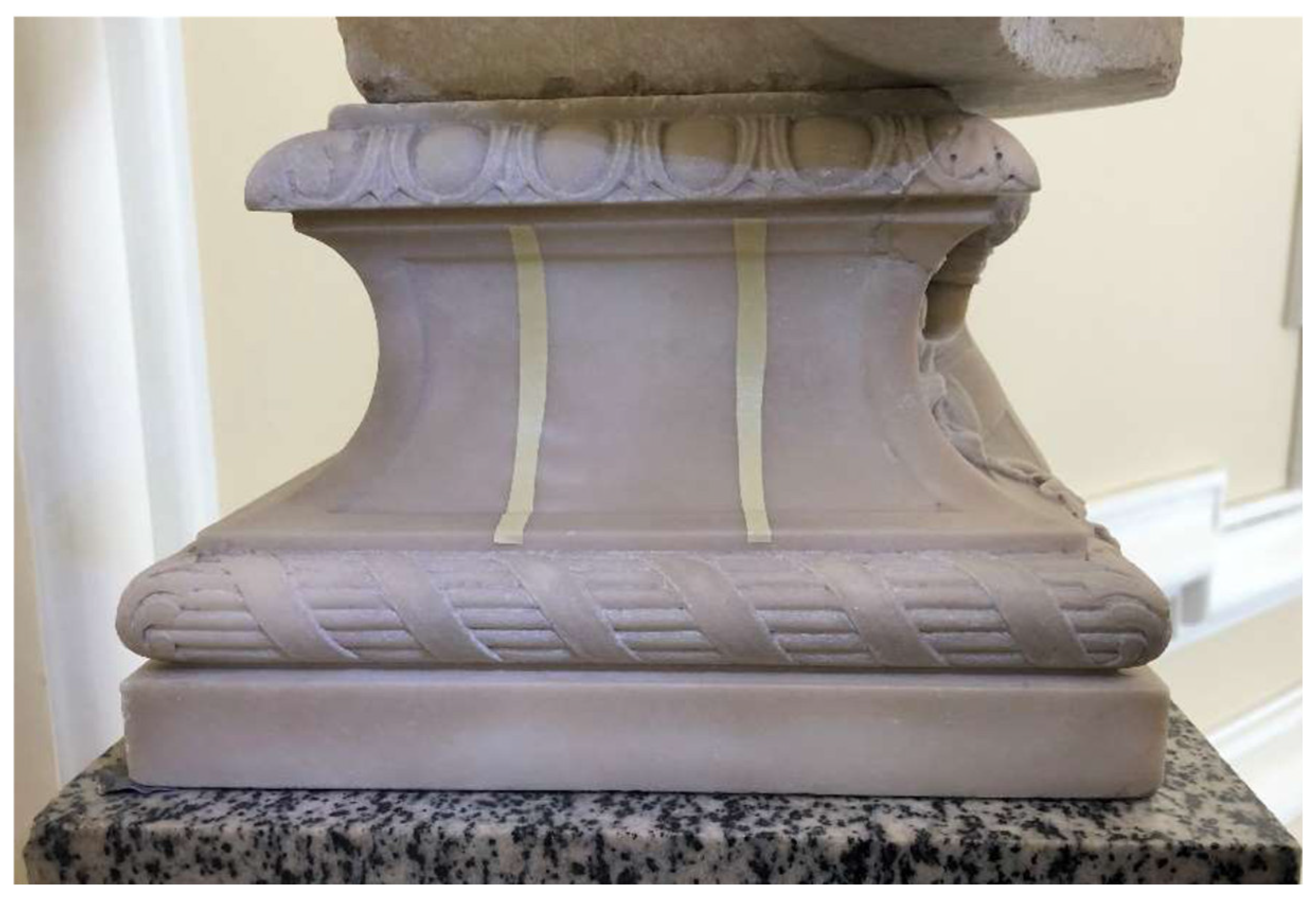
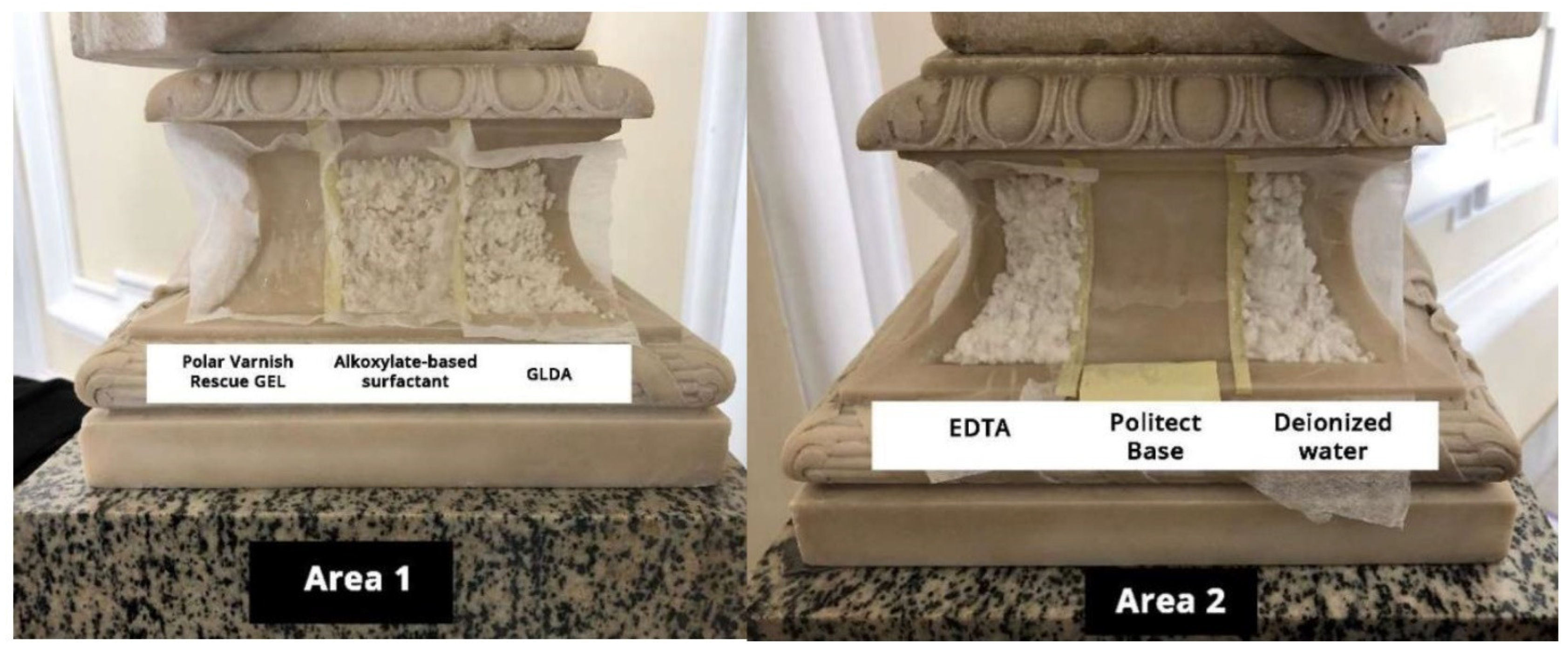
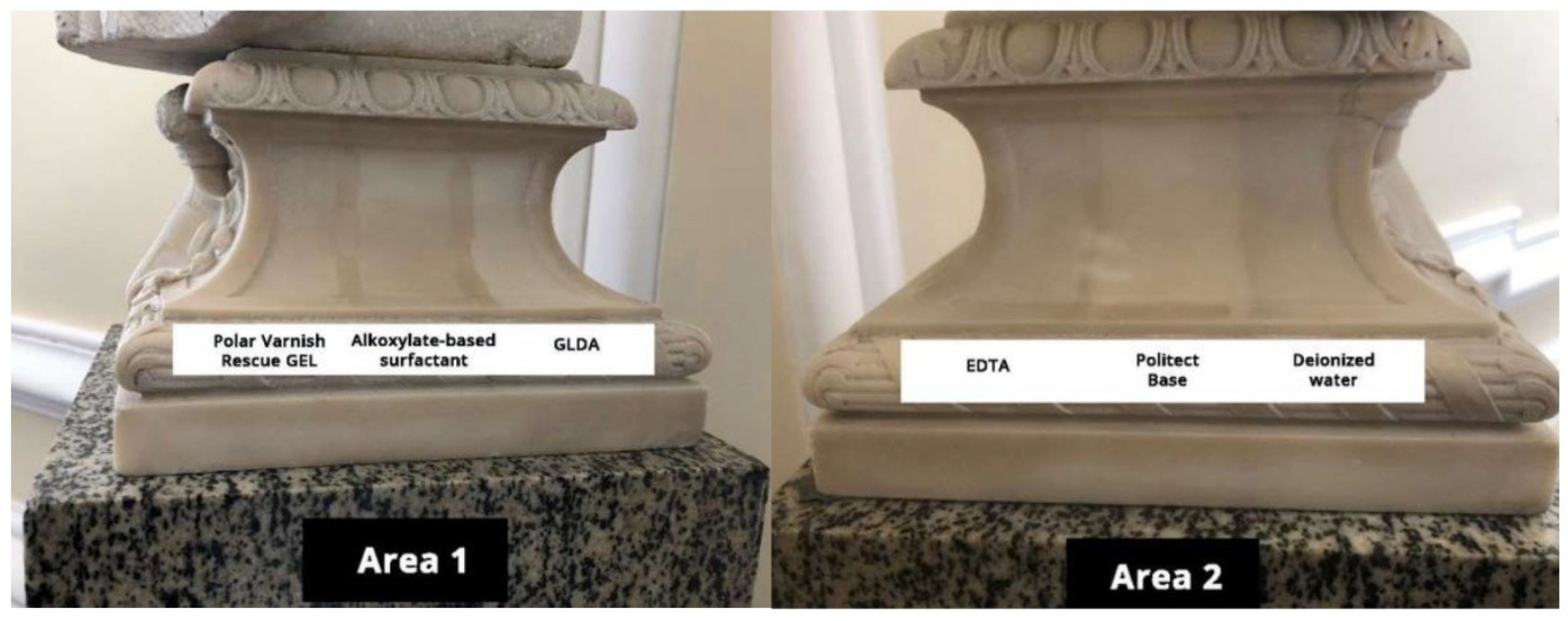
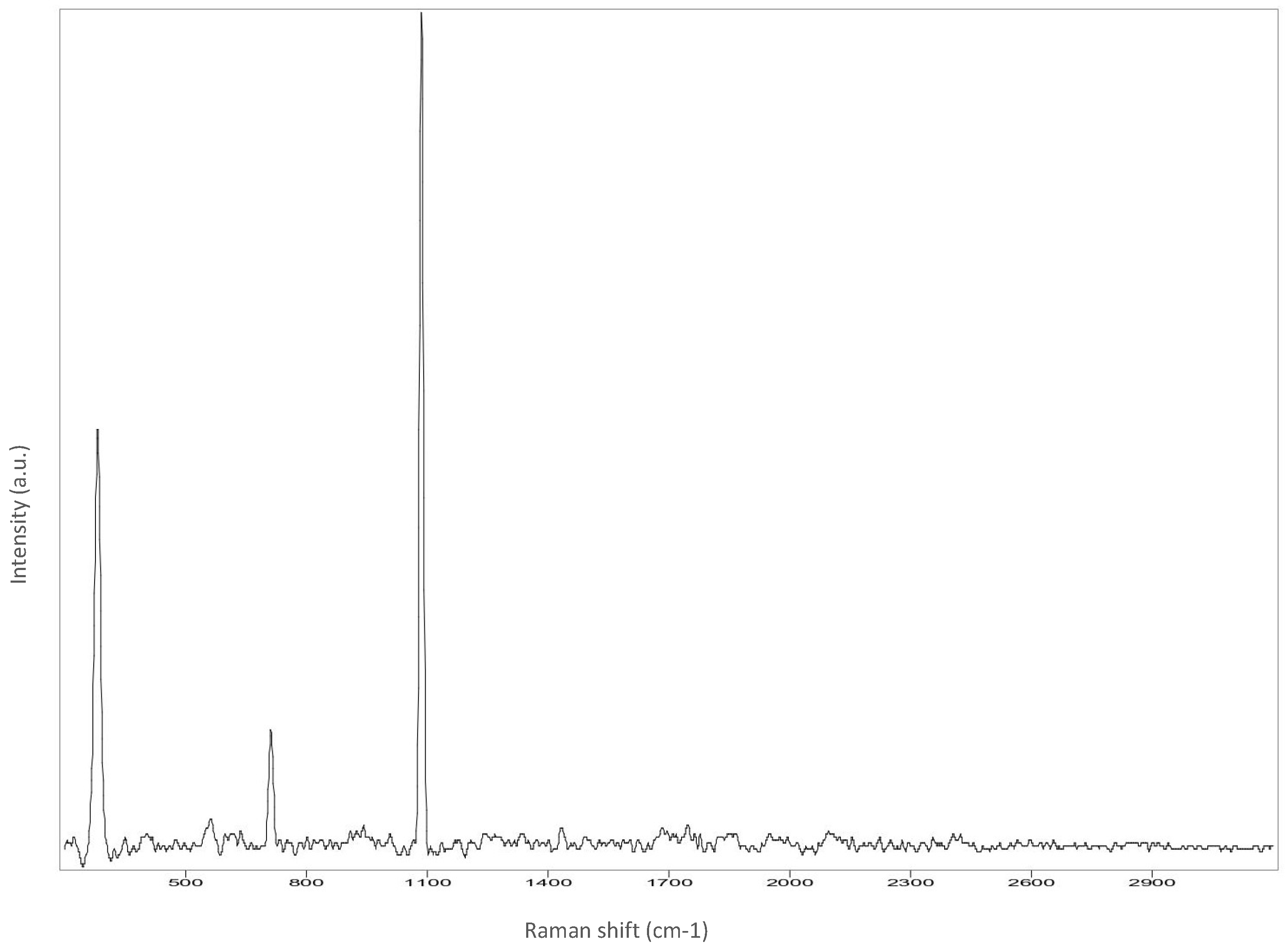
2.2. Cleaning Tests
- Ability to effectively remove the particulate matter that has penetrated the porosity;
- Toxicological and chemical-physical properties in compliance with the principles of green chemistry;
- Selectivity towards the material to be removed to preserve the surface of the artwork;
- Ability to achieve maximum removal of the particulate matter by operating in the least invasive way possible;
- Easy application and ability to control the cleaning method.
Application Method
3. Results
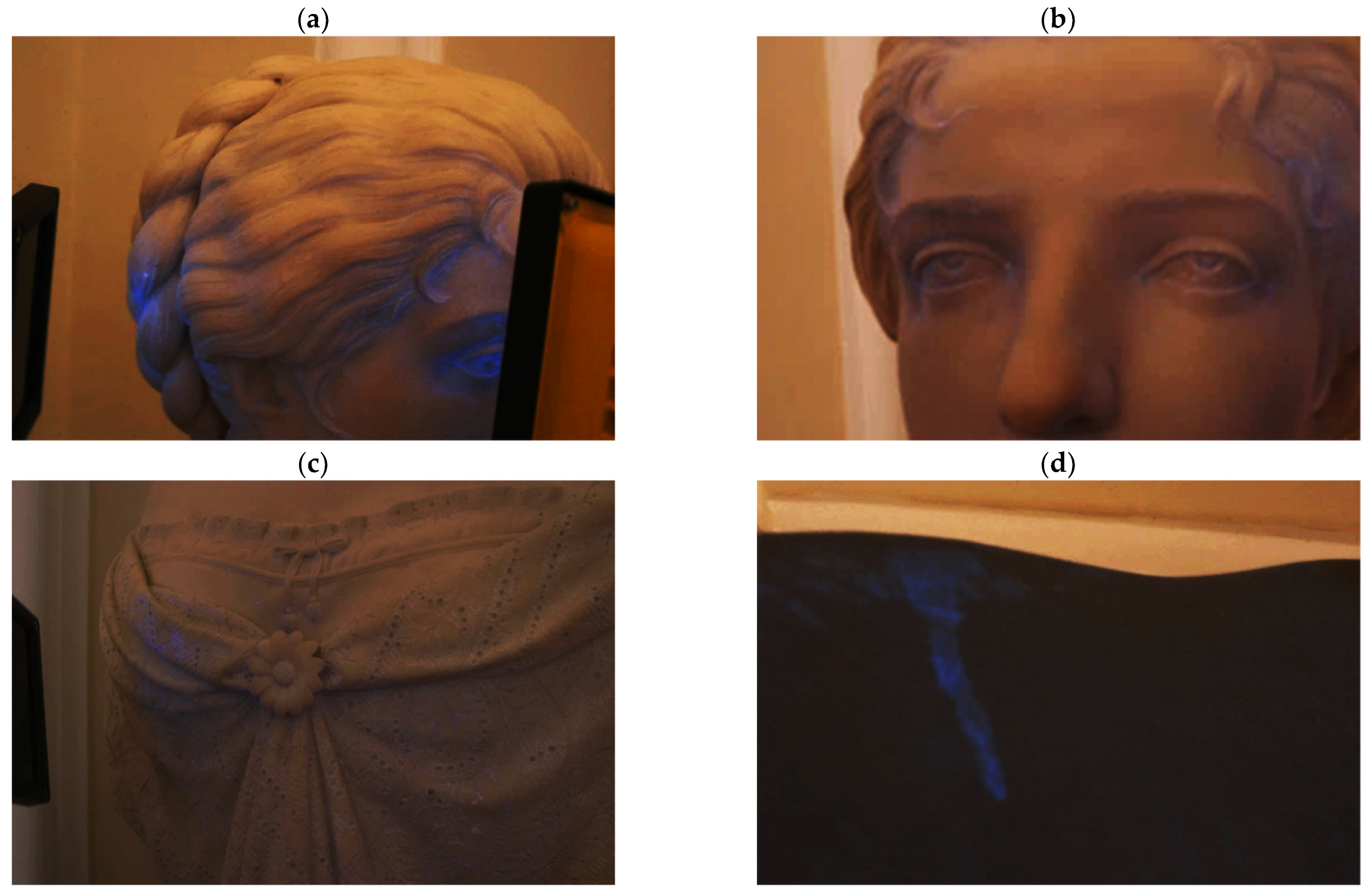
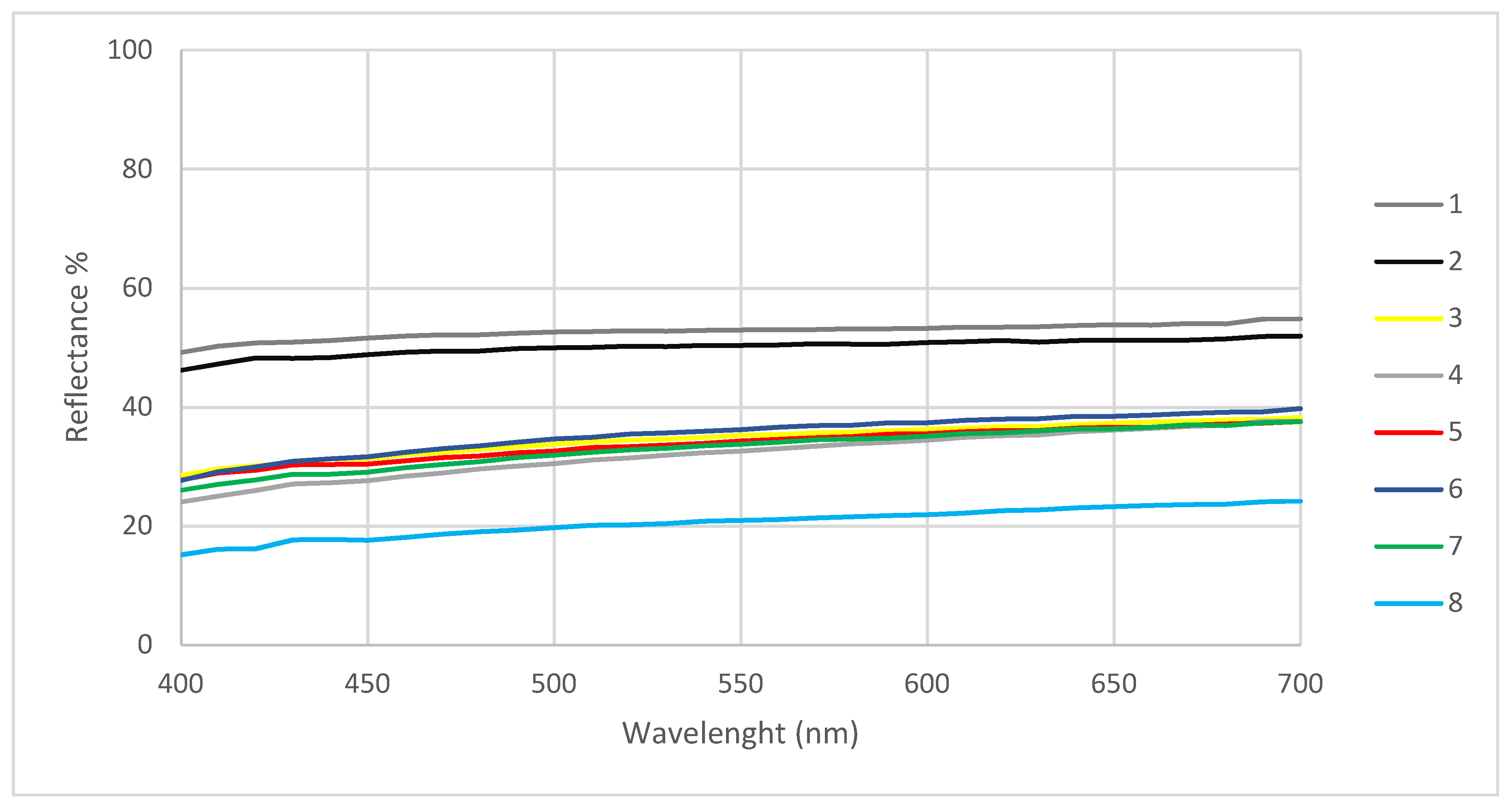
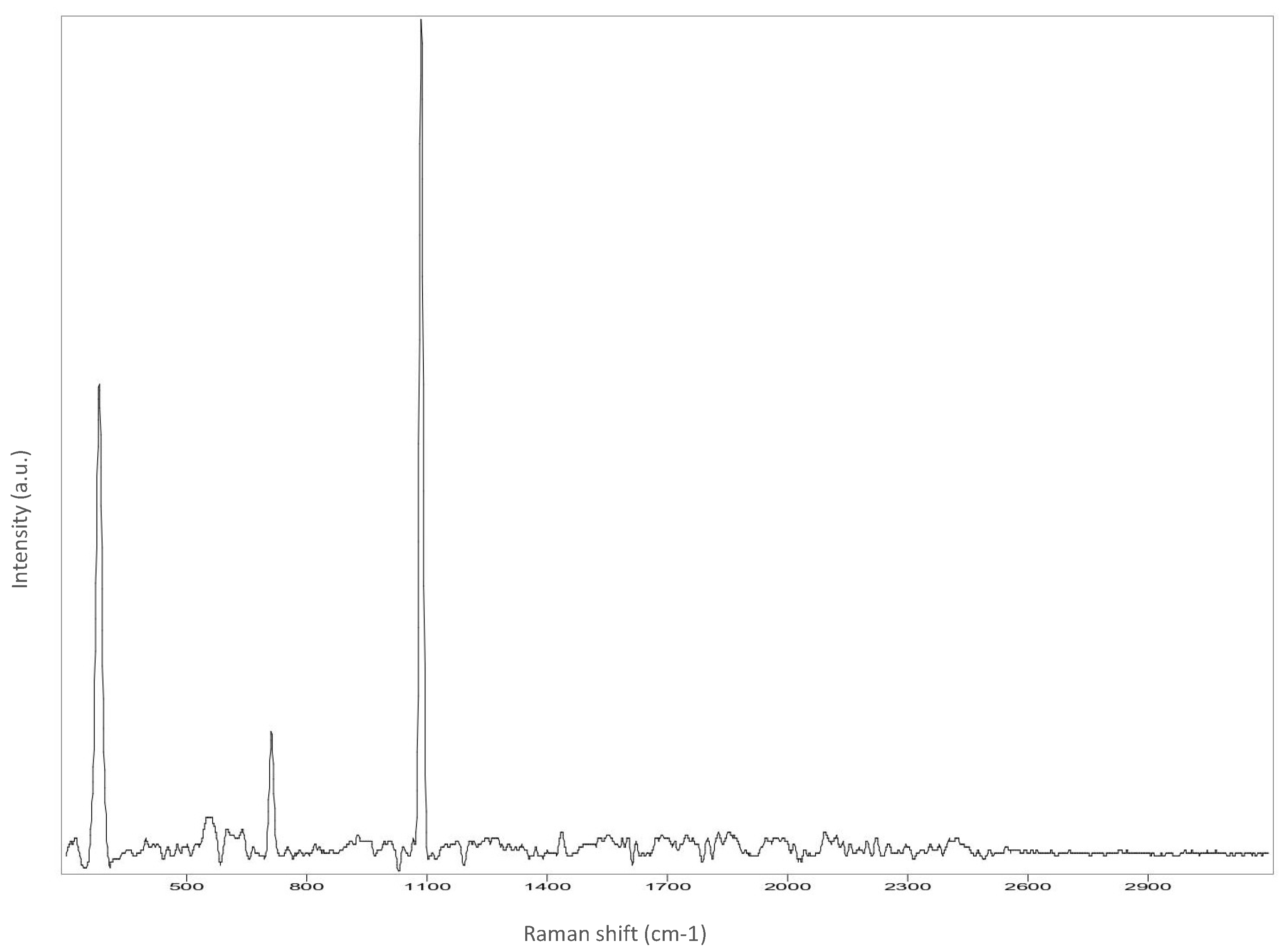
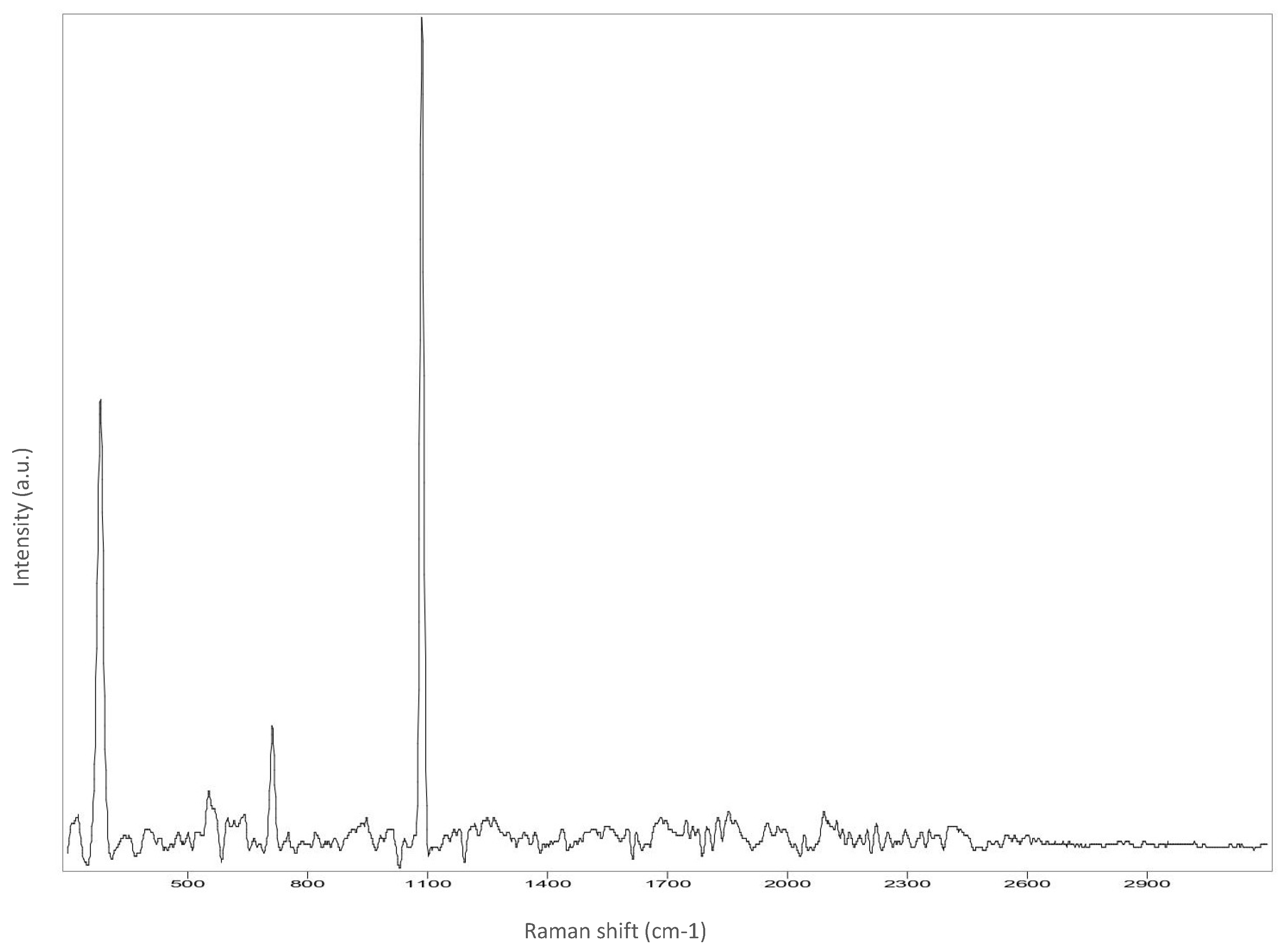
3.1. Non-Invasive Preliminary Analysis
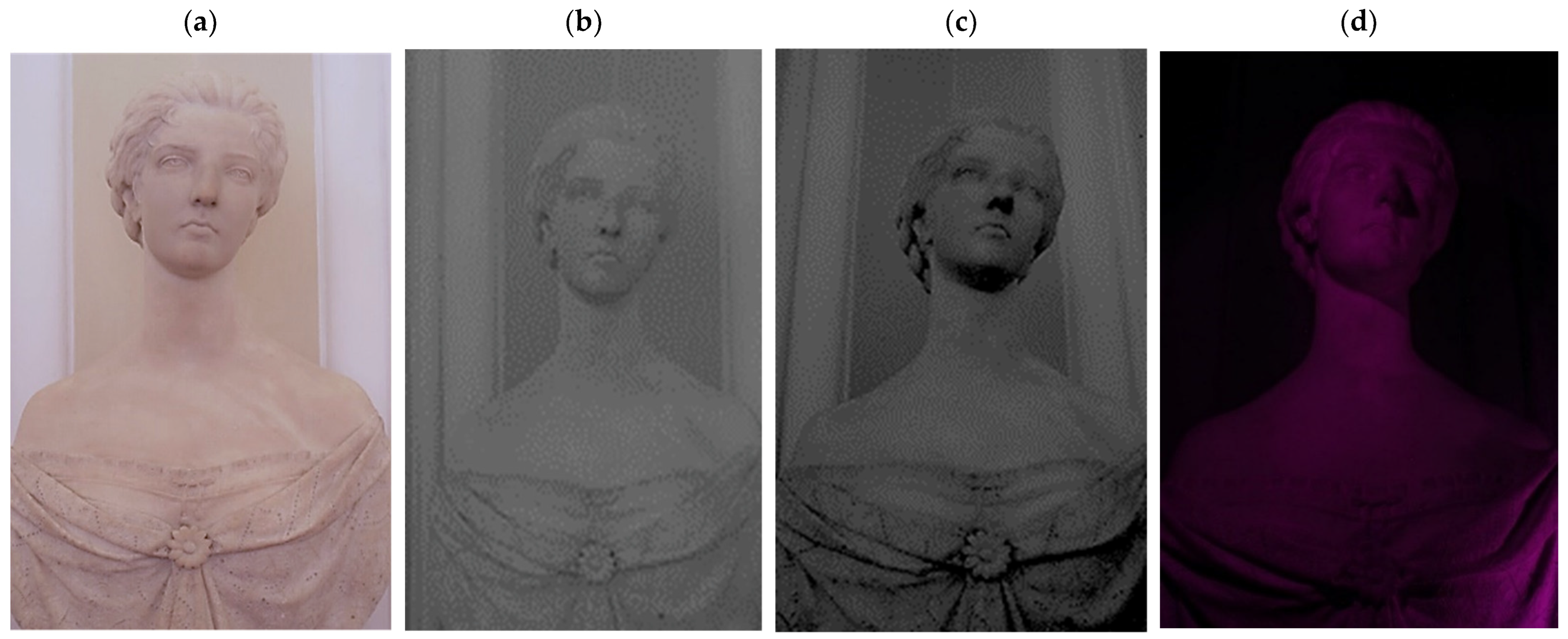
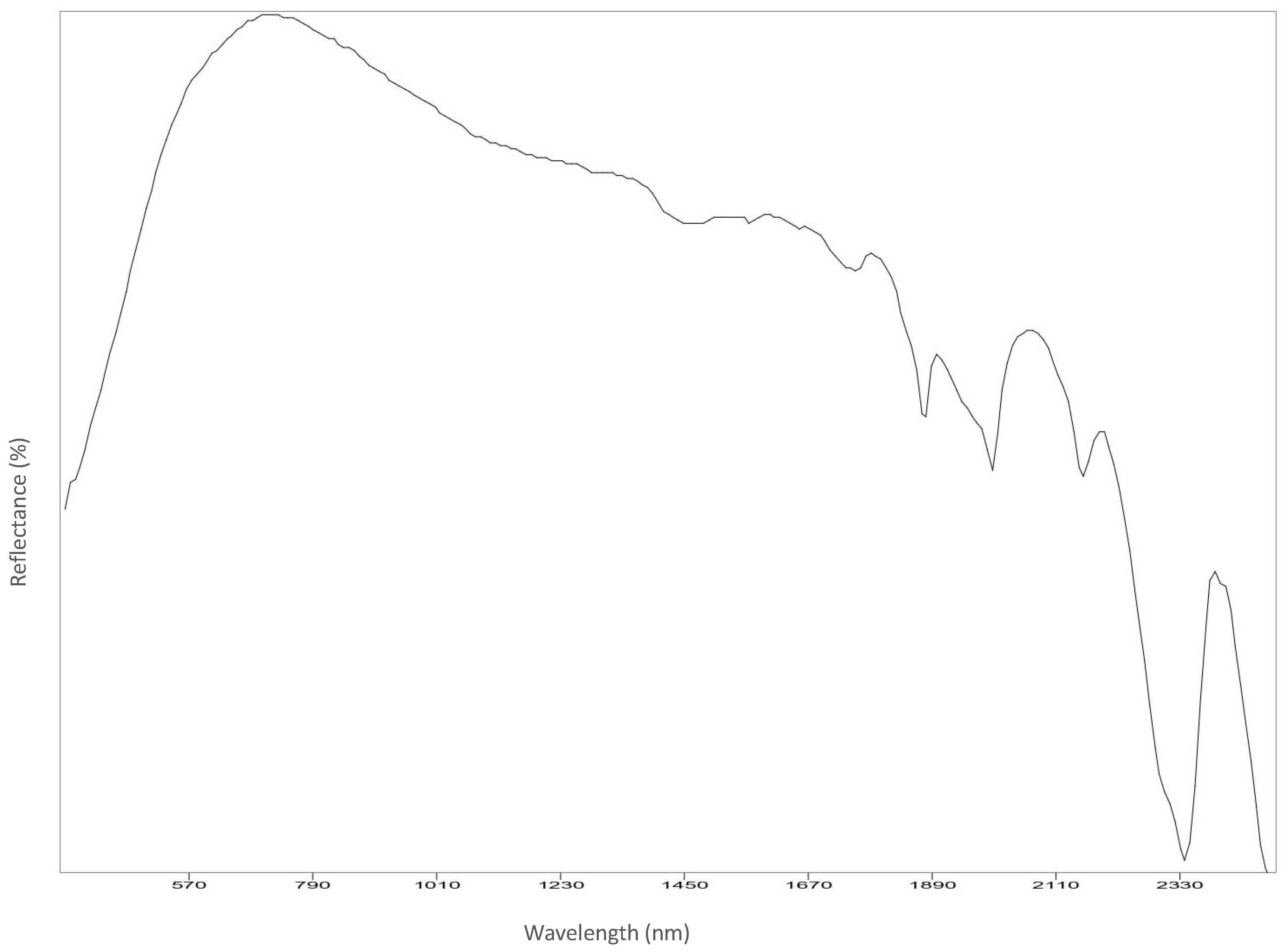
- Areas with a blue induced fluorescence could be related to superficial structural variations from calcium carbonate polymorphs. As Toffolo et al. showed in their work [35], calcium carbonate polymorphs like aragonite and pyrogenic calcite in fact have a visible fluorescence in the blue region (Figure 4a,b). Also, by varying light angles, the analysis showed different fluorescence colors, thus revealing the inorganic nature of the substance;
- A blue induced fluorescence located on the back and front of the sculpture reveals dripping residues of an unidentified product (Figure 4c,d);
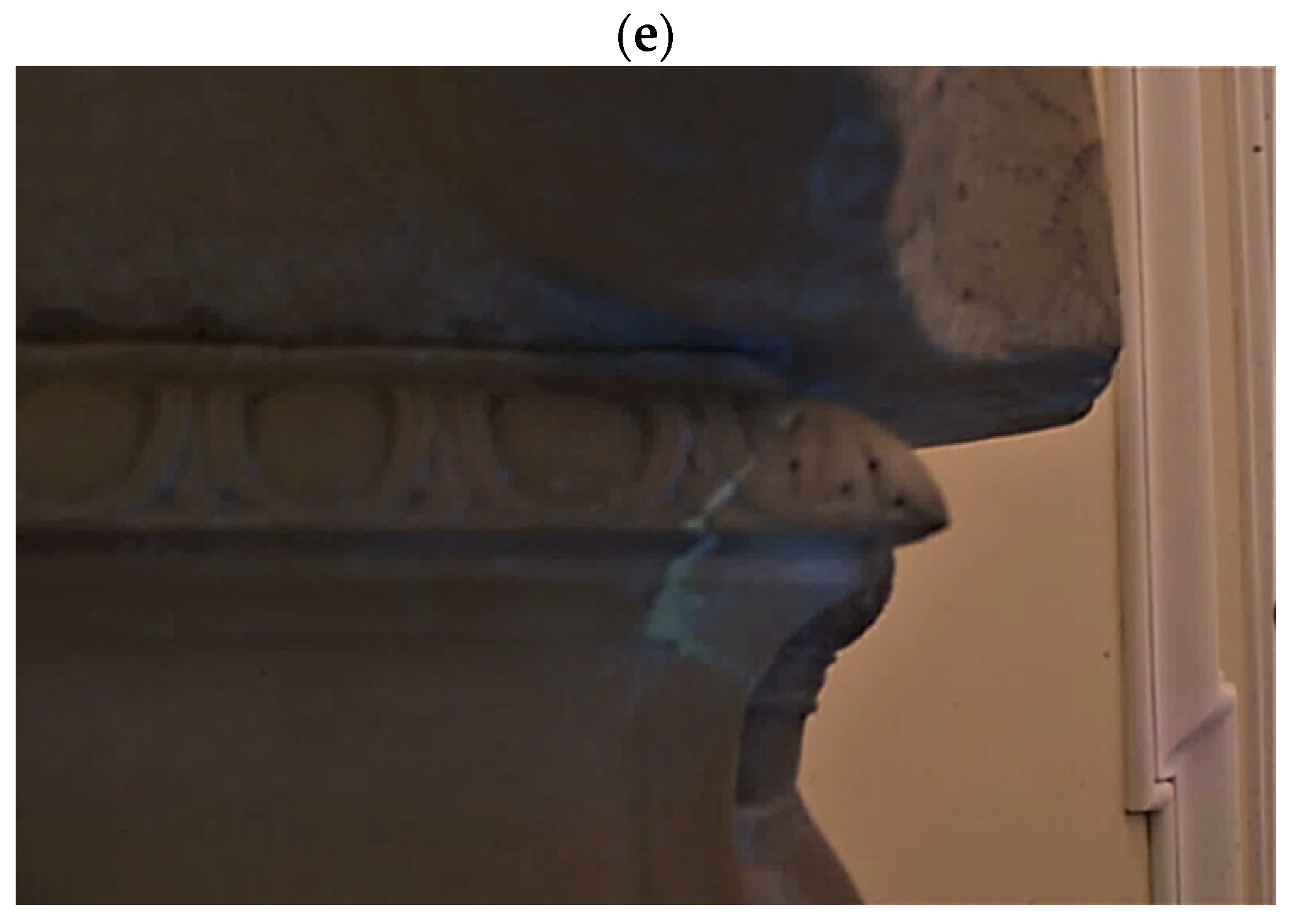
- A yellowish induced fluorescence around the base corner may be related to an organic adhesive used in previous reintegration work (Figure 4e).
3.2. Cleaning Tests
4. Discussion
5. Conclusions
Author Contributions
Funding
Institutional Review Board Statement
Informed Consent Statement
Data Availability Statement
Conflicts of Interest
Appendix A


Appendix B


References
- Maravelaki-Kalaitzaki, P. Black crusts and patinas on Pentelic marble from the Parthenon and Erechtheum (Acropolis, Athens): Characterization and origin. Anal. Chim. Acta 2005, 532, 187–198. [Google Scholar] [CrossRef]
- Bugini, R.; Tabasso, M.L.; Realini, M. Rate of formation of black crusts on marble. A case study. J. Cult. Herit. 2000, 1, 111–116. [Google Scholar] [CrossRef]
- Comite, V.; Pozo-Antonio, J.S.; Cardell, C.; Randazzo, L.; La Russa, M.F.; Fermo, P. A multi-analytical approach for the characterization of black crusts on the facade of an historical cathedral. Microchem. J. 2020, 158, 105121. [Google Scholar] [CrossRef]
- Mitsos, D.; Kantarelou, V.; Palamara, E.; Karydas, A.G.; Zacharias, N.; Gerasopoulos, E. Characterization of black crust on archaeological marble from the Library of Hadrian in Athens and inferences about contributing pollution sources. J. Cult. Herit. 2022, 53, 236–243. [Google Scholar] [CrossRef]
- Bergin, M.H.; Tripathi, S.N.; Jai Devi, J.; Gupta, T.; McKenzie, M.; Rana, K.S.; Shafer, M.M.; Villalobos, A.M.; Schauer, J.J. The discoloration of the Taj Mahal due to particulate carbon and dust deposition. Environ. Sci. Technol. 2015, 49, 808–812. [Google Scholar] [CrossRef]
- Arola, A.; Schuster, G.; Myhre, G.; Kazadzis, S.; Dey, S.; Tripathi, S.N. Inferring absorbing organic carbon content from AERONET data. Atmos. Chem. Phys. 2011, 11, 215–225. [Google Scholar] [CrossRef] [Green Version]
- Funke, A.; Poole, L.; Church, J.; Striegel, M.; Singer, M. The use of medical chelating agents for the removal of iron stains from marble. Objects Specialty Group Postprints 2017, 24, 235–249. [Google Scholar]
- Kamally, H.A. Orange, Yellow, Brownish Stains and Alteration on White Marble at El Montazah in Alexandria, Egypt. Int. J. Archit. Herit. 2021, 15, 1942–1958. [Google Scholar] [CrossRef]
- Sansonetti, A.; Bertasa, M.; Corti, C.; Rampazzi, L.; Monticelli, D.; Scalarone, D.; Sassella, A.; Canevali, C. Optimization of Copper Stain Removal from Marble through the Formation of Cu (II) Complexes in Agar Gels. Gels 2021, 7, 111. [Google Scholar] [CrossRef]
- Kate, K.; Wood, R.K.L. Digital Imaging of Artefacts: Developments in Methods and Aims; Archaeopress Publishing Ltd.: Oxford, UK, 2018. [Google Scholar]
- Campanella, L.; Cardellicchio, F.; Dell’Aglio, E.; Reale, R.; Salvi, A.M. A green approach to clean iron stains from marble surfaces. Herit. Sci. 2022, 10, 79. [Google Scholar] [CrossRef]
- Marvasi, M.; Donnarumma, F.; Frandi, A.; Mastromei, G.; Sterflinger, K.; Tiano, P.; Perito, B. Black microcolonial fungi as deteriogens of two famous marble statues in Florence, Italy. Int. Biodeterior. Biodegrad. 2012, 68, 36–44. [Google Scholar] [CrossRef]
- Santo, A.P.; Cuzman, O.A.; Petrocchi, D.; Pinna, D.; Salvatici, T.; Perito, B. Black on white: Microbial growth darkens the external marble of Florence cathedral. Appl. Sci. 2021, 11, 6163. [Google Scholar] [CrossRef]
- Cuzman, O.A.; Vettori, S.; Fratini, F.; Cantisani, E.; Ciattini, S.; Chelazzi, L.; Ricci, M.; Garzonio, C.A. Alteration of marble stones by red discoloration phenomena. In Science and Art: A Future for Stone: Proceedings of the 13th International Congress on the Deterioration and Conservation of Stone, Paisley, UK, 6–10 September 2016; University of the West of Scotland: Glasgow, UK, 2016; p. 75. [Google Scholar]
- Pinzari, F.; Zotti, M.; De Mico, A.; Calvini, P. Biodegradation of inorganic components in paper documents: Formation of calcium oxalate crystals as a consequence of Aspergillus terreus Thom growth. Int. Biodeterior. Biodegrad. 2010, 64, 499–505. [Google Scholar] [CrossRef]
- Pinna, D.; Bracci, S.; Magrini, D.; Salvadori, B.; Andreotti, A.; Colombini, M.P. Deterioration and discoloration of historical protective treatments on marble. Environ. Sci. Pollut. Res. 2022, 29, 20694–20710. [Google Scholar] [CrossRef] [PubMed]
- Gherardi, F.; Kapridaki, C.; Roveri, M.; Gulotta, D.; Maravelaki, P.N.; Toniolo, L. The deterioration of Apuan white marble in contemporary architectural context. J. Cult. Herit. 2019, 35, 297–306. [Google Scholar] [CrossRef]
- Maxwell, I. Stone Cleaning: For Better or Worse? An Overview. In Stone Cleaning and the Nature, Soiling and Decay Mechanisms of Stone, Proceedings of the International Conference, Edinburgh, UK, 14–16 April 1992; Routledge: London, UK, 1992; pp. 3–49. [Google Scholar]
- Fassina, V. General Criteria for the Cleaning of Stone: Theoretical Aspects and Methodology of Application. In Stone Material in Monuments: Diagnosis and Conservation, Proceedings of the Scuola Universitaria CUM Conservazione dei Monumenti, Heraklion, Greek, 24–30 May 1993; pp. 131–138. Available online: https://www.researchgate.net/profile/Vasco-Fassina-2/publication/345726155_GENERAL_CRITERIA_FOR_THE_CLEANING_OF_STONE_THEORETICAL_ASPECTS_AND_METHODOLOGY_OF_APPLICATION_by_Vasco_Fassina/links/5fabde92299bf18c5b64de40/GENERAL-CRITERIA-FOR-THE-CLEANING-OF-STONE-THEORETICAL-ASPECTS-AND-METHODOLOGY-OF-APPLICATION-by-Vasco-Fassina.pdf (accessed on 20 August 2022).
- Natali, I.; Carretti, E.; Angelova, L.; Baglioni, P.; Weiss, R.G.; Dei, L. Structural and mechanical properties of “peelable” organoaqueous dispersions with partially hydrolyzed poly (vinyl acetate)-borate networks: Applications to cleaning painted surfaces. Langmuir 2011, 27, 13226–13235. [Google Scholar] [CrossRef]
- Zhenova, A. Challenges in the development of new green solvents for polymer dissolution. Polym. Int. 2020, 69, 895–901. [Google Scholar] [CrossRef]
- Andreotti, A.; Colombini, M.P.; De Cruz, A. Er: YAG laser cleaning of a marble Roman urn. J. Inst. Conserv. 2020, 43, 12–24. [Google Scholar] [CrossRef]
- Aldrovandi, A.; Lalli, C.; Lanterna, G.; Matteini, M. Laser cleaning: A study on greyish alteration induced on non-patinated marbles. J. Cult. Herit. 2000, 1, S55–S60. [Google Scholar] [CrossRef]
- Ranalli, G.; Zanardini, E. Biocleaning on Cultural Heritage: New frontiers of microbial biotechnologies. J. Appl. Microbiol. 2021, 131, 583–603. [Google Scholar] [CrossRef]
- Benocci, C. Villa Ludovisi; Istituto Poligrafico e Zecca dello Stato: Roma, Italy, 2010. [Google Scholar]
- Broggi, L. Le Residenze di s. M. La Regina Madre d’Italia, Margherita di Savoia; Istituto d’Arti Grafiche: Bergamo, Italy, 1909; ISBN 1910 480. [Google Scholar]
- Brunori, V. Art evening at palazzo Margherita. In A Walk through the History and Art Collection of the Embassy of the United States of America in Rome; Gangemi editore: Roma, Italy, 2006; ISBN 9788849210279. [Google Scholar]
- Marchi, C. Palazzo Margherita: The Embassy of the United States of America in Rome; De Luca: Roma, Italy, 1980. [Google Scholar]
- Schiavo, A. Villa Ludovisi and Palazzo Margherita; Roma amor per conto della Banca Nazionale del Lavoro: Roma, Italy, 1981. [Google Scholar]
- Macchia, A.; Biribicchi, C.; Carnazza, P.; Montorsi, S.; Sangiorgi, N.; Demasi, G.; Prestileo, F.; Cerafogli, E.; Colasanti, I.A.; Aureli, H.; et al. Multi-Analytical Investigation of the Oil Painting “Il Venditore di Cerini” by Antonio Mancini and Definition of the Best Green Cleaning Treatment. Sustainability 2022, 14, 3972. [Google Scholar] [CrossRef]
- Gervais, C.; Grissom, C.A.; Little, N.; Wachowiak, M.J. Cleaning marble with ammonium citrate. Stud. Conserv. 2010, 55, 164–176. [Google Scholar] [CrossRef]
- Giraud, T.; Gomez, A.; Lemoine, S.; Pelé-Meziani, C.; Raimon, A.; Guilminot, E. Use of gels for the cleaning of archaeological metals. Case study of silver-plated copper alloy coins. J. Cult. Herit. 2021, 52, 73–83. [Google Scholar] [CrossRef]
- Macchia, A.; Colasanti, I.A.; Rivaroli, L.; Favero, G.; de Caro, T.; Pantoja Munoz, L.; Campanella, L.; La Russa, M.F. Natural based products for cleaning copper and copper alloys artefacts. Nat. Prod. Res. 2021. [Google Scholar] [CrossRef] [PubMed]
- Delegou, E.T.; Avdelidis, N.P.; Karaviti, E.; Moropoulou, A. NDT&E techniques and SEM-EDS for the assessment of cleaning interventions on Pentelic marble surfaces. X-ray Spectrom. Int. J. 2008, 37, 435–443. [Google Scholar]
- Toffolo, M.B.; Ricci, G.; Caneve, L.; Kaplan-Ashiri, I. Luminescence reveals variations in local structural order of calcium carbonate polymorphs formed by different mechanisms. Sci. Rep. 2019, 9, 16170. [Google Scholar] [CrossRef] [Green Version]
- Vandenabeele, P.; Bodé, S.; Alonso, A.; Moens, L. Raman spectroscopic analysis of the Maya wall paintings in Ek’Balam, Mexico. Spectrochim. Acta Part A Mol. Biomol. Spectrosc. 2005, 61, 2349–2356. [Google Scholar] [CrossRef]
- Bishop, J.L.; King, S.J.; Lane, M.D.; Brown, A.J.; Lafuente, B.; Hiroi, T.; Roberts, S.; Swayze, G.A.; Lin, J.-F.; Sánchez Román, M. Spectral properties of anhydrous carbonates and nitrates. Earth Space Sci. 2021, 8, e2021EA001844. [Google Scholar] [CrossRef]
- Morbidelli, L. Le Rocce e i Loro Costituenti. Scienze e Lettere; Bardi Editore: Roma, Italy, 2003; ISBN 88-88620-20-6. [Google Scholar]
- Gunasekaran, S.; Anbalagan, G.; Pandi, S. Raman and infrared spectra of carbonates of calcite structure. J. Raman Spectrosc. 2006, 37, 892–899. [Google Scholar] [CrossRef]
- Monte, M. Oxalate film formation on marble specimens caused by fungus. J. Cult. Herit. 2003, 4, 255–258. [Google Scholar] [CrossRef]
- Rampazzi, L.; Andreotti, A.; Bonaduce, I.; Colombini, M.P.; Colombo, C.; Toniolo, L. Analytical investigation of calcium oxalate films on marble monuments. Talanta 2004, 63, 967–977. [Google Scholar] [CrossRef] [PubMed]
- Anastas, P.T.; Warner, J.C. Green chemistry. Frontiers 1998, 640, 1998. [Google Scholar]
- Mahmoud, M.A.; Nasr-El-Din, H.A.; De Wolf, C.A.; Alex, A.K. Effect of Lithology on the Flow of Chelating Agents in Porous Media during Matrix Acid Treatments. In Proceedings of the SPE Production and Operations Symposium, Oklahoma City, OK, USA, 13–23 March 2011; OnePetro: Washington, DC, USA, 2011. [Google Scholar]
- Adenuga, O.O.; Nasr-El-Din, H.A.; Sayed, M.A.I. Reactions of Simple Organic Acids and Chelating Agents with Dolomite. In Proceedings of the SPE Production and Operations Symposium, Oklahoma City, OK, USA, 23–26 March 2013; OnePetro: Washington, DC, USA, 2013. [Google Scholar]
- Rabie, A.I. Reaction of Calcite and Dolomite with In-Situ Gelled Acids, Organic Acids, and Environmentally Friendly Chelating Agent (GLDA). Ph.D. Thesis, Texas A&M University, College Station, TX, USA, 2012. [Google Scholar]
- Fredd, C.N.; Fogler, H.S. The influence of chelating agents on the kinetics of calcite dissolution. J. Colloid Interface Sci. 1998, 204, 187–197. [Google Scholar] [CrossRef] [PubMed]
- Oviedo, C.; Rodríguez, J. EDTA: The chelating agent under environmental scrutiny. Quim. Nova 2003, 26, 901–905. [Google Scholar] [CrossRef]
- Available online: https://echa.europa.eu/it/substance-information/-/substanceinfo/100.112.462 (accessed on 20 August 2022).


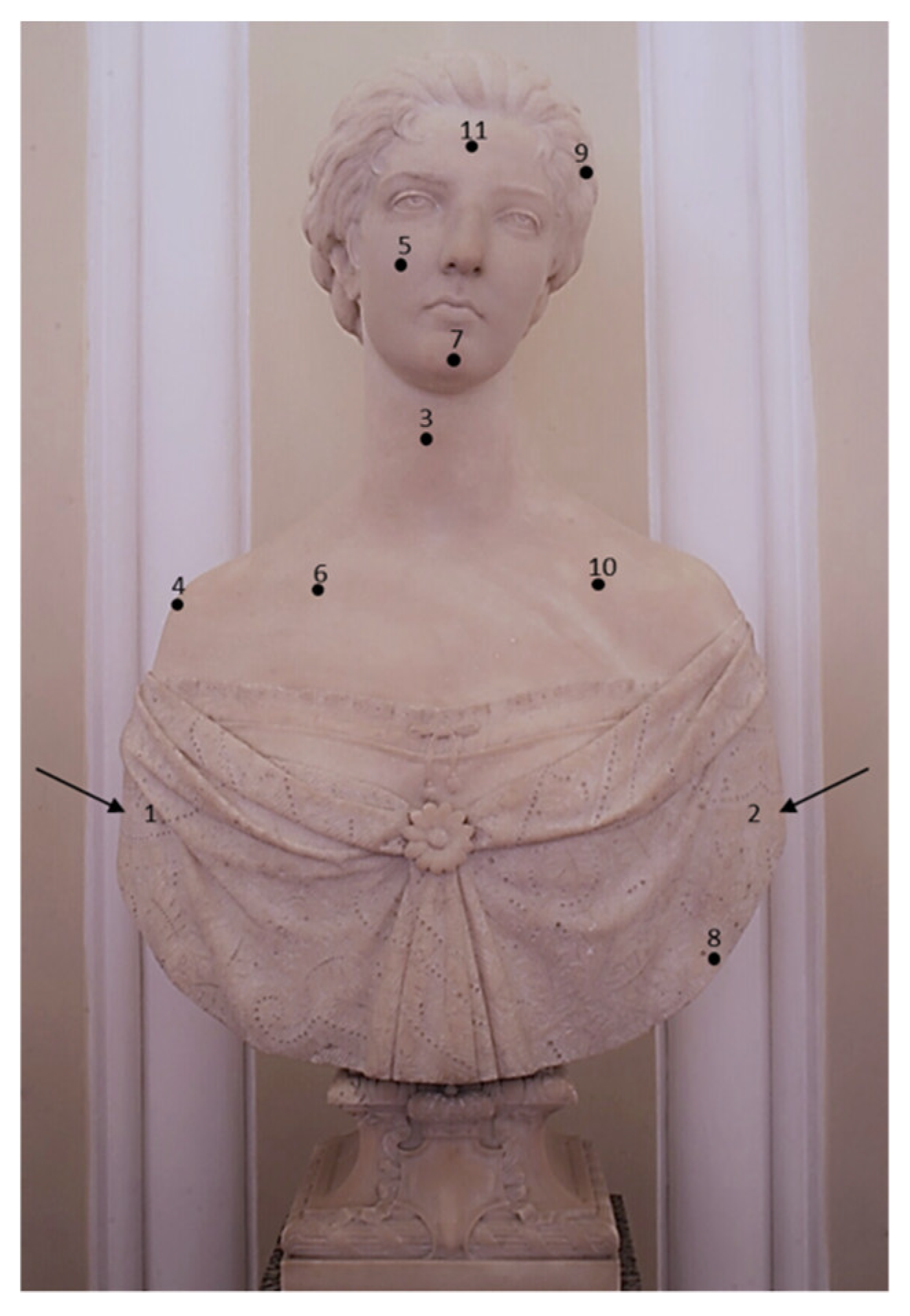
| N. | Statue Areas | Analytical Techniques | ||
|---|---|---|---|---|
| Colorimeter | Raman Spectroscopy | Reflectance Spectroscopy | ||
| 1 | Back left | X | X | X |
| 2 | Back right | X | X | X |
| 3 | Neck | X | X | |
| 4 | Left shoulder | X | X | |
| 5 | Left cheek | X | X | X |
| 6 | Left chest | X | X | |
| 7 | Chin | X | X | X |
| 8 | Dress | X | X | X |
| 9 | Hair | X | ||
| 10 | Right chest | X | ||
| 11 | Forehead | X | ||
| ID | Product | Manufacturer |
|---|---|---|
| 1 | Polar Varnish Rescue GEL | YOCOCU APS |
| 2 | Alkoxyde based surfactant | Sigma Aldritch |
| 3 | GLDA (5% in H2O w/v) | Nouryon |
| 4 | Disodium EDTA (5% in H2O w/v) | Sigma Aldritch |
| 5 | Politect base® | Politect |
| 6 | Deionized water as reference | - |
| Point | 1 | 2 | 3 | 4 | 5 | 6 | 7 | 8 |
|---|---|---|---|---|---|---|---|---|
| Description | Left back | Right back | Neck | Left shoulder | Left cheek | Left chest | Chin | Dress |
| C.S. | #C1C0BE | #BDBCB9 | #A39F97 | #A09A8E | #A29D95 | #A6A298 | #A19C92 | #837D74 |
| L* | 77.84 | 76.3 | 65.81 | 63.83 | 65.11 | 66.6 | 64.68 | 52.83 |
| a* | −0.05 | −0.03 | 0.33 | 1.07 | 0.71 | 0.23 | 0.6 | 0.75 |
| b* | 1.38 | 1.63 | 4.77 | 6.88 | 4.85 | 5.72 | 6.06 | 5.85 |
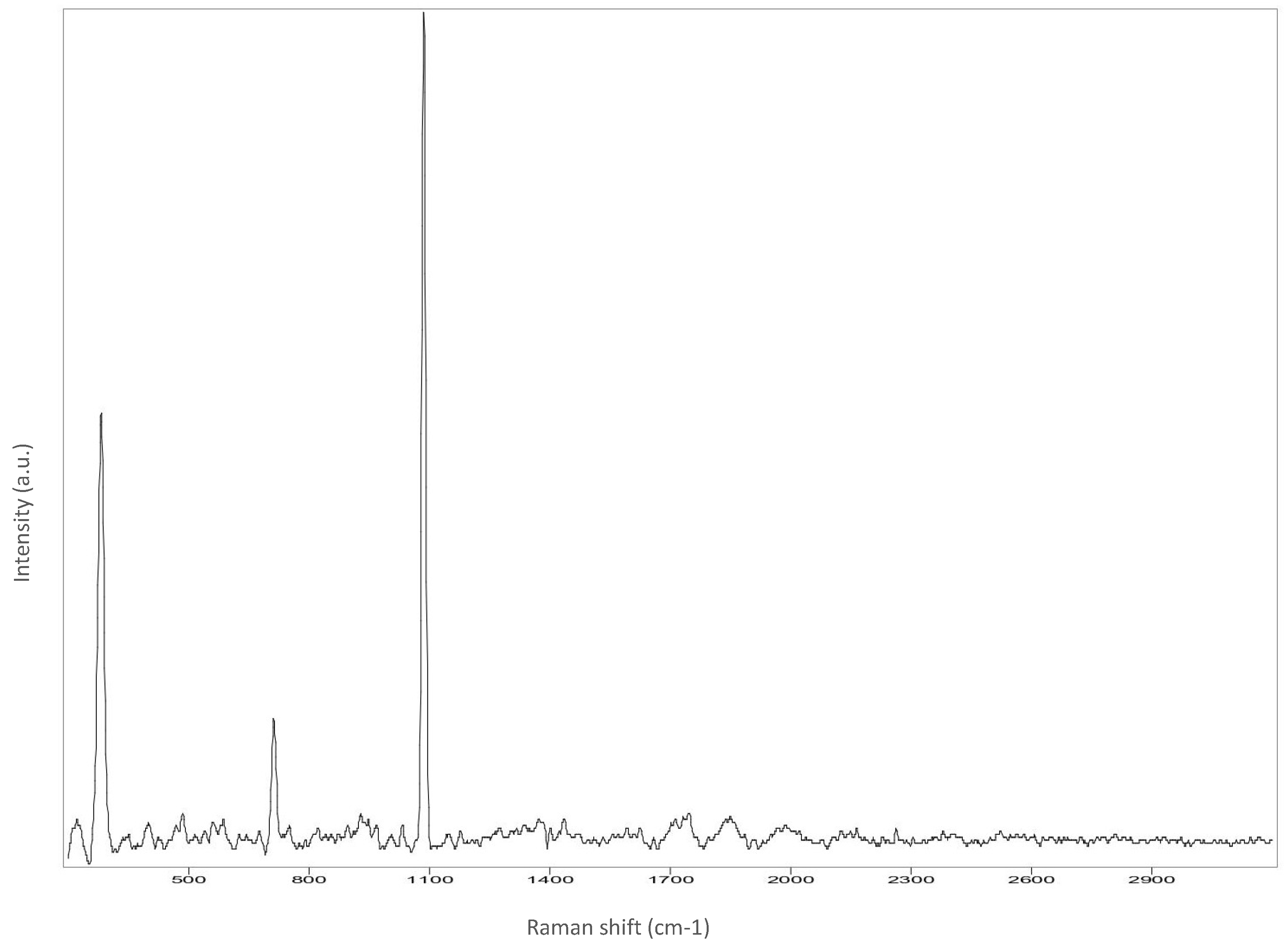
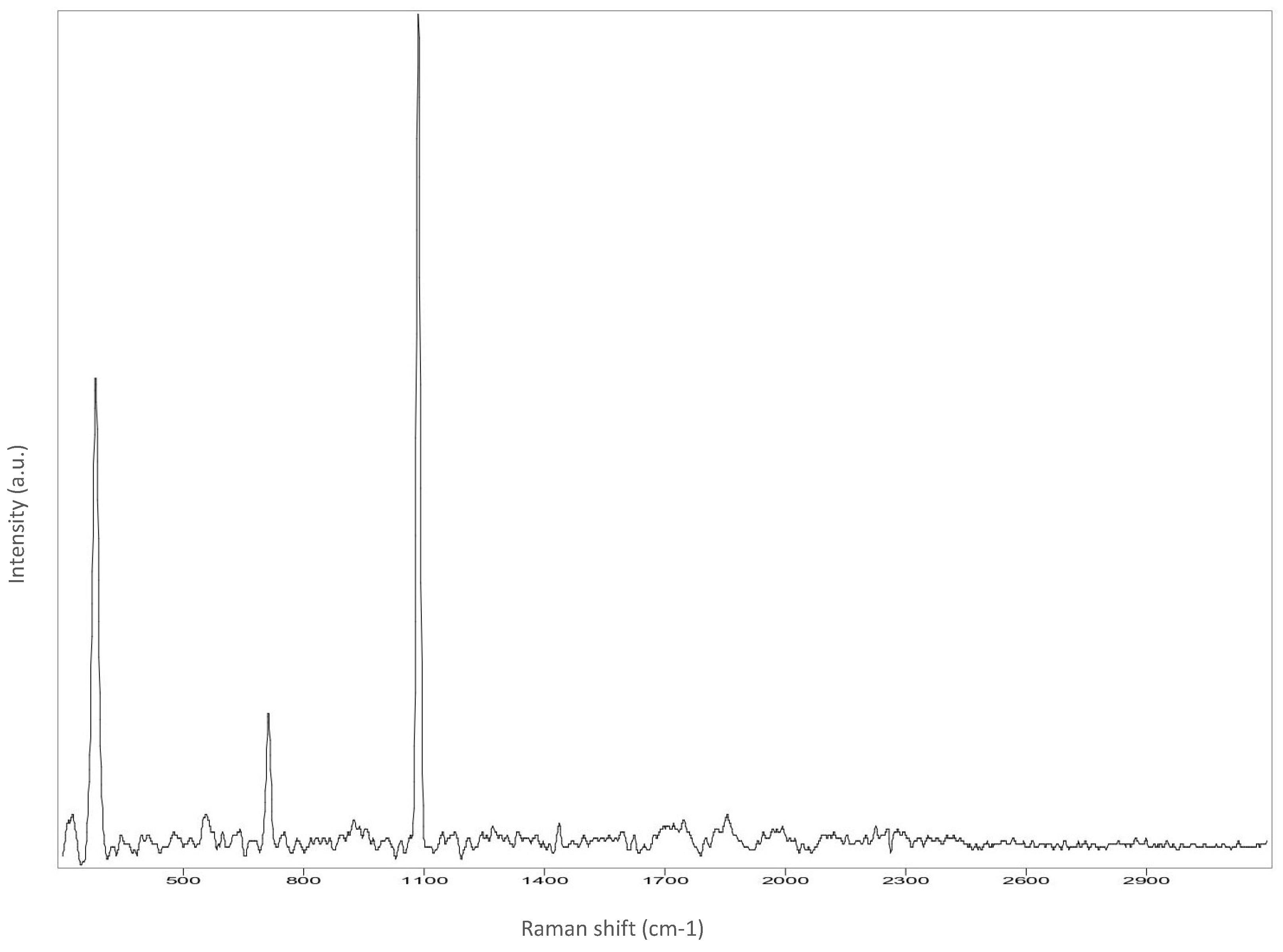
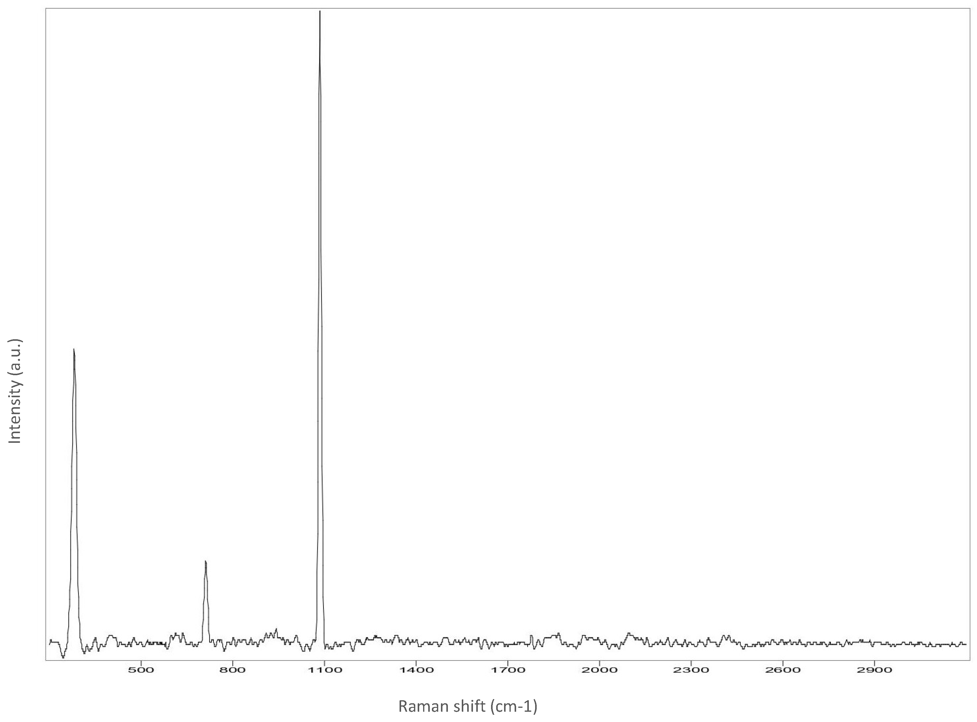
| Statue Areas | Sample Bands (cm−1) |
|---|---|
| Left back | 280; 711; 1085 |
| Right back | 282; 710; 1085 |
| Left cheek | 281; 712; 1085 |
| Chin | 282; 713; 1085 |
| Dress | 281; 712; 1085 |
| Hair | 281; 712; 1085 |
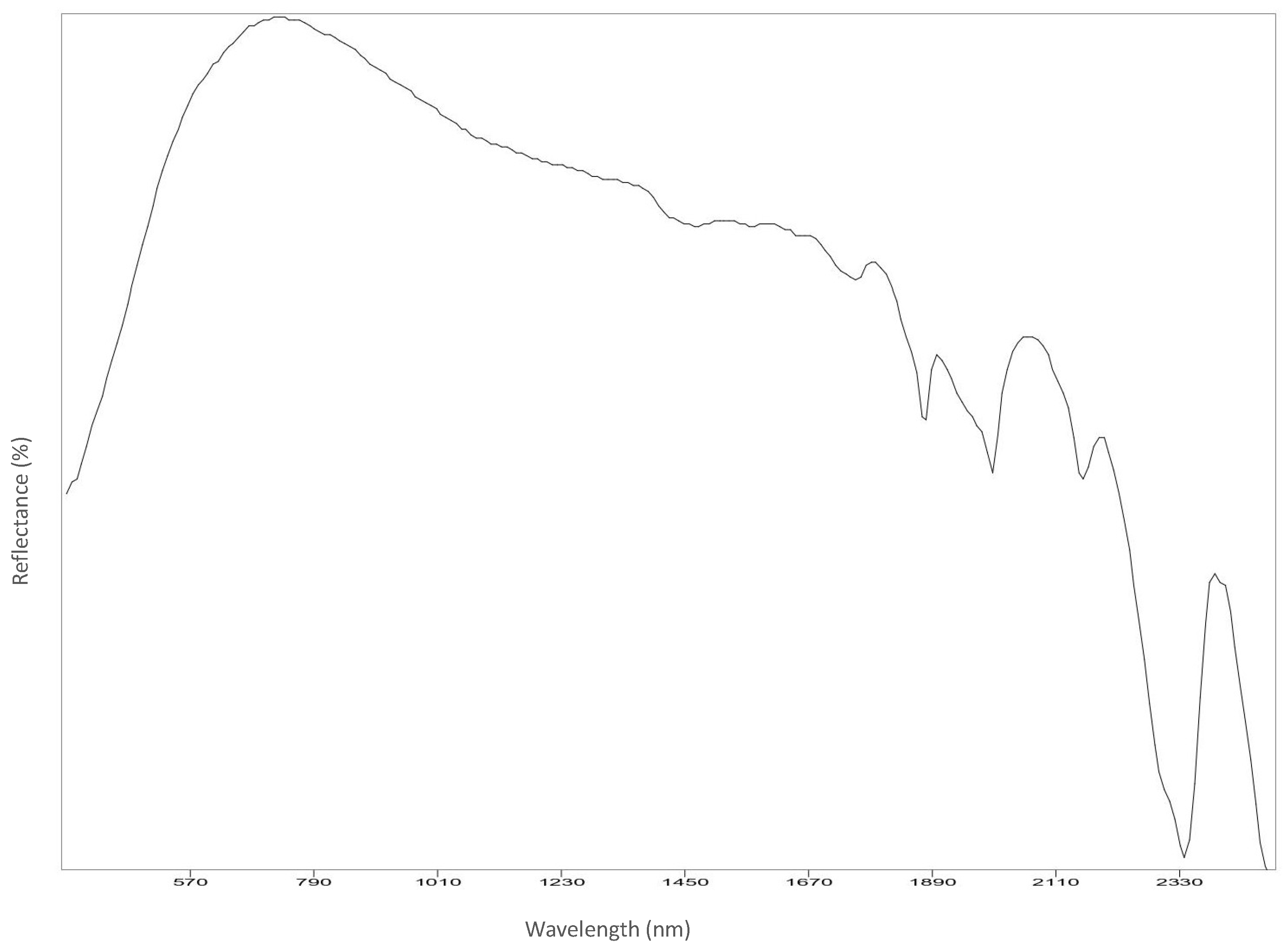
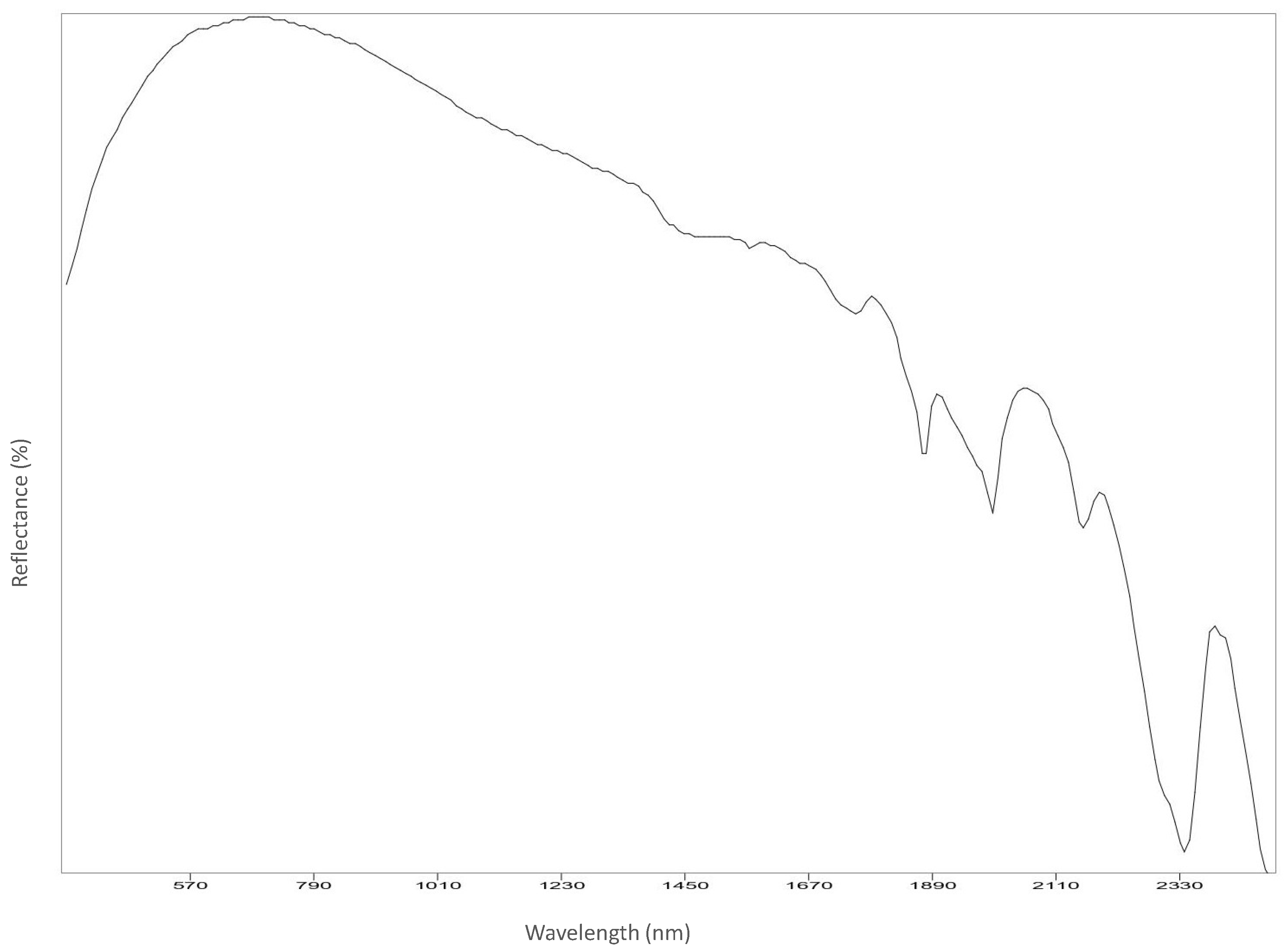
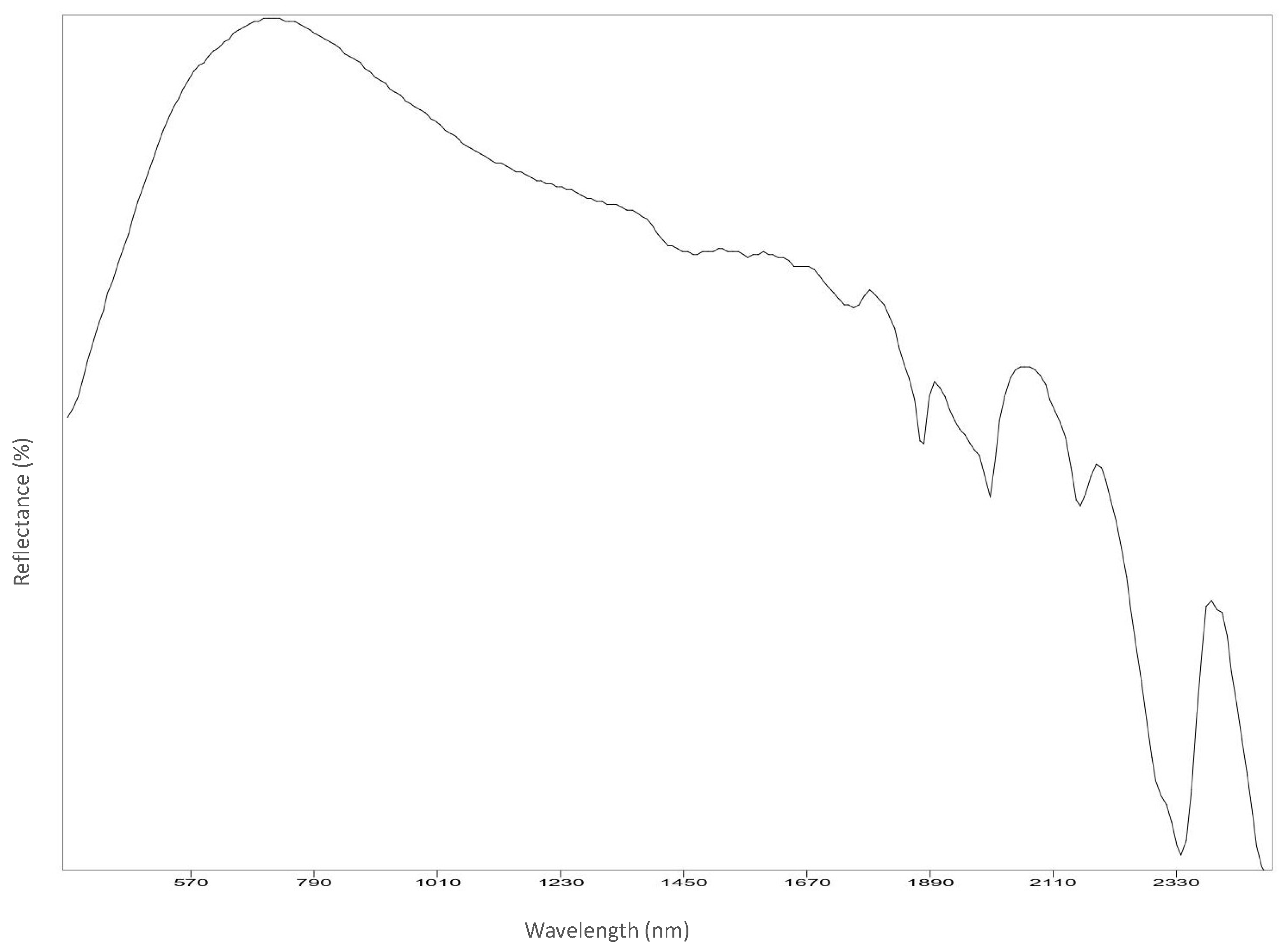
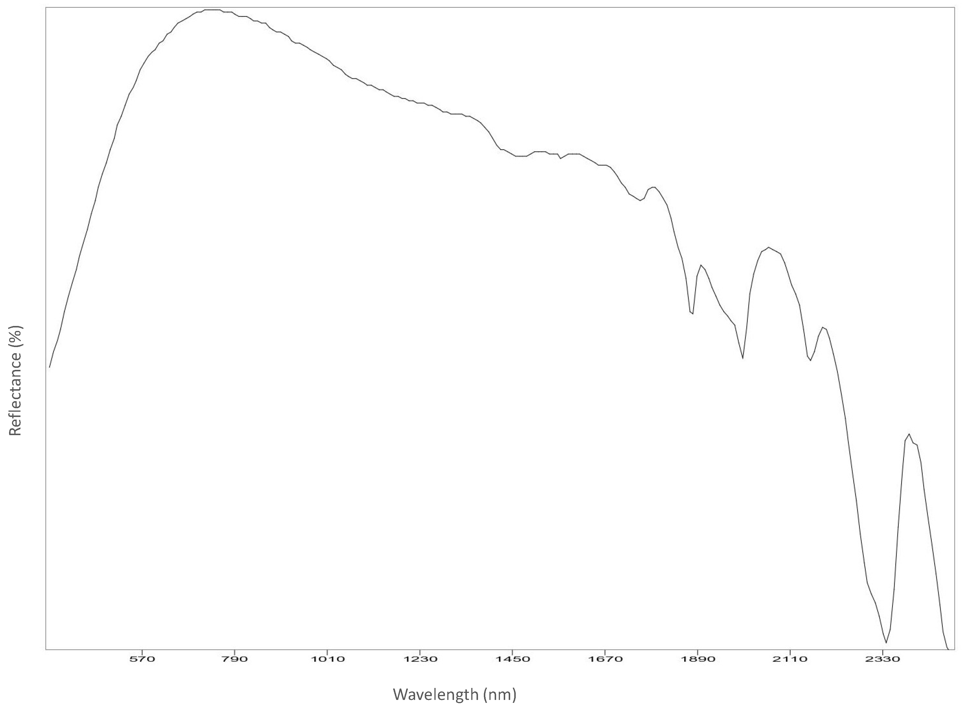
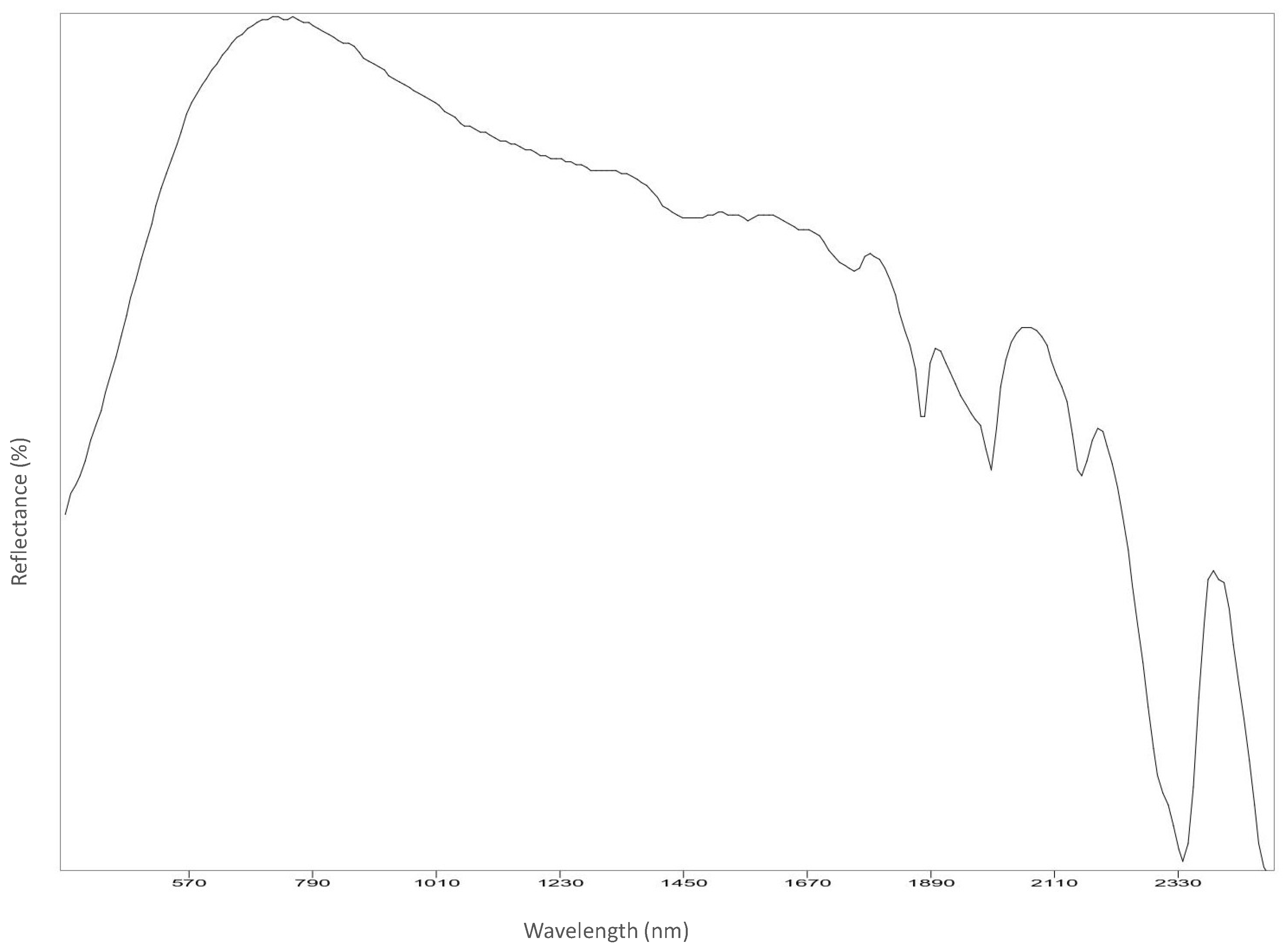
| Statue Areas | Sample Bands (nm) |
|---|---|
| Left back | 1449; 1565; 1650; 1757; 1875; 1994; 2153; 2337; 2402 |
| Right back | 1453; 1565; 1650; 1755; 1875; 1994; 2157; 2337; 2400 |
| neck | 1475; 1567; 1650; 1757; 1875; 1996; 2157; 2335; 2400 |
| Left shoulder | 1459; 1565; 1648; 1757; 1875; 1994; 2155; 2337; 2400 |
| Left cheek | 1457; 1565; 1648; 1757; 1875; 1994; 2155; 2337; 2402 |
| Left chest | 1461; 1565; 1648; 1757; 1875; 1996; 2155; 2337; 2398 |
| Chin | 1465; 1567; 1650; 1757; 1875; 1996; 2155; 2337; 2400 |
| Dress | 1455; 1565; 1650; 1757; 1875; 1996; 2157; 2339; 2396 |
| Right chest | 1451; 1567; 1656; 1757; 1875; 1948; 1994; 2157; 2339; 2402 |
| Forehead | 1449; 1567; 1654; 1759; 1875; 1994; 2157; 2337; 2404 |
| Before Cleaning | After Cleaning | |
|---|---|---|
| 1-Polar Varnish Rescue GEL |  |  |
| 2-Alkoxylated-based surfactant |  |  |
| 3-GLDA |  |  |
| 4-EDTA |  |  |
| 5-Politect base |  |  |
| Polar Varnish Rescue | Alkoxylated-Based Surfactant | GLDA | EDTA | Politect® Base | ||||||
|---|---|---|---|---|---|---|---|---|---|---|
| Before | After | Before | After | Before | After | Before | After | Before | After | |
| L* | 78.89 | 79.21 | 77.20 | 77.45 | 78.53 | 83.88 | 76.33 | 78.44 | 77.10 | 84.1 |
| a* | 1.04 | 1.11 | 1.15 | 1.07 | 1.02 | −0.04 | 1.11 | −0.07 | 1.02 | 0.83 |
| b* | 2.90 | 2.61 | 2.60 | 2.71 | 2.40 | 0.81 | 2.44 | 3.63 | 2.63 | 1.63 |
Publisher’s Note: MDPI stays neutral with regard to jurisdictional claims in published maps and institutional affiliations. |
© 2022 by the authors. Licensee MDPI, Basel, Switzerland. This article is an open access article distributed under the terms and conditions of the Creative Commons Attribution (CC BY) license (https://creativecommons.org/licenses/by/4.0/).
Share and Cite
Macchia, A.; Cerafogli, E.; Rivaroli, L.; Colasanti, I.A.; Aureli, H.; Biribicchi, C.; Brunori, V. Marble Chromatic Alteration Study Using Non-Invasive Analytical Techniques and Evaluation of the Most Suitable Cleaning Treatment: The Case of a Bust Representing Queen Margherita di Savoia at the U.S. Embassy in Rome. Analytica 2022, 3, 406-429. https://doi.org/10.3390/analytica3040028
Macchia A, Cerafogli E, Rivaroli L, Colasanti IA, Aureli H, Biribicchi C, Brunori V. Marble Chromatic Alteration Study Using Non-Invasive Analytical Techniques and Evaluation of the Most Suitable Cleaning Treatment: The Case of a Bust Representing Queen Margherita di Savoia at the U.S. Embassy in Rome. Analytica. 2022; 3(4):406-429. https://doi.org/10.3390/analytica3040028
Chicago/Turabian StyleMacchia, Andrea, Eleonora Cerafogli, Laura Rivaroli, Irene Angela Colasanti, Hélène Aureli, Chiara Biribicchi, and Valeria Brunori. 2022. "Marble Chromatic Alteration Study Using Non-Invasive Analytical Techniques and Evaluation of the Most Suitable Cleaning Treatment: The Case of a Bust Representing Queen Margherita di Savoia at the U.S. Embassy in Rome" Analytica 3, no. 4: 406-429. https://doi.org/10.3390/analytica3040028