Article

An Exploratory Study of Early Immune Response Markers for Pembrolizumab in Urothelial Tract Cancer

by
Dag Rune Stormoen
1,2,3,4,*,†,
Lise Høj Omland
1,†,
Kent William Mouw
2,3,5,
Zoltan Szallasi
6,7,8,9,
Sisse Rye Ostrowski
4,10,
Susanne Dam Nielsen
4,11 and
Helle Pappot
1,4
1
Department of Oncology, Rigshospitalet, Blegdamsvej 9, 2200 Copenhagen, Denmark
2
Harvard Medical School, Boston, MA 02115, USA
3
Department of Radiation Oncology, Dana-Farber Cancer Institute, Boston, MA 02115, USA
4
Department of Clinical Medicine, Faculty of Health and Medical Sciences, University of Copenhagen, 2200 Copenhagen, Denmark
5
Department of Radiation Oncology, Brigham & Women’s Hospital, Boston, MA 02115, USA
6
Danish Cancer Society Research Center, 2200 Copenhagen, Denmark
7
Computational Health Informatics Program, Boston Children’s Hospital, Boston, MA 02115, USA
8
SE NAP, Brain Metastasis Research Group, 2nd Department of Pathology, Semmelweis University, 1085 Budapest, Hungary
9
Department of Bioinformatics, Semmelweis University, 1085 Budapest, Hungary
10
Department of Clinical Immunology, Rigshospitalet, 2200 Copenhagen, Denmark
11
Department of Infectious Diseases, Rigshospitalet, 2200 Copenhagen, Denmark
*
Author to whom correspondence should be addressed.
These authors contributed equally to this work.
Uro 2024, 4(1), 1-11; https://doi.org/10.3390/uro4010001
Submission received: 29 November 2023 / Revised: 5 January 2024 / Accepted: 8 January 2024 / Published: 12 January 2024
(This article belongs to the Special Issue Urologic Oncology: Analysis and Treatment)

Abstract

:
Background: This prospective pilot study explored the potential of the innate immune system’s response to cancer-related immuno-stimulants as a predictive biomarker for Immune Checkpoint Inhibitor (ICI) effectiveness, using pembrolizumab-treated metastatic urothelial tract cancer (mUTC) patients as the study population. Methods: We included ten mUTC patients and assessed their innate immune responses before the first and second pembrolizumab cycles with the TruCulture® immunoassay. We also executed survival analysis and compared cytokine release. Results: R848-induced IFNα and HKCA-induced IL-10 values decreased in patients with disease progression (n = 7), while these values increased in non-progressing patients (n = 3), denoting a significant difference (p = 0.00192 and p = 0.00343, respectively). Further, an increased R848-induced IFNα response correlated with extended survival (log-rank p-value of 0.048). Conclusion: Our small study identified distinct immune response patterns following pembrolizumab’s first cycle in mUTC patients, hypothesizing the potential of an increased R848-induced IFNα response for improved survival outcomes. Further confirmatory studies are in progress.

1. Introduction

The advent of Immune Checkpoint Inhibitors (ICIs) has revolutionized the treatment paradigm for patients with locally advanced, unresectable, or metastatic urothelial tract cancer (mUTC), providing a new standard of care [1]. With ICI, durable responses have been observed in both the first-line and second-line treatment settings for patients with mUTC [2,3]. However, the efficacy of ICIs has been limited to roughly 20% of patients [4,5,6,7], highlighting the imperative need for predictive biomarkers to enhance clinical decision-making [8,9]. Early and accurate identification of responders carries multifaceted advantages: it prevents clinical deterioration due to disease progression during ineffective treatment, mitigates unnecessary side effects, and potentially curtails healthcare costs by enabling continued treatment solely for identified responders [9].
PD-L1 immunohistochemistry presently serves as the only predictive biomarker for pembrolizumab treatment selection and solely among cisplatin-ineligible patients in the first-line setting [3,10,11,12]. Despite a positive association between PD-L1 expression and treatment response [13], PD-L1 status alone cannot reliably predict treatment response [4,5], as the ICI effect is seen in both PD-L1 positive and negative tumors [14]. This underscores the necessity to discover new predictive biomarkers for application in mUTC and also for emerging treatments [9,14,15,16,17].
Several candidate predictive biomarkers evaluating response to ICI have been or are currently being investigated [9]. Mutation rates are high in mUTC, and tumor mutational burden (TMB) has been associated with response to ICI in mUTC and across other cancer types [9]. TMB and PD-L1 expression do not correlate, suggesting their combined use may improve efficacy [18]. Furthermore, mutational signatures affiliated with the APOBEC family of cytidine deaminases commonly found in mUTC have been tied to ICI response [19,20,21]. Other potential indicators, such as specific somatic mutations, gene expression signatures, and FGFR aberrations, have been scrutinized as potential ICI response predictors in mUTC [9]. Despite these findings, none have yet been routinely incorporated into clinical practice.
The innate immune system and tumor microenvironment are essential in producing cytokine-directing antigen-recognizing T-cells in the tumor. Cytokines are messenger molecules of the immune system. An increased concentration of stimulatory cytokines in tissue can result in inflammation, infiltration of macrophages and neutrophils, and subsequent anti-tumor activity mediated by effector T-cells [22]. It has been shown that exhausted lymphocytes with reduced cytokine production could provide part of the explanation for poor response to ICI [23,24,25] and that both the intrinsic (cell–cell contact) and extrinsic cytokine activation of T-cells play an essential role in producing an effective anti-tumor T-cell response [26]. The extrinsic activation pathway is regulated by different cytokines (including IL-6, IL-8, IL-10, IFNα, IFNγ, and TNFα) produced by tumor-infiltrating T-cells [26]. The extent of cytokine production by tumor-infiltrating T-cells is correlated with the peripheral T-cells’ extent of cytokine production [27,28], making the peripheral lymphocytes’ ability to produce cytokines for regulating anti-tumor T-cell response a possible predictive marker for ICI [27].
TruCulture® is a standardized immunoassay assessing the induced immune response as a surrogate for immune function through an ex vivo stimulation of innate immunologic signaling pathways on whole blood [29,30]. Using TruCulture®, the immune function has been characterized in healthy individuals and across diseases and surgical and medical procedures [29,30,31,32,33,34,35,36,37]. TruCulture® reveals the induced innate and adaptive immune response in whole blood after 22 h of stimulation by quantifying the release of soluble immune activation products (cytokines, chemokines, soluble receptors, etc.) in the supernatant. Five stimuli (I–V) are applied to screen the function of different immunologic signaling pathways, such as Toll-like receptors (TLRs) (Figure S1).
This pilot study aimed to investigate the innate immune system’s response to cancer-relevant immuno-stimulants using TruCulture® in patients with mUTC before and after one pembrolizumab cycle and examine if changes following one treatment cycle were associated with tumor response at the first evaluation scan. With this approach, we hope to identify predictive biomarkers for treatment effects in patients with mUTC.

2. Materials and Methods

2.1. Study Design

We performed a prospective, observational pilot study that included patients with mUTC referred for pembrolizumab. The study is part of a large interdisciplinary prospective observational trial (Immuno-Mo) conducted in collaboration with PERSIMUNE, Center of Excellence for Personalized Medicine of Infectious Complications in Immune Deficiency. Immuno-Mo was approved by The Regional Ethics Committee (project no: H-17024315). All patients provided written informed consent.

2.2. Patients and Data Collection

Patients with histologically verified mUTC referred for first-line or second-line pembrolizumab at the Department of Oncology, Rigshospitalet, Denmark, were eligible for study inclusion. Patients were included as a convenience sample of the first ten patients consenting to study participation after recruitment started in October 2019.
Patients had whole blood sampled immediately before the first cycle and the second cycle. Patients previously treated with ICI were excluded.
Data on age, gender, primary tumor location, histology, PD-L1 expression determined via combined positive score (CPS), location of metastases, European Cooperative Oncology Group performance status (ECOG PS), number of treatment cycles, reasons for pembrolizumab discontinuation, and date of death were collected from electronic medical records (EMRs). Tumor responses were assessed on computed tomography (CT) scans performed as part of routine treatment evaluation and analyzed by local radiologists specialized in urologic oncology using iRECIST as guideline. According to local practice, the first CT scan is performed after three treatment cycles or earlier in case of treatment discontinuation, and responses are categorized as complete response (CR), partial response (PR), stable disease (SD), or progressive disease (PD) according to radiologist’s assessment. Baseline CT scan should be as close to treatment start as possible, and a maximum of 4 weeks before treatment start. For the present study, we define non-progressive disease as CR, PR, or SD and progressive disease as PD.

2.3. TruCulture®

TruCulture® (Myriad RBM; Austin, TX, USA) was conducted per the manufacturer’s instructions. The method is implemented at Rigshospitalet Copenhagen for clinical and research use, as described in detail [35]. In brief, whole blood was sampled in lithium heparin tubes and transferred to prewarmed TruCulture tubes one hour (±15 min) after blood sampling. Next, we applied a custom-designed TruCulture® panel with the following immune stimuli: (1) polyinosinic: polycytidylic acid (POLY IC), a double-stranded RNA analog and TLR3 agonist; (2) heat-killed Candida Albicans (HKCA), a whole microbe inducing a complex immune response through Toll-like receptor (TLR) 1, TLR2, TLR4, TLR6, and Dectin-1; (3) resiquimod (R848), a synthetic TLR7/8 agonist of TLR7/8 mimicking single-stranded RNA; (4) lipopolysaccharide (LPS), an endotoxin from Escherichia coli O111:B4 and TLR4 agonist; and (5) no stimulation/NULL, containing only cell culture medium Next, TruCulture® tubes were incubated in a digital dry block heater (WWE International A/S, Stamford, CT, USA) at 37 °C for 22 h (±30 min). Following incubation, centrifuged supernatants were harvested and frozen at −20 °C for 1–7 days, and after that at −80 °C until analyses for the following cytokines (9-plex Luminex assay, Luminex 200 instrument (R&D Systems, BIO-Techne LTD, Minneapolis, MN, USA)): interferon (IFN)-α, IFN-γ, interleukin (IL)-10, IL-12, IL-17A, IL-1β, IL-6, IL-8, and tumor necrosis factor-α (TNFα) (process described in Figure S1). An in-house reference interval from healthy individuals was provided based on the 2.5% to 97.5% percentile range.

2.4. Statistics

To compare the change in TruCulture-induced cytokine release from the pre-first cycle to the pre-second cycle between the progression and non-progression groups, we applied an unpaired two-sided Welch t-test. Normal distribution was assessed through a visual inspection of histograms and Q–Q plots. Results were adjusted for family-wise error with the Bonferroni–Holm method and presented as adjusted and unadjusted values (unadjusted in text). Survival curves were constructed using the Kaplan–Meier method, and groups were stratified by increase or decrease in cytokine release from the first to second measurement (positive vs. negative delta). A non-parametric log-rank test was performed to compare the groups.
A p-value of <0.05 was considered statistically significant. Statistical analysis and plots were made using R software version 4.02.

3. Results

3.1. Patients

Between October 2019 and September 2020, 10 patients were included in the study: nine men and one woman. The median age was 68 (range 54–76) (Table 1). One patient received pembrolizumab in the first-line treatment setting, and nine in the second-line following platin-based chemotherapy (all cisplatin/gemcitabine).
The median time interval between the first and the second blood sampling was 21 days (20–28 days), with pembrolizumab being administered at day 0 for all patients.

3.2. Tumor Responses

The first CT evaluation was performed after a median of three treatment cycles (range 1–3 cycles). Seven patients had progressive disease and discontinued treatment, and three had non-progressive disease (two PR, one SD). Patients with progression received a median of three cycles (range 2–3 cycles), and non-progression received a median of nine cycles (range 8–24 cycles). All patients with progression also had clinical worsening of their disease.

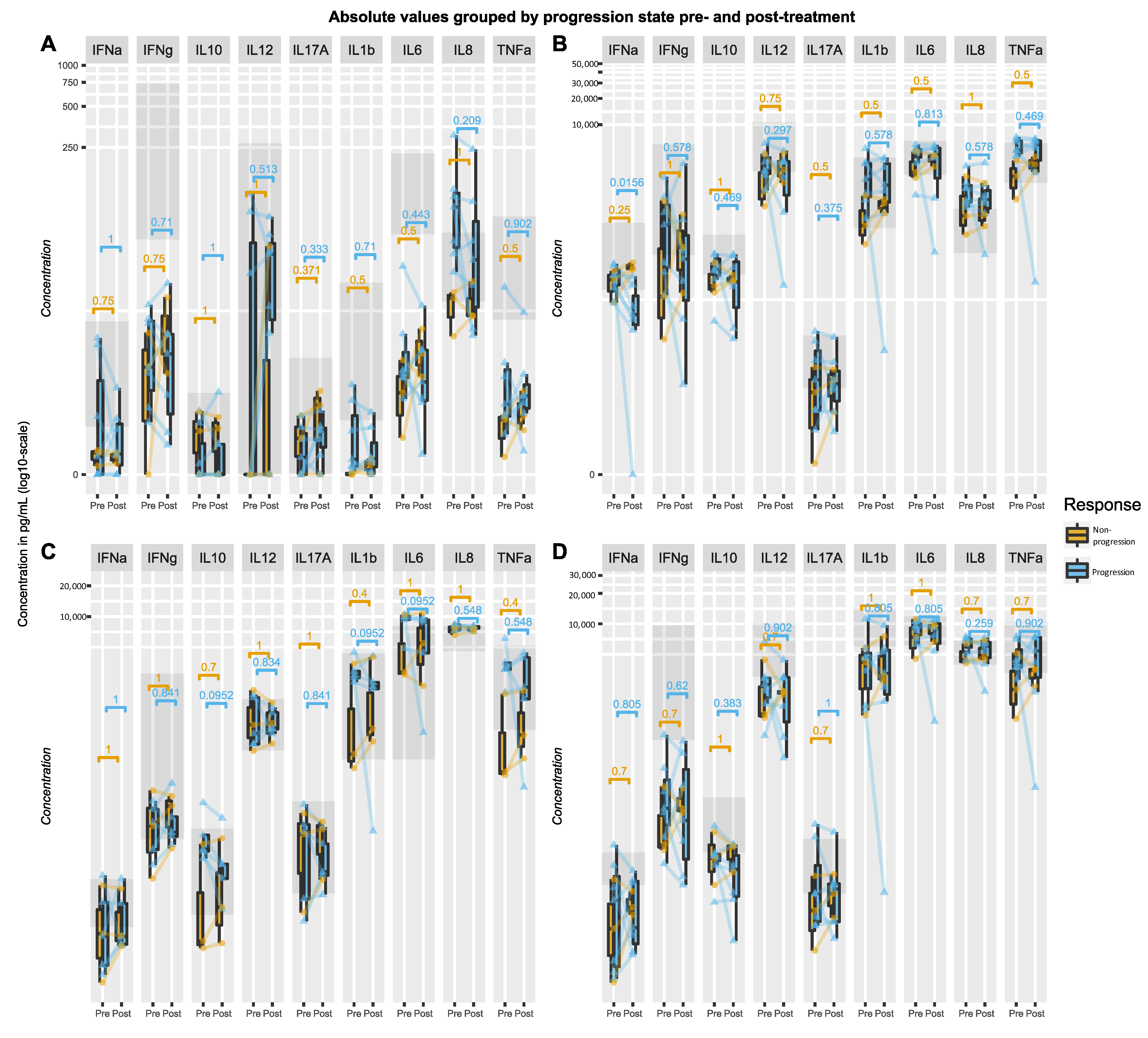
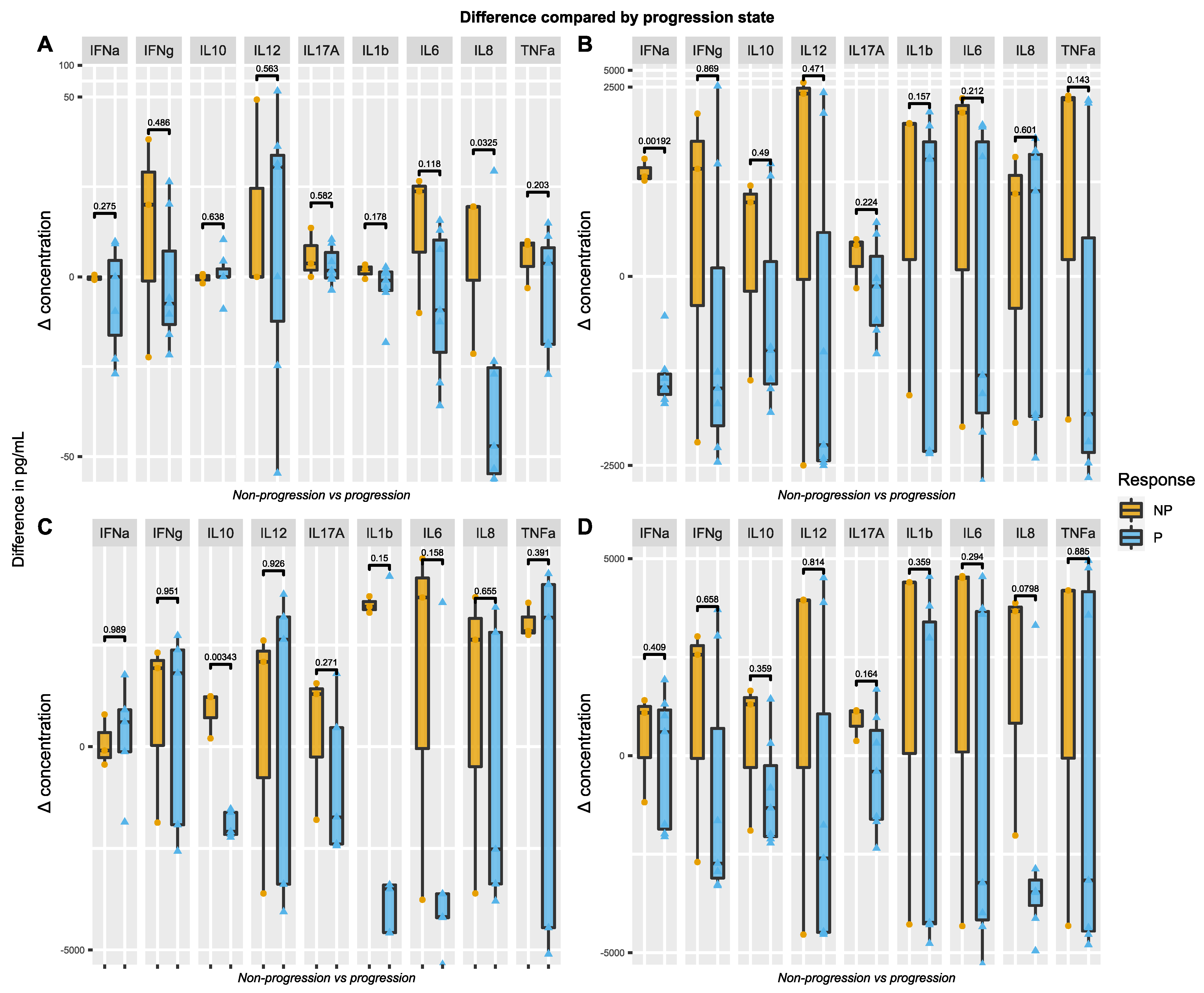
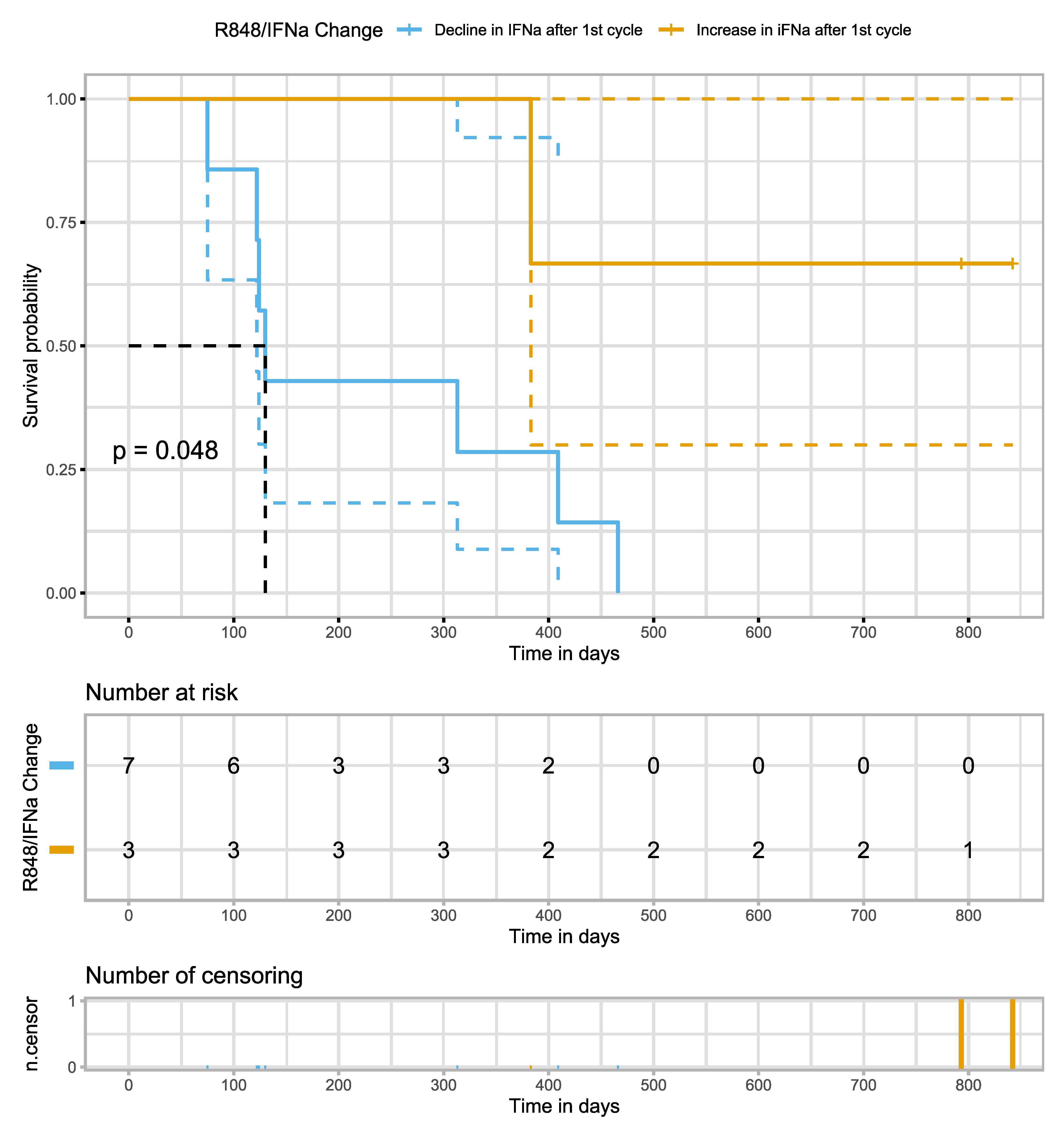
3.3. Stimulated Innate Immune Responses

Blood samples were collected from all patients for TruCulture® analysis, both immediately before the first treatment cycle and immediately before the second. In the unstimulated TruCulture® tubes, cytokine responses were negligible (near zero) for all ten patients at both time points. Consequently, there was no significant difference in the levels of unstimulated cytokine release between the measurements taken before the first and second cycles (p = 1.0), as indicated in the data not shown (refer to Figure 1).
Upon stratifying the patients based on tumor response (progression versus non-progression), distinct cytokine release patterns emerged between the samples taken before the first and second cycles. In patients experiencing tumor progression, the R848-induced IFNα response decreased in all seven cases. Conversely, in patients without progression, an increase in R848-induced IFNα was observed (see Table S1 and Figure 1B). Notably, the variation in R848-induced IFNα release between the two sampling points differed significantly between patients with and without progression, being more pronounced in the latter group (p = 0.00192) (refer to Table S2 and Figure 2B). However, no other significant differences were noted in the R848-induced response between the two sampling times.
In the case of the HKCA-induced IL-10 response, a decrease was observed from the first to the second sample in patients with progression (p = 0.0156), whereas an increase was noted in patients without progression (p = 0.25) (refer to Table S1 and Figure 1C). Additionally, the mean change (delta) in IL-10 levels was significantly different between the progressed and non-progressed groups, being more elevated in the latter (p = 0.00343) (see Table S2 and Figure 2C).
The POLY-IC-induced IL-8 response showed a borderline significant difference in mean change (delta) between patients with and without progression, with a higher increase observed in patients without progression (p = 0.0325) (refer to Table S2 and Figure 1A).
No significant differences were observed in the LPS-induced immune responses between patients with progression and those without (see Figure 1D and Figure 2D).
Furthermore, survival analysis, considering the induced immune response (increased or decreased) as a dependent variable, revealed that an increased R848-induced IFNα response correlated with a higher likelihood of survival (log-rank p-value of 0.048) (refer to Figure 3).

4. Discussion

In this preliminary exploratory study, we focused on the innate immune response in patients with metastatic urothelial-cell carcinoma (mUTC) before and after a single cycle of pembrolizumab treatment. Utilizing the TruCulture® system, we examined the absolute values and the variations in cytokine release induced by the first treatment cycle. Patients were stratified based on tumor response (progression vs. non-progression) as determined by the initial evaluation CT scan. Our findings revealed notable differences between the groups: non-progressors exhibited a higher R848-induced IFNα, HKCA-induced IL-10, and marginally increased POLY-IC-induced IL-8 levels. Additionally, survival analysis indicated a potential survival advantage in patients exhibiting an augmented R848-induced IFNα response (Figure 3).
To the best of our knowledge, this investigation is the inaugural study to assess both the alterations in and the prospective predictive value of the innate immune response, as measured via the TruCulture® system, following Immune Checkpoint Inhibitor (ICI) therapy in mUTC patients.
We noted that patients demonstrating progression at the first evaluation CT scan exhibited a diminished R848-induced IFNα response (n = 7) and a decreased HKCA-induced IL-10 response (n = 5). Conversely, patients showing non-progression experienced an increased R848-induced IFNα response (n = 3) and an augmented HKCA-induced IL-10 response (n = 3). The observed fluctuations in cytokine release within the TruCulture® system could serve as surrogates for immune function [36] and may, therefore, qualify as potential predictive markers for the efficacy of ICI treatment. However, peripheral immune markers encompass various cytokines, and the immunogenic pattern of cytokine release could be disease-specific, influenced by the stimuli used [38]. Consequently, the replicability of our observed pattern in other cancer types remains uncertain.
In a study by Gjærde et al., in which TruCulture® was used to measure induced cytokine response pre- and post-hematopoietic stem-cell transplantation, they generally observed an increase in most cytokines post-therapy [35]. Notably, this observation was performed in patients with a hematological disease undergoing extensive therapy, changing the immune system. This might explain why we did not observe this general increase in our cohort.
The mean variation in cytokine release between the initial and subsequent samples demonstrated significant differences in response groups, specifically for R848-induced IFNα and HKCA-induced IL-10 releases. A decrease in immune response was noted in patients exhibiting tumor progression, contrasting with an increase in those without progression. R848, a potent synthetic TLR7/8 agonist, is known to elevate cytokine release, including TNFα, IL-6, and IFNα, from target cells, thereby exhibiting significant antiviral and anti-tumor effects [39,40,41,42]. The augmented R848-induced cytokine release in non-progressors could suggest a rejuvenation of the immune system facilitated by ICI, potentially restoring some capacity for an anti-tumor response. Conversely, despite ICI treatment, the decrease in R848-induced IFNα in progressors might be attributed to persistent lymphocyte and overall immune cell exhaustion, reflecting an inadequate anti-tumor response [23,43].
HKCA, mimicking fungal microbial presence, activates the β-glucan-specific dectin-1 receptor and several extracellular TLRs on phagocytes. This activation leads to reactive oxygen species generation and NF-κB activation, culminating in pro-inflammatory cytokine secretion [44,45]. The attenuated pathway in HKCA-induced unspecific pro-inflammatory cytokine production could account for the less-pronounced difference between progressors and non-progressors compared to the R848-induced IFNα response.
The predictive value of peripheral cytokines in ICI responses has been the subject of several studies, yet the findings remain inconsistent and inconclusive. Generally, higher baseline circulating cytokine levels correlate with improved survival or response [46]. Research in non-small-cell lung cancer and malignant melanoma has identified a link between pre-treatment IFNγ levels and ICI response, with elevated plasma IFNγ levels associated with better response and prolonged survival [22,47,48,49]. Regrettably, our study could not establish a relationship between pre-treatment-induced cytokine levels and treatment response in mUTC patients, possibly due to the small sample size and methodological differences, as other studies focused on circulating levels rather than induced cytokine release. Notably, the only patient with SCC had long-term response. It could be interesting to see a larger cohort of both SCC and UC histology and their cytokine release patterns.
The principal limitation of this exploratory study is the small patient cohort. Nonetheless, a consistent pattern and difference in the R848- and HKCA-induced IFNα and IL-10 releases, respectively, were observed between progressors and non-progressors. Pseudo-progression at the initial CT scan is a potential concern; however, the clinical deterioration and short survival observed in all patients experiencing progression render this unlikely.
Therefore, while the results are exciting and hypothesis-generating, they should be interpreted cautiously due to the limited sample size. Further investigations in larger cohorts are essential to validate these findings, potentially serving as early response markers to ICI in clinical practice, contingent upon successful validation studies.

5. Conclusions

Our study identified consistent differences in the mean changes of R848-induced IFNα and HKCA-induced IL-10 following the initial cycle of pembrolizumab treatment in patients with metastatic urothelial-cell carcinoma (mUTC). Specifically, progressors exhibited lower levels of these cytokines, while non-progressors demonstrated higher levels. Furthermore, our analysis suggests a higher survival probability in patients experiencing an increase in R848-induced IFNα response compared to those with a decline in IFNα release. Based on these preliminary findings, we are conducting a more comprehensive study to explore the potential of these cytokine responses as early biomarkers for the efficacy of Immune Checkpoint Inhibitor (ICI) therapy further.

Supplementary Materials

The following supporting information can be downloaded at: https://www.mdpi.com/article/10.3390/uro4010001/s1, Figure S1: TruCulture® principle and stimuli; Table S1: Absolute value–p-value; Table S2: p-values of difference(delta) between progression vs non-progression.

Author Contributions

D.R.S.: Writing of first draft, data processing, statistical analysis, graphs and tables, interpretation of data, revisions of the manuscript, finalizing the manuscript. L.H.O.: Data collection and revisions of the manuscript. S.R.O.: Revision of the manuscript, interpretation of data, the conceptualization of applied methods for immune assays. S.D.N.: Manuscript revision, data interpretation, and project conceptualization. K.W.M.: Revision of manuscript, interpretation of data. Z.S.: Revision of manuscript, interpretation of data. H.P.: Funding, revision of the manuscript, ethical approvals, interpretation of data, the conceptualization of the project. All authors have read and agreed to the published version of the manuscript.

Funding

This trial is investigator-initiated by medical doctors employed at Rigshospitalet, without commercial or economic interests in the trial. The trial is funded by Departments at Rigshospitalet participating in the trial (Dept. of Clinical Immunology, Dept. of Infectious Diseases), by PERSIMUNE Centre of Excellence and by public funds (The Capital Region Research Foundation for Health Research, FSS, Novo Nordisk Fonden, Augustinus Fonden).

Institutional Review Board Statement

The study was conducted in accordance with the Declaration of Helsinki, and approved by the Danish Ethics Committee under project no. H-17024315.

Informed Consent Statement

Written and oral informed consent was obtained from all subjects involved in the study.

Data Availability Statement

Data from the corresponding author are available upon reasonable request.

Conflicts of Interest

The authors declare no conflict of interest.

References

  1. Witjes, J.A.; Bruins, H.M.; Cathomas, R.; Compérat, E.M.; Cowan, N.C.; Gakis, G.; Hernández, V.; Linares Espinós, E.; Lorch, A.; Neuzillet, Y.; et al. European Association of Urology Guidelines on Muscle-invasive and Metastatic Bladder Cancer: Summary of the 2020 Guidelines. Eur. Urol. 2021, 79, 82–104. [Google Scholar] [CrossRef] [PubMed]
  2. Hepp, Z.; Shah, S.N.; Smoyer, K.; Vadagam, P. Epidemiology and treatment patterns for locally advanced or metastatic urothelial carcinoma: A systematic literature review and gap analysis. J. Manag. Care Spec. Pharm. 2021, 27, 240–255. [Google Scholar] [CrossRef] [PubMed]
  3. Witjes, J.A.; Babjuk, M.; Bellmunt, J.; Bruins, H.M.; De Reijke, T.M.; De Santis, M.; Gillessen, S.; James, N.; Maclennan, S.; Palou, J.; et al. EAU-ESMO Consensus Statements on the Management of Advanced and Variant Bladder Cancer—An International Collaborative Multistakeholder Effort†. Eur. Urol. 2020, 77, 223–250. [Google Scholar] [CrossRef] [PubMed]
  4. Balar, A.V.; Castellano, D.; O’Donnell, P.H.; Grivas, P.; Vuky, J.; Powles, T.; Plimack, E.R.; Hahn, N.M.; de Wit, R.; Pang, L.; et al. First-line pembrolizumab in cisplatin-ineligible patients with locally advanced and unresectable or metastatic urothelial cancer (KEYNOTE-052): A multicentre, single-arm, phase 2 study. Lancet Oncol. 2017, 18, 1483–1492. [Google Scholar] [CrossRef] [PubMed]
  5. Balar, A.V.; Galsky, M.D.; Rosenberg, J.E.; Powles, T.; Petrylak, D.P.; Bellmunt, J.; Loriot, Y.; Necchi, A.; Hoffman-Censits, J.; Perez-Gracia, J.L.; et al. Atezolizumab as first-line treatment in cisplatin-ineligible patients with locally advanced and metastatic urothelial carcinoma: A single-arm, multicentre, phase 2 trial. Lancet 2017, 389, 67–76. [Google Scholar] [CrossRef] [PubMed]
  6. Bellmunt, J.; de Wit, R.; Vaughn, D.J.; Fradet, Y.; Lee, J.-L.; Fong, L.; Vogelzang, N.J.; Climent, M.A.; Petrylak, D.P.; Choueiri, T.K.; et al. Pembrolizumab as Second-Line Therapy for Advanced Urothelial Carcinoma. N. Engl. J. Med. 2017, 376, 1015–1026. [Google Scholar] [CrossRef]
  7. Rosenberg, J.E.; Hoffman-Censits, J.; Powles, T.; van der Heijden, M.S.; Balar, A.V.; Necchi, A.; Dawson, N.; O’Donnell, P.H.; Balmanoukian, A.; Loriot, Y.; et al. Atezolizumab in patients with locally advanced and metastatic urothelial carcinoma who have progressed following treatment with platinum-based chemotherapy: A single-arm, multicentre, phase 2 trial. Lancet 2016, 387, 1909–1920. [Google Scholar] [CrossRef]
  8. Parikh, M.; Powles, T. Immune Checkpoint Inhibition in Advanced Bladder and Kidney Cancer: Responses and Further Management. Am. Soc. Clin. Oncol. Educ. Book 2021, 41, e182–e189. [Google Scholar] [CrossRef]
  9. Jun, T.; Anker, J.; Galsky, M.D. Biomarkers for therapy selection in metastatic urothelial cancer. J. Cancer Metastasis Treat. 2022, 8, 1. [Google Scholar] [CrossRef]
  10. Szabados, B.; Prendergast, A.; Jackson-Spence, F.; Choy, J.; Powles, T. Immune Checkpoint Inhibitors in Front-line Therapy for Urothelial Cancer. Eur. Urol. Oncol. 2021, 4, 943–947. [Google Scholar] [CrossRef]
  11. Schulz, G.B.; Todorova, R.; Braunschweig, T.; Rodler, S.; Volz, Y.; Eismann, L.; Pfitzinger, P.; Jokisch, F.; Buchner, A.; Stief, C.; et al. PD-L1 expression in bladder cancer: Which scoring algorithm in what tissue? Urol. Oncol. Semin. Orig. Investig. 2021, 39, 734.e1–734.e10. [Google Scholar] [CrossRef]
  12. EMA Restricts Use of Keytruda and Tecentriq in Bladder Cancer. Available online: https://www.ema.europa.eu/en/news/ema-restricts-use-keytruda-tecentriq-bladder-cancer (accessed on 15 November 2023).
  13. Rouanne, M.; Radulescu, C.; Adam, J.; Allory, Y. PD-L1 testing in urothelial bladder cancer: Essentials of clinical practice. World J. Urol. 2021, 39, 1345–1355. [Google Scholar] [CrossRef]
  14. Powles, T.; Park, S.H.; Voog, E.; Caserta, C.; Valderrama, B.P.; Gurney, H.; Kalofonos, H.; Radulović, S.; Demey, W.; Ullén, A.; et al. Avelumab Maintenance Therapy for Advanced or Metastatic Urothelial Carcinoma. N. Engl. J. Med. 2020, 24, 1218–1230. [Google Scholar] [CrossRef] [PubMed]
  15. Powles, T.; Rosenberg, J.E.; Sonpavde, G.P.; Loriot, Y.; Durán, I.; Lee, J.L.; Matsubara, N.; Vulsteke, C.; Castellano, D.; Wu, C.; et al. Enfortumab Vedotin in Previously Treated Advanced Urothelial Carcinoma. N. Engl. J. Med. 2021, 384, 1125–1135. [Google Scholar] [CrossRef] [PubMed]
  16. Loriot, Y.; Necchi, A.; Park, S.H.; Garcia-Donas, J.; Huddart, R.; Burgess, E.; Fleming, M.; Rezazadeh, A.; Mellado, B.; Varlamov, S.; et al. Erdafitinib in Locally Advanced or Metastatic Urothelial Carcinoma. N. Engl. J. Med. 2019, 381, 338–348. [Google Scholar] [CrossRef]
  17. Powles, T.; Bellmunt, J.; Comperat, E.; De Santis, M.; Huddart, R.; Loriot, Y.; Necchi, A.; Valderrama, B.P.; Ravaud, A.; Shariat, S.F.; et al. Bladder cancer: ESMO Clinical Practice Guideline for diagnosis, treatment and follow-up. Ann. Oncol. 2021, 33, S0923753421048274. [Google Scholar] [CrossRef] [PubMed]
  18. Yarchoan, M.; Albacker, L.A.; Hopkins, A.C.; Montesion, M.; Murugesan, K.; Vithayathil, T.T.; Zaidi, N.; Azad, N.S.; Laheru, D.A.; Frampton, G.M.; et al. PD-L1 expression and tumor mutational burden are independent biomarkers in most cancers. JCI Insight 2019, 4, e126908. [Google Scholar] [CrossRef]
  19. Alexandrov, L.B.; Nik-Zainal, S.; Wedge, D.C.; Aparicio, S.A.J.R.; Behjati, S.; Biankin, A.V.; Bignell, G.R.; Bolli, N.; Borg, A.; Børresen-Dale, A.-L.; et al. Signatures of mutational processes in human cancer. Nature 2013, 500, 415–421. [Google Scholar] [CrossRef]
  20. Galsky, M.D.; Banchereau, R.; Hamidi, H.R.; Leng, N.; Harris, W.; O’Donnell, P.H.; Kadel, E.E.; Yuen, K.C.Y.; Jin, D.; Koeppen, H.; et al. Tumor, immune, and stromal characteristics associated with clinical outcomes with atezolizumab (atezo) + platinum-based chemotherapy (PBC) or atezo monotherapy (mono) versus PBC in metastatic urothelial cancer (mUC) from the phase III IMvigor130 study. J. Clin. Oncol. 2020, 38, 5011. [Google Scholar] [CrossRef]
  21. Litchfield, K.; Reading, J.L.; Puttick, C.; Thakkar, K.; Abbosh, C.; Bentham, R.; Watkins, T.B.K.; Rosenthal, R.; Biswas, D.; Rowan, A.; et al. Meta-analysis of tumor- and T cell-intrinsic mechanisms of sensitization to checkpoint inhibition. Cell 2021, 184, 596–614.e14. [Google Scholar] [CrossRef]
  22. Boutsikou, E.; Domvri, K.; Hardavella, G.; Tsiouda, D.; Zarogoulidis, K.; Kontakiotis, T. Tumour necrosis factor, interferon-gamma and interleukins as predictive markers of antiprogrammed cell-death protein-1 treatment in advanced non-small cell lung cancer: A pragmatic approach in clinical practice. Ther. Adv. Med. Oncol. 2018, 10, 175883591876823. [Google Scholar] [CrossRef]
  23. Davoodzadeh Gholami, M.; Kardar, G.A.; Saeedi, Y.; Heydari, S.; Garssen, J.; Falak, R. Exhaustion of T lymphocytes in the tumor microenvironment: Significance and effective mechanisms. Cell. Immunol. 2017, 322, 1–14. [Google Scholar] [CrossRef]
  24. Wherry, E.J. T cell exhaustion. Nat. Immunol. 2011, 12, 492–499. [Google Scholar] [CrossRef] [PubMed]
  25. McLane, L.M.; Abdel-Hakeem, M.S.; Wherry, E.J. CD8 T Cell Exhaustion During Chronic Viral Infection and Cancer. Annu. Rev. Immunol. 2019, 37, 457–495. [Google Scholar] [CrossRef] [PubMed]
  26. Schneider, A.K.; Chevalier, M.F.; Derré, L. The multifaceted immune regulation of bladder cancer. Nat. Rev. Urol. 2019, 16, 613–630. [Google Scholar] [CrossRef] [PubMed]
  27. Iwahori, K.; Shintani, Y.; Funaki, S.; Yamamoto, Y.; Matsumoto, M.; Yoshida, T.; Morimoto-Okazawa, A.; Kawashima, A.; Sato, E.; Gottschalk, S.; et al. Peripheral T cell cytotoxicity predicts T cell function in the tumor microenvironment. Sci. Rep. 2019, 9, 2636. [Google Scholar] [CrossRef] [PubMed]
  28. McNamara, M.J.; Hilgart-Martiszus, I.; Barragan Echenique, D.M.; Linch, S.N.; Kasiewicz, M.J.; Redmond, W.L. Interferon-γ Production by Peripheral Lymphocytes Predicts Survival of Tumor-Bearing Mice Receiving Dual PD-1/CTLA-4 Blockade. Cancer Immunol. Res. 2016, 4, 650–657. [Google Scholar] [CrossRef] [PubMed]
  29. Duffy, D.; Rouilly, V.; Libri, V.; Hasan, M.; Beitz, B.; David, M.; Urrutia, A.; Bisiaux, A.; LaBrie, S.T.; Dubois, A.; et al. Functional Analysis via Standardized Whole-Blood Stimulation Systems Defines the Boundaries of a Healthy Immune Response to Complex Stimuli. Immunity 2014, 40, 436–450. [Google Scholar] [CrossRef]
  30. Duffy, D.; Rouilly, V.; Braudeau, C.; Corbière, V.; Djebali, R.; Ungeheuer, M.-N.; Josien, R.; LaBrie, S.T.; Lantz, O.; Louis, D.; et al. Standardized whole blood stimulation improves immunomonitoring of induced immune responses in multi-center study. Clin. Immunol. 2017, 183, 325–335. [Google Scholar] [CrossRef]
  31. Herberth, M.; Rahmoune, H.; Schwarz, E.; Koethe, D.; Harris, L.W.; Kranaster, L.; Witt, S.H.; Spain, M.; Barnes, A.; Schmolz, M.; et al. Identification of a Molecular Profile Associated with Immune Status in First-Onset Schizophrenia Patients. Clin. Schizophr. Relat. Psychoses 2014, 7, 207–215. [Google Scholar] [CrossRef]
  32. Rodrigues, K.B.; Dufort, M.J.; Llibre, A.; Speake, C.; Rahman, M.J.; Bondet, V.; Quiel, J.; Linsley, P.S.; Greenbaum, C.J.; Duffy, D.; et al. Innate immune stimulation of whole blood reveals IFN-1 hyper-responsiveness in type 1 diabetes. Diabetologia 2020, 63, 1576–1587. [Google Scholar] [CrossRef] [PubMed]
  33. Duffy, D.; Nemes, E.; Llibre, A.; Rouilly, V.; Musvosvi, M.; Smith, N.; Filander, E.; Africa, H.; Mabwe, S.; Jaxa, L.; et al. Immune Profiling Enables Stratification of Patients with Active Tuberculosis Disease or Mycobacteriu m tuberculosis Infection. Clin. Infect. Dis. 2021, 73, e3398–e3408. [Google Scholar] [CrossRef] [PubMed]
  34. Aasvang, E.K.; Pitter, S.; Hansen, C.P.; Storkholm, J.H.; Krohn, P.S.; Burgdorf, S.K.; Von Stemann, J.H.; Lundgren, J.D.; Nielsen, S.D.; Kehlet, H.; et al. Preoperative TruCulture® whole blood cytokine response predicts post-operative inflammation in pancreaticoduodenectomy patients—A pilot cohort study. Scand. J. Immunol. 2020, 92, e12930. [Google Scholar] [CrossRef]
  35. Gjærde, L.K.; Brooks, P.T.; Andersen, N.S.; Friis, L.S.; Kornblit, B.; Petersen, S.L.; Schjødt, I.; Nielsen, S.D.; Ostrowski, S.R.; Sengeløv, H. Functional immune reconstitution early after allogeneic haematopoietic cell transplantation: A comparison of pre- and post-transplantation cytokine responses in stimulated whole blood. Scand. J. Immunol. 2021, 94, e13042. [Google Scholar] [CrossRef]
  36. Ørbæk, M.; Gynthersen, R.M.M.; Mens, H.; Stenør, C.; Wiese, L.; Brandt, C.; Ostrowski, S.R.; Nielsen, S.D.; Lebech, A.-M. Stimulated Immune Response by TruCulture® Whole Blood Assay in Patients With European Lyme Neuroborreliosis: A Prospective Cohort Study. Front. Cell. Infect. Microbiol. 2021, 11, 666037. [Google Scholar] [CrossRef] [PubMed]
  37. Todberg, T.; Loft, N.; Møller, D.L.; Ostrowski, S.R.; Nielsen, S.D.; Skov, L. Impact of methotrexate and adalimumab on immune function of patients with psoriasis. Dermatol. Ther. 2022, 35, e15284. [Google Scholar] [CrossRef] [PubMed]
  38. Liu, T.; Wang, S.; Wornow, M.; Altman, R.B. Construction of disease-specific cytokine profiles by associating disease genes with immune responses. PLoS Comput. Biol. 2022, 18, e1009497. [Google Scholar] [CrossRef]
  39. Dockrell, D.H. Imiquimod and resiquimod as novel immunomodulators. J. Antimicrob. Chemother. 2001, 48, 751–755. [Google Scholar] [CrossRef]
  40. Bishop, G.A.; Hsing, Y.; Hostager, B.S.; Jalukar, S.V.; Ramirez, L.M.; Tomai, M.A. Molecular Mechanisms of B Lymphocyte Activation by the Immune Response Modifier R-848. J. Immunol. 2000, 165, 5552–5557. [Google Scholar] [CrossRef]
  41. Chi, H.; Li, C.; Zhao, F.S.; Zhang, L.; Ng, T.B.; Jin, G.; Sha, O. Anti-tumor Activity of Toll-Like Receptor 7 Agonists. Front. Pharmacol. 2017, 8, 304. [Google Scholar] [CrossRef]
  42. Javaid, N.; Choi, S. Toll-like Receptors from the Perspective of Cancer Treatment. Cancers 2020, 12, 297. [Google Scholar] [CrossRef] [PubMed]
  43. Liu, Y.; Zeng, G. Cancer and Innate Immune System Interactions: Translational Potentials for Cancer Immunotherapy. J. Immunother. 2012, 35, 299–308. [Google Scholar] [CrossRef]
  44. Brown, G.D.; Herre, J.; Williams, D.L.; Willment, J.A.; Marshall, A.S.J.; Gordon, S. Dectin-1 Mediates the Biological Effects of β-Glucans. J. Exp. Med. 2003, 197, 1119–1124. [Google Scholar] [CrossRef]
  45. Gantner, B.N.; Simmons, R.M.; Canavera, S.J.; Akira, S.; Underhill, D.M. Collaborative Induction of Inflammatory Responses by Dectin-1 and Toll-like Receptor 2. J. Exp. Med. 2003, 197, 1107–1117. [Google Scholar] [CrossRef] [PubMed]
  46. Wang, M.; Zhai, X.; Li, J.; Guan, J.; Xu, S.; Li, Y.; Zhu, H. The Role of Cytokines in Predicting the Response and Adverse Events Related to Immune Checkpoint Inhibitors. Front. Immunol. 2021, 12, 670391. [Google Scholar] [CrossRef] [PubMed]
  47. Yamazaki, N.; Kiyohara, Y.; Uhara, H.; Iizuka, H.; Uehara, J.; Otsuka, F.; Fujisawa, Y.; Takenouchi, T.; Isei, T.; Iwatsuki, K.; et al. Cytokine biomarkers to predict antitumor responses to nivolumab suggested in a phase 2 study for advanced melanoma. Cancer Sci. 2017, 108, 1022–1031. [Google Scholar] [CrossRef] [PubMed]
  48. Hirashima, T.; Kanai, T.; Suzuki, H.; Yoshida, H.; Matsushita, A.; Kawasumi, H.; Samejima, Y.; Noda, Y.; Nasu, S.; Tanaka, A.; et al. The Levels of Interferon-gamma Release as a Biomarker for Non-small-cell Lung Cancer Patients Receiving Immune Checkpoint Inhibitors. Anticancer Res. 2019, 39, 6231–6240. [Google Scholar] [CrossRef]
  49. Costantini, A.; Julie, C.; Dumenil, C.; Hélias-Rodzewicz, Z.; Tisserand, J.; Dumoulin, J.; Giraud, V.; Labrune, S.; Chinet, T.; Emile, J.-F.; et al. Predictive role of plasmatic biomarkers in advanced non-small cell lung cancer treated by nivolumab. OncoImmunology 2018, 7, e1452581. [Google Scholar] [CrossRef]
Figure 1. Absolute concentration values of cytokines by stimuli (A) PolyIC, (B) R848, (C) HKCA, (D) LPS at first and second measurement grouped by response state at first CT scan. The light-gray area shows the reference area. Colored lines show concentration for individual patients. Non-parametric paired Wilcoxon test, unadjusted for family-wise error. Gray-shade marks reference area. Adjusted p-values in Table S2. NP = Non-progression; p = progression. Pre = Pre-treatment measurement; Post = post first-cycle treatment measurement.
Uro 04 00001 g001
Figure 2. Showing the four different stimuli (A) PolyIC, (B) R848, (C) HKCA, (D) LPS, with the difference (delta) between first and second measurement of the different cytokine concentrations, grouped by the response at the first CT scan at the x-axis (non-progression (0) and progression (1)). Paired two-sided Welch T-test obtained p-values to examine for difference (delta) between the groups. The delta for each patient is also plotted individually according to the response group. NP = Non-progression. P = Progression. Adjusted p-values in Table S2.
Uro 04 00001 g002
Figure 3. The Kaplan–Meier curve shows the survival probability of all ten patients divided by the decline or increase in IFNa after the first cycle. Non-parametric Mantel–Cox test is performed comparing incline vs. decline (p = 0.048) on the difference in survival.
Uro 04 00001 g003
Table 1. Table showing patient characteristics.
Pt. IDAgeGenderPS at BaselinePost Pembro TreatmentDisease BurdenCycles before CT ScanResponse at 1st CT ScanNumber of Cycles Reason for EOTTreatment LineHistologyDays between
Measurements
PD-L1 Status
1 male1NoneLbD,Car,Adr PD2Decline in PS UC <10
2 maleNAVinflunineSt PR9PD UC <10
3 male1NoneLbD, Pen PD3PD UC NA
4 male1EV,VinflunineLbD PD3PD UC NA
5 female0NoneLbD SD24EOT (2 years) SCC >10
6 male1NoneLbD PD3PD UC NA
7 male1NoneAdr PD2PD UC >10
8 male0NoneSt, Lu PD3PD UC >10
9 male1NoneLbD PR8Decline in PS UC NA
10 male0NoneLu, Bo PD3PD UC >10
PS = ECOG performance status; EOT = end of treatment; PD = progressive disease; SD = stable disease; PR = partial response; UC = urothelial carcinoma; SCC = squamous-cell carcinoma; NA = not available. LbD = Lymph nodes below diaphragm. Car = Peritoneal carcinosis. St = Soft tissue metastasis pelvis. Adr = Adrenal metastasis. Lu = Lung metastasis. Bo = Bone metastasis. Pen = Penile metastasis.
Disclaimer/Publisher’s Note: The statements, opinions and data contained in all publications are solely those of the individual author(s) and contributor(s) and not of MDPI and/or the editor(s). MDPI and/or the editor(s) disclaim responsibility for any injury to people or property resulting from any ideas, methods, instructions or products referred to in the content.

