Article

Inactivating Host Bacteria for Characterization and Use of Phages

1
Department of Molecular Microbiology and Immunology, The University of Texas at San Antonio, San Antonio, TX 78249, USA
2
Department of Biochemistry and Structural Biology, UT Health, San Antonio, TX 78229, USA
3
Department of Pathology and Laboratory Medicine, UT Health, San Antonio, TX 78229, USA
*
Author to whom correspondence should be addressed.
Biophysica 2023, 3(4), 558-568; https://doi.org/10.3390/biophysica3040038
Submission received: 31 July 2023 / Revised: 4 September 2023 / Accepted: 8 September 2023 / Published: 27 September 2023
(This article belongs to the Special Issue Biomedical Optics)

Abstract

:
Phage characterization for research and therapy can involve newly isolated phages propagated in pathogenic bacteria. If so, characterization requires safety-managing the bacteria. In the current study, we adapt a common and inexpensive reagent, PrimeStore (Longhorn Vaccines and Diagnostics, San Antonio, TX, USA), to safety-manage bacteria in 20 min by selectively inactivating the bacteria. No bacterial survivors are observed among >109 bacteria per ml for a representative of both Gram-negative bacteria (Escherichia coli) and Gram-positive bacteria (Bacillus thuringiensis). This procedure causes no detected inactivation of podophage T3, myophage T4 and siphophage 0105phi7-2. Margins of safety for PrimeStore concentration exist for bacterial inactivation and phage non-inactivation. Thus, general applicability is expected. Subsequent dialysis is used to block long-term effects on phages. Nonetheless, comparable tests should be performed for each pathogenic bacterial strain/phage. Electron microscopy of thin sections reveals inactivation-altered bacterial cytoplasm and a non-disintegrated bacterial envelope (ghosts). Ghosting of E. coli includes re-arrangement of the cytoplasm and the release of endotoxin. The activity of the released endotoxin is >99% reduced after subsequent dialysis, which also removes PrimeStore components. Ghosting of B. thuringiensis includes apparent phase separation within the cytoplasm. The primary application envisaged is biophysical and other screening of phages for therapy of infectious disease.

1. Introduction

Environmental bacteriophages (phages) are screened for potential use in both procedures of biomedicine and studies of evolution. In the case of phage therapy of bacterial disease, phages are typically propagated in pathogenic hosts [1,2,3,4,5]. Thus, pre-use phage transportation, physical/chemical characterization and subsequent purification present the problem of removing residual bacterial pathogenicity.
Pre-use characterization of phages has advantages that are emphasized by our recent discovery that phages, even related phages, vary by four orders of magnitude in their titers after 4–5 h in the blood of mice [6,7]. Potentially, an animal-independent, more efficient (in cost and time) procedure can be developed to determine the probability that a phage will have high persistence, 4–6 h at least, in humans. Thus, we need a high-throughput, simple, inexpensive strategy for managing contaminating bacteria.
Potential strategies include (1) removal of bacteria by either filtration [8,9] or centrifugation [10,11], and (2) isolation of personnel from procedures of characterization. However, use of filtration has the risk of phage loss via adherence to filters [12,13]. Use of centrifugation is relatively expensive and presents infection hazards. Isolation of personnel from procedures requires expensive modification of whatever equipment is used. More efficient and reliable is treatment of phage preparations with bacteria-killing, but phage-preserving, reagents. However, such reagents are not currently available, to our knowledge.
Our preliminary thinking in this area began with the fact that the phages used for phage therapy were lytic and typically not membrane-enclosed. Thus, a procedure designed to disrupt membranes would be a good place to start searching for a bacteria-killing procedure that preserves phages used for phage therapy. An example is the mixture called PrimeStore (Longhorn Vaccines and Diagnostics; San Antonio, TX, USA), which has been used for inactivating membrane-enclosed viruses and preserving viral RNA for PCR [14,15,16,17]. PrimeStore also both kills bacteria and inactivates RNases, the latter being relatively resistant to inactivation [14,17].
Thus, we tested a diluted PrimeStore-based reagent for inactivation of both a Gram-negative bacterium, Escherichia coli, and a Gram-positive bacterium, Bacillus thuringiensis. We found that we could inactivate both these bacteria (1) with no bacteria detected in a preparation that started with over 109 colony formers per ml and (2) without causing detectable loss in the titer of podophage T3, myophage T4 and siphophage 0105phi7-2. Thus, we have found one procedure that satisfies the requirements in these cases and probably many more. The details are described here.

2. Materials and Methods

2.1. Bacterial and Phage Strains: Purification of Phages

E. coli BB/1, often used for propagating coliphages (e.g., [6]), was the Gram-negative bacterial strain used in this study. A locally isolated B. thuringiensis, also previously used for phage propagation [7], was the Gram-positive bacterial strain.
Bacterial colonies were obtained by streaking on hard agar: 1.0% Bacto-agar in T broth, 10 g Bacto-tryptone, 5 g KCl per liter of water. Liquid cultures were started with a single colony. The liquid medium for E. coli was 2xLB broth: 20 g Bacto-tryptone, 10 g Bacto-yeast extract, 5 g NaCl per liter of water. The liquid medium for B. thuringiensis was T-Broth, with a 1000 dilution of 1.0 M CaCl2 added after separate autoclaving. Cultures were grown to stationary phase by overnight (~16 h) incubation with aeration at 37 °C for E. coli and 22 °C for B. thuringiensis.
Phage T3 was liquid culture-propagated and purified in cesium chloride density gradients as previously described [18]. Phages T4 (D strain) [19] and 0105phi7-2 [7] were agarose-overlay-propagated in Petri plates and purified by rate zonal centrifugation in a sucrose gradient as previously described (T4 [19]; 0105phi7-2 [7]).

2.2. Counting of Bacteria and Phages

Determination of bacterial single-colony-producing units (CPU) per ml was initiated by dilution of bacteria in liquid culture medium. Then, 100 µL of diluted bacteria, in a sterile glass micropipette, was transferred to a hard agar surface in a Petri plate. The bacteria were spread using an alcohol-dipped flame-sterilized L-shaped glass rod. Colonies were counted using a colony counter (Stuart CC-200, Cole-Parmer, Vernon Hills, IL, USA).
Determination of phage plaque-forming units (PFU) per ml was initiated by diluting the phages. The phages in the final dilution were mixed, at 50 °C, with growth medium and molten agarose (Seakem Gold agarose; Lonza, Rockland, ME, USA). The concentration of agarose is indicated in the text; the growth medium was the medium used for propagation of the host. The incubation time and temperature were as follows: phage T3, 4–5 h, 37 °C; phage T4, 16–18 h, 30 °C; phage 0105phi7-2, 16–18 h, 22 °C. Plaques were counted using the plate reader used for bacteria.

2.3. Effects of PrimeStore-Based Inactivation Reagent on Bacterial and Phage Viability

The active ingredient of the inactivation reagent was PrimeStore (Longhorn Vaccine and Diagnostics, San Antonio, TX, USA). PrimeStore had the following composition: 2.1 M Guanidine thiocyanate, 0.500 mM TCEP (Tris(2-carboxyethyl)phosphine hydrochloride), 8.5 mM sodium citrate, 0.375% (w/v) sodium N-Lauroylsarcosine, 50 ppm antifoam A, 50 mM Tris, 0.095 mM EDTA, 22% (v/v) ethanol, 38 mM HCl (see Acknowledgments). Tests of inactivation were performed on 50 μL of bacteria, added to the following inactivation reagent: 50 μL of PrimeStore previously diluted with broth to achieve the final concentration indicated. This mixture was left at room temperature (22 ± 2 °C) for the time indicated. The same procedure was used for tests of the inactivation of phages.
Following incubation, the mixture was placed on ice. Then, 100 µL portions were removed, diluted and used to determine CFU per ml; 10 µL portions were removed, diluted and used to determine PFU per ml.
To increase the concentration of bacteria, pelleting was performed at 8000 rpm for 5 min in a JA25.50 rotor (maximum g = 7720). The supernatant was decanted. For testing of inactivation, the bacteria were processed as described above after concentration and subsequent resuspension. For electron microscopy, bacterial pellets were resuspended in either broth (control) or broth containing 1:10 diluted PrimeStore and incubated for 20 min at room temperature with mixing.

2.4. Electron Microscopy (EM) of Bacteria Propagated in Liquid Culture

Electron microscopy (EM) of thin sections was used to determine changes in structure of the inactivated and the control, pelleting-concentrated bacteria from Section 2.3. A first fixation of bacteria was performed by adding 1.0 mL of bacteria to 9.0 mL of the following fixative solution and incubating for 1.0 h at room temperature: 4.0% formaldehyde, 1.0% glutaraldehyde, 0.11 M sodium phosphate, pH 7.3.
Fixed cells were pelleted by centrifugation for 5 min at 8000 rpm in a JA25.50 rotor. The supernatant was decanted, and the pelleted bacteria brought to ~5 °C. The bacteria were then gel-embedded by resuspension in 100 µL molten (50° C) 1.2% (m/v) Seakem Gold agarose that had been dissolved in 2xLB broth, in the case of E. coli, and T broth, in the case of B. thuringiensis. The embedded cells (~0.6% agarose, final) were transferred to a 96-well plate on a coolant pack at −20 °C.
A second fixation was performed by removing the resulting semisolid gel with a spatula. The gel was transferred to a 0.6 mL conical microfuge centrifuge tube to which 0.4 mL of the above fixative solution was added. The embedded bacteria were incubated with fixative for 1.0 h at room temperature.
After the second fixation, the bacteria were dehydrated and embedded in Epon 812. Sections 100 nm thick were cut. The procedures have been previously described [20]. Electron microscopy was performed using a JEOL JEM-1400 electron microscope. Images were recorded using an AMT Image Capture Engine Version 7 in the Department of Pathology at UT Health, San Antonio.

2.5. Electron Microscopy of Phage T4 Plaques

Plaques of phage T4, after formation at 37 °C in a 0.6% supporting agarose gel, were prepared for EM via the following procedure. The clear region of a plaque, extending to and including a small segment of the surrounding bacterial lawn, was excised with a glass micropipette and transferred to a piece of parafilm. After separation from the lower agar gel, the upper agarose overlay was immersed in 0.5 mL 2xLB broth containing 10% (v/v) PrimeStore inactivation reagent and incubated at room temperature for 1.0 h. Following transfer of the inactivation-reagent treated agar slice to another piece of parafilm, 0.5 mL of the above fixative solution was added. Fixation was performed and was followed by dehydration, embedding, thin-sectioning and electron microscopy as in Section 2.4.

2.6. Detection of Endotoxin

Preparations of 10% PrimeStore-inactivation-reagent treated E. coli were subjected to pelleting of bacteria-sized inactivation products by centrifugation at 8000 rpm (maximum g = 7720) for 5 min at 15 °C. The supernatant was quantitatively tested for endotoxin using a ToxinSensor Chromogenic LAL Endotoxin Assay Kit (GenScript, Piscataway, NJ, USA). The manufacturer’s instructions were followed. Subsequent dialysis was performed using Spectra 25,000 MWCO (molecular weight cut-off) cellulose dialysis tubing and 4 changes (2 L) of deionized water at 5 °C for 8–10 h.

3. Results

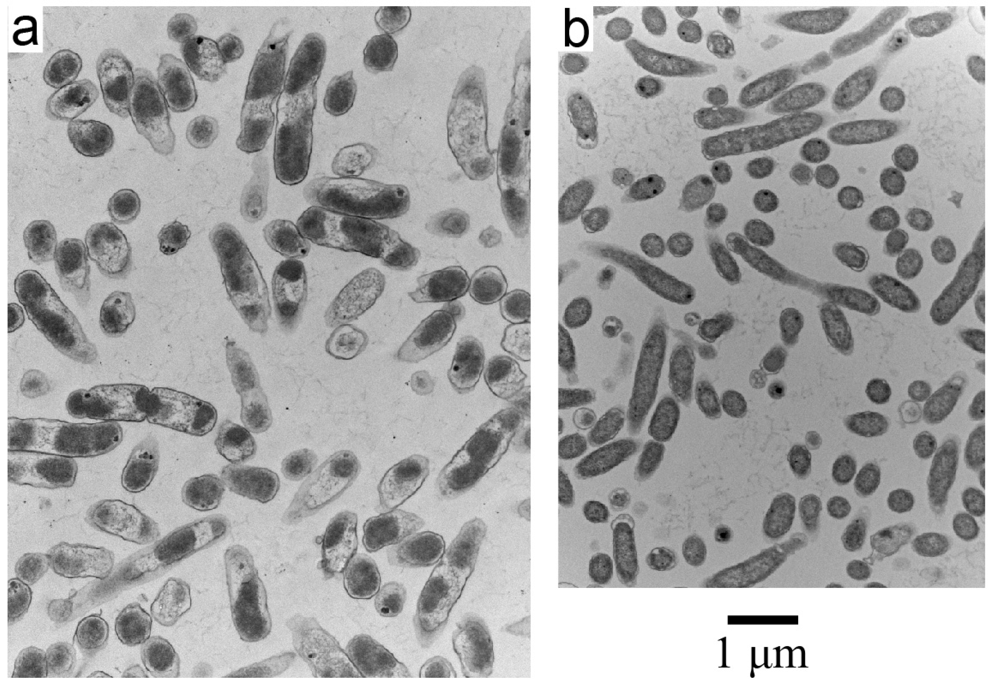
3.1. Inactivation of Gram-Negative and Gram-Positive Bacteria in Growth Medium

A diluted, 10% PrimeStore-inactivation-reagent was effective in killing both E. coli and B. thuringiensis, leaving no CFU among these bacteria at concentrations above those of a stationary phase culture (Table 1a). The concentration of bacteria in Table 1a was increased by pelleting and resuspension. The same result was observed when 5% PrimeStore reagent was used (Table 1a).
In contrast, the 10% PrimeStore-inactivation-reagent had no effect on the PFU per ml of the phages tested. Podophage T3, myophage T4 and siphophage 0105phi7-2 did not lose titer when PrimeStore-reagent-treated for 20 min (Table 1b) in a separate experiment. Simultaneously, the 10% PrimeStore-inactivation-reagent killed all E. coli and B. thuringiensis in preparations that had not been pre-concentrated.

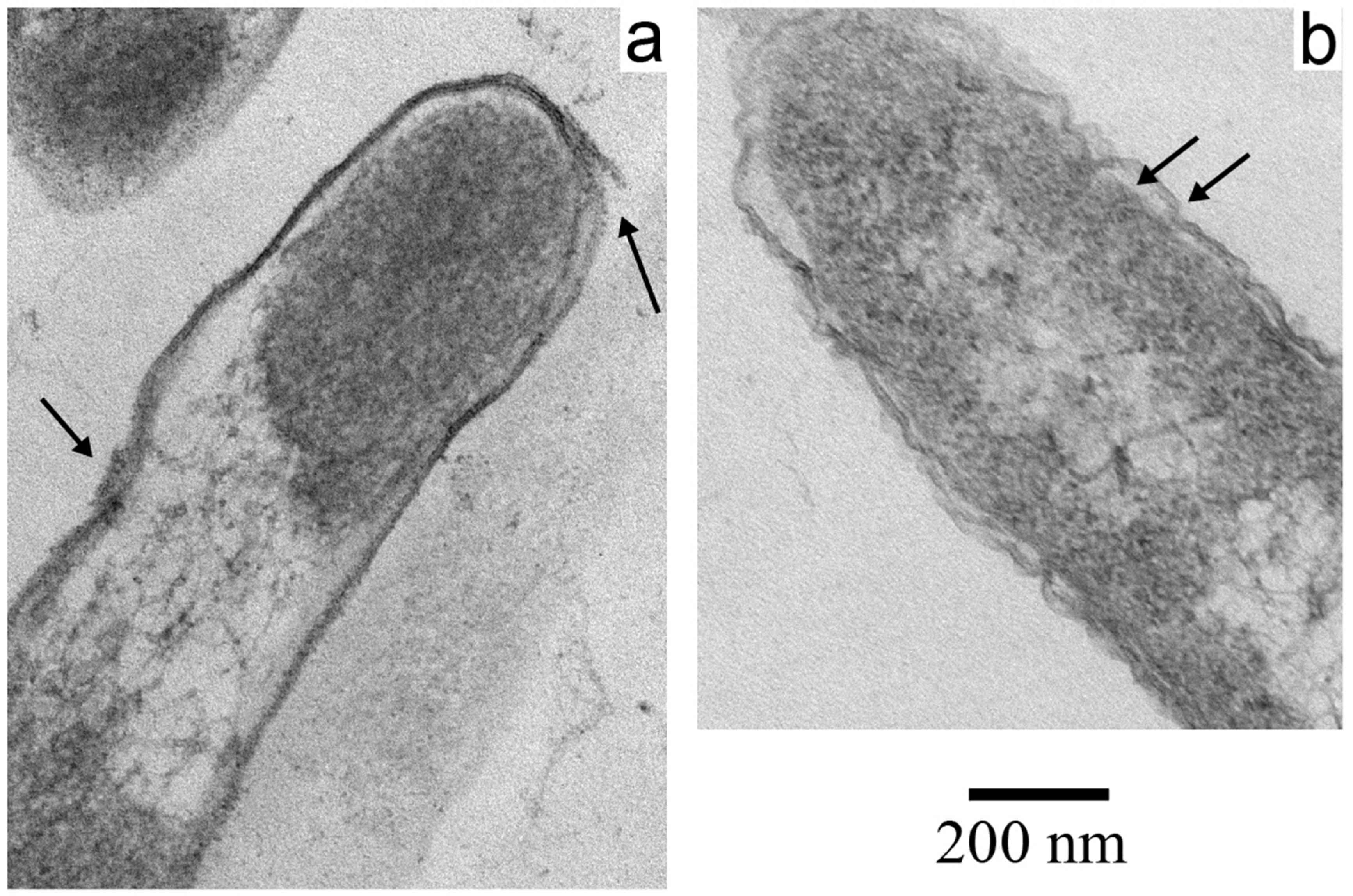
3.2. Electron Microscopy of Inactivated E. coli

Electron microscopy of thin sections revealed that the inactivation of pelleting-concentrated E. coli was not accompanied by general bacterial disintegration. Images of particles with bacterial cell-like envelopes were over 90% of the particle images observed. A low-magnification example is shown in Figure 1a. Background, extracellular fibers were the embedding agarose gel. A low-magnification control image of cells prepared in the absence of treatment with 10% PrimeStore-reagent is shown in Figure 1b. The first inactivation-induced change observed was that the cell envelopes had become rounded and wider at the ends, as seen by comparison of Figure 1a with Figure 1b. The magnifications in such low-magnification images are not precise enough to compare sizes.
Closer examination revealed additional effects of inactivation. First, the following change was observed. Images of both sagittal and transverse sections of the control typically had relatively low-density, DNA-fiber-containing cytoplasm embedded in higher-density cytoplasm, as seen in Figure 1b (referred to as class 1 images), with a higher magnification example in Figure 2b. In contrast, images of both sagittal and transverse sections of inactivated E. coli typically revealed lower-density cytoplasm in segments along the long axis. The lower-density cytoplasm was not embedded in the higher-density cytoplasm (class 2 image; Figure 1a and Figure 2a). Quantitatively, class 2 images were over 95% of the images in the PrimeStore-reagent treated sample; class 1 images were not observed in over 1000 ghosts. These numbers were 10% and 86% for the control sample. Thus, at the least, inactivation rearranged the E. coli cytoplasm. These differences were observed in two independent trials.
In addition, inactivation associated damage to the E. coli envelope was observed. The three layers of the control were, as previously shown [21,22,23], outer membrane/cell wall (lower arrow in Figure 2b) and inner membrane (upper arrow in Figure 2b). These three layers were also sometimes seen in the inactivated sample.
However, evidence of damage was also seen in the inactivated sample, as indicated by the two arrows in Figure 2a. Sometimes, damage to the envelope was sufficient to expel a section of low-density cytoplasm into the extracellular space, without severing the connection to the rest of the cytoplasm. An example is shown in Figure 3a.
The most dramatic damage to the cell envelope was observed when a T4 plaque was examined after treatment with 10% PrimeStore-reagent. Several T4 phages were sometimes observed with tails pointed toward the central region of material, presumably cytoplasmic, that appeared to be holding the phages together. The phages had contracted tails, an indication that they had previously attached to the cell envelope and had at least started to inject DNA. However, no layers of the cell envelope were observed. The phages were apparently being held in place by partial or low-density cytoplasm without an envelope, the envelope presumably having been removed by the 10% PrimeStore-reagent. Also seen in this image are apparent segments of high-density cytoplasm made extracellular (leftmost and rightmost arrows in Figure 3b) and membrane-covered vesicles, presumably released from the outer bacterial membrane (central arrow in Figure 3b).

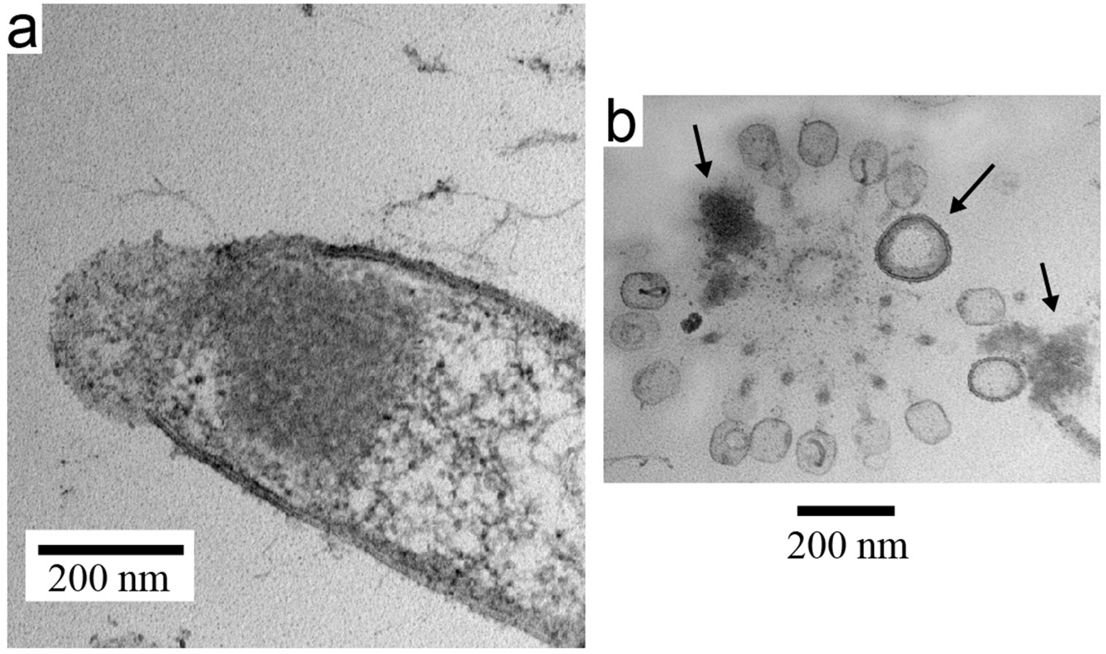
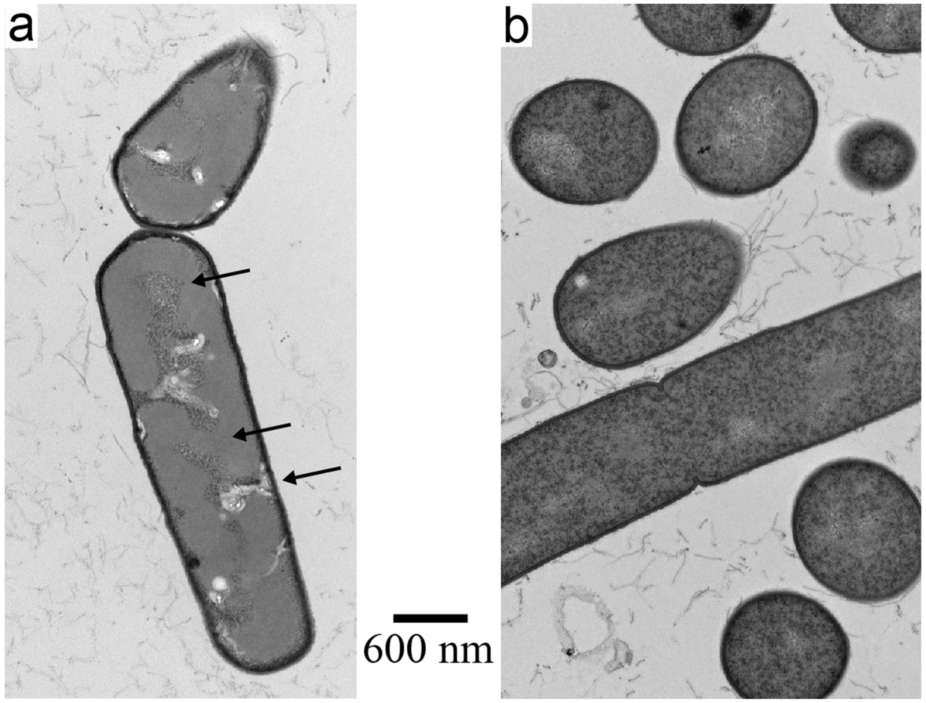
3.3. Electron Microscopy of Inactivated B. thuringiensis

Electron microscopy revealed that inactivation of B. thuringiensis left a bacterial envelope (>95% of the total particles observed), as was the case for E.coli. Similarly, the distribution of relatively low-density cytoplasm was changed; an example is shown in Figure 4a and a control image is shown in Figure 4b.
However, the following observations distinguished inactivated B. thuringiensis from inactivated E. coli. First, the images of inactivated B. thuringiensis cytoplasm had three, rather than two, densities. The lowest-density cytoplasm had fibrous material assumed to be the bacterial DNA (bottom arrow in Figure 4a), as was the case for the low-density cytoplasm of E. coli. Intermediate-density cytoplasm, with no counterpart in the control and without any analog for E. coli, was present (intermediate arrow in Figure 4a). Finally, unlike what was seen with E. coli, the high-density cytoplasm was embedded in the intermediate-density cytoplasm (top arrow in Figure 4a). The high-density B. thuringiensis cytoplasm was significantly more granular than the intermediate-density cytoplasm. These features were observed throughout specimens from two independent trials. Nonetheless, E. coli and B. thuringiensis both lost the capacity to propagate.
Finally, the observed inactivation-induced E. coli envelope damage was not observed for B. thuringiensis. Specifically, obvious envelope breaks and leaked cytoplasm were not observed. The three envelope layers were seen in some regions of both control and inactivated B. thuringiensis. The envelope appeared to be thicker in the inactivated cells. These features were observed throughout specimens from two independent trials.

3.4. Margin for Preservation of Phage Infectivity

The three phages tested also survived, without detectable PFU/mL loss, when inactivation was increased from 20 min to 1.0 h in 10% PrimeStore-reagent (Table 2a). However, a significant loss of phage titer occurred at 8 h for T3 (40%), T4 (25%) and 0105phi7-2 (40%) (Table 2b).
Reducing the PrimeStore concentration to 2.5 and 5% reduced the PFU/mL loss at 24.0 h to a marginal amount (Table 2c,d). Alternatively, the loss of titer 24 h after incubation in 10% PrimeStore reagent was reduced by dilution by a factor of 10 immediately after treatment with the 10% PrimeStore reagent (Table 2e).
Increasing the PrimeStore concentration to 15% (50% increase in PrimeStore concentration) caused dramatic loss of phage titer after 20 min: T3 (67%), T4 (93%) and 0105phi7-2 (98%). This loss was less dramatic, but still potentially problematic, at 12.5%: T3 (50%), T4 (41%) and 0105phi7-2 (61%).

3.5. Effect of Inactivation on Release of Endotoxin

To determine whether endotoxin (i.e., cell membrane associated lipopolysaccharide [24,25,26,27]) was released from ghosted E. coli, both ghosted cells and un-inactivated cells were pelleted and, thus, separated from released endotoxin. Assay of endotoxin in the supernatant revealed that ghosted cells released 1.1 × 10⁹ units compared to the 2.7 × 10⁸ units released by un-inactivated cells (Table 3). This observation agrees with the observation made by EM that outer membrane damage occurred in inactivated cells. However, the data do not reveal further details of the correlation of structure with endotoxin release.
In addition, after dialysis, the following effect of 10% PrimeStore-reagent was noted. The level of endotoxin activity was lowered by 99.8% (Table 3). The level of endotoxin activity in the untreated sample was dialysis-lowered by 25.5%. Thus, PrimeStore-reagent treatment, followed by dialysis, has an effect that would be positive for phage therapy if the in vitro endotoxin activity loss also occurs in vivo.

4. Discussion

The 10% PrimeStore-reagent has an approximate 2× margin of safety for bacterial inactivation. Thus, this reagent and the above procedure are likely to be adaptable to many, if not all, bacterial pathogens. The PrimeStore concentration can be reduced, if need be, to extend the time of phage stability. Nonetheless, given the known [24,25,28,29] variability in the envelope of bacteria, separate tests should be performed for each bacterial strain (and phage) used. These tests, as described here, are simple and inexpensive. For example, these tests do not require enzymatic digestion of cell envelopes (with lysozyme, for example [30,31]). We note that previous data with PrimeStore-based and other inactivation-reagents typically are unlike the data presented here in that they do not include conditions in which viruses are left alive (e.g., [32,33]).
A second advantage is in preparative phage isolation, for the making of phage therapy cocktails, for example. The formation of ghosts implies susceptibility to (hazard-free) pelleting- or filtration-removal of all bacterial components that are not released from ghosts. In contrast, use of cell wall-digesting enzymes/detergents for bacterial inactivation often has the disadvantage of more complete destruction of bacteria [30,31]. However, endotoxin is released from E. coli, and presumably other Gram-negative bacteria, during PrimeStore-reagent treatment. This problem can be resolved if found that dialysis, post-PrimeStore-reagent treatment, removes in vivo endotoxin activity as thoroughly as found here to remove in vitro endotoxin activity.
The reason for the endotoxin activity removal effect of PrimeStore-reagent treatment and dialysis is possibly either (1) adherence of endotoxin to the dialysis membrane or (2) its passage through the dialysis membrane after disassembly of noncovalently assembled lipopolysaccharide chains to form either smaller, non-covalently assembled aggregates or monomers [24,26], the latter with a molecular weight as low as 3 Kd [34]. Loss of in vitro endotoxin activity has previously been found to be associated with disassembly of lipopolysaccharide chains to form monomers [26].
After ghosts are removed and the specimen dialyzed, the phages are in the presence of primarily proteins and nucleic acids that are released from the bacterial cells. The question of further phage purification for phage therapy is not definitively answered here. However, the following suggests that further purification may not always be necessary, especially in the case of an emergency. The components released from bacteria in the phage preparation are likely to have also been released in the patient because of infection of the patient with the same bacteria. Thus, whether harmful or not, injecting these components may not have a major effect (if any) on the status of the patient.
Given the potential for emergencies, bacterial pandemics for example, the following is an assessment of how rapidly a phage therapy response can be generated in the case of a bacterial pathogen for which no current therapy exists. Using in-gel propagation techniques [35,36], the isolation, with 3× cloning, of a phage takes about 5 days; typically, multiple phages (over 50 in some cases) would be simultaneously isolated. Phage preparation via plate stock takes another day. Treatment with PrimeStore-inactivation-reagent, followed by dialysis, takes another 5 h. Electrophoretic phage characterization occupies 5–20 h, depending on the details. Thus, a phage therapy response can be generated in less than a week. In an extreme emergency, this time can be shortened by restricting the number of times the phage is cloned. We know of nothing that is likely to be effective and that is even close to this in speed. We imagine that having reserve capacity to do this would be appropriate, even if phage therapy is not the dominant method of anti-bacterial defense.
The current study is, to our knowledge, the first in which the chemical inactivation of Gram-positive and Gram-negative bacteria, without harming phages, has been achieved in a time as short as 20 min. A previous study [37], designed for the isolation of sequencing-quality phage DNA, approached this objective, but was limited by a 2-day time requirement, the use of only a Gram-negative bacterium and inactivation that was not shown to be as complete as that achieved here.

Author Contributions

Conceptualization, J.P.C. and P.S.; methodology, J.P.C., E.T.W., B.H. and P.S.; validation, J.P.C. and P.S.; formal analysis, J.P.C. and P.S.; investigation, J.P.C., E.T.W., B.H. and P.S.; resources, J.P.C., B.H. and P.S.; writing—original draft preparation, P.S.; writing—review and editing, J.P.C. and P.S; visualization, B.H., J.P.C. and P.S.; project administration, J.P.C. and P.S.; funding acquisition, J.P.C. and P.S. All authors have read and agreed to the published version of the manuscript.

Funding

This research was funded by the Morrison Trust (2021 and 2022) and the San Antonio Medical Foundation (2022).

Data Availability Statement

The data presented in this study are all available within one or more of the following: the figures and text.

Acknowledgments

The authors thank Longhorn Vaccines and Diagnostics for their donation of the PrimeStore used in this study and Luke Daum for providing details of PrimeStore composition.

Conflicts of Interest

P.S. and J.P.C. are founding members of a company, Phage Refinery LLC, that has plans to use the data presented here for the purposes of the company. J.P.C. is a partial owner of Longhorn Vaccine and Diagnostics. The above funders had no role in the design of the study; in the collection, analyses or interpretation of the data; in the writing of the manuscript; or in the decision to publish the results.

References

  1. Hatfull, G.F.; Dedrick, R.M.; Schooley, R.T. Phage therapy for antibiotic-resistant bacterial infections. Annu. Rev. Med. 2022, 73, 197–211. [Google Scholar] [CrossRef] [PubMed]
  2. Strathdee, S.A.; Hatfull, G.F.; Mutalik, V.K.; Schooley, R.T. Phage therapy: From biological mechanisms to future directions. Cell 2023, 186, 17–31. [Google Scholar] [CrossRef] [PubMed]
  3. Żaczek, M.; Górski, A.; Grzybowski, A.; Pasternak, E.; Weber-Dąbrowska, B.; Międzybrodzki, R. How interest in phages has bloomed into a leading medical research activity in Poland. Viruses 2022, 14, 2617. [Google Scholar] [CrossRef]
  4. Górski, A.; Borysowski, J.; Międzybrodzki, R. Phage therapy: Towards a successful clinical trial. Antibiotics 2020, 9, 827. [Google Scholar] [CrossRef] [PubMed]
  5. Diallo, K.; Dublanchet, A. A century of clinical use of phages: A literature review. Antibiotics 2023, 12, 751. [Google Scholar] [CrossRef]
  6. Serwer, P.; Wright, E.T.; De La Chapa, J.; Gonzales, C.B. Basics for improved use of phages for therapy. Antibiotics 2021, 10, 723. [Google Scholar] [CrossRef]
  7. Roberts, S.M.; Aldis, M.; Wright, E.T.; Gonzales, C.B.; Lai, Z.; Weintraub, S.T.; Hardies, S.C.; Serwer, P. Siphophage 0105phi7-2 of Bacillus thuringiensis: Novel propagation, DNA, and genome-implied assembly. Int. J. Mol. Sci. 2023, 24, 8941. [Google Scholar] [CrossRef]
  8. Goswami, K.P.; Pugazhenthi, G. Credibility of polymeric and ceramic membrane filtration in the removal of bacteria and virus from water: A review. J. Environ. Manag. 2020, 268, 110583. [Google Scholar] [CrossRef]
  9. Cevallos-Mendoza, J.; Amorim, C.G.; Rodríguez-Díaz, J.M.; Montenegro, M.d.C.B.S.M. Removal of contaminants from water by membrane filtration: A review. Membranes 2022, 12, 570. [Google Scholar] [CrossRef]
  10. Mortimer, M.; Petersen, E.J.; Buchholz, B.A.; Holden, P.A. Separation of bacteria, protozoa and carbon nanotubes by density gradient centrifugation. Nanomaterials 2016, 6, 181. [Google Scholar] [CrossRef]
  11. Hyman, P. Phages for phage therapy: Isolation, characterization, and host range breadth. Pharmaceuticals 2019, 12, 35. [Google Scholar] [CrossRef] [PubMed]
  12. Karczewska, M.; Strzelecki, P.; Szalewska-Pałasz, A.; Nowicki, D. How to tackle bacteriophages: The review of approaches with mechanistic insight. Int. J. Mol. Sci. 2023, 24, 4447. [Google Scholar] [CrossRef] [PubMed]
  13. Farber, F.E.; Gradwohl, S.E.; Sanford, P.B.; Tobin, M.J.; Lee, K.J.; Gerba, C.P. Bacteriophage concentration from water by filter chromatography. J. Virol. Meth. 1983, 7, 297–304. [Google Scholar] [CrossRef] [PubMed]
  14. Daum, L.T.; Worthy, S.A.; Yim, K.C.; Nogueras, M.; Schuman, R.F.; Choi, Y.W.; Fischer, G.W. A clinical specimen collection and transport medium for molecular diagnostic and genomic applications. Epidemiol. Infect. 2011, 139, 1764–1773. [Google Scholar] [CrossRef]
  15. Daum, L.T.; Fischer, G.W. Rapid and safe detection of SARS-CoV-2 and influenza virus RNA using onsite quantitative PCR diagnostic testing from clinical specimens collected in molecular transport medium. J. Appl. Lab. Med. 2021, 6, 1409–1416. [Google Scholar] [CrossRef]
  16. Clarke, C.; Smith, K.; Goldswain, S.J.; Helm, C.; Cooper, D.V.; Kerr, T.J.; Kleynhans, L.; van Helden, P.D.; Warren, R.M.; Miller, M.A.; et al. Novel molecular transport medium used in combination with Xpert MTB/RIF ultra provides rapid detection of Mycobacterium bovis in African buffaloes. Sci. Rep. 2021, 11, 7061. [Google Scholar] [CrossRef]
  17. Fischer, G.W.; Daum, L.T. Biological Specimen Collection/Transport Compositions and Methods. U.S. Patent 8,080,645 B2, 20 December 2011. [Google Scholar]
  18. Serwer, P.; Wright, E.T.; Liu, Z.; Jiang, W. Length quantization of DNA partially expelled from heads of a bacteriophage T3 mutant. Virology 2014, 456–457, 157–170. [Google Scholar] [CrossRef]
  19. Serwer, P.; Wright, E.T. Gated ethidium- and bleomycin-loading in phage T4 that is subsequently purified leak-free. Biophysica 2022, 2, 366–380. [Google Scholar] [CrossRef]
  20. Serwer, P.; Hunter, B.; Wright, E.T. Electron microscopy of in-plaque phage T3 assembly: Proposed analogs of neurodegenerative disease triggers. Pharmaceuticals 2020, 13, 18. [Google Scholar] [CrossRef]
  21. Silhavy, T.J.; Kahne, D.; Walker, S. The bacterial cell envelope. Cold Spring Harb. Perspect. Biol. 2010, 2, a000414. [Google Scholar] [CrossRef]
  22. Bahadur, R.; Chodisetti, P.K.; Reddy, M. Cleavage of Braun’s lipoprotein Lpp from the bacterial peptidoglycan by a paralog of l,d-transpeptidases, LdtF. Proc. Natl. Acad. Sci. USA 2021, 118, e2101989118. [Google Scholar] [CrossRef]
  23. Baquero, F.; Martínez, J.-L.; Sánchez, A.; Fernández-de-Bobadilla, M.D.; San-Millán, A.; Rodríguez-Beltrán, J. Bacterial subcellular architecture, structural epistasis, and antibiotic resistance. Biology 2023, 12, 640. [Google Scholar] [CrossRef]
  24. Fux, A.C.; Casonato Melo, C.; Michelini, S.; Swartzwelter, B.J.; Neusch, A.; Italiani, P.; Himly, M. Heterogeneity of lipopolysaccharide as source of variability in bioassays and LPS-binding proteins as remedy. Int. J. Mol. Sci. 2023, 24, 8395. [Google Scholar] [CrossRef]
  25. Correa, W.; Brandenburg, K.; Zähringer, U.; Ravuri, K.; Khan, T.; Von Wintzingerode, F. Biophysical analysis of lipopolysaccharide formulations for an understanding of the low endotoxin recovery (LER) phenomenon. Int. J. Mol. Sci. 2017, 18, 2737. [Google Scholar] [CrossRef]
  26. Mueller, M.; Lindner, B.; Kusumoto, S.; Fukase, K.; Schromm, A.B.; Seydel, U. Aggregates are the biologically active units of endotoxin. J. Biol. Chem. 2004, 279, 26307–26313. [Google Scholar] [CrossRef]
  27. Gioannini, T.L.; Teghanemt, A.; Zhang, D.; Levis, E.N.; Weiss, J.P. Monomeric endotoxin:protein complexes are essential for TLR4-dependent cell activation. J. Endotoxin Res. 2005, 11, 117–123. [Google Scholar] [CrossRef]
  28. Vermassen, A.; Leroy, S.; Talon, R.; Provot, C.; Popowska, M.; Desvaux, M. Cell wall hydrolases in bacteria: Insight on the diversity of cell wall amidases, glycosidases and peptidases toward peptidoglycan. Front. Microbiol. 2019, 10, 331. [Google Scholar] [CrossRef]
  29. Frirdich, E.; Vermeulen, J.; Biboy, J.; Vollmer, W.; Gaynor, E.C. Multiple Campylobacter jejuni proteins affecting the peptidoglycan structure and the degree of helical cell curvature. Front. Microbiol. 2023, 14, 1162806. [Google Scholar] [CrossRef]
  30. Shehadul Islam, M.; Aryasomayajula, A.; Selvaganapathy, P.R. A review on macroscale and microscale cell lysis methods. Micromachines 2017, 8, 83. [Google Scholar] [CrossRef]
  31. Grigorov, E.; Kirov, B.; Marinov, M.B.; Galabov, V. Review of microfluidic methods for cellular lysis. Micromachines 2021, 12, 498. [Google Scholar] [CrossRef]
  32. Welch, S.R.; Davies, K.A.; Buczkowski, H.; Hettiarachchi, N.; Green, N.; Arnold, U.; Jones, M.; Hannah, M.J.; Evans, R.; Burton, C.; et al. Analysis of inactivation of SARS-CoV-2 by specimen transport media, nucleic acid extraction reagents, detergents, and fixatives. J. Clin. Microbiol. 2020, 58, e01713–e01720. [Google Scholar] [CrossRef]
  33. Olejnik, J.; Hume, A.J.; Ross, S.J.; Scoon, W.A.; Seitz, S.; White, M.R.; Slutzky, B.; Yun, N.E.; Mühlberger, E. Art of the kill: Designing and testing viral inactivation procedures for highly pathogenic negative sense RNA viruses. Pathogens 2023, 12, 952. [Google Scholar] [CrossRef]
  34. Sali, W.; Patoli, D.; Pais de Barros, J.P.; Labbé, J.; Deckert, V.; Duhéron, V.; Le Guern, N.; Blache, D.; Chaumont, D.; Lesniewska, E.; et al. Polysaccharide chain length of lipopolysaccharides from Salmonella minnesota is a determinant of aggregate stability, plasma residence time and proinflammatory propensity in vivo. Front. Microbiol. 2019, 10, 1774. [Google Scholar] [CrossRef]
  35. Serwer, P.; Hayes, S.J.; Zaman, S.; Lieman, K.; Rolando, M.; Hardies, S.C. Improved isolation of undersampled bacteriophages: Finding of distant terminase genes. Virology 2004, 329, 412–424. [Google Scholar] [CrossRef]
  36. Serwer, P.; Hayes, S.J.; Thomas, J.A.; Hardies, S.C. Propagating the missing bacteriophages: A large bacteriophage in a new class. Virol. J. 2007, 4, 21. [Google Scholar] [CrossRef]
  37. Serwer, P.; Hayes, S.J.; Thomas, J.A.; Griess, G.A.; Hardies, S.C. Rapid determination of genomic DNA length for new bacteriophages. Electrophoresis 2007, 28, 1896–1902. [Google Scholar] [CrossRef]
Figure 1. Low-magnification EM images of (a) inactivation-reagent treated and (b) control (untreated) E. coli BB/1.
Biophysica 03 00038 g001
Figure 2. High-magnification EM images of (a) inactivation-reagent treated and (b) control (untreated) E. coli BB/1. In (a): arrows, damaged cell envelope. In (b): upper arrow, inner membrane; lower arrow, cell wall and outer membrane.
Biophysica 03 00038 g002
Figure 3. High-magnification EM images of inactivated (a) E. coli BB/1, which has partially leaked cytoplasm and (b) particles in a plaque of phage T4. In (b): left and right arrows, segments of high-density cytoplasm; central arrow, membrane-covered vesicle.
Biophysica 03 00038 g003
Figure 4. High-magnification EM images of (a) inactivated and (b) control (untreated) B. thuringiensis. In (a): bottom arrow, lowest density cytoplasm; intermediate arrow, intermediate density cytoplasm; top arrow, high-density cytoplasm.
Biophysica 03 00038 g004
Table 1. Effects of 10% and 5% PrimeStore-inactivation-reagent. (a) CPU per ml for representative Gram-negative and -positive organisms, (b) PFU per ml for phages. Details are given in the Materials and Methods Section.
a.Control-CFU/mLTreat-(5 %) CFU/mLTreat-(10 %) CFU/mL
Bacteria
E. coli9.0 × 1010<101<101
B. thuringiensis3.5 × 109<101<101
b.
PhagesControl-PFU/mLTreat-(10 %) PFU/mL
T31.8 × 10101.8 × 1010
T46.6 × 1097.0 × 109
0105phi 7-22.2 × 1082.2 × 108
Table 2. Range of effectiveness on PFU/mL. (a,b) Effect of time, as indicated, after treatment with 10% PrimeStore-inactivation-reagent; (c,d) effect of % PrimeStore, as indicated, after treatment for 24 h; (e) comparison of uninterrupted 24 h treatment with 10% PrimeStore-reagent (Treat) to 24 h treatment with 1:10 dilution at 20 min (1:10).
a. Inactivation (1 h)b. Inactivation (8 h)
PhagesControlTreatPhagesControlTreat
T31.0 × 1071.0 × 107T31.1 × 1076.6 × 106
T48.4 × 1068.6 × 106T47.0 × 1065.3 × 106
0105phi 7-25.1 × 1065.2 × 1060105phi 7-23.5 × 1062.1 × 106
c. Inactivation (2.5% PS)d. Inactivation (5.0% PS)
PhagesControlTreatPhagesControlTreat
T31.4 × 1071.5 × 107T31.2 × 1071.1 × 107
T48.9 × 1069.0 × 106T49.2 × 1069.1 × 106
0105phi 7-23.8 × 1063.7 × 1060105phi 7-23.4 × 1063.2 × 106
e. Effect of PS Dilution
PhageControlTreat1:10
T42.1 × 1071.1 × 1051.5 × 107
Table 3. Effect of inactivation on release of E. coli endotoxin. An overnight culture of E. coli was divided into equal portions, one untreated and incubated with 10% (v/v) inactivation reagent for 20 min and the other incubated with broth. The supernatant was assayed for endotoxin both before and after dialysis.
Pre-DialysisPost-Dialysis
ControlTreat ControlTreat
2.7 × 1081.1 × 1096.9 × 1071.9 × 106
Disclaimer/Publisher’s Note: The statements, opinions and data contained in all publications are solely those of the individual author(s) and contributor(s) and not of MDPI and/or the editor(s). MDPI and/or the editor(s) disclaim responsibility for any injury to people or property resulting from any ideas, methods, instructions or products referred to in the content.

