Article

The Counteraction of Cultivated Cistus creticus L. (Rock Rose) Plants to the Strain Imposed by a Long-Term Exposure to Non-Ionizing Radiation and the Role of DDC

by
Aikaterina L. Stefi
1,2,*,
Georgia Kalouda
1,
Aikaterini S. Skouroliakou
3,
Dido Vassilacopoulou
2 and
Nikolaos S. Christodoulakis
1
1
Section of Botany, Faculty of Biology, National and Kapodistrian University of Athens, 15701 Athens, Greece
2
Section of Biochemistry and Molecular Biology, Faculty of Biology, National and Kapodistrian University of Athens, 15701 Athens, Greece
3
Department of Biomedical Engineering, University of West Attica, 12243 Athens, Greece
*
Author to whom correspondence should be addressed.
Biophysica 2022, 2(3), 248-265; https://doi.org/10.3390/biophysica2030024
Submission received: 24 July 2022 / Revised: 15 August 2022 / Accepted: 30 August 2022 / Published: 1 September 2022
(This article belongs to the Collection Feature Papers in Biophysics)

Abstract

:
Two groups of Cistus creticus seedlings were grown in two chambers under controlled environmental conditions. In one of the chambers, a continuously emitting base unit of a wireless telephone was placed. After fifty days of culture, the two groups of plants were removed and thoroughly investigated and compared. The aboveground parts of the exposed plants were retarded in development while their roots exhibited increased biomass, compared to the controls. There was a minor decrease in the absorbance of the photosynthetic pigments in exposed plants, while an overproduction of Reactive Oxygen Species (ROS) ROS in their leaves and roots was detected. The expression of the L-Dopa decarboxylase (DDC) seemed to “erupt” following the exposure to radiation in both shoots and roots of the stressed plants, and their roots slow down their secondary development; strangely, the phenolic content is reduced in their leaves, the external topography of which indicates a rather xeromorphic response. We may suggest that Cistus creticus plants, forced by the radiation stress, can finely tune their metabolic pathways in a way that can be useful in the pharmaceutical industry.

1. Introduction

The seasonally dimorphic subshrubs (phrygana, garrigue, or batha) and the evergreen sclerophyllous species (chaparral or maquis) which compose the two main plant formations in the Mediterranean region [1] are imposed into two severe, timely separated strains [2,3]: the unfavorable environmental conditions of the hot, arid summer and the brief but chilling temperatures of the winter [4]. All these plants have developed unique, interesting strategies to either evade or escape the stressing conditions [5]. On the poor soils of the Mediterranean terrain, many species of the Cistaceae family grow. Among them, the rock rose (Cistus creticus (The International Plant Name Index: Cistaceae, Cistus creticus L.—Sp. Pl., ed. 2. 1: 738. [Sep 1762] IPNI (2004)); synonym: Cistus incanus) [6] (Figure 1a), a small, woody, seasonally dimorphic shrub, distributed along the coast of the Central-Eastern Mediterranean, Northern Africa, and Western Asia, being absent in France and the Iberian Peninsula [7].
Information is available for C. creticus concerning the physiology of its leaves [8], its uses in traditional folk medicine [9], the antimicrobial, antioxidant, antitumor, antinociceptive, and analgesic effects of its leaf extracts [10,11,12,13], as well as their antiproliferative effect on human prostate cells [14]. Extracts and purified substances from C. creticus have been tested and proven to have selective activities against the influenza virus due to the bioactive secondary metabolites they contain, such as flavonoids, aromatic compounds, phenolics, and tannins [15,16]. The later have been investigated separately and C. creticus is considered as the plant with the most extensive accumulation of phenolic compounds in its leaves [12,13].
Concerning the morphology and the anatomy of this species, data are available on seasonal dimorphism [17], the leaf structure and the secreting activity of the trichomes [18,19], the constituents of the commercial labdanum oil [20,21], the types of trichomes [22], as well as on the variations between summer and winter leaves [23].
During the last decades, wireless networks and millions of mobile phone users have had a substantial contribution to the settled “cloud” of non-ionizing radiation, which imposes a non-intended, irradiated life on all organisms. The rapidly increasing use of the cellular technology and the consequent increase of a stress factor—the electromagnetic radiation in the atmosphere [24]—drastically affect the Earth’s natural radiofrequency environment. This change triggered a constantly expanding confrontation concerning the effects of this additional radiation to human life and environmental health [24,25,26,27]. Some concern was given to plant reactions as well [28,29,30]. Since radiofrequency seems to constitute a genuine environmental stimulus for plants and given that plants can exploit sunlight, they must also have some means of protecting their living cells from the damages [31]. Electromagnetic radiation can evoke specific responses, many of them being similar to those observed after a natural stress or an in vitro stressful treatment [32,33]. In addition, plants prove to be an outstanding model for studying such interactions since their architecture (high surface/volume ratio) optimizes their co-action with the environment. Plants remain exposed to radiation at a constant orientation within an electromagnetic field, due to their inability to move [34], while it is possible to easily document changes over time, such as disturbed cell structure, retarded growth, and yield shortage [33,35,36].
On the other hand, it has been well-documented that, among the escape reactions of the Mediterranean plants from the impact of combined environmental stresses, the production of certain secondary metabolites whose biosynthesis is shared by animals and plants seems to be of major significance [37,38,39].
Among these metabolites, some animal neurotransmitters are included, with a noted example being that of acetylcholine, in the stinging trichomes of the stinging nettle (Urtica spp.) [40,41,42]. Another secondary metabolite synthesized by both plants and animals [37,38] is the extremely toxic L-3,4-dihydroxyphenylalanine (L-DOPA) [43,44]. Plant cells rapidly eliminate L-DOPA, due to its toxicity, through an enzymatic conversion catalyzed by DDC. The product of this reaction is another renowned mammal neurotransmitter [45], the catecholamine dopamine [42]. Dopamine is a precursor of many alkaloids, regulates carbohydrate metabolism, and protects the plants against various pathogens [45]. Recently, an interesting review has been released, describing the biosynthesis and the functions of dopamine in plants as well as its role in plant growth and development [46].
Considering all of the above, as well as the fact that: (a) all plant species, investigated so far, appear to be seriously disturbed and/or deformed when exposed to mobile phone irradiation [33,35,36,47,48], (b) Cistus creticus is a seasonally dimorphic plant of high resistance to environmental stress, (c) many specific secondary metabolites of pharmaceutical interest are synthesized by this plant [49,50], (d) the DDC expression and dopamine conversion are boosted after exposure to various stressing factors, in many plant species so far investigated [39,44,48,51], and (e) the need to further investigate the effects of dopamine against environmental stresses, intending to improve the yield of eco-friendly crops and ensure food security, we launched this research to probe the overall response—including the expression of DDC—of a tolerant, highly appreciated pharmaceutical plant to an emerging “pollutant”, and add a piece of knowledge to this major, “hard-nut-to-crack” issue. Following this investigation, we suggest that Cistus creticus individuals, after long-term exposure to a wireless phone radiation, increase their ROS and present a significant increase of the DDC expression and a substandard accumulation of phenolics within the mesophyll cells. It seems that the exposed plants can finely tune the metabolic pathways within a stressing environment, a feature that can be exploited by the pharmaceutical industry.

2. Materials and Methods

2.1. Plant Material and Exposure Setup

Seeds of Cistus creticus were collected in late October 2020 from certain individuals growing wild on a north-facing slope of the “Ioulia and Alexandros Diomidis Botanical Garden”, west of Athens Metropolitan area (38°00′91.92′′ Ν, 23°64′75.86′′ E, at an elevation of 149 m). Scarification of seeds was performed by thermal treatment in boiling water for 10 s due to their water-impermeable hardy nature [52]. Then, they were placed in a petri-dish with 3 mL of distilled water and incubated at 20 °C (70% humidity) in the dark for about 4 weeks, until sprouting.
Sprouts, at the cotyledon stage, were sown in 80 × 80 mm, 400 mL pots, filled with damp Potgrond P medium (Klasmann–Deilmann, Geeste, Germany), at pH 6.0. Two young plants were accommodated in each pot (Figure 1b). Twelve pots, in two groups of six, were collocated in each of the two Faraday cages (40 × 40 × 25 cm, covered with 0.8 mm mesh, 0.1 mm stainless-steel wire). The cages were thoroughly checked, after their construction, for their ability to isolate any radiation either incoming or emitted from within. They had a built-in light source (Philips, Amsterdam, Netherlands, CorePro LED bulb, 11.5 W = 75 W, at 2700 K, 105 mA) producing 2500 lux radiation (photosynthetically active radiation = 60 μmol m−2 s−1) at the surface of the pots.
Both cages were placed in a ventilated, adjustable temperature, P-Selecta incubator (Model No. 2000238, Barcelona, Spain), where they remained at 25 °C for seven weeks (26 October to 14 December 2020) (experiment 1) (Figure 1c). In the middle of one of the cages, the base unit of a Digital Enhanced Cordless Telecommunications (DECT) telephone apparatus (General Electric, Boston, MA, USA, Model 123) was appropriately positioned (yellow arrow in Figure 1c,d). The DECT base was in a 24 h/7 days a week pulsed transmission mode, at 1882 MHz, as described elsewhere [53], while the light/dark program of the chamber was adjusted to a 16/8 cycle [47].
The experiment was repeated one more time for cross-checking the results (experiment 2; Figures 2–5 and 7). The setup was identical, and the incubation period remained the same (4 January to 22 February 2021).
Radiation was measured in the two cages with the NARDA SRM3000 (Narda Safety Test Solutions, GmbH, Pfullingen, Germany) spectrum analyzer while the DECT device was transmitting within one of them. Τhe corresponding electrical field intensity (average and peak), in each experimental setup, was measured for a 6 min period according to ICNIRP [54] guidelines, as follows:
CAGEAverageMaximum
Control73.67 mV/m548 mV/m
Exposed2.072 V/m11.32 V/m
Supplementary measurements were performed, in the control cage, with a broadband field meter (TES-92, 50 MHz–3.5 GHz, electromagnetic radiation detector—TES Electrical Electronic Corp., Taipei, Taiwan) at the value of 490.1 mV/m. In the nearby cage (exposed), radiation reached the value of 27.46 V/m (27.460 mV/m, at 1882 MHz) (55-fold higher).

2.2. Microscopy

At the end of each experiment (Figure 1d), the pots were removed from the cages, and the culture medium was smashed in the water to release the young plants which were then washed to remove any remnants of the Potgrond P medium from the roots. Free from the medium debris, they were placed on a filter paper (Figure 2) to dry at 60 °C, for three days. All plants were weighed for their aboveground part and their root system. A small part from the center of the leaf, adjacent to the mid-vein, was removed from three leaves taken at random, cut into small pieces (1 × 1 mm), and fixed in phosphate buffered 3% glutaraldehyde (Merck KGaA, Darmstadt, Germany) (pH 6.8) at 0 °C for 2 h [55]. A few pieces were dehydrated in graded acetone series, critical point dried, coated with gold, and viewed with a JEOL (Tokyo, Japan) JSM-6360 Scanning Electron Microscope. The rest of the tissue was post-fixed in 1% osmium tetroxide (Merck KGaA, Darmstadt, Germany) in phosphate buffer, dehydrated in graded ethanol series, and embedded in Durcupan ACM (Fluka, Steinheim, Switzerland). Semi-thin sections obtained from a LKB (LKB-Produkter AB, Bromma, Sweden) Ultrotome III were placed on glass slides and stained with 0.5 toluidine blue O (in 1% borax solution) (Merck KGaA, Darmstadt, Germany), as a general stain, for light microscopic observations.
The same fixation, dehydration, and embedding process was followed (a) for small pieces of roots dissected from the elongation zone of numerous primary roots, about 1 cm above the apical meristem, and (b) for C. creticus leaves detached from plants growing wild in a nearby Mediterranean formation of seasonal dimorphics, at the western slope of mount Hymettus (east of Athens: 37°57′58.1′′ N, 23°47′15.5′′ E, altitude 220 m), since it seems crucial to compare the attributes of the wild-growing leaves to those of the leaves from the cultured plants.
Fixation was repeated after each experiment and the embedded tissues were sectioned and observed for cross-checking the results. Literature for double fixation is cited in detail in [56].

2.3. Pigments Protocol

Chlorophyll pigments were extracted and measured for their spectral absorbance using a VWR 1200 VIS Spectrophotometer (VWR International GmbH, Darmstadt, Germany) according to the protocol described in detail in [57]. The absorption of chlorophylls was measured considering the leaf mass, and all measurements were normalized per 50 mg of leaf fresh weight (average absorbance of 4 weighed leaf samples, each one originating from a different individual, for each experiment—control and exposed).

2.4. Protein Extraction and Determination of Protein Concentration

After weighing the mass of the fresh plants, four mature leaves were detached from four plants (the older leaf on the stem) out of each group (control and exposed). These plants were picked at random. The leaves were separately ground, immediately after being detached, in liquid nitrogen. Protein extraction was performed using RIPA lysis buffer (Thermo Fisher Scientific Inc.—Waltham, MA, USA), samples were centrifuged (12,000× g, 30 min, 4 °C), and the supernatants were stored at −20 °C. Total protein concentration was determined according to the method of Bradford [58], using Bovine Serum Albumin (BSA-Merck KGaA, Darmstadt, Germany) as a standard.

2.5. SDS-Polyacrylamide Gel Electrophoresis and Immunoblotting

SDS-polyacrylamide gel electrophoresis (PAGE) and immunoblotting was performed on a “Biorad Mini Protean” electrophoresis apparatus, as described by Laemmli [59], using a 12% (w/v) polyacrylamide slab gel. Electrophoresis was carried out at 120 V for 140 min at room temperature. Following electrophoresis, samples were transferred onto nitrocellulose membrane [60]. Immunological detection of the proteins was performed according to Batteiger’s method [61] using the antibody described below. For monitoring protein separation during SDS-PAGE and estimation of proteins’ molecular weight, the BlueStar pre-stained protein marker (Nippon Genetics Co., Tokyo, Japan) was used. The chromogenic alkaline-phosphate detection method was performed on the nitrocellulose membrane. Nitro Blue Tetrazolium/5-Bromo-4-Chloro-3-Indolyl Phosphate (NBT/BCIP-Merck KGaA, Darmstadt, Germany) substrate was used as the detection method.

2.6. Antibodies

The specific anti-DDC antibody (against the last 22 C-terminal amino acids of human DDC) was raised in the laboratory of Dr D. Vassilacopoulou. The secondary antibody (MFCD00162782) was purchased from Sigma-Aldrich™, Milan, Italy.

2.7. Quantification

The software “Image Pro Plus” v.10.0 (Media Cybernetics, Rockville, MD, USA) was employed for the quantification of the immunodetection bands. The intensity of each band was measured 5 times to obtain the statistically needed repetition for the standard error of the mean, and data (in pixels) were illustrated in bar graphs, using the software “OriginLab Pro” v.9.0 (Northampton, MA, USA).

2.8. Total Reactive Oxygen Species (ROS) Estimation

ROS levels were measured using the oxidant-sensitive fluorescent acetyl ester CM-H2DCFDA (5-(and-6)-chloromethyl-2′,7′-dichloro-dihydro-fluorescein diacetate—Merck KGaA, Darmstadt, Germany), as described in detail in [39]. Total ROS were expressed as fluorescent units/μg of protein extracts [62].

2.9. Total Phenolic Content

Phenolics are more than abundant in the leaves of the Mediterranean plants. Biosynthesis of phenolics, some of them of great pharmaceutical importance, seems to be a “Mediterranean habit”. The adopted extraction method was introduced as the most effective since it was repeatedly tested in numerous physiological and pharmaceutical investigations [63,64,65]. Total phenolic content was determined using the Folin–Ciocalteu method, based on the reduction of phosphor-wolframate-phosphomolybdate complex by phenolics to a blue product [66,67]. Then, 0.1 g from dry leaves’ tissues was pulverized and 10 mL of 50% methanol solution was added. The samples were incubated in a water bath at 40 °C for three hours, and afterwards, the samples were filtered. Then, 0.25 mL (v/v) of Folin–Ciocalteu reagent (Merck KGaA, Darmstadt, Germany), diluted 100 times, was mixed with 0.75 mL of saturated sodium carbonate (20% w/v), and 50 μL of the samples (filtered samples) were added. Afterwards, the mixture was heated for 30 min at 45 °C and left to cool at room temperature prior to the absorbance measurement at 765 nm using the UV/Vis spectrophotometer (Shimadzu PC-1800, Kyoto, Japan). The results were calculated by comparing a standard curve (μg gallic acid—98% gallic acid/mL) to the absorbance curve of each sample gallica acid was purchased from Merck KGaA, Darmstadt, Germany).
Three biological replicates were performed.

2.10. Statistical Analysis
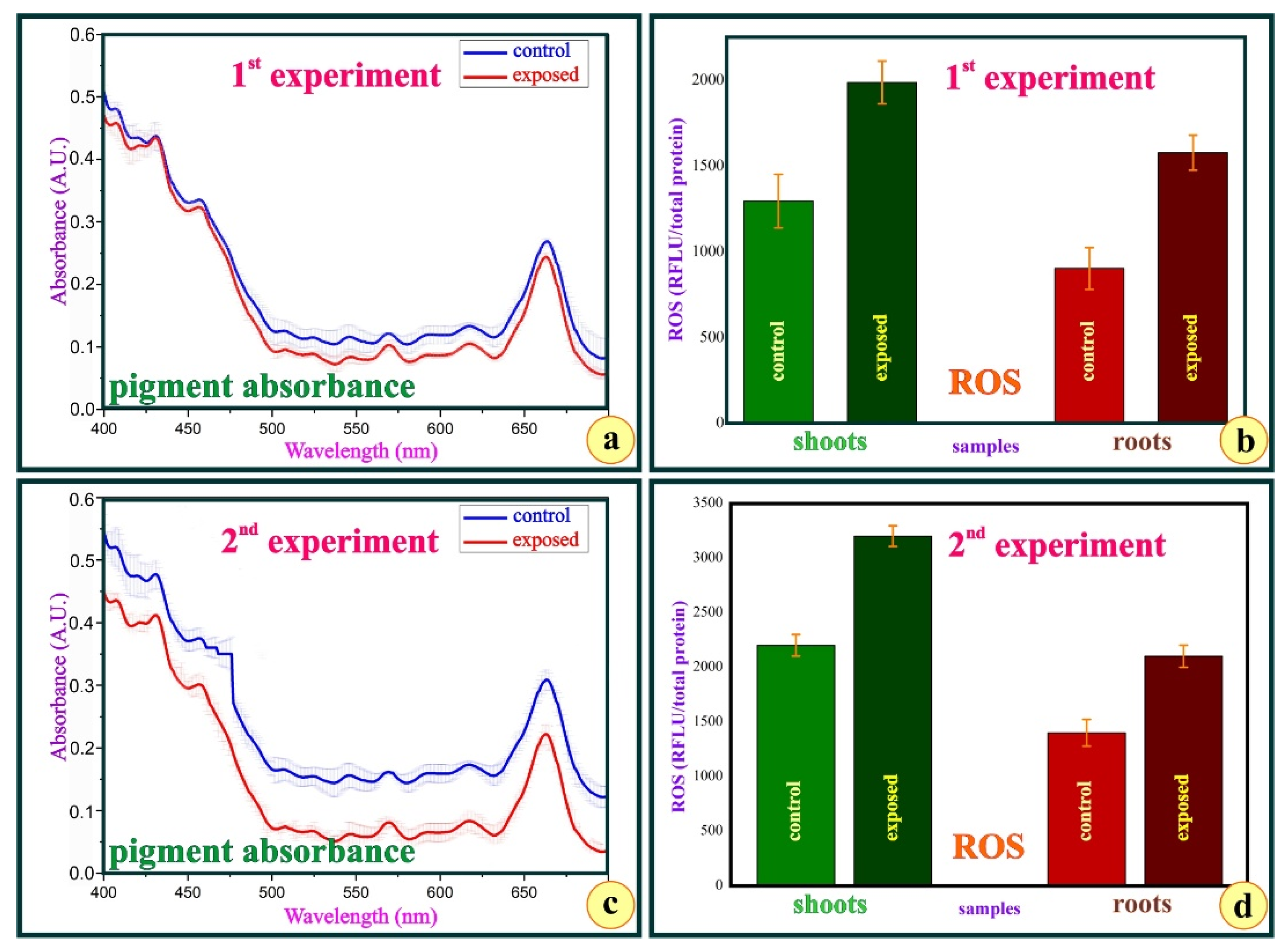
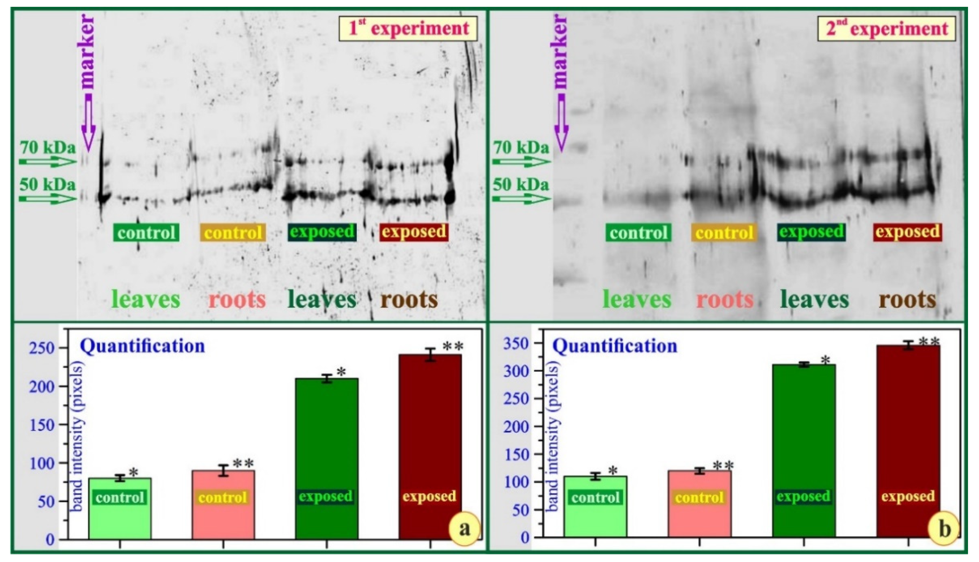
All numerical data, being subject to statistical analysis, were processed using the SPSS v.21.0 software (SPSS Inc., Chicago, IL, USA). The analysis was performed by employing paired t-tests comparing control samples to each one of the exposed samples. Data were previously checked for their normality. Data were expressed as mean ± standard error of the mean. The differences were considered statistically significant when p < 0.05 (*). N: number of biological samples, and n: number of biological experiments.

3. Results

Considering the biomass of the cultured plants from both experiments, we observed, at a first glance, that the two groups of plants looked rather similar (Figure 2). However, the biomass of the aboveground part of the control, compared to that of the exposed plants, seemed to differ significantly. In the first experiment, the exposed plants failed to match the productivity of their control counterparts, presenting a yield of 12% inferior for the aboveground parts, but it seems that they shifted their biomass production towards the root. The exposed plants produced 37% more biomass for their belowground parts (first experiment) (Figure 3) compared to the control plants. The corresponding values, for the second experiment, were −14% for the stem and +20% for the roots.
The pigment absorbance also seemed to be affected. The graphs of the pigment absorbance after the first experiment indicate that the leaves of the exposed plants presented reduced absorbance. This probably happens because the strain reduces their photosynthetic pigments through the damage of chloroplasts by active oxygen species [68] (Figure 4a). The same is true, to a higher extent, for the second experiment (Figure 4c).
A careful investigation of the oxidative stress, in the form of ROS, indicated that both the shoots and the roots of the exposed plants sustained severe oxidative stress. In the first experiment, the shoots of the exposed plants presented a statistically significant increase of the oxidative stress at a value of about 30% compared to the control plants (Figure 4b, left). A significant increase at a value of about 40% was recorded for the roots of the exposed plants, respectively (Figure 4b, right). The corresponding values were 36% (stems) and 45% (roots) for the second experiment (Figure 4d).
Finally, the immunodetection of DDC in the leaf samples of control and exposed plants indicated that, after the exposure, the levels of DDC protein expression rose remarkably in both the shoots and the roots. The immunodetected enzymes for the exposed plants, compared to the corresponding from the control ones, differed significantly in both experiments (Figure 5a,b), a fact clearly demonstrated by the quantification of the bands (Figure 5a,b, lower). Interestingly, the obtained data indicated the presence of two DDC immunoreactive signals in every sample tested. The presence of the higher MW DDC immunoreactive band could represent a yet unknown DDC isoform or post-translational modification. The same is true for the roots of the exposed plants (Figure 5a,b, roots). The upper band in all specimens (Figure 5a,b) probably indicates a weak expression of another transcript of the DDC, with an about similar molecular weight.
Observations of leaf cross-sections revealed some major differences among the two leaf types. Both leaf types (Figure 6a) were of limited thickness, with sized epidermal cells and mesophyll tissues loosely kept apart. This is a structure far from typical for a xeromorphic plant, probably because water stress does not exist in the culture chambers. The “control” leaves exhibited smaller cells in the adaxial epidermis, numerous cells accumulating condensed, osmiophilic phenolics, a few idioblasts with crystals, weak mechanical tissue, and stomata on both surfaces.
On the contrary, the “exposed” leaves (Figure 6b) were more or less similar in thickness, with thinner epidermal cells, distinct but single-layered palisade, and loose spongy parenchyma. Many mesophyll cells seem to accumulate loose, granular phenolic contents, while the mechanical tissue escorting the conductive bundles is hardly developed. Both leaf types possessed stomata on both surfaces: they were numerous on the surface of the “exposed” leaves, and thus could be observed even on the upper epidermis, in cross-sections of the leaves (red arrow in Figure 6b). Paradermal sections confirmed the fact that “control” leaves, although not exposed to the radiation stress, compared to their “exposed” counterparts, appeared to accumulate large quantities of condensed phenolics in the vacuoles of their mesophyll cells (compare Figure 6c,d).
Considering the biosynthesis of secondary metabolites as an interesting aspect of “plant life” when the primary metabolic pathways are unable to operate, mainly due to unfavorable environmental conditions, we assessed the phenolic content of both leaf types. It seems rather controversial, but it is totally true that the leaves of the non-stressed “control” plants seemed to accumulate as much as almost double the phenolics located in the leaves of the exposed plants, in both experiments (compare Figure 7a,b).
To establish the differences in the various attributes of the leaves after the seven weeks of culture and distinguish the effects of radiation on the exposed plants, we carefully observed the surfaces (adaxial and abaxial epidermis) of the leaves from wild-growing plants. This approach would provide some pieces of information on the effect the almost favorable conditions could have in the culture plants and verify the true effect of radiation on the exposed, cultured individuals.
The dorsiventral, hairy leaves of the wild-growing plants of Cistus creticus exhibited certain xeromorphic characteristics and have been thoroughly studied regarding their secretory trichomes and the difference of the mesophyll structure, the secretive function, and the secondary metabolites’ accumulation between the summer and the winter leaves of the plant [19]. The epidermal features of these leaves indicate the stress they sustain from the environment. The adaxial surface is wavy, with deep fissures and numerous, multicellular, spiny hair (Figure 8a). The abaxial surface is characterized by the projections of the leaf nerves, and the large number of trichomes which can be either spiny, multicellular-protective, or short, multicellular-secretive. The hair cover of the abaxial surface is thick (Figure 8b).
Among the cultured plants, the leaves of the “control” individuals seemed to retain a xeromorphic structure, yet their adaxial (upper) surface had few trichomes of rather simple structure (Figure 8c) compared to those of the leaves from the wild-growing plants. The same is true for the abaxial (lower) epidermis, which was rather flat, with no projection of the leaf nerves (Figure 8e). Both surfaces of the “control” leaves appeared to be less xeromorphic than those of the naturally growing plants. Concerning the “exposed” leaves, we easily observed the increased pubescence (Figure 8e,f), especially on the lower surface (Figure 8f), although trichomes appeared simple and fragile as in the “control” plants, being significantly different compared to those on the leaves of the wild-growing plants. An interesting difference between the “control” and the “exposed” leaves is the stomatal density.
The number of stomata at the surfaces of the “control” leaves was 17 ± 6 mm2 for the upper and 52 ± 9 mm2 for the lower epidermis (Figure 8c,d). In the exposed plants, the stomatal frequency rose significantly at the value of 89 ± 10 mm2 for the upper and 121 ± 12 mm2 for the lower epidermis (Figure 8e,f). This could somehow be considered as a reaction towards a xeromorphic assembly.
A detailed observation and counting of stomata on the lower (abaxial) surfaces from both the “control” and the “exposed” plants easily confirmed the significant difference in the number of stomata (circles in Figure 9a,b). The increase in stomatal frequency of the exposed plants was about 420% for the adaxial and 130% for the abaxial (Figure 9a,b) epidermis. Similar results were recorded in the second experiment (384% and 118% increase of stomatal frequency for the adaxial and abaxial surfaces, respectively).
Concerning the primary roots, sectioned at exactly the same length (2 mm) from the root tip and after numerous observations, we revealed that the roots of the exposed individuals seemed to delay their secondary development, although they finally surpassed the biomass of the roots from the “control” plants. As depicted in Figure 10, in all roots—main and lateral—of the “control” plants (Figure 10a), the development of the secondary xylem, and the secondary structure in general, appeared closer to the root tip. However, all the roots of their “exposed” counterparts (Figure 10b) seemed to retain their primary structure within a longer length above the root tip. This is an interesting observation to be discussed in the next section. Both root types appeared to be lined with a layer of phenolic-containing cells just where the endodermis and the pericycle are expected to be located.

4. Discussion

Prior to the discussion on the results of the current investigation, we must point out that the culture conditions within each of the chambers, compared to the normal Mediterranean environment, were far from being stressful concerning temperature, light intensity, and water availability, and thus, the plants were able to proceed with their development, adopting a more mesomorphic structure. This turnaround was primarily reflected in the most malleable organs of the plant, the leaves. The differences of the leaf structure of the cultivated plants, when compared to the wild-growing plants [19], were significant. However, in one of the chambers, despite the favorable growing conditions, a stressing environmental factor emerged: the radiation emitted from the base unit of the DECT apparatus.
The comparison of the biomass between the two groups of plants revealed an interesting response. At a first glance, the “phenotype” for both plant groups seemed similar, but when it comes to biomass (dry weight), interesting differences were revealed. Reduced leaf compactness of the stressed plants may be responsible for that. In the case of C. creticus, the aboveground biomass was reduced in the exposed plants, in both experiments, while the roots of these plants exhibited significantly higher development. The decrease of the aboveground biomass had a mean value of about −12.5% in both experiments, while the increase of the root biomass was about +37%. This is not a common phenomenon. In previous investigations for the effect of the non-ionizing radiation on plant species from various taxa (pinophyte, monocots, eudicots), the exposed plants exhibited lower biomass for both their aboveground parts and their roots [33,35]. An exception was reported for Zea mays, of which the exposed plants exhibited higher root biomass than their control counterparts [36]. The significant increase of the root biomass of the exposed plants may suggest that certain metabolic pathways responsible for the stem growth are retarded by radiation, while the roots take advantage of the protection offered within the soil micro-environment. Since this interpretation cannot be attributed to the plants mentioned above and reported in previous investigations, we can assume that the roots of the stress-tolerant C. creticus may have an ability to exploit the occasional “friendship” of the soil environment and take advantage of it.
The response of C. creticus to the radiation emitted within the chamber was also investigated in detail. We observed the structural features of the leaf, in both light and scanning electron micrographs, along with the root and some physiological parameters, such as the pigment absorbance, the levels of the stressful reactive oxygen species, the expression of the stress-relieving DDC, and other secondary metabolites.
The measurement of the absorbance spectrum for the photosynthetic pigments can provide basic information on the physiological status of a plant. Drought stress causes a major decline in the total chlorophyll content in all sunflower varieties investigated [69]. The decrease in chlorophyll under drought stress is also reported to be caused through the damage of chloroplasts by active oxygen species [68]. According to our data, the absorbance of the photosynthetic pigments appeared to decrease, and this reduction can be significant for a developing plant. Given that the graphs were created after normalizing per leaf tissue, the minor divergence in control vs. exposed samples at 400 nm in the second experiment can be explained.
It is well-documented that under stress conditions, the easily ruined organelles such as chloroplasts, mitochondria, and peroxisomes become the major ROS generation sites in plants [70,71,72]. Therefore, the photosynthetic apparatus is damaged by ROS generation under environmental stresses [72]. We observed that in C. creticus, the severely stressed leaves sustained ROS overproduction and suffered a reduction of their photosynthetic pigments, as indicated by the absorbance spectrum.
It has also been reported, repeatedly, that the oxidative stress is considered responsible for the production of phenolics, which play an important role for plants under stress conditions [73,74,75]. Assuming the ROS levels in the leaves of C. creticus trigger the secondary metabolic pathways, we observed that the stressed leaves appeared less prone to the phenolic accumulation habit (Figure 6 and Figure 7). This response is rather difficult to explain. It seems, to the best of our knowledge, unique among the Mediterranean plants investigated so far, and probably indicates a special strategy, introducing, for this species, a kind of specific tolerance against a variety of unfavorable environmental conditions.
Concerning the significant increase of the DDC expression in the stressed leaves, it has been reported that it is due to the urgent need for the decarboxylation of L-DOPA. This is an extremely toxic secondary metabolite whose biosynthesis is shared by animals and plants [37,38]. L-DOPA is converted to the well-known mammal neurotransmitter [45], the catecholamine dopamine [42], an antioxidant that can regulate carbohydrate metabolism. It can also serve as a precursor of some alkaloids, protect the plants against certain pathogens, and control nitrogen detoxification [45]. DDC expression seems to “erupt” after environmental stress since dopamine, the output of the DDC activity, is a potent antioxidant, acting as a ROS scavenger [76]. Therefore, it can help the plants defend themselves against stressing environmental factors, including pollutants [77], high solar irradiance, water deficit [38], and non-ionizing radiation [51], which cause excessive ROS production. A significant increase in DDC expression was also reported for myrtle (Myrtus communis L.) leaves after long-term exposure to mobile phone-generated, non-ionizing radiation [48], for Phlomis fruticosa after short-term exposure to severe environmental summer strain [39], as well as for the leaves of Olea europaea L. under combined environmental stress [44]. As far as the roots are concerned, the occurrence of L-DOPA decarboxylase has already been demonstrated in Cytisus scoparius [37] and Vigna unguiculata [78]. In Nerium oleander, the expression levels of DDC in roots exposed to a mobile phone-generated, non-ionizing radiation, were surprisingly increased up to 200%, as compared to the hardly detected band for their control counterparts [49]. In the case of C. creticus, exposed leaves and roots, in both experiments, exhibited massive DDC expression (Figure 5). Presumably, the stress was not “ignored”, and the defense mechanism was launched. The result of this defense strategy is among the impressive hits of the evolution. Under all circumstances, a secondary band, below that of 53 kDa, was apparent, and it probably depicts the expression of another transcript having a rather similar molecular weight. This observation is a matter for further investigation.
The tannin paradox of C. creticus also seems worthy to discuss. The “exposed” leaves seemed to scorn the common trait of the stressed Mediterranean plants, while their “control” counterparts, although experiencing the conditions of a “humid” culture, appeared to accumulate phenolics, in their condensed form, in all mesophyll cells. Since the biosynthesis of phenolics, in Mediterranean plants, is a very common “escape valve” to overcome such problems, the evaluation of the phenolic content of both leaf types could prove valuable. The biosynthesis of phenolics is genetically determined and has been “imprinted” in the genetic material of all Mediterranean plants. The accumulation of these metabolically inert compounds can be observed, to a great extent, in all Mediterranean plants, no matter what the environmental conditions are [12,13,15,16,79]. The more stressing the environmental conditions appear, the higher the rate of phenolic accumulation. In the case of C. creticus, we can assume that the radiation within the culture chamber probably creates a kind of relieving condition against some other stressful environmental factor, i.e., light intensity or temperature, and thus shifts plant metabolism to a more mesomorphic profile.
The above-described mesomorphic profile of the leaves cannot be exhibited in scanning electron micrographs, where the “exposed” leaves appeared to possess traits supporting the survival A higher stomatal frequency actually has an effect on the rate of CO2 uptake as well as the rate of transpiration [80]. Increased CO2 uptake can enhance CO2 fixation and, consequently, plant growth [81]. Moreover, stomatal short-term behavior and long-term developmental (e.g., stomatal size and its density) responses to environmental changes might also affect photosynthesis [82,83]. On the other hand, the reduced absorbance recorded in our investigation for the photosynthetic pigments does not seem to be in favor of high photosynthetic rates, eliminating the advantage of the higher stomatal frequency. The higher stomatal frequency and the increased CO2 uptake may be the reasons why exposed plants do not suffer extreme reductions of biomass production compared to their control counterparts. This incompatibility of the mesophyll structure and the surface topography, concerning the expression of xeromorphic traits, is difficult to explain and should be a matter of further, detailed investigation.

5. Conclusions

Cultivated plants of Cistus creticus exhibited a controversial response to the severe environmental stress of a long-term exposure to mobile phone-generated, non-ionizing radiation. They followed the common reaction of most Mediterranean plants concerning the considerable increase of ROS, the recession of the absorption spectrum of the photosynthetic pigments, the significant increase of the DDC expression, and the presence of some xeromorphic traits on both leaf surfaces. However, on the other hand, the higher root biomass of the “exposed” plants, the mesophyll structure of the “exposed” leaves, and the substandard accumulation of phenolics within the mesophyll cells do not testify to the existence of strain conditions. Probably, the plant can finely tune the metabolic pathways and its development, taking advantage of whatever seems in favor and opposing whatever seems hostile. Therefore, the application of radiation under these “hydroponic” conditions of the culture can produce plants of bilateral structure, being somehow xeromorphic and somehow mesomorphic.

Author Contributions

Conceptualization, A.L.S., D.V. and N.S.C.; data curation, A.S.S., D.V. and N.S.C.; formal analysis, A.L.S. and A.S.S.; investigation, A.L.S., G.K., D.V. and N.S.C.; methodology, A.L.S., G.K., D.V. and N.S.C.; project administration, N.S.C.; resources, D.V. and N.S.C.; software, A.S.S.; supervision, D.V. and N.S.C.; validation, A.S.S. and N.S.C.; visualization, A.L.S., G.K. and D.V.; writing—original draft, A.L.S., A.S.S., D.V. and N.S.C.; writing—review and editing, A.L.S. and N.S.C. All authors have read and agreed to the published version of the manuscript.

Funding

This research received no funding.

Institutional Review Board Statement

Not applicable.

Informed Consent Statement

Not applicable.

Data Availability Statement

Not applicable.

Acknowledgments

The authors deeply thank Maria Triantaphyllou for providing the JEOL JSM 6360 Scanning Electron Microscope facilities.

Conflicts of Interest

The authors declare no conflict of interest.

References

  1. Mitrakos, K. A theory of Mediterranean plant life. Oecologia Plant 1980, 1, 245–252. [Google Scholar]
  2. Mooney, H.A.; Parsons, D.J. Structure and Function in the California Chaparral—An Example from San Dimas. In Mediterranean Type Ecosystems. Origin and Structure; di Castri, F., Mooney, H.A.M., Eds.; Springer: Berlin, Germany, 1973; pp. 83–112. [Google Scholar]
  3. Christodoulakis, N.S.; Kogia, D.; Mavroeidi, D.; Fasseas, C. Anatomical and cytochemical investigation on the leaf of Teucrium polium L., a pharmaceutical shrub of the Greek phryganic formations. J. Biol. Res. 2010, 14, 199–209. [Google Scholar]
  4. Daget, P. Le bioclimat mediterraneen: Caracteres generaux, modes de caracterisation. Vegetatio 1977, 34, 1–20. [Google Scholar] [CrossRef]
  5. Nahal, I. The Mediterranean Climate from a Biological Viewpoint. In Ecosystems of the World, Mediterranean-Type Shrublands; de Castri, F., Goodall, D.W., Specht, R.L., Eds.; Elsevier Scientific Publishing Company: Amsterdam, The Netherlands, 1981; pp. 63–86. [Google Scholar]
  6. Hutchinson, J. The Families of Flowering Plants: Arranged According to a New System Based on Their Probable Phylogeny, 3rd ed.; Clarendon: Oxford, UK, 1973; ISBN 0-19-854377-8. [Google Scholar]
  7. Edlmann, M.L.A.; de Luca, L.; Lazzeri, S. Atlante Anatomico Degli Alberi ed Arbusti Della Macchia Mediterranea; (Relazioni e Monografie Agrarie Subtropicali e Tropicali, Nuova Serie 114.); Istituto Agronomico per l’ Oltremare: Firenze, Italy, 1994. [Google Scholar]
  8. De Micco, V.; Arena, C.; Vitale, L.; Aronne, G.; De Santo, A.V. Anatomy and photochemical behaviour of Mediterranean Cistus incanus winter leaves under natural outdoor and warmer indoor conditions. Botany 2011, 89, 677–688. [Google Scholar] [CrossRef]
  9. Dioscurides, A.C. De Materia Medica; Militos Publications: Athens, Greece, 2001. [Google Scholar]
  10. De Andrés, A.I.; Gómez-Serranillos, M.P.; Iglesias, I.; Villar, A.M. Effects of extract of Cistus populifolius L. on the central nervous system. Phytother. Res. 1999, 13, 575–579. [Google Scholar] [CrossRef]
  11. Küpeli, E.; Yesilada, E. Flavonoids with anti-inflammatory and antinociceptive activity from Cistus laurifolius L. leaves through bioassay-guided procedures. J. Ethnopharmacol. 2007, 112, 524–530. [Google Scholar] [CrossRef]
  12. Barrajõn-Catalán, E.; Fernández-Arroyo, S.; Roldán, C.; Guillén, E.; Saura, D.; Segura-Carretero, A.; Micol, V. A systematic study of the polyphenolic composition of aqueous extracts deriving from several Cistus genus species: Evolutionary relationship. Phytochem. Anal. 2011, 22, 303–312. [Google Scholar] [CrossRef]
  13. Barrajón-Catalán, E.; Fernández-Arroyo, S.; Saura, D.; Guillén, E.; Fernández-Gutiérrez, A.; Segura-Carretero, A.; Micol, V. Cistaceae aqueous extracts containing ellagitannins show antioxidant and antimicrobial capacity, and cytotoxic activity against human cancer cells. Food Chem. Toxicol. 2010, 48, 2273–2282. [Google Scholar] [CrossRef]
  14. Vitali, F.; Pennisi, G.; Attaguile, G.; Savoca, F.; Tita, B. Antiproliferative and cytotoxic activity of extracts from Cistus incanus L. and Cistus monspeliensis L. on human prostate cell lines. Nat. Prod. Res. 2011, 25, 188–202. [Google Scholar] [CrossRef]
  15. Droebner, K.; Ehrhardt, C.; Poetter, A.; Ludwig, S.; Planz, O. CYSTUS052, a polyphenol-rich plant extract, exerts anti-influenza virus activity in mice. Antivir. Res. 2007, 76, 1–10. [Google Scholar] [CrossRef]
  16. Chon, H. Medicinal herbs and plant extracts for influenza: Bioactivity, mechanism of anti-influenza effects, and modulation of immune responses. Stud. Nat. Prod. Chem. 2012, 38, 305–323. [Google Scholar]
  17. Aronne, G.; De Micco, V. Seasonal dimorphism in the Mediterranean Cistus incanus L. subsp. incanus. Ann. Bot. 2001, 87, 789–794. [Google Scholar] [CrossRef]
  18. Gülz, P.-G.; Herrmann, T.; Hangst, K. Leaf trichomes in the genus Cistus. Flora 1996, 191, 85–104. [Google Scholar] [CrossRef]
  19. Christodoulakis, N.S.; Georgoudi, M.; Fasseas, C. Leaf structure of Cistus creticus L. (rock rose), a medicinal plant widely used in folk remedies since ancient times. J. Herbs Spices Med. Plants 2014, 20, 103–114. [Google Scholar] [CrossRef]
  20. Demetzos, C.; Harvala, C.; Philianos, S.M.; Skaltsounis, A.L. A new labdane-type diterpene and other compounds from the leaves of Cistus incanus ssp. creticus. J. Nat. Prod. 1990, 53, 1365–1368. [Google Scholar] [CrossRef]
  21. Weyerstahl, P.; Marschall, H.; Weirauch, M.; Thefeld, K.; Surburg, H. Constituents of commercial Labdanum oil. Flavour Fragr. J. 1998, 13, 295–318. [Google Scholar] [CrossRef]
  22. Fahn, A.; Cutler, D.F. Xerophytes. In Encyclopedia of Plant Anatomy; Borntraeger: Berlin, Germany, 1992; Volume XIII. [Google Scholar]
  23. Catoni, R.L.; Gratani, L.; Varone, L. Physiological, morphological and anatomical trait variations between winter and summer leaves of Cistus species. Flora 2012, 207, 442–449. [Google Scholar] [CrossRef]
  24. Sharma, S.; Parihar, L. Effect of mobile phone radiation on nodule formation in the leguminous plants. Curr. World Environ. J. 2014, 9, 145–155. [Google Scholar] [CrossRef]
  25. Roux, D.; Vian, A.; Girard, S.; Bonnet, P.; Paladian, F.; Davies, E.; Ledoigt, G. Electromagnetic fields (900 MHz) evoke consistent molecular responses in tomato plants. Physiol. Plant. 2006, 128, 283–288. [Google Scholar] [CrossRef]
  26. Pietruszewski, S.; Muszyñski, S.; Dziwulska, A. Electromagnetic fields and electromagnetic radiation as non-invasive external stimulants for seeds (selected methods and responses). Int. Agrophysics 2007, 21, 95–100. [Google Scholar]
  27. Sheridan, S.; Kalkstein, L.; Kalkstein, A.; Greene, S. Heat-related Mortality and Heat Watch-warning Systems in the United States: Recent Developments. In Proceedings of the ISEE 22nd Annual Conference, Seoul, Korea, 28 August–1 September 2010. [Google Scholar]
  28. Vian, A.; Roux, D.; Girard, S.; Bonnet, P.; Paladian, F.; Davies, E.; Ledoigt, G. Microwave irradiation affects gene expression in plants. Plant Signal. Behav. 2006, 1, 67–70. [Google Scholar] [CrossRef] [PubMed]
  29. Haggerty, K. Adverse influence of radio frequency background on trembling aspen seedlings: Preliminary observations. Int. J. For. Res. 2010, 2010, 836278. [Google Scholar] [CrossRef]
  30. Kumar, A.; Singh, H.P.; Batish, D.R.; Kaur, S.; Kumar-Kohli, R. EMF radiations (1800 MHz)-inhibited early seedling growth of maize (Zea mays) involves alterations in starch and sucrose metabolism. Protoplasma 2015, 253, 1043–1099. [Google Scholar] [CrossRef]
  31. Hollósy, F. Effects of ultraviolet radiation on plant cells. Micron 2002, 33, 179–197. [Google Scholar] [CrossRef]
  32. Waldmann-Selsam, C.; Balmori-de la Puente, A.; Breunig, H.; Balmori, A. Radiofrequency radiation injures trees around mobile phone base stations. Sci. Total Environ. 2016, 572, 554–569. [Google Scholar] [CrossRef]
  33. Stefi, A.L.; Margaritis, L.H.; Christodoulakis, N.S. The effect of the non-ionizing radiation on exposed, laboratory cultivated maize (Zea mays L.) plants. Flora 2017, 233, 22–30. [Google Scholar] [CrossRef]
  34. Vian, A.; Davies, E.; Gendraud, M.; Bonnet, P. Plant responses to high frequency electromagnetic fields. Biomed. Res. Int. 2016, 2016, 1830262. [Google Scholar] [CrossRef]
  35. Stefi, A.L.; Margaritis, L.H.; Christodoulakis, N.S. The effect of the non-ionizing radiation on exposed, laboratory cultivated upland cotton (Gossypium hirsutum L.) plants. Flora 2017, 226, 55–64. [Google Scholar] [CrossRef]
  36. Stefi, A.L.; Margaritis, L.H.; Christodoulakis, N.S. The aftermath of long-term exposure to non-ionizing radiation on laboratory cultivated pine plants (Pinus halepensis M.). Flora 2017, 234, 173–186. [Google Scholar] [CrossRef]
  37. Tocher, R.D.; Tocher, C.S. Dopa decarboxylase in Cytisus scoparius. Phytochemistry 1972, 11, 1661–1667. [Google Scholar] [CrossRef]
  38. Iriti, M. Plant neurobiology, a fascinating perspective in the field of research on plant secondary metabolites. Int. J. Mol. Sci. 2013, 14, 10819–10821. [Google Scholar] [CrossRef] [PubMed]
  39. Stefi, A.L.; Vassilacopoulou, D.; Christodoulakis, N.S. Environmentally stressed summer leaves of the seasonally dimorphic Phlomis fruticosa and the relief through the L-Dopa decarboxylase (DDC). Flora 2019, 251, 11–19. [Google Scholar] [CrossRef]
  40. Jaffe, M.J. Evidence for the regulation of phytochrome-mediated processes in bean roots by the neurohumor, acetylcholine. Plant Physiol. 1970, 46, 768–777. [Google Scholar] [CrossRef] [PubMed]
  41. Barlow, R.B.; Dixon, R.O.D. Choline acetyltransferase in the nettle Urtica dioica L. Biochem. J. 1973, 132, 15–18. [Google Scholar] [CrossRef] [Green Version]
  42. Odjakova, M.; Hadjiivanova, C. Animal neurotransmitter substances in plants. Bulg. J. Plant Physiol. 1997, 23, 94–102. [Google Scholar]
  43. Soares, A.R.; Marchiosi, R.; Siqueira-Soares, R.d.C.; Lima, R.B.d.; Santos, W.D.d.; Ferrarese-Filho, O. The role of L-DOPA in plants. Plant Signal. Behav. 2014, 9, e28275. [Google Scholar] [CrossRef]
  44. Stefi, A.L.; Vassilacopoulou, D.; Routsi, E.; Stathopoulos, P.; Argyropoulou, A.; Skaltsounis, A.-L.; Christodoulakis, N.S. The Combined Environmental Stress on the Leaves of Olea europaea L. and the Relief Mechanism Through Biosynthesis of Certain Secondary Metabolites. J. Plant Growth Regul. 2020, 40, 1044–1059. [Google Scholar] [CrossRef]
  45. Kulma, A.; Szopa, J. Catecholamines are active compounds in plants. Plant Sci. 2007, 172, 433–440. [Google Scholar] [CrossRef]
  46. Liu, Q.; Gao, T.; Liu, Q.; Liu, Y.; Zhao, Y.; Liu, Y.; Li, W.; Ding, K.; Ma, F.; Li, C. Functions of dopamine in plants: A review. Plant Signal. Behav. 2020, 15, e1827782. [Google Scholar] [CrossRef]
  47. Stefi, A.L.; Margaritis, L.H.; Christodoulakis, N.S. The effect of the non-ionizing radiation on cultivated plants of Arabidopsis thaliana (Col.). Flora 2016, 223, 114–120. [Google Scholar] [CrossRef]
  48. Stefi, A.L.; Vassilacopoulou, D.; Margaritis, L.H.; Christodoulakis, N.S. Oxidative stress and an animal neurotransmitter synthesizing enzyme in the leaves of wild growing myrtle after exposure to GSM radiation. Flora 2018, 243, 67–76. [Google Scholar] [CrossRef]
  49. Aničić, N.; Patelou, E.; Papanikolaou, A.; Kanioura, A.; Valdesturli, C.; Arapitsas, P.; Skorić, M.; Dragićević, M.; Gašić, U.; Koukounaras, A.; et al. Comparative Metabolite and Gene Expression Analyses in Combination with Gene Characterization Revealed the Patterns of Flavonoid Accumulation During Cistus criticus subsp. creticus Fruit Development. Front. Plant Sci. 2021. [Google Scholar] [CrossRef] [PubMed]
  50. Atsalakis, E.; Chinou, I.; Makropoulou, M.; Karabournioti, S.; Graikou, K. Evaluation of Phenolic Compounds in Cistus creticus Bee Pollen from Greece. Antioxidant and Antimicrobial Properties. Nat. Prod. Commun. 2017. [Google Scholar] [CrossRef]
  51. Stefi, A.L.; Mitsigiorgi, K.; Vassilacopoulou, D.; Christodoulakis, N.S. Response of young Nerium oleander plants to long-term non-ionizing radiation. Planta 2020, 251, 108–125. [Google Scholar] [CrossRef] [PubMed]
  52. Doussi, M.A.; Thanos, C.A. Post-fire regeneration of hard seeded plants: Ecophysiology of seed germination. In Proceedings of the 2nd International Conference Forest Fire Research, Coimbra, Portugal, 21–24 November 1994. [Google Scholar]
  53. Margaritis, L.; Manta, A.K.; Kokkaliaris, K.D.; Schiza, D.; Alimisis, K.; Barkas, G.; Georgiou, E.; Giannakopoulou, O.; Kollia, I.; Kontogianni, G.; et al. Drosophila oogenesis as a bio-marker responding to EMF sources. Electromagn. Biol. Med. 2014, 33, 165–189. [Google Scholar] [CrossRef] [PubMed]
  54. ICNIRP. Guidelines for limiting exposure to time-varying electric, magnetic and electromagnetic fields (up to 300 GHz). Health Phys. 1998, 74, 494–522. [Google Scholar]
  55. Sabatini, D.; Bensch, K.; Barrnett, B. Cytochemistry and electron microscopy. The preservation of cellular ultrastructure and enzymatic activity by aldehyde fixation. J. Cell Biol. 1963, 17, 208–212. [Google Scholar]
  56. Christodoulakis, N.S.; Tsiarta, M.; Fasseas, C. Leaf structure and histochemical investigation in Papaver rhoeas L. (corn poppy). J. Herbs Spices Med. Plants 2013, 19, 119–131. [Google Scholar] [CrossRef]
  57. Gechev, T.; Mehterov, N.; Denev, I.; Hille, J. A Simple and Powerful Approach for Isolation of Arabidopsis Mutants with Increased Tolerance to H2O2 Induced Cell Death. Methods Enzymol. 2013, 527, 203–220. [Google Scholar] [CrossRef]
  58. Bradford, M.M. A rapid and sensitive method for the quantitation of microgram quantities of protein utilizing the principle of protein-dye binding. Ann. Biochem. 1976, 72, 248–254. [Google Scholar] [CrossRef]
  59. Laemmli, U.K. Cleavage of structural proteins during the assembly of the head of bacteriophage T4. Nature 1970, 227, 680–685. [Google Scholar] [CrossRef]
  60. Towbin, H.; Staehelin, T.; Gordon, J. Electrophoretic transfer of proteins from polyacrylamide gels to nitrocellulose sheets: Procedure and some applications. Proc. Natl. Acad. Sci. USA 1979, 76, 4350–4354. [Google Scholar] [CrossRef] [PubMed]
  61. Batteiger, B.; Newhall, W.J.; Jones, R.B. The use of Tween 20 as a blocking agent in the immunological detection of proteins transferred to nitrocellulose membranes. J. Immunol. Methods 1982, 55, 297–307. [Google Scholar] [CrossRef]
  62. Sunkar, R. Review: MicroRNAs with macro-effects on plant stress responses. Semin. Cell Dev. Biol. 2010, 21, 805–811. [Google Scholar] [CrossRef] [PubMed]
  63. Athanasakis, G.; Aligiannis, N.; Gonou-Zagou, Z.; Skaltsounis, A.-L.; Fokialakis, N. Antioxidant Properties of the Wild Edible Mushroom Lactarius salmonicolor. Full Communications. J. Med. Food 2013, 16, 760–764. [Google Scholar] [CrossRef] [PubMed]
  64. Kukula-Koch, W.; Aligiannis, N.; Halabalaki, M.; Skaltsounis, A.-L.; Glowniak, K.; Kalpoutzakis, E. Influence of extraction procedures on phenolic content and antioxidant activity of Cretan barberry herb. Food Chem. 2013, 138, 406–413. [Google Scholar] [CrossRef]
  65. Kouka, P.; Priftis, A.; Stagos, D.; Angelis, A.; Stathopoulos, P.; Xinos, N.; Skaltsounis, A.-L.; Mamoulakis, C.; Tsatsakis, A.M.; Spandidos, D.; et al. Assessment of the antioxidant activity of an olive oil total polyphenolic fraction and hydroxytyrosol from a Greek Olea europaea variety in endothelial cells and myoblasts. Int. J. Mol. Med. 2017, 40, 703–712. [Google Scholar] [CrossRef] [Green Version]
  66. Singleton, V.L.; Rossi, J.A., Jr. Colorimetry of total phenolics with phosphomolybdic-phosphotungtic acid reagent. Am. J. Enol. Vitic. 1965, 16, 144–158. [Google Scholar]
  67. Salluca, T.G.; Penarrieta, J.M.; Alvarado, J.A.; Bergenstahl, B. Determination of total phenolic compounds content and the antioxidant capacity of Andean tubers and roots (isaño, oca, ulluco and arracacha). Rev. Bol. Quim. 2008, 25, 59–62. [Google Scholar]
  68. Smirnoff, N. Antioxidant Systems and Plant Response to the Environment. In Environment and Plant Metabolism: Flexibility and Acclimation; Smirnoff, V., Ed.; BIOS Scientific Publishers: Oxford, UK, 1995; pp. 217–243. [Google Scholar]
  69. Manivannan, P.; Jaleel, C.A.; Sankar, B.; Kishorekumar, A.; Somasundaram, R.; Lakshmanan, G.M.A.; Panneerselvam, R.N. Growth, biochemical modifications and proline metabolism in Helianthus annuus L. as induced by drought stress. Colloids Surf. B Biointerfaces 2007, 59, 141–149. [Google Scholar] [CrossRef]
  70. Kratsch, H.A.; Wise, R.R. The ultrastructure of chilling stress. Plant Cell Environ. 2000, 23, 337–350. [Google Scholar] [CrossRef]
  71. Mittler, R.; Vanderauwera, S.; Gollery, M.; van Breusegem, F. Reactive oxygen gene network of plants. Trends Plant Sci. 2004, 9, 490–498. [Google Scholar] [CrossRef] [PubMed]
  72. Yamamoto, Y.; Aminaka, R.; Yoshioka, M.; Khatoon, M.; Komayama, K.; Takenaka, D.; Yamashita, A.; Nijo, N.; Inagawa, K.; Morita, N.; et al. Quality control of photosystem II: Impact of light and heat stresses. Photosynth. Res. 2008, 98, 589–608. [Google Scholar] [CrossRef]
  73. Stephanou, M.; Manetas, Y. The effects of seasons, exposure, enhanced UV-B radiation, and water stress on leaf epicuticular and internal UV-B absorbing capacity of Cistus creticus: A Mediterranean field study. J. Exp. Bot. 1997, 48, 1977–1985. [Google Scholar] [CrossRef]
  74. Nikolakaki, A.; Christodoulakis, N.S. Secreting formations and cytochemical investigation of the leaf of Phlomis fruticosa, a seasonally dimorphic subshrub. Secreting activity of the leaf-originating calluses. Flora 2007, 202, 429–436. [Google Scholar] [CrossRef]
  75. Weidner, S.; Karolac, M.; Karamać, M.; Kosińska, A.; Amarowitcz, R. Phenolic compounds and properties of antioxidants in grapevine roots (Vitis vinifera L.) under drought stress followed by recovery. Acta Soc. Bot. Pol. 2009, 78, 97–103. [Google Scholar] [CrossRef]
  76. Kanazawa, K.; Sakakibara, H. High content of dopamine, a strong antioxidant, in Cavendish banana. J. Agric. Food Chem. 2000, 48, 844–848. [Google Scholar] [CrossRef]
  77. Pedroso, A.N.V.; Bussotti, F.; Tani, C.; Papini, A.; Domingos, M. Pollution emissions from a petrochemical complex and other environmental stressors induce structural and ultrastructural damage in leaves of a biosensor tree species from the Atlantic Rain Forest. Ecol. Indic. 2016, 67, 215–226. [Google Scholar] [CrossRef]
  78. Kaur, A.; Thurkral, A.K. Effect of animal hormones on the growth, protein and sugar contents of Vigna unguiculate L. seedlings. Indian J. Plant Physiol. 1990, 33, 259–261. [Google Scholar]
  79. Christodoulakis, N.S.; Fasseas, C. Air pollution effects on the leaf structure of Laurus nobilis, an injury resistant species. Bull. Environ. Contam. Toxicol. 1990, 44, 276–281. [Google Scholar] [CrossRef]
  80. Xu, Z.; Jiang, Y.; Jia, B.; Zhou, G. Elevated-CO2 Response of Stomata and Its Dependence on Environmental Factors. Front. Plant Sci. 2016, 7, 657. [Google Scholar] [CrossRef] [PubMed]
  81. Ainsworth, E.A.; Rogers, A. The response of photosynthesis and stomatal conductance to rising [CO2]: Mechanisms and environmental interactions. Plant Cell Environ. 2007, 30, 258–270. [Google Scholar] [CrossRef] [PubMed]
  82. Haworth, M.; Elliott-Kingston, C.; McElwain, J.C. Co-ordination of physiological and morphological responses of stomata to elevated [CO2] in vascular plants. Oecologia 2013, 171, 71–82. [Google Scholar] [CrossRef] [PubMed]
  83. DaMatta, F.M.; Godoy, A.G.; Menezes-Silva, P.E.; Martins, S.C.V.; Sanglard, L.M.V.P.; Morais, L.E.; Torre-Neto, A.; Ghini, R. Sustained enhancement of photosynthesis in coffee trees grown under free-air CO2 enrichment conditions: Disentangling the contributions of stomatal, mesophyll, and biochemical limitations. J. Exp. Bot. 2016, 67, 341–352. [Google Scholar] [CrossRef] [Green Version]
Figure 1. (a) A wild-growing individual of Cistus creticus. (b) The plastic pots with the young plants, ready for incubation. (c) The two cages accommodated within the P-Selecta incubator at the beginning of the experiment. (d) The two cages removed from the incubator at the end of the experiment, after seven weeks. The left cage contains the irradiated plants. The yellow arrow points at the DECT base unit.
Biophysica 02 00024 g001
Figure 2. The two groups of cultured, fresh individuals of C. creticus. Primarily, the differences in biomass.
Biophysica 02 00024 g002
Figure 3. Biomass for the two groups of plants (a) after the first experiment (left): the aboveground parts of the exposed plants (shoots) presented 12% lower productivity, while the roots appeared to be 37% higher in biomass compared to their control counterparts. (b) After the second experiment (right): the aboveground parts of the exposed plants presented 14% lower productivity, while the roots were 20% more productive in biomass.
Biophysica 02 00024 g003
Figure 4. (a) The absorption spectrum of the photosynthetic pigments after the first experiment. (b) Bar graphs depicting measured reactive oxygen species (ROS) levels after the first experiment. (c) The absorption spectrum of the photosynthetic pigments after the second experiment. (d) Bar graphs depicting measured ROS levels after the second experiment. Differences were considered as statistically significant when p < 0.05 (*). Error bars represent the standard error of the mean (SEM, N = 4, n = 2).
Biophysica 02 00024 g004
Figure 5. Immunodetection of L-Dopa DeCaboxylase (DDC) in leaf samples of “control” and “exposed” plants of C. creticus using the polyclonal specific anti-human DDC C-terminal antibody. Error bars represent the standard error of the mean (SEM). Subfigures (a) and (b) depict the quantification of the lower bands of each sample is provided through the graphs. Differences were considered as statistically significant when p < 0.05. Asterisks indicate statistically significant differences: * comparison between control and exposed leaves, and ** between control and exposed roots.
Biophysica 02 00024 g005
Figure 6. Cross-sections of a leaf from (a) a control plants and (b) an exposed plant. Paradermal sections from a leaf of (c) a control plant and (d) an exposed plant. pp = Palisade parenchyma, sp = spongy parenchyma. Red arrow points at a stoma on the adaxial surface, yellow arrows point at idioblasts with crystals. (All scale bars are 100 μm).
Biophysica 02 00024 g006
Figure 7. The total phenolic content of (a) the leaves of the control (left) and the exposed (right) plants from the first experiment and (b) the leaves of the control (left) and the exposed (right) plants from the second experiment. Differences were considered as statistically significant if p > 0.05. Asterisks (*) indicate statistically significant differences.
Biophysica 02 00024 g007
Figure 8. (a) The upper epidermis of the leaf from a wild-growing plant. Stomata are absent. (b) The lower epidermis of the leaf from a wild-growing plant. Notice the thick pubescence. Some of the stomata are pointed out by arrows. (c) The upper epidermis of the leaves of the “control” plants. Hair and stomata appear sporadically. (d) The lower epidermis of the leaves of the “control” plants. Multicellular, spiny hair can be observed along with stomata. (e) The upper epidermis of the leaves of the “exposed” plants appears. Trichomes and stomata can be observed. (f) The lower epidermis of the leaves of the “exposed” plants. Many trichomes and numerous stomata can be observed. Yellow arrows, in all figures, point at stomata, selectively (i.e., in (f), stomata are marked only for a central area of the picture). In (cf) the stomatal frequency is given (down, left) as number of stomata/mm2. (All scale bars are 200 μm).
Biophysica 02 00024 g008
Figure 9. (a) Detail of the lower epidermis of a “control” plant. Each stoma is traced by a circle around it. (b) Detail from the lower epidermis of an “exposed” plant. The stomata can easily be observed. (All scale bars are 50 μm).
Biophysica 02 00024 g009
Figure 10. Cross-sections of the roots at 2 mm above the root tip. (a) The root of the “control” plant. The developed xylem can easily be observed. (b) The root of an “exposed” plant. The xylem is hardly developed, while many conductive elements still possess non-lignified walls. In both sections, the layer of tannin-containing cells, surrounding the central cylinder, can easily be distinguished. (All scale bars are 100 μm).
Biophysica 02 00024 g010
Publisher’s Note: MDPI stays neutral with regard to jurisdictional claims in published maps and institutional affiliations.

