Article

On the Molecular Driving Force of Protein–Protein Association

Dipartimento di Scienze e Tecnologie, Università del Sannio, Via Francesco de Sanctis, 82100 Benevento, Italy
*
Author to whom correspondence should be addressed.
Biophysica 2022, 2(3), 240-247; https://doi.org/10.3390/biophysica2030023
Submission received: 30 July 2022 / Revised: 20 August 2022 / Accepted: 22 August 2022 / Published: 25 August 2022
(This article belongs to the Collection Feature Papers in Biophysics)

Abstract

:
The amount of water-accessible-surface-area, WASA, buried upon protein–protein association is a good measure of the non-covalent complex stability in water; however, the dependence of the binding Gibbs free energy change upon buried WASA proves to be not trivial. We assign a precise physicochemical role to buried WASA in the thermodynamics of non-covalent association and perform close scrutiny of the contributions favoring and those contrasting protein–protein association. The analysis indicates that the decrease in solvent-excluded volume, an entropic effect, described by means of buried WASA, is the molecular driving force of non-covalent association in water.

1. Introduction

Protein–protein association is an archetypal example of molecular recognition whose relevance in governing almost all biological processes is well-established [1,2,3]. In the last years, it emerged that molecular recognition is fundamental for the growth of amyloid-beta fibrils [4], other types of fibrils [5], and the formation of biomolecular condensates through liquid-liquid phase separation [6]. Proteins do not have eyes, but do have very rugged surfaces; nevertheless, they are able to find the right partner to construct the correct non-covalent complex. Notwithstanding the huge number of experimental studies carried out by means of different techniques to determine the structural features of the complexes and the thermodynamics of their formation, a general consensus on the molecular driving force of non-covalent association is still lacking. The present work is not a review of this huge matter, but a study aimed at offering a partial explanation of the important results obtained by Lynne Regan some years ago. She and co-workers [7] tried to make a step forward by performing a detailed analysis on 113 non-covalent heterodimers whose structures have been solved at a resolution of better than 3 Å and are present in the Protein Data Bank, PDB [8], and whose binding constants have carefully been determined and are present in the PDBbind v2011 database [9]; they found that the magnitude of the binding constant increases with the amount of buried WASA [10], ΔWASA, but did not find a clear correlation with the chemical nature (i.e., polar or nonpolar) of buried WASA. Note, in this respect, that buried WASA in the 113 heterodimers proved to be, on the average, 60% of nonpolar nature; this finding is not surprising nor unexpected (even though it is generally believed that nonpolar side chains are clustered in the protein interior), because it is in line with the average value determined for the nonpolar fraction of the surface of the native structure of a large set of globular proteins [11,12] (i.e., the buried WASA that drives protein–protein association has nothing special); moreover, Regan and co-workers constructed a plot of the ratio of the binding Gibbs free energy changes, at 298 K, to the buried WASA, ΔGb/ΔWASA, versus the buried WASA for the 113 heterodimers, obtaining the following, somewhat unexpected, results (look at Figure 1, that is a reconstruction of Figure 1B of the article by Regan and co-workers): ΔGb/ΔWASA decreases almost linearly for |ΔWASA| < 2000 Å2, and remains practically constant for |ΔWASA| ≥ 2000 Å2; this means that 1 Å2 of buried WASA contributes more to the binding constant when |ΔWASA| < 2000 Å2. A tentative explanation is that when the buried WASA is not large, there are few hot spots that are enough to render tight the non-covalent complex; in contrast, when the buried surface increases, the number of interaction sites is large, and there is no need of hot spots to have a tight association. In fact, for |ΔWASA| ≥ 2000 Å2, ΔGb/ΔWASA ≈ 17 J mol−1Å−2 so that, at 300 K, Kb = 8.3 × 10⁵ M−1 for ΔWASA = −2000 Å2, and Kb = 7.6 × 10⁸ M−1 for ΔWASA = −3000 Å2. The above Kb estimates are in line with experimentally determined values of the binding constant [7]; note, in this respect, that Kb cannot be too large because non-covalent complexes have to be as stable as necessary to perform their biological function and to dissociate when requested by the cell.
The dependence of ΔGb/ΔWASA versus |ΔWASA| should merit special attention because it contains the rules governing the thermodynamics of protein–protein association (even though in a non-transparent fashion); however, to the best of our knowledge, no explanation has been provided up to now for this interesting trend. Buried WASA is a fundamental factor in driving protein–protein association, but other contributions play an important role. To shed light on the matter, it is necessary to devise a theoretical approach able to provide a molecular-level rationalization of the heuristic finding that buried WASA is a fundamental factor; this is exactly the aim of the present study.

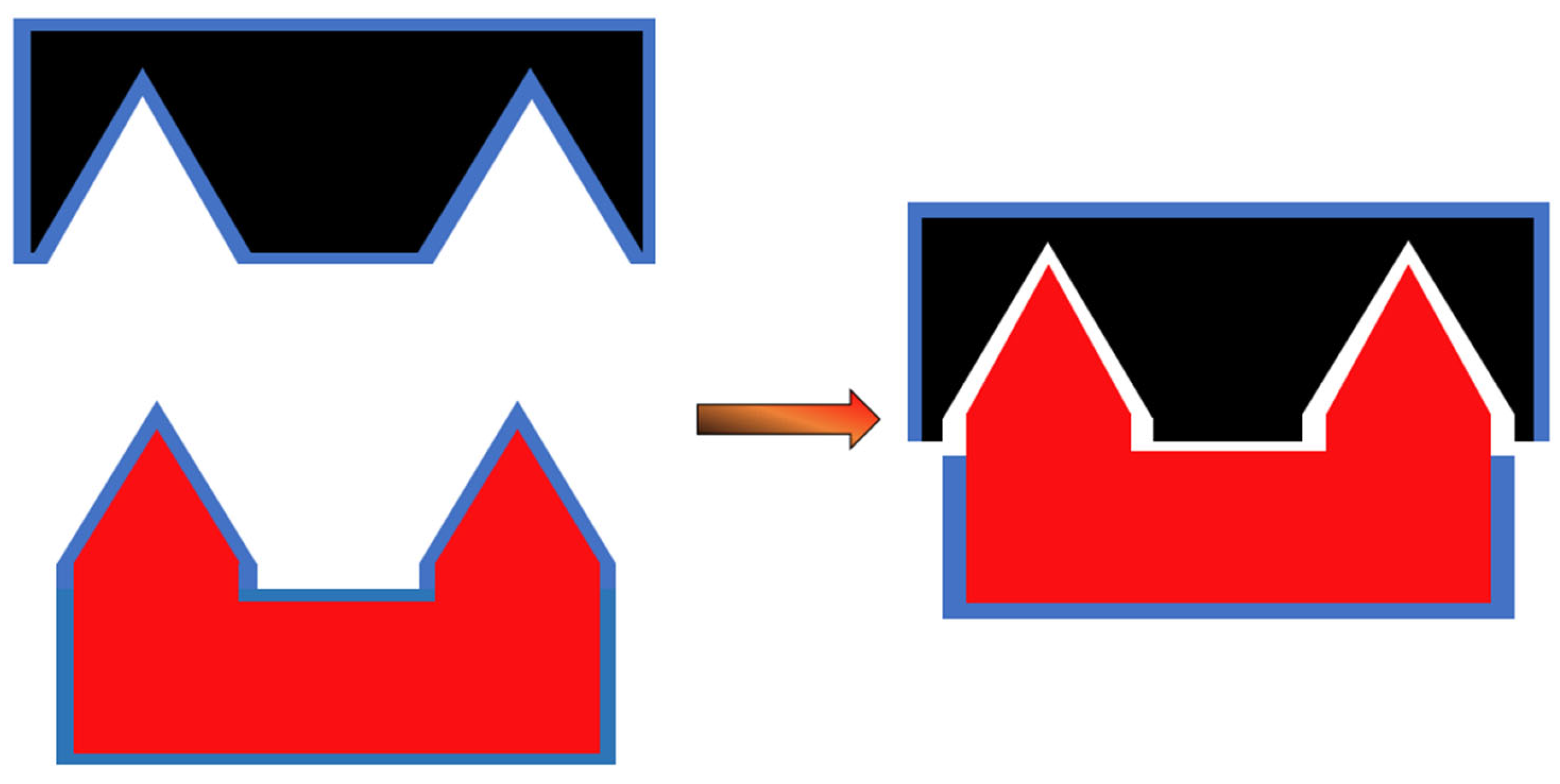
2. The Solvent-Excluded Volume Effect

The presence of a solute molecule, at a fixed position, in a liquid causes a solvent-excluded volume effect because the center of solvent molecules cannot penetrate the solvent-accessible-surface-area of the solute molecule (exactly because the latter occupies that space) [12]. The solvent-accessible-surface-area is the surface generated by a sphere, corresponding to the solvent molecule, that rolls over the van der Waals surface of the solute molecule [10]. In water, it is called water-accessible-surface-area, WASA, and the rolling sphere usually has a radius of 1.4 Å. On inserting a solute molecule in water, keeping constant pressure and temperature, the liquid volume increases by the partial molar volume of the solute, but this fact does not cancel the solvent-excluded volume effect, because the latter is caused by the impossibility of the center of water molecules to penetrate the solute WASA; it corresponds to a decrease in the configurational space accessible to water molecules (i.e., solvent molecules in the general case), and so to a loss in the translational entropy of water molecules (not only the ones that contact the solute van der Waals surface, because the residence time in the hydration shell is very very short). The important question is: how is it possible to account for the solvent-excluded volume effect in theoretical treatments of processes and phenomena occurring in water or other liquids? The answer is that the solvent-excluded volume effect has been associated with the theoretical concept of cavity creation in a liquid [13]. The existence of a cavity in water, due to the occurrence of molecular scale density fluctuations at equilibrium, requires that the center of water molecules cannot enter the cavity WASA. Therefore, a loss in translational entropy of water molecules is associated with cavity creation and determines the corresponding Gibbs free energy cost. All this reasoning implies that the ΔGc magnitude increases, even keeping constant the van der Waals volume of the cavity, with cavity WASA (i.e., WASA of the solute molecule to be hosted), and this expectation is confirmed by calculations [14,15]. Since the solvent-excluded volume effect is ubiquitous in processes occurring in liquids, especially in water, it should not be a surprise that several experimental measurements have shown a strong dependence on solute WASA.
Among others the experimental determination of the binding constant for the formation of protein–protein heterodimers, as pointed out by Lynne Regan and co-workers [7]; moreover, the geometric origin of the solvent-excluded volume effect implies that the distinction between nonpolar WASA and polar one is irrelevant (the chemical nature does not matter); in fact, no correlation between Kimplies that the and the buried nonpolar WASA was detected by Regan and co-workers [7]. The large WASA decrease associated with the formation of a non-covalent complex is schematically shown in Figure 2. WASA burial leads to a re-gain of translational entropy of water molecules (i.e., it corresponds to an increase in the configurational space accessible to water molecules), and provides an always negative Gibbs free energy change driving association.

3. Theory Section

Several theoretical approaches have been developed to quantitatively describe molecular recognition phenomena [16,17,18]. In particular, the ones devised by Honig and co-workers [19], and by Jackson and Sternberg [20] are similar to that described below; the main difference lies in the idea of and the role assigned to the solvent-excluded volume effect. In order to devise a theoretical framework to describe the formation of a protein–protein non-covalent complex, it is useful to consider the process of bringing two protein molecules from a fixed position at infinite separation to a fixed position at contact distance in water, keeping constant pressure and temperature [21]. In this manner, the translational and rotational entropy loss due to non-covalent complex formation is neglected (but it has to be considered later to make a correct comparison with experimental data that also account for this entropy loss). The binding Gibbs free energy change proves to be:
ΔGb = ΔG(dir) + ΔG(ind)
where ΔG(dir) represents the Gibbs free energy change due to the direct interaction of the two protein molecules in the non-covalent complex, and is independent of water; it consists of both an energetic contribution and an entropic one:
ΔG(dir) = E(M-M) − T⋅ΔS(M-M)
where E(M-M) is the energy gain due to direct Monomer-Monomer interactions in the complex, and ΔS(M-M) represents the loss in conformational entropy of the side chains exposed on the surfaces as a consequence of non-covalent complex formation (i.e., the interdigitation of side chains protruding from the surfaces of the two Monomers causes both an energy gain and an entropy loss; this situation is clearly different from that considered for the association of two rigid bodies, such as two large and flat plates, in which the ΔS(M-M) term is neglected [22]). ΔG(ind) represents the indirect part of the Gibbs free energy necessary to carry out the association, and accounts for the features of water, the liquid in which the non-covalent complex (i.e., the Dimer) formation takes place. ΔG(ind) is exactly related to the Ben-Naim standard hydration Gibbs free energy of the Dimer and Monomer, respectively [22]:
ΔG(ind) = ΔG(D/w) − 2⋅ΔG(M/w)
where ΔG is the Gibbs free energy change associated with the transfer of a solute molecule from a fixed position in the ideal gas phase to a fixed position in the water, at constant temperature and pressure. Statistical mechanics allows the exact division of ΔG in two contributions [13]:
ΔG = ΔGc + Ea
where ΔGc is the Gibbs free energy change associated with the creation, at a fixed position in the water, of a cavity, adapt to host the solute molecule, and Ea is the energy gain associated with switching on the solute-water attractive interactions. Clearly, solute insertion in water causes a structural reorganization of water-water H-bonds; the latter process is characterized by an almost complete enthalpy-entropy compensation [13]:
ΔHr = T⋅ΔSr
and so it does not affect the binding Gibbs free energy change. Note that the validity of Equation (5) has directly been verified for the pocket-ligand association by means of molecular dynamics simulations in the TIP4P water model by McCammon and colleagues [23,24]. Inserting Equation (4) into Equation (3), the latter becomes:
ΔG(ind) = [ΔGc(D/w) − 2⋅ΔGc(M/w)] + [Ea(D/w) − 2⋅Ea(M/w)]
To use this equation, it is necessary: (a) to know the structure of the two Monomers and that of the Dimer; (b) to calculate ΔGc and Ea for these structures in water, and to perform the requested differences; this would be a formidable task, especially because the calculation of the reversible work of cavity creation in water is not computationally feasible for large and complex molecules such as globular proteins; however, analyses performed by means of classic scaled particle theory [25], SPT, indicated that ΔGc scales linearly with cavity WASA for simple cavity shapes [15]; the latter scaling has been confirmed by means of computer simulations in detailed water models [14,26]; moreover, it holds also for the Ea quantity [14,27]. On this basis, Equation (6) can be re-written as:
ΔG(ind) = [(ΔGc/WASA) + (Ea/WASA)]⋅ΔWASA
The (ΔGc/WASA) ratio is always a positive quantity, the (Ea/WASA) ratio is always a negative quantity, and ΔWASA is a negative quantity for the formation of a non-covalent complex. If the two protein molecules interact through two large and complementary surfaces, it is feasible to conclude that non-covalent complex formation causes a large WASA burial with the loss of a large fraction of protein-water attractive interactions and the gain of a lot of protein–protein attractive interactions (i.e., the occurrence of well-designed attractions between the two surfaces is an obligatory necessity to partially balance the energy loss due to the breaking of protein-water attractions, for instance, a large number of protein-water H-bonds). A reliable assumption is that these two contrasting contributions compensate each other to a large extent, so that:
E(M-M) ≈ f⋅(Ea/WASA)⋅ΔWASA
where f is a number smaller than 1, representing the fraction of the Monomer-water interaction energy that is compensated for by the direct Monomer-Monomer attractions. In this manner, the overall binding Gibbs free energy change is:
ΔGb = [(ΔGc/WASA) + (1 − f)⋅(Ea/WASA)]⋅ΔWASA − T⋅ΔS(M-M)
Now, it is necessary to assign numerical values to the different contributions present in Equation (9) and to make a comparison with the experimental ΔGb data of Regan and co-workers [7].

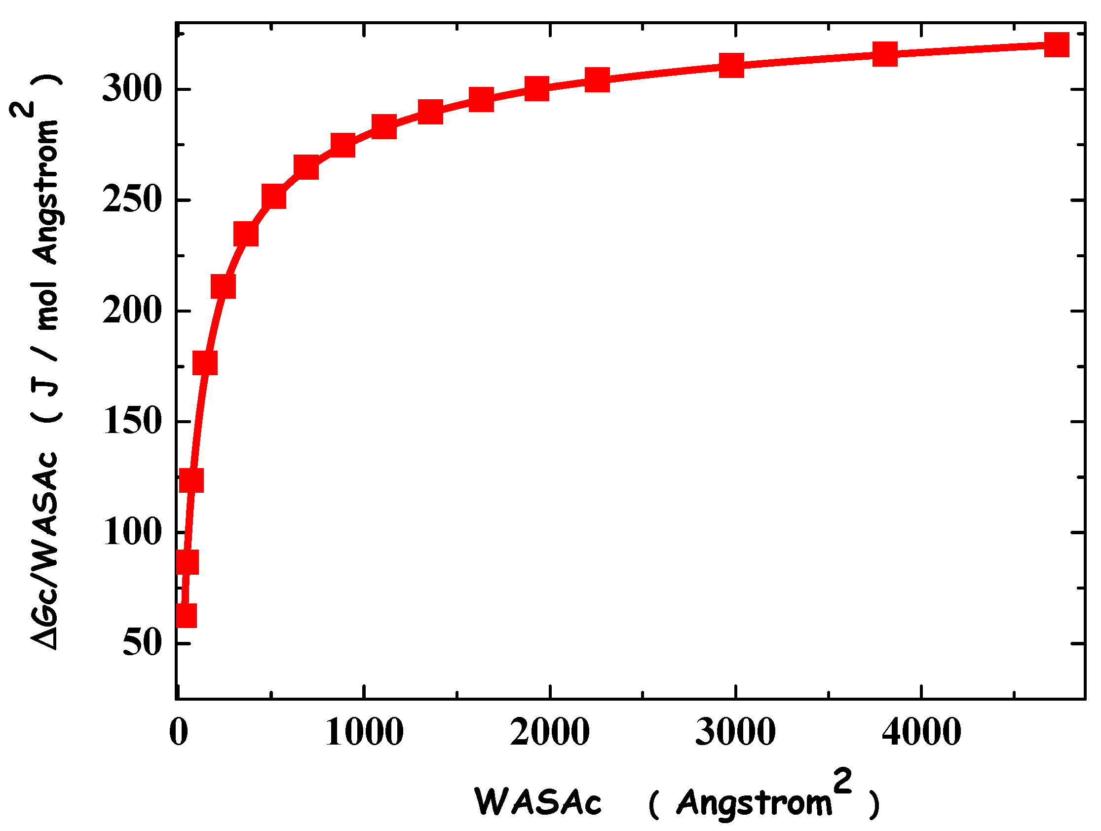
4. Results and Discussion

Using classic SPT relationships [15,25], it is straightforward to calculate ΔGc for spherical cavities of increasing radius, and to construct a plot of ΔGc/WASAc versus WASAc. The plot is shown in Figure 3 and refers to 298 K and 1 atm; it is characterized by a linear region for small cavities and a plateau region for large cavities, as it is firmly established [14]; this trend seems to be the mirror image of that shown in Figure 1. At the plateau, the ratio ΔGc/WASAc ≈ 300 J mol−1Å−2 and so it is markedly larger than the plateau of ΔGb/ΔWASA ≈ 17 J mol−1Å−2. WASA burial implies a decrease in the solvent-excluded volume effect and so a re-gain of translational entropy of water molecules; the latter provides a favorable contribution to the formation of a non-covalent complex. The above numerical comparison, however, indicates that WASA burial also leads to unfavorable contributions that are able to almost cancel the ΔGc/WASAc term. First of all, the two surfaces that produce the non-covalent complex have good energetic attractions among each other, but, in all probability, these attractions are not able to fully compensate for the loss of Monomer-water attractions due to WASA burial. At 298 K, the ratio (Ea/WASA) ≈ −250 J mol−1Å−2 for n-alkanes in water [27]; for a protein surface that is 60% nonpolar and 40% polar, the ΔEa/WASA) ratio can amount to about −350 J mol−1Å−2; it should be reliable to assume that about 2/5 of this value, −150 J mol−1Å−2, are not compensated by E(M-M) and must be subtracted to the (ΔGc/WASAc) contribution. On the other hand, WASA burial and side chain interdigitation lead to a conformational entropy loss whose magnitude rises on increasing ΔWASA (i.e., the freezing of side chain dihedral angles is more effective in enlarging the interacting surfaces). By taking into account the existing estimates of side chain conformational entropy [28] and the side chain WASA [29], the T⋅ΔS(M-M)/WASA] ratio should amount to about −100 J mol−1Å−2, at 298 K.
Finally, it is necessary to account for the loss of translational and rotational entropy caused by the formation of a non-covalent complex (i.e., two molecules become a single object), a contribution neglected in the theoretical treatment because it is convenient to assume the two molecules be in a fixed position. According to Janin and Finkelstein [30], this entropy loss produces a positive Gibbs free energy change of about 60 kJ mol−1 at 298 K. If ΔWASA = −2000 Å2, this unfavorable contribution would be −30 J mol−1Å−2; if ΔWASA = −3000 Å2, this unfavorable contribution would be −20 J mol−1Å−2. Therefore, putting together all our estimates, one obtains:
ΔGb/ΔWASA = 300 − 150 − 100 − 30 (20) = 20 (30) J mol−1Å−2
These two numbers are surprisingly close to the plateau value in Figure 1, as originally determined by Regan and co-workers [7]. In all probability, such a good agreement is in part the consequence of error cancelation. Nevertheless, the theoretical analysis is correct in singling out the contributions favoring and those contrasting non-covalent association in water; it is interesting that all the terms in Equation (9) depend strongly on buried WASA, confirming the pivotal role played by this geometric measure in shedding light on both structural and thermodynamic features of proteins [10]; this is the physical ground of the several computational procedures devised to quantitatively characterize the energetics of molecular recognition phenomena [31,32,33].

5. Conclusions

Protein–protein association to form a non-covalent complex is an archetypal example of molecular recognition; it seems to be ruled by the amount of WASA buried in the complex, such as other non-covalent association phenomena in water (i.e., micelle formation). Regan and co-workers performed an interesting analysis of the dependence of the ΔGb/ΔWASA ratio upon ΔWASA for 113 non-covalent heterodimers [7]. Since the database is large, the results are robust and merit a tentative rationalization. We have highlighted that the amount of buried WASA is a measure of the decrease in solvent-excluded volume effect, and so a measure of the gain in translational entropy of water molecules upon complex formation. The latter entropy gain is the molecular driving force of non-covalent association, even though contrasting contributions are operative and produce the unexpected plateau in the trend of ΔGb/ΔWASA versus |ΔWASA|.

Author Contributions

Conceptualization, G.G.; methodology, G.G. and R.R.; validation, G.G. and R.R.; writing—original draft preparation, R.R. and G.G.; writing—review and editing, R.R. and G.G.; supervision, G.G.; project administration, G.G.; funding acquisition, G.G. All authors have read and agreed to the published version of the manuscript.

Funding

This research was funded by Università degli Studi del Sannio, FRA 2021.

Data Availability Statement

Not applicable.

Conflicts of Interest

The authors declare no conflict of interest.

References

  1. Chothia, C.; Janin, J. Principles of protein-protein recognition. Nature 1975, 256, 705–708. [Google Scholar] [CrossRef] [PubMed]
  2. Marcotte, E.M.; Pellegrini, M.; Ng, H.L.; Rice, D.W.; Yeates, T.O.; Eisenberg, D. Detecting protein function and protein-protein interactions from genome sequences. Science 1999, 285, 751–753. [Google Scholar] [CrossRef] [PubMed]
  3. Wodak, S.J.; Vlasblom, J.; Turinsky, A.L.; Pu, S. Protein-protein interaction networks: The puzzling riches. Curr. Opin. Struct. Biol. 2013, 23, 941–953. [Google Scholar] [CrossRef] [PubMed]
  4. Xu, Y.; Knapp, K.; Le, K.N.; Schafer, N.P.; Safari, M.S.; Davtyan, A.; Wolynes, P.G.; Vekilov, P.G. Frustrated peptide chains at the fibril tip control the kinetics of growth of amyloid-beta fibrils. Proc. Natl. Acad. Sci. USA 2021, 118. [Google Scholar] [CrossRef]
  5. Kumar, A.; Chakraborty, D.; Mugnai, M.L.; Straub, J.E.; Thirumalai, D. Sequence Determines the Switch in the Fibril Forming Regions in the Low-Complexity FUS Protein and Its Variants. J. Phys. Chem. Lett. 2021, 12, 9026–9032. [Google Scholar] [CrossRef] [PubMed]
  6. Ahlers, J.; Adams, E.M.; Bader, V.; Pezzotti, S.; Winklhofer, K.F.; Tatzelt, J.; Havenith, M. The key role of solvent in condensation: Mapping water in liquid-liquid phase-separated FUS. Biophys. J. 2021, 120, 1266–1275. [Google Scholar] [CrossRef]
  7. Chen, J.; Sawyer, N.; Regan, L. Protein-protein interactions: General trends in the relationship between binding affinity and interfacial buried surface area. Protein Sci. 2013, 22, 510–515. [Google Scholar] [CrossRef] [PubMed]
  8. Bernstein, F.C.; Koetzle, T.F.; Williams, G.J.; Meyer, E.F., Jr.; Brice, M.D.; Rodgers, J.R.; Kennard, O.; Shimanouchi, T.; Tasumi, M. The Protein Data Bank: A computer-based archival file for macromolecular structures. J. Mol. Biol. 1977, 112, 535–542. [Google Scholar] [CrossRef]
  9. Wang, R.; Fang, X.; Lu, Y.; Wang, S. The PDBbind database: Collection of binding affinities for protein-ligand complexes with known three-dimensional structures. J. Med. Chem. 2004, 47, 2977–2980. [Google Scholar] [CrossRef]
  10. Lee, B.; Richards, F.M. The interpretation of protein structures: Estimation of static accessibility. J. Mol. Biol. 1971, 55, 379-IN374. [Google Scholar] [CrossRef]
  11. Miller, S.; Janin, J.; Lesk, A.M.; Chothia, C. Interior and surface of monomeric proteins. J. Mol. Biol. 1987, 196, 641–656. [Google Scholar] [CrossRef]
  12. Merlino, A.; Pontillo, N.; Graziano, G. A driving force for polypeptide and protein collapse. Phys. Chem. Chem. Phys. 2016, 19, 751–756. [Google Scholar] [CrossRef]
  13. Graziano, G. Contrasting the hydration thermodynamics of methane and methanol. Phys Chem. Chem. Phys. 2019, 21, 21418–21430. [Google Scholar] [CrossRef]
  14. Chandler, D. Interfaces and the driving force of hydrophobic assembly. Nature 2005, 437, 640–647. [Google Scholar] [CrossRef]
  15. Graziano, G. The Gibbs energy cost of cavity creation depends on geometry. J. Mol. Liq. 2015, 211, 1047–1051. [Google Scholar] [CrossRef]
  16. Luo, H.; Sharp, K. On the calculation of absolute macromolecular binding free energies. Proc. Natl. Acad. Sci. USA 2002, 99, 10399–10404. [Google Scholar] [CrossRef]
  17. Zhou, H.X.; Gilson, M.K. Theory of free energy and entropy in noncovalent binding. Chem. Rev. 2009, 109, 4092–4107. [Google Scholar] [CrossRef]
  18. Pan, A.C.; Jacobson, D.; Yatsenko, K.; Sritharan, D.; Weinreich, T.M.; Shaw, D.E. Atomic-level characterization of protein-protein association. Proc. Natl. Acad. Sci. USA 2019, 116, 4244–4249. [Google Scholar] [CrossRef]
  19. Froloff, N.; Windemuth, A.; Honig, B. On the calculation of binding free energies using continuum methods: Application to MHC class I protein-peptide interactions. Protein Sci. 1997, 6, 1293–1301. [Google Scholar] [CrossRef]
  20. Jackson, R.M.; Sternberg, M.J. A continuum model for protein-protein interactions: Application to the docking problem. J. Mol. Biol. 1995, 250, 258–275. [Google Scholar] [CrossRef]
  21. Ben-Naim, A. Temperature and Pressure Dependence of the Hydrophobic Interactions. In Hydrophobic Interactions; Springer: Boston, MA, USA, 1980; pp. 185–258. [Google Scholar]
  22. Graziano, G. Dimerization thermodynamics of large hydrophobic plates: A scaled particle theory study. J. Phys. Chem. B 2009, 113, 11232–11239. [Google Scholar] [CrossRef] [PubMed]
  23. Setny, P.; Baron, R.; McCammon, J.A. How Can Hydrophobic Association Be Enthalpy Driven? J. Chem. Theory Comput. 2010, 6, 2866–2871. [Google Scholar] [CrossRef] [PubMed]
  24. Graziano, G. Molecular driving forces of the pocket–ligand hydrophobic association. Chem. Phys. Lett. 2012, 533, 95–99. [Google Scholar] [CrossRef]
  25. Pierotti, R.A. A scaled particle theory of aqueous and nonaqueous solutions. Chem. Rev. 1976, 76, 717–726. [Google Scholar] [CrossRef]
  26. Wallqvist, A.; Berne, B.J. Molecular Dynamics Study of the Dependence of Water Solvation Free Energy on Solute Curvature and Surface Area. J. Phys. Chem. 1995, 99, 2885–2892. [Google Scholar] [CrossRef]
  27. Underwood, R.; Tomlinson-Phillips, J.; Ben-Amotz, D. Are long-chain alkanes hydrophilic? J. Phys. Chem. B 2010, 114, 8646–8651. [Google Scholar] [CrossRef] [PubMed]
  28. Doig, A.J.; Sternberg, M.J. Side-chain conformational entropy in protein folding. Protein Sci. 1995, 4, 2247–2251. [Google Scholar] [CrossRef] [PubMed]
  29. Samanta, U.; Bahadur, R.P.; Chakrabarti, P. Quantifying the accessible surface area of protein residues in their local environment. Protein Eng. 2002, 15, 659–667. [Google Scholar] [CrossRef] [PubMed]
  30. Finkelstein, A.V.; Janin, J. The price of lost freedom: Entropy of bimolecular complex formation. Protein Eng. Des. Sel. 1989, 3, 1–3. [Google Scholar] [CrossRef] [PubMed]
  31. Cramer, C.J.; Truhlar, D.G. Implicit Solvation Models:  Equilibria, Structure, Spectra, and Dynamics. Chem. Rev. 1999, 99, 2161–2200. [Google Scholar] [CrossRef] [PubMed]
  32. Gohlke, H.; Kiel, C.; Case, D.A. Insights into protein-protein binding by binding free energy calculation and free energy decomposition for the Ras-Raf and Ras-RalGDS complexes. J. Mol. Biol. 2003, 330, 891–913. [Google Scholar] [CrossRef]
  33. Miller, B.R., 3rd; McGee, T.D., Jr.; Swails, J.M.; Homeyer, N.; Gohlke, H.; Roitberg, A.E. MMPBSA.py: An Efficient Program for End-State Free Energy Calculations. J. Chem. Theory Comput. 2012, 8, 3314–3321. [Google Scholar] [CrossRef]
Figure 1. The values of the binding Gibbs free energy change normalized to the buried WASA are plotted versus the values of buried WASA for the 113 non-covalent heterodimers analyzed by Regan and co-workers [7]; the two curves enclose the region where experimental values occur.
Biophysica 02 00023 g001
Figure 2. Picture showing the formation of a non-covalent heterodimer through the contact packing of two complementary surfaces. The blue shell represents WASA; the latter markedly diminishes upon association.
Biophysica 02 00023 g002
Figure 3. Trend of ΔGc/WASAc versus WASAc at 298 K, obtained by calculating the ΔGc values for spherical cavities of increasing radius in water, by means of classic SPT relationships. The experimental density of water at 298 K, and an effective radius of 1.4 Å for water molecules have been used in performing classic SPT calculations.
Biophysica 02 00023 g003
Publisher’s Note: MDPI stays neutral with regard to jurisdictional claims in published maps and institutional affiliations.

