Article

Blue Light Enhances Fluoride Anticariogenic Activity against Streptococcus mutans

Department of Prosthodontics, Goldschleger School of Dental Medicine, Sackler Faculty of Medicine, Tel-Aviv University, Tel-Aviv 6997801, Israel
*
Author to whom correspondence should be addressed.
Biophysica 2022, 2(3), 168-173; https://doi.org/10.3390/biophysica2030017
Submission received: 16 June 2022 / Revised: 1 July 2022 / Accepted: 6 July 2022 / Published: 7 July 2022
(This article belongs to the Collection Feature Papers in Biophysics)

Abstract

:
Previous studies have shown that sub-lethal exposure of blue light caused increased bacterial cell membrane permeability. We hypothesized that combining blue light exposure with other antibacterial agents may increase their efficacy. The aim of the present study was to test the combined effect of blue light and sodium fluoride against dental caries pathogen Streptococcus mutans. Sm biofilms were exposed to blue light (400–500 nm) with or without sodium fluoride. Exposed and non-exposed samples were studied for acid production (lactate assay kit), acid tolerance (ATPase assay kit) and bacterial cell membrane damage (fluorescence microscopy). Results showed that the combined treatment significantly reduced the virulence of Sm concomitant with an increase in bacterial cell membrane permeability. Taken together, these results suggest that adjacent blue light exposure may increase fluoride caries preventive properties.

1. Introduction

Dental caries is one of the most prevalent diseases among children and adolescents, affecting 45% of the population aged between 6 and 19 years [1]. This disease is caused by the metabolic breakdown of sugars by oral bacteria under anaerobic conditions that yields the production of lactic acid, which in turn causes the demineralization and destruction of the tooth’s enamel and dentine [2]. Among these bacteria Streptococcus mutans is considered the principal pathogen [3], mainly due to this bacterium’s abilities to produce highly adhesive biofilm structures, to efficiently produce lactic acid (acidogenic) and withstand acidic conditions (aciduric).
Due to its anticariogenic activities [4], fluoride is one of the most commonly used active ingredients in many dentifrices aimed to prevent dental caries. It is able to reduce demineralization by inhibiting the ability of cariogenic bacteria to produce acid and tolerate acidic conditions and tooth surface adhesion via exopolysaccharide (EPS) production [5]. Once inside the bacterial cell, fluoride ions are strong enzymatic inhibitors resulting in impaired ATP production and inability to maintain intracellular pH [6]. However, fluoride may also have neurotoxic side effects [7] and can only be used safely in limited concentrations, which reduces its efficacy.
Previous studies have suggested that the use of sub-lethal blue light may increase bacterial membrane permeability by inducing membrane damage, thus allowing antibacterial agents to enter the cell more easily and increase their effect [8,9]. Therefore, the aim of the present study was to test the premise that exposing Streptococcus mutans biofilm to blue light might induce membrane damage and increase the efficacy of fluoride to impair its abilities to produce and endure lactic acid.

2. Materials and Methods

2.1. Light Source

A high-intensity non-coherent visible light, known in dentistry as the plasma arc, i.e., a xenon light source supplemented with a filter (wavelengths, 400–500 nm) (Sapphire® Supreme, Den-Mat®, Lompoc, CA, USA) fitted with a 9 mm-diameter light-guiding tip, was applied. The average light power (1500 mW/cm2, ‘SC’ mode) was measured with the unit’s built in power meter prior to each experiment.

2.2. Bacterial Strain and Growth Conditions

Streptococcus mutans (ATCC 27351) was cultured in BHI media supplemented with sucrose (5% w/v) at 37 °C under anaerobic conditions using an anaerobic jar and kit (GasPack® EZ, BD, UK).

2.3. Experimental Protocol

Bacterial suspensions (0.4 OD, 40 μL) were placed at the bottoms of a 48-well plate (Nunc) with a 2-well gap between samples, and BHI media (0.6 mL) supplemented with sucrose (5%) was gently added to each well. Microplates were incubated anaerobically at 37 °C for 24 h to allow for biofilm formation. Following incubation, the supernatant was discarded, and wells were added with saline (0.2 mL) with or without sodium fluoride (0.05% w/v) and exposed to a sub-lethal dose (preliminary data, not shown) of intermittent blue light (5 s) from a constant distance (5 mm) for a total exposure time of 120 s, equivalent to light fluence of 164 J/cm2, as indicated. Following light exposure, samples were studied for cell membrane damage or incubated for an additional 24 h with fresh growth media and studied for lactic acid production and ATPase activity as described below.

2.4. Membrane Integrity

Membrane damage was determined using Bacteria Live/Dead Staining Kit® (Promokine, Heidelberg, Germany) comprising DMAO, a green fluorescent dye that binds nucleic acids of both membrane-intact and membrane-damaged bacterial cells, and Ethidium homodimer-III (EthD-III), a red fluorescent dye that binds only to nucleic acids of membrane-damaged bacterial cells. Bacterial samples (100 µL) were added with 1 µL of the dye mixture and incubated in the dark for 15 min at room temperature. Samples (10 µL) were wet-mounted on slides, covered with cover slips and studied under a fluorescent microscope (X1000, L3201LED, MRC) using an excitation light of 460–470 nm and a blue LED filter with cutoff of 500 nm. Digital images of six random fields were taken using a mounted camera (AM7023, DinoEye®, Anmo, Taiwan). Results were recorded as a percentage of red fluorescent dyed bacteria of the total counts.

2.5. Colorimetric Analysis for Lactate Production and ATPase Activity

Biofilms were studied for lactic acid production and ATPase activity [10] using lactate and ATPase colorimetric assay kits (abcam). Kits were used according to the manufacturer’s instructions, within the linear range of the kit, and results were recorded using a microplate reader at 600 nm.

2.6. Statistical Analysis

To compare the effect of the various treatments on measured parameters, ANOVA was applied with post hoc pairwise comparisons according to Dunnet and Scheffe. Tests applied were two-tailed and p ≤ 0.05 was considered statistically significant. Experiments were conducted in six replicates.

3. Results

3.1. Lactate and ATPase

The mean results of lactic acid production and ATPase activity in the treated biofilms are presented in Figure 1 and Figure 2. These results show that the exposure of the biofilms to blue light significantly increased the effect of sodium fluoride by reducing 65% of lactic acid production and 48% of ATPase activity as compared with sodium fluoride alone (p < 0.001).

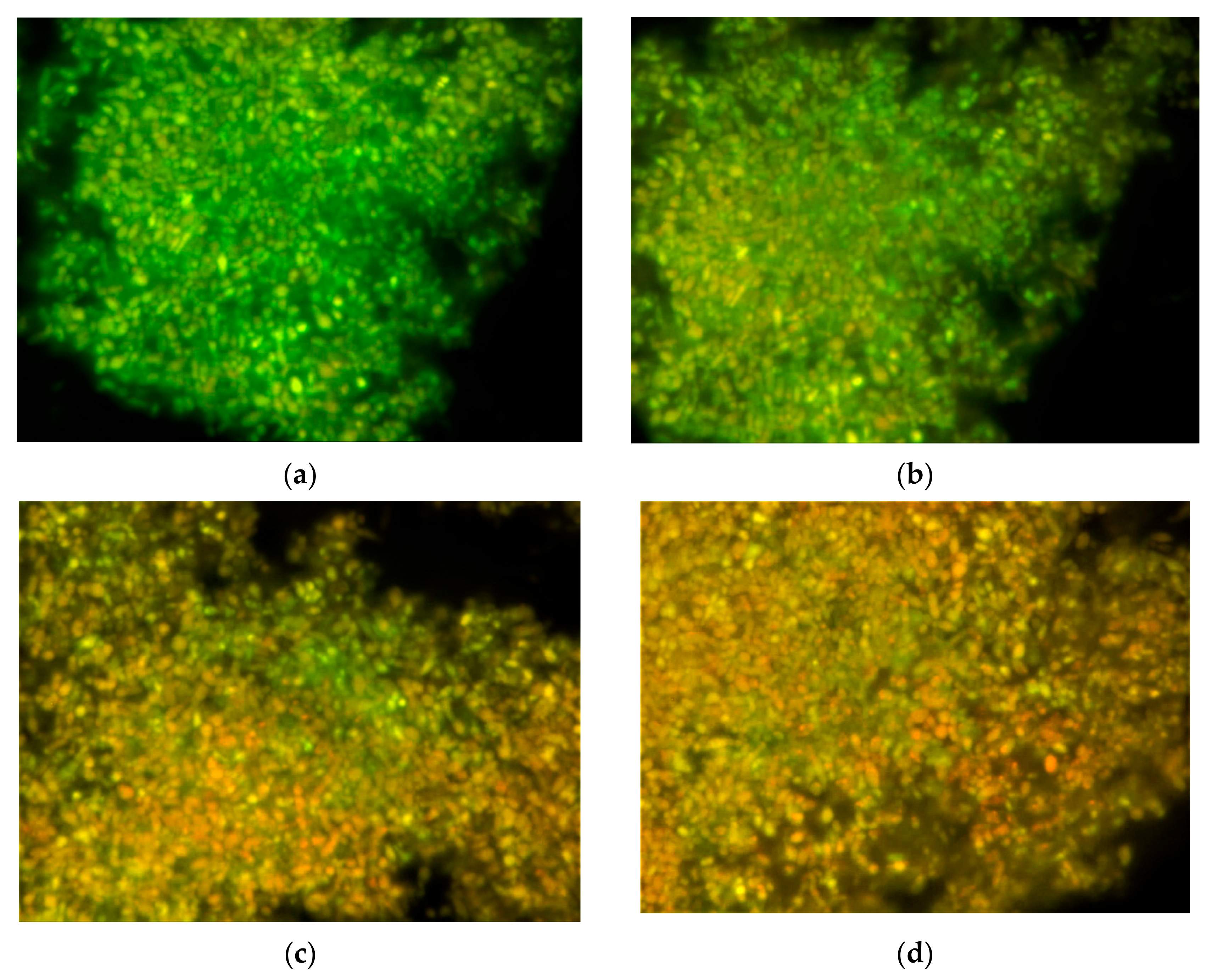
3.2. Membrane Integrity

Results of the fluorescent microscopy and membrane-damaged cell analysis are presented in Figure 3 and Figure 4. These results show a significant 29% increase in proportions of bacterial cells with membrane damage following blue light exposure as compared with sodium fluoride alone (p < 0.001).

4. Discussion

Previously, we have shown that exposing bacterial cells to a sub-lethal dosage of blue light increases their cell membrane permeability [9,11], and this suggested that this effect may increase the efficacy of antibacterial agents. In this study, we examined the combined effect of sub-lethal blue light and sodium fluoride on the cariogenicity of Streptococcus mutans in a biofilm configuration. The results of the present study showed that blue light exposure caused a significant increase in the anti-cariogenic effect of sodium fluoride against Streptococcus mutans’ acidogenic and aciduric properties.
Fluoride is one of the most prevalent active ingredients in caries preventive products such as toothpastes and mouthwashes. Its caries-preventive activities include reducing the tooth’s susceptibility to demineralization by the formation of fluorhydroxyapatite [12] as well as its antibacterial activity. Fluoride has been demonstrated as a potent inhibitor of key enzymes in the aciduric mechanisms of cariogenic bacteria such as Urease and F-ATPase and acidogenic mechanisms such as Enolase [13].
The increasing problem of bacterial resistance to antibacterial chemotherapy has created an urgent need for development of new treatment approaches towards antibacterial therapy. One such approach is a bacterial crippling strategy also known as anti-virulence therapy [14] aimed to cripple the bacterium’s virulence rather than its vitality, thus eliminating the selective stress, which promotes bacterial resistance while reducing pathogenicity.
Given our frequent exposure to fluoride via drinking water and the daily use of dentifrices containing fluoride, it is not surprising that fluoride resistance mechanisms have been reported in some strains of Streptococcus mutans [13]. These include sustained resistance properties due to genetic mutations in various loci, including the upregulation of the gene encoding for fluoride export (i.e., fluoride antiporters) [15]. These observations further stress the importance of devising new strategies for caries prevention. Blue light application during tooth brushing (recommended time 2 min) might facilitate the influx of fluoride into cariogenic bacterial cells.
Within the limitations of an in vitro study, the results of the present study suggest that using sub-lethal blue light exposure combined with sodium fluoride may increase the anticariogenic efficacy of fluoride against Streptococcus mutans. However, in addition to its beneficial antibacterial phototoxic effect, blue light may also have disruptive effects on mammalian cells [16], possibly mediated by light-induced ROS. Therefore, in any prospective clinical trial, exposure time should be minimized to reduce any risk to the oral soft tissues. Further studies are on the way.

Author Contributions

Conceptualization, N.S.; methodology, N.S. and U.J.; formal analysis, U.J. and S.D.; investigation, N.S. and S.L.; resources, U.J.; data curation, N.S. and S.D.; writing—original draft preparation, N.S.; writing—review and editing S.L.; visualization, S.D.; supervision, N.S.; project administration, U.J. All authors have read and agreed to the published version of the manuscript.

Funding

This research received no external funding.

Conflicts of Interest

The authors declare no conflict of interest.

References

  1. Fleming, E.; Afful, J. Prevalence of Total and Untreated Dental Caries among Youth: United States, 2015–2016. NCHS Data Brief 2018, 307, 1–8. [Google Scholar]
  2. Touger-Decker, R.; van Loveren, C. Sugars and dental caries. Am. J. Clin. Nutr. 2003, 78, 881S–892S. [Google Scholar] [CrossRef] [PubMed] [Green Version]
  3. Krzyściak, W.; Jurczak, A.; Kościelniak, D.; Bystrowska, B.; Skalniak, A. The virulence of Streptococcus mutans and the ability to form biofilms. Eur. J. Clin. Microbiol. Infect. Dis. 2014, 33, 499–515. [Google Scholar] [CrossRef] [PubMed] [Green Version]
  4. Cui, T.; Luo, W.; Xu, L.; Yang, B.; Zhao, W.; Cang, H. Progress of Antimicrobial Discovery Against the Major Cariogenic Pathogen Streptococcus mutans. Curr. Issues Mol. Biol. 2019, 32, 601–644. [Google Scholar] [CrossRef] [PubMed]
  5. Han, Y. Effects of brief sodium fluoride treatments on the growth of early and mature cariogenic biofilms. Sci. Rep. 2021, 11, 18290. [Google Scholar] [CrossRef] [PubMed]
  6. Marquis, R.E.; Clock, S.A.; Mota-Meira, M. Fluoride and organic weak acids as modulators of microbial physiology. FEMS Microbiol. Rev. 2003, 26, 493–510. [Google Scholar] [CrossRef] [PubMed]
  7. Grandjean, P. Developmental fluoride neurotoxicity: An updated review. Environ. Health A Glob. Access Sci. Source 2019, 18, 110. [Google Scholar] [CrossRef] [PubMed] [Green Version]
  8. McKenzie, K.; Maclean, M.; Grant, M.H.; Ramakrishnan, P.; MacGregor, S.J.; Anderson, J.G. The effects of 405 nm light on bacterial membrane integrity determined by salt and bile tolerance assays, leakage of UV-absorbing material and SYTOX green labelling. Microbiology 2016, 162, 1680–1688. [Google Scholar] [CrossRef] [PubMed] [Green Version]
  9. Jeffet, U.; Dagan, N.; Sterer, N. Effect of Sublethal Blue Light on Herbal Extract Activity against Volatile Sulfide Compound Production by Fusobacterium nucleatum. Photochem. Photobiol. 2021, 97, 443–447. [Google Scholar] [CrossRef] [PubMed]
  10. Wang, Y.; Wang, X.; Jiang, W.; Wang, K.; Luo, J.; Li, W.; Zhou, X.; Zhang, L. Antimicrobial peptide GH12 suppresses cariogenic virulence factors of Streptococcus mutans. J. Oral Microbiol. 2018, 10, 1442089. [Google Scholar] [CrossRef] [PubMed] [Green Version]
  11. Jeffet, U.; Shimon, R.; Sterer, N. Effect of High Intensity Blue Light on Fusobacterium nucleatum Membrane Integrity. Photochem. Photobiol. 2020, 96, 178–181. [Google Scholar] [CrossRef]
  12. Cardoso, C.; Lacerda, B.; Mangueira, D.F.; Charone, S.; Olympio, K.P.; Magalhães, A.C.; Pessan, J.P.; Vilhena, F.V.; Sampaio, F.C.; Buzalaf, M.A. Mechanisms of action of fluoridated acidic liquid dentifrices against dental caries. Arch. Oral Biol. 2015, 60, 23–28. [Google Scholar] [CrossRef] [PubMed]
  13. Liao, Y.; Brandt, B.W.; Li, J.; Crielaard, W.; Van Loveren, C.; Deng, D.M. Fluoride resistance in Streptococcus mutans: A mini review. J. Oral Microbiol. 2017, 9, 1344509. [Google Scholar] [CrossRef] [PubMed] [Green Version]
  14. Fleitas Martínez, O.; Cardoso, M.H.; Ribeiro, S.M.; Franco, O.L. Recent Advances in Anti-virulence Therapeutic Strategies with a Focus on Dismantling Bacterial Membrane Microdomains, Toxin Neutralization, Quorum-Sensing Interference and Biofilm Inhibition. Front. Cell. Infect. Microbiol. 2019, 9, 74. [Google Scholar] [CrossRef] [PubMed]
  15. Liao, Y.; Chen, J.; Brandt, B.W.; Zhu, Y.; Li, J.; van Loveren, C.; Deng, D.M. Identification and functional analysis of genome mutations in a fluoride-resistant Streptococcus mutans strain. PLoS ONE 2015, 10, e0122630. [Google Scholar] [CrossRef] [PubMed] [Green Version]
  16. Wataha, J.C.; Lockwood, P.E.; Lewis, J.B.; Rueggeberg, F.A.; Messer, R.L. Biological effects of blue light from dental curing units. Dent. Mater. 2004, 20, 150–157. [Google Scholar] [CrossRef]
Figure 1. Effect of the various treatments on Streptococcus mutans lactic acid production: Saline, as negative control; Blue light (BL); Sodium fluoride (NaF); and combination of blue light and sodium fluoride (BL + NaF). Results (±standard deviation) measured using a colorimetric assay are presented as absorbance at 600 nm.
Biophysica 02 00017 g001
Figure 2. Effect of the various treatments on Streptococcus mutans ATPase activity: Saline, as negative control; Blue light (BL); Sodium fluoride (NaF); and combination of blue light and sodium fluoride (BL + NaF). Results (±standard deviation) measured using a colorimetric assay are presented as absorbance at 600 nm.
Biophysica 02 00017 g002
Figure 3. Fluorescent microscopy images showing bacterial cells with membrane damage (stained red) and without damage (stained green) following the various treatments: (a) Saline; (b) Blue light; (c) Sodium fluoride; and (d) Combination of blue light and sodium fluoride.
Biophysica 02 00017 g003
Figure 4. Effect of the various treatments on Streptococcus mutans cell membrane damage: Saline, as negative control; Blue light (BL); Sodium fluoride (NaF); and combination of blue light and sodium fluoride (BL + NaF). Results (±standard deviation) are presented as percent of red stained bacterial cells from the total counts.
Biophysica 02 00017 g004
Publisher’s Note: MDPI stays neutral with regard to jurisdictional claims in published maps and institutional affiliations.

