
Application of Non-Destructive Testing Techniques (NDTT) to Characterize Nanocarriers Used for Drug Delivery: A Mini Review

 ,
,  ,
,
Abstract
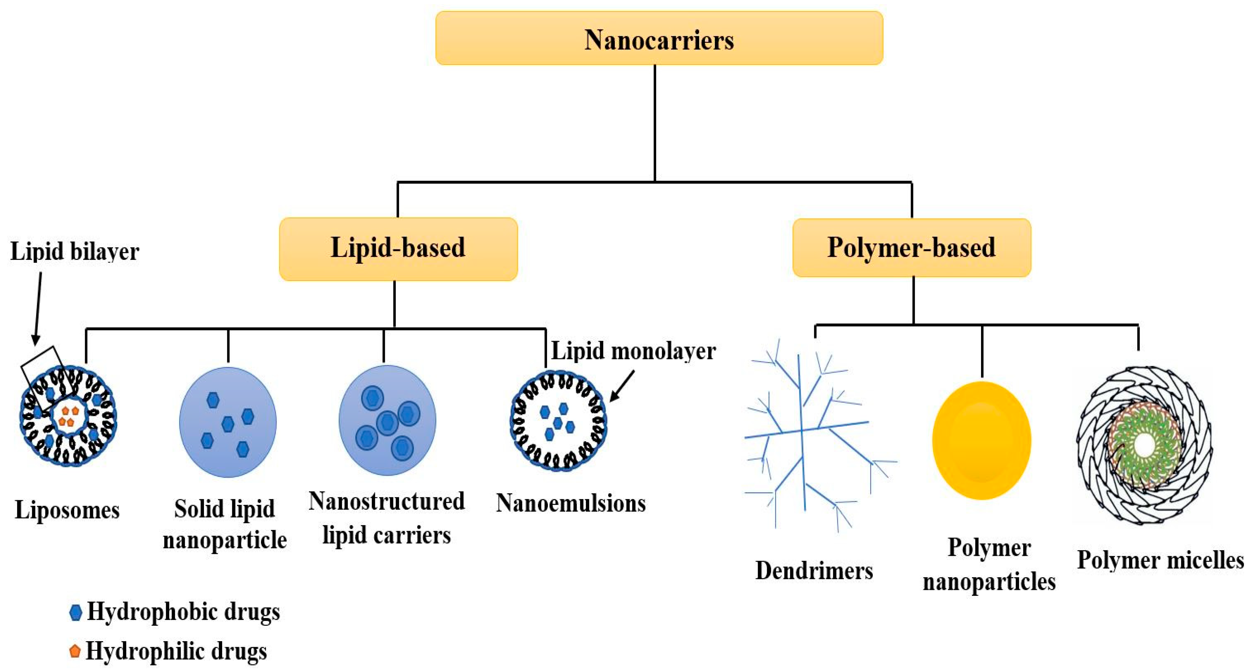
:1. Introduction
2. Non-Destructive Testing Techniques
2.1. X-ray Diffraction
2.1.1. Working Principle
2.1.2. Application
2.2. Ultraviolet-Visible Spectroscopy
2.2.1. Working Principle
2.2.2. Application
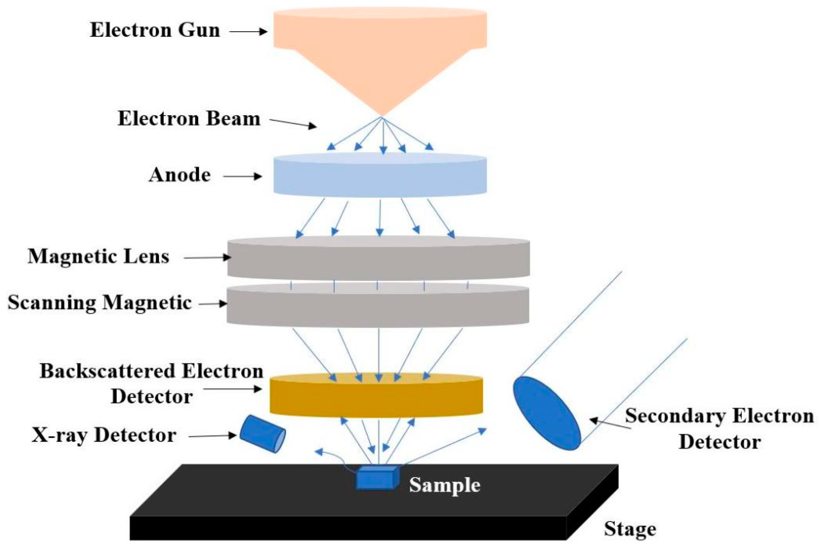
2.3. Scanning Electron Microscopy with Energy Dispersive X-ray Spectroscopy
2.3.1. Working Principle
2.3.2. Application
2.4. Nuclear Magnetic Resonance Spectroscopy
2.4.1. Working Principle
2.4.2. Application
2.5. Dynamic Light Scattering
2.5.1. Working Principle
2.5.2. Application
2.6. Confocal Laser Scanning Microscopy
2.6.1. Working Principle
2.6.2. Application
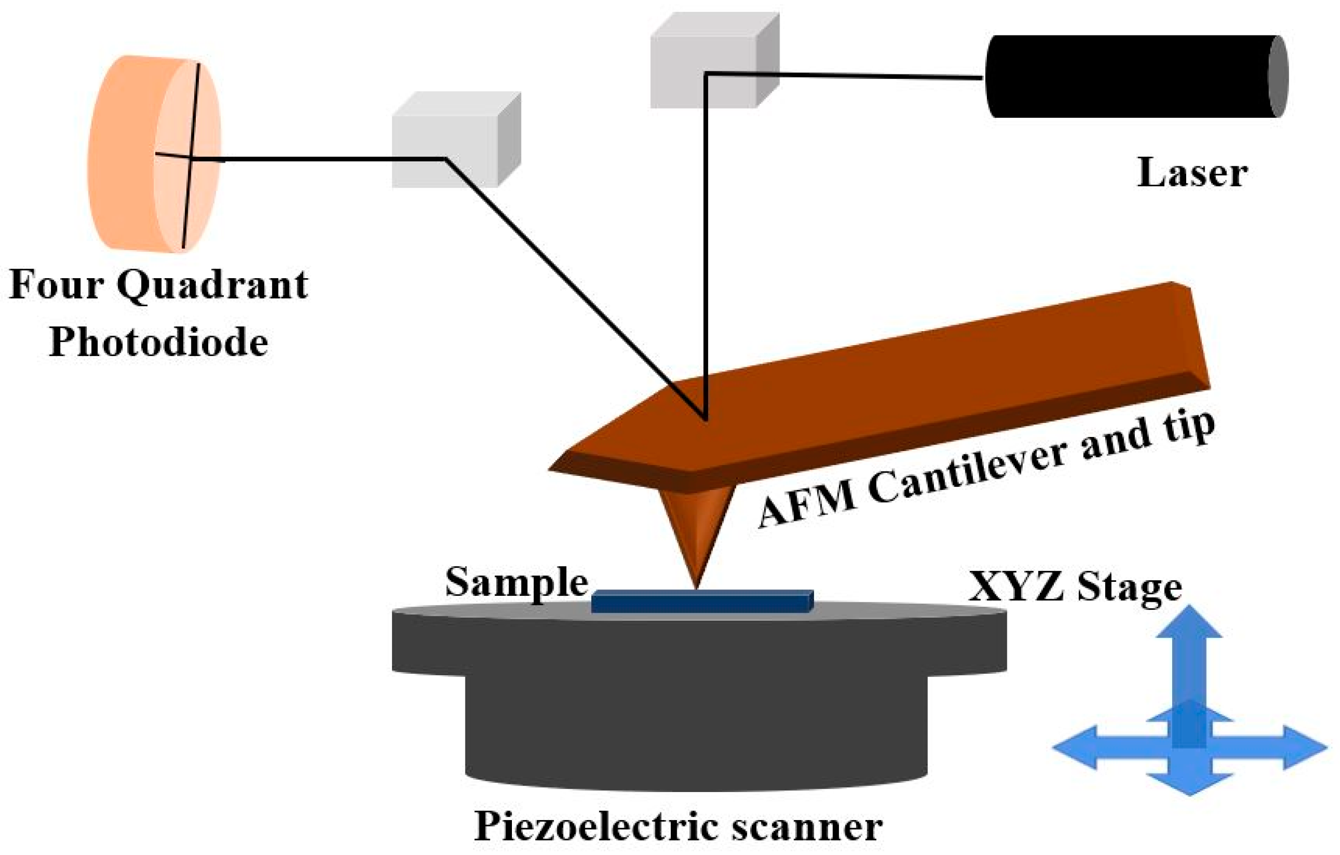
2.7. Atomic Force Microscopy
2.7.1. Working Principle
2.7.2. Application
3. Future Perspectives
4. Conclusions
Author Contributions
Funding
Institutional Review Board Statement
Informed Consent Statement
Data Availability Statement
Acknowledgments
Conflicts of Interest
References
- Khan, I.; Saeed, K.; Khan, I. Nanoparticles: Properties, applications and toxicities. Arab. J. Chem. 2019, 12, 908–931. [Google Scholar] [CrossRef]
- Senthilkumar, N.; Sharma, P.K.; Sood, N.; Bhalla, N. Designing magnetic nanoparticles for in vivo applications and understanding their fate inside human body. Coord. Chem. Rev. 2021, 445, 214082. [Google Scholar] [CrossRef]
- Manaia, E.B.; Abuçafy, M.P.; Chiari-Andréo, B.G.; Silva, B.L.; Junior, J.A.O.; Chiavacci, L.A. Physicochemical characterization of drug nanocarriers. Int. J. Nanomed. 2017, 12, 4991. [Google Scholar] [CrossRef] [Green Version]
- Nicolas, J.; Mura, S.; Brambilla, D.; Mackiewicz, N.; Couvreur, P. Design, functionalization strategies and biomedical applications of targeted biodegradable/biocompatible polymer-based nanocarriers for drug delivery. Chem. Soc. Rev. 2013, 42, 1147–1235. [Google Scholar] [CrossRef]
- Gupta, S.; Bansal, R.; Gupta, S.; Jindal, N.; Jindal, A. Nanocarriers and nanoparticles for skin care and dermatological treatments. Indian Dermatol. Online J. 2013, 4, 267. [Google Scholar] [CrossRef]
- Nazila, K.; Yameen, B.; Wu, J.; Farokhzad, O. Nanoparticles: Mechanisms of controlling drug release Nazila. Chem. Rev. 2016, 116, 2602–2663. [Google Scholar]
- Raval, N.; Maheshwari, R.; Kalyane, D.; Youngren-Ortiz, S.R.; Chougule, M.B.; Tekade, R.K. Importance of Physicochemical Characterization of Nanoparticles in Pharmaceutical Product Development. In Basic Fundamentals of Drug Delivery; Academic Press: Cambridge, MA, USA, 2019. [Google Scholar]
- Beloqui, A.; Solinís, M.Á.; Rodríguez-Gascón, A.; Almeida, A.J.; Préat, V. Nanostructured lipid carriers: Promising drug delivery systems for future clinics. Nanomed. Nanotechnol. Biol. Med. 2016, 12, 143–161. [Google Scholar] [CrossRef]
- Bailey, C.M.; Kamaloo, E.; Waterman, K.L.; Wang, K.F.; Nagarajan, R.; Camesano, T.A. Size dependence of gold nanoparticle interactions with a supported lipid bilayer: A QCM-D study. Biophys. Chem. 2015, 203, 51–61. [Google Scholar] [CrossRef] [Green Version]
- Jain, A.K.; Thareja, S. In vitro and in vivo characterization of pharmaceutical nanocarriers used for drug delivery. Artif. Cells Nanomed. Biotechnol. 2019, 47, 524–539. [Google Scholar] [CrossRef] [Green Version]
- Reich, G. Near-infrared spectroscopy and imaging: Basic principles and pharmaceutical applications. Adv. Drug Deliv. Rev. 2005, 57, 1109–1143. [Google Scholar] [CrossRef]
- Awotwe-Otoo, D.; Zidan, A.S.; Rahman, Z.; Habib, M.J. Evaluation of anticancer drug-loaded nanoparticle characteristics by nondestructive methodologies. AAPS PharmSciTech 2012, 13, 611–622. [Google Scholar] [CrossRef] [Green Version]
- Gowen, A.; O’donnell, C.; Cullen, P.J.; Bell, S. Recent applications of chemical imaging to pharmaceutical process monitoring and quality control. Eur. J. Pharm. Biopharm. 2008, 69, 10–22. [Google Scholar] [CrossRef] [Green Version]
- Rahman, Z.; Zidan, A.S.; Khan, M.A. Non-destructive methods of characterization of risperidone solid lipid nanoparticles. Eur. J. Pharm. Biopharm. 2010, 76, 127–137. [Google Scholar] [CrossRef]
- Lim, M.K.; Cao, H. Combining multiple NDT methods to improve testing effectiveness. Constr. Build. Mater. 2013, 38, 1310–1315. [Google Scholar] [CrossRef]
- Ilavarasi Jeyamalar, J.; Krishnaveni, M.; Kannan, C. Synthesis and characterization of Ni-incorporated mesoporous silica material for its potential applications in oligomerization of glycerol. Phosphorus Sulfur Silicon Relat. Elem. 2022, 197, 1–7. [Google Scholar] [CrossRef]
- Moradi, S.; Najjar, R.; Hamishehkar, H.; Lotfi, A. Triple-responsive drug nanocarrier: Magnetic core-shell nanoparticles of Fe3O4@poly(N-isopropylacrylamide)-grafted-chitosan, synthesis and in vitro cytotoxicity evaluation against human lung and breast cancer cells. J. Drug Deliv. Sci. Technol. 2022, 72, 103426. [Google Scholar] [CrossRef]
- Karimi Jabali, M.; Allafchian, A.R.; Jalali, S.A.H.; Shakeripour, H.; Mohammadinezhad, R.; Rahmani, F. Design of a pDNA nanocarrier with ascorbic acid modified chitosan coated on superparamagnetic iron oxide nanoparticles for gene delivery. Colloids Surf. A Physicochem. Eng. Asp. 2022, 632, 127743. [Google Scholar] [CrossRef]
- Weiss, A.-V.; Fischer, T.; Iturri, J.; Benitez, R.; Toca-Herrera, J.L.; Schneider, M. Mechanical properties of gelatin nanoparticles in dependency of crosslinking time and storage. Colloids Surf. B Biointerfaces 2019, 175, 713–720. [Google Scholar] [CrossRef]
- Theodoropoulos, D.; Rova, A.; Smith, J.R.; Barbu, E.; Calabrese, G.; Vizirianakis, I.S.; Tsibouklis, J.; Fatouros, D.G. Towards boron neutron capture therapy: The formulation and preliminary in vitro evaluation of liposomal vehicles for the therapeutic delivery of the dequalinium salt of bis-nido-carborane. Bioorg. Med. Chem. Lett. 2013, 23, 6161–6166. [Google Scholar] [CrossRef]
- Danışman-Kalındemirtaş, F.; Birman, H.; Karakuş, S.; Kilislioğlu, A.; Erdem-Kuruca, S. Preparation and biological evaluation of novel 5-Fluorouracil and Carmofur loaded polyethylene glycol / rosin ester nanocarriers as potential anticancer agents and ceramidase inhibitors. J. Drug Deliv. Sci. Technol. 2022, 73, 103456. [Google Scholar] [CrossRef]
- Taheri-Kafrani, A.; Shirzadfar, H.; Abbasi Kajani, A.; Kudhair, B.K.; Jasim Mohammed, L.; Mohammadi, S.; Lotfi, F. Functionalized graphene oxide/Fe3O4 nanocomposite: A biocompatible and robust nanocarrier for targeted delivery and release of anticancer agents. J. Biotechnol. 2021, 331, 26–36. [Google Scholar] [CrossRef] [PubMed]
- Carrera Espinoza, M.J.; Lin, K.-S.; Weng, M.-T.; Kunene, S.C.; Wang, S.S.-S. In vitro studies of Pluronic F127 coated magnetic silica nanocarriers for drug delivery system targeting liver cancer. Eur. Polym. J. 2021, 153, 110504. [Google Scholar] [CrossRef]
- Kaliyamoorthi, K.; Ramasamy, S.; Pillai, A.S.; Alexander, A.; Arivarasu, A.; Enoch, I.V.M.V. Camptothecin-loaded holmium ferrite nanocarrier. Expanded activity on breast cancer cells. Mater. Lett. 2021, 285, 129164. [Google Scholar] [CrossRef]
- Baghbanbashi, M.; Pazuki, G.; Khoee, S. One Pot Silica Nanoparticle Modification and Doxorubicin Encapsulation as pH-Responsive Nanocarriers, Applying PEG/Lysine Aqueous Two Phase System. J. Mol. Liq. 2022, 349, 118472. [Google Scholar] [CrossRef]
- Raj, V.; Kim, Y.; Kim, Y.-G.; Lee, J.-H.; Lee, J. Chitosan-gum arabic embedded alizarin nanocarriers inhibit biofilm formation of multispecies microorganisms. Carbohydr. Polym. 2022, 284, 118959. [Google Scholar] [CrossRef]
- Ghanbari, N.; Salehi, Z.; Khodadadi, A.A.; Shokrgozar, M.A.; Saboury, A.A. Glucosamine-conjugated graphene quantum dots as versatile and pH-sensitive nanocarriers for enhanced delivery of curcumin targeting to breast cancer. Mater. Sci. Eng. C 2021, 121, 111809. [Google Scholar] [CrossRef]
- Alvarez-Román, R.; Naik, A.; Kalia, Y.; Guy, R.H.; Fessi, H. Skin penetration and distribution of polymeric nanoparticles. J. Control. Release 2004, 99, 53–62. [Google Scholar] [CrossRef]
- Verma, D.D.; Verma, S.; Blume, G.; Fahr, A. Particle size of liposomes influences dermal delivery of substances into skin. Int. J. Pharm. 2003, 258, 141–151. [Google Scholar] [CrossRef]
- Arias, L.S.; Pessan, J.P.; de Souza Neto, F.N.; Lima, B.H.R.; de Camargo, E.R.; Ramage, G.; Delbem, A.C.B.; Monteiro, D.R. Novel nanocarrier of miconazole based on chitosan-coated iron oxide nanoparticles as a nanotherapy to fight Candida biofilms. Colloids Surf. B Biointerfaces 2020, 192, 111080. [Google Scholar] [CrossRef]
- Alp, E.; Damkaci, F.; Guven, E.; Tenniswood, M. Starch nanoparticles for delivery of the histone deacetylase inhibitor CG-1521 in breast cancer treatment. Int. J. Nanomed. 2019, 14, 1335. [Google Scholar] [CrossRef] [Green Version]
- Wang, T.; Meng, Q.; Lin, L.; Yang, L.; Zhao, W.; Sun, D. Self-assembled dehydropeptide nanocarrier as a delivery system for antitumor drug temozolomide. Bioorg. Chem. 2022, 124, 105842. [Google Scholar] [CrossRef] [PubMed]
- Zohreh, N.; Rastegaran, Z.; Hosseini, S.H.; Akhlaghi, M.; Istrate, C.; Busuioc, C. pH-triggered intracellular release of doxorubicin by a poly(glycidyl methacrylate)-based double-shell magnetic nanocarrier. Mater. Sci. Eng. C 2021, 118, 111498. [Google Scholar] [CrossRef] [PubMed]
- Thodeti, S.; Reddy, R.; Kumar, J. Synthesis and characterization of pure and indium doped SnO2 nanoparticles by sol-gel methods. Int. J. Sci. Eng. Res 2016, 7, 310–317. [Google Scholar]
- Sawyer, L.; Grubb, D.T.; Meyers, G.F. Polymer Microscopy; Springer Science & Business Media: Berlin/Heidelberg, Germany, 2008. [Google Scholar]
- Hansford, G.M.; Turner, S.; Degryse, P.; Shortland, A.J. High-resolution X-ray diffraction with no sample preparation. Acta Crystallogr. Sect. A Found. Adv. 2017, 73, 293–311. [Google Scholar] [CrossRef] [PubMed] [Green Version]
- Sabbagh, H.A.K.; Hussein-Al-Ali, S.H.; Hussein, M.Z.; Abudayeh, Z.; Ayoub, R.; Abudoleh, S.M. A Statistical Study on the Development of Metronidazole-Chitosan-Alginate Nanocomposite Formulation Using the Full Factorial Design. Polymers 2020, 12, 772. [Google Scholar] [CrossRef] [PubMed] [Green Version]
- Rachmawati, H.; Yanda, Y.L.; Rahma, A.; Mase, N. Curcumin-loaded PLA nanoparticles: Formulation and physical evaluation. Sci. Pharm. 2016, 84, 191–202. [Google Scholar] [CrossRef] [PubMed] [Green Version]
- Boddolla, S.; Thodeti, S. A review on characterization techniques of nanomaterials. Int. J. Eng. Sci. Math. 2018, 7, 169–175. [Google Scholar]
- Begum, R.; Farooqi, Z.H.; Naseem, K.; Ali, F.; Batool, M.; Xiao, J.; Irfan, A. Applications of UV/Vis spectroscopy in characterization and catalytic activity of noble metal nanoparticles fabricated in responsive polymer microgels: A review. Crit. Rev. Anal. Chem. 2018, 48, 503–516. [Google Scholar] [CrossRef]
- Pang, S.C.; Chin, S.F.; Nadirah, A.; Tay, S.H.; Yazid, S.N.A.M. Fabrication of polysaccharide-based nanoparticles as drug delivery nanocarriers. ECS Trans. 2015, 66, 15. [Google Scholar] [CrossRef]
- Hung, H.-S.; Bau, D.-T.; Yeh, C.-A.; Kung, M.-L. Evaluation of cellular uptake mechanisms for AuNP-collagen-Avemar nanocarrier on transformed and non-transformed cell lines. Colloids Surf. A Physicochem. Eng. Asp. 2019, 580, 123791. [Google Scholar] [CrossRef]
- Musa, A.; Ahmad, M.B.; Hussein, M.Z.; Saiman, M.I.; Sani, H.A. Effect of gelatin-stabilized copper nanoparticles on catalytic reduction of methylene blue. Nanoscale Res. Lett. 2016, 11, 438. [Google Scholar] [CrossRef] [PubMed] [Green Version]
- Akhtar, K.; Khan, S.A.; Khan, S.B.; Asiri, A.M. Scanning electron microscopy: Principle and applications in nanomaterials characterization. In Handbook of Materials Characterization; Springer: Berlin/Heidelberg, Germany, 2018; pp. 113–145. [Google Scholar]
- Scimeca, M.; Bischetti, S.; Lamsira, H.K.; Bonfiglio, R.; Bonanno, E. Energy Dispersive X-ray (EDX) microanalysis: A powerful tool in biomedical research and diagnosis. Eur. J. Histochem. EJH 2018, 62, 2841. [Google Scholar] [CrossRef] [PubMed]
- Rezaei, S.; Kashanian, S.; Bahrami, Y.; Cruz, L.J.; Motiei, M. Redox-sensitive and hyaluronic acid-functionalized nanoparticles for improving breast cancer treatment by cytoplasmic 17α-methyltestosterone delivery. Molecules 2020, 25, 1181. [Google Scholar] [CrossRef] [Green Version]
- Wu, M.; Hou, P.; Dong, L.; Cai, L.; Chen, Z.; Zhao, M.; Li, J. Manganese dioxide nanosheets: From preparation to biomedical applications. Int. J. Nanomed. 2019, 14, 4781. [Google Scholar] [CrossRef] [PubMed] [Green Version]
- Singh, M.K.; Singh, A. Chapter 14—Nuclear magnetic resonance spectroscopy. In Characterization of Polymers and Fibres; Singh, M.K., Singh, A., Eds.; Woodhead Publishing: Sawston, UK, 2022; pp. 321–339. [Google Scholar]
- Anantachaisilp, S.; Smith, S.M.; Treetong, A.; Pratontep, S.; Puttipipatkhachorn, S.; Ruktanonchai, U.R. Chemical and structural investigation of lipid nanoparticles: Drug–lipid interaction and molecular distribution. Nanotechnology 2010, 21, 125102. [Google Scholar] [CrossRef]
- Pecora, R. Dynamic Light Scattering: Applications of Photon Correlation Spectroscopy; Springer Science & Business Media: Berlin/Heidelberg, Germany, 1985. [Google Scholar]
- Lim, J.; Yeap, S.P.; Che, H.X.; Low, S.C. Characterization of magnetic nanoparticle by dynamic light scattering. Nanoscale Res. Lett. 2013, 8, 381. [Google Scholar] [CrossRef] [Green Version]
- Instruments, M. Dynamic Light Scattering: An Introduction in 30 Minutes; Technical Note Malvern MRK656-01; Malvern Instruments: Worcestershire, UK, 2012. [Google Scholar]
- Li, Z. Critical particle size where the Stokes-Einstein relation breaks down. Phys. Rev. E 2009, 80, 061204. [Google Scholar] [CrossRef] [Green Version]
- Goldstein, J.; Joy, D.C.; Romig, A.D., Jr. Principles of Analytical Electron Microscopy; Springer Science & Business Media: Berlin/Heidelberg, Germany, 2013. [Google Scholar]
- Echlin, P. Handbook of Sample Preparation for Scanning Electron Microscopy and X-ray Microanalysis; Springer Science & Business Media: Berlin/Heidelberg, Germany, 2011. [Google Scholar]
- Mahmood, S.; Taher, M.; Mandal, U.K. Experimental design and optimization of raloxifene hydrochloride loaded nanotransfersomes for transdermal application. Int. J. Nanomed. 2014, 9, 4331. [Google Scholar]
- Lin, P.-C.; Lin, S.; Wang, P.C.; Sridhar, R. Techniques for physicochemical characterization of nanomaterials. Biotechnol. Adv. 2014, 32, 711–726. [Google Scholar] [CrossRef] [Green Version]
- Rasmussen, K.; Rauscher, H.; Mech, A.; Sintes, J.R.; Gilliland, D.; González, M.; Kearns, P.; Moss, K.; Visser, M.; Groenewold, M. Physico-chemical properties of manufactured nanomaterials-Characterisation and relevant methods. An outlook based on the OECD Testing Programme. Regul. Toxicol. Pharmacol. 2018, 92, 8–28. [Google Scholar] [CrossRef]
- Caminade, A.-M.; Laurent, R.; Majoral, J.-P. Characterization of dendrimers. Adv. Drug Deliv. Rev. 2005, 57, 2130–2146. [Google Scholar] [CrossRef] [PubMed]
- Sapsford, K.E.; Tyner, K.M.; Dair, B.J.; Deschamps, J.R.; Medintz, I.L. Analyzing nanomaterial bioconjugates: A review of current and emerging purification and characterization techniques. Anal. Chem. 2011, 83, 4453–4488. [Google Scholar] [CrossRef] [PubMed]
- Lundqvist, M.; Sethson, I.; Jonsson, B.-H. Transient interaction with nanoparticles “freezes” a protein in an ensemble of metastable near-native conformations. Biochemistry 2005, 44, 10093–10099. [Google Scholar] [CrossRef] [PubMed]
- Mahmood, S.; Mandal, U.K.; Chatterjee, B.; Taher, M. Advanced characterizations of nanoparticles for drug delivery: Investigating their properties through the techniques used in their evaluations. Nanotechnol. Rev. 2017, 6, 355–372. [Google Scholar] [CrossRef]
- Sattler, E.; Maier, T.; Hoffmann, V.; Hegyi, J.; Ruzicka, T.; Berking, C. Noninvasive in vivo detection and quantification of Demodex mites by confocal laser scanning microscopy. Br. J. Dermatol. 2012, 167, 1042–1047. [Google Scholar] [CrossRef]
- Allison, D.P.; Mortensen, N.P.; Sullivan, C.J.; Doktycz, M.J. Atomic force microscopy of biological samples. Wiley Interdiscip. Rev. Nanomed. Nanobiotechnol. 2010, 2, 618–634. [Google Scholar] [CrossRef]
- Müller, D.J.; Dufrene, Y.F. Atomic force microscopy as a multifunctional molecular toolbox in nanobiotechnology. Nanosci. Technol. Collect. Rev. Nat. J. 2010, 3, 269–277. [Google Scholar] [CrossRef]
- El Kirat, K.; Burton, I.; Dupres, V.; Dufrene, Y. Sample preparation procedures for biological atomic force microscopy. J. Microsc. 2005, 218, 199–207. [Google Scholar] [CrossRef]
- Moreno-Herrero, F.; Colchero, J.; Gomez-Herrero, J.; Baro, A. Atomic force microscopy contact, tapping, and jumping modes for imaging biological samples in liquids. Phys. Rev. E 2004, 69, 031915. [Google Scholar] [CrossRef] [Green Version]
- Boufi, S.; Haaj, S.B.; Magnin, A.; Pignon, F.; Impéror-Clerc, M.; Mortha, G. Ultrasonic assisted production of starch nanoparticles: Structural characterization and mechanism of disintegration. Ultrason. Sonochem. 2018, 41, 327–336. [Google Scholar] [CrossRef]
- Meyabadi, T.F.; Dadashian, F.; Sadeghi, G.M.M.; Asl, H.E.Z. Spherical cellulose nanoparticles preparation from waste cotton using a green method. Powder Technol. 2014, 261, 232–240. [Google Scholar] [CrossRef]
- Huang, X.; Shen, S.; Zhang, Z.; Zhuang, J. Cross-linked polyethylenimine–tripolyphosphate nanoparticles for gene delivery. Int. J. Nanomed. 2014, 9, 4785. [Google Scholar] [CrossRef] [PubMed] [Green Version]
- Murphy, D.B. Fundamentals of Light Microscopy and Electronic Imaging; John Wiley & Sons: Hoboken, NJ, USA, 2002. [Google Scholar]
- Longo, C.; Casari, A.; Beretti, F.; Cesinaro, A.M.; Pellacani, G. Skin aging: In vivo microscopic assessment of epidermal and dermal changes by means of confocal microscopy. J. Am. Acad. Dermatol. 2013, 68, e73–e82. [Google Scholar] [CrossRef]
- Zhang, Z.; Wo, Y.; Zhang, Y.; Wang, D.; He, R.; Chen, H.; Cui, D. In vitro study of ethosome penetration in human skin and hypertrophic scar tissue. Nanomed. Nanotechnol. Biol. Med. 2012, 8, 1026–1033. [Google Scholar] [CrossRef] [PubMed]
- Venuganti, V.V.; Sahdev, P.; Hildreth, M.; Guan, X.; Perumal, O. Structure-skin permeability relationship of dendrimers. Pharm. Res. 2011, 28, 2246–2260. [Google Scholar] [CrossRef]
- Wang, T.; Bai, J.; Jiang, X.; Nienhaus, G.U. Cellular uptake of nanoparticles by membrane penetration: A study combining confocal microscopy with FTIR spectroelectrochemistry. ACS Nano 2012, 6, 1251–1259. [Google Scholar] [CrossRef]
- Wan, L.; Manickam, D.S.; Oupický, D.; Mao, G. DNA release dynamics from reducible polyplexes by atomic force microscopy. Langmuir 2008, 24, 12474–12482. [Google Scholar] [CrossRef]
- Sarwar, M.S.; Huang, Q.; Ghaffar, A.; Abid, M.A.; Zafar, M.S.; Khurshid, Z.; Latif, M. A smart drug delivery system based on biodegradable chitosan/poly (allylamine hydrochloride) blend films. Pharmaceutics 2020, 12, 131. [Google Scholar] [CrossRef] [Green Version]
- Ezhilarasi, P.; Karthik, P.; Chhanwal, N.; Anandharamakrishnan, C. Nanoencapsulation techniques for food bioactive components: A review. Food Bioprocess Technol. 2013, 6, 628–647. [Google Scholar] [CrossRef]


| Technique | Nanocarriers Examined | Parameters Examined | References |
|---|---|---|---|
| Scanning electron microscopy | Mesoporous silica materials | Structure/shape, size distribution, and particle size | [16] |
| Scanning electron microscopy | Core-shell nanoparticles of magnetic Fe3O4-poly (N-isopropylacrylamide) grafted with chitosan | Structure/shape, size distribution, and particle size | [17] |
| Scanning electron microscopy | Ascorbic acid-modified chitosan based superparamagnetic iron oxide nanoparticles | Identification of elemental composition, structure/shape, size distribution, and particle size | [18] |
| Dynamic light scattering | Gelatin nanoparticles | Size distribution, particle size, and surface charge | [19] |
| Dynamic light scattering | Liposomes consisting of phosphatidylcholine or dimyristoylphosphatidylcholine | Size distribution and particle size | [20] |
| Dynamic light scattering | 5-Fluorouracil and Carmofur loaded polyethylene glycol/rosin ester nanocarriers | Size distribution and particle size | [21] |
| X-ray diffraction | Graphene oxide/Fe3O4 nanocomposite | Phase and structure | [22] |
| X-ray diffraction | Pluronic F127 coated magnetic silica nanocarriers | Crystalline phase and structure | [23] |
| X-ray diffraction | Camptothecin-loaded holmium ferrite nanocarrier | Phase and structure | [24] |
| Atomic force microscopy | Silica nanoparticle | Aggregation topography, Size, shape, and structure | [25] |
| Atomic force microscopy | Chitosan-gum arabic embedded alizarin nanocarriers | Surface topography and uniformity | [26] |
| Atomic force microscopy | Glucosamine-conjugated graphene quantum dots | Surface topography and morphology | [27] |
| Confocal laser scanning microscopy | Polystyrene nanoparticles | Distribution, size, and shape | [28] |
| Confocal laser scanning microscopy | Liposomes | Effect of penetration ability and distribution | [29] |
| Confocal laser scanning microscopy | Miconazole based on chitosan-coated iron oxide nanoparticles | Structure/shape | [30] |
| Nuclear magnetic resonance spectroscopy | Starch nanoparticles | Purity, structure, and composition | [31] |
| Nuclear magnetic resonance spectroscopy | Dehydropeptide nanocarrier | Molecular conformation | [32] |
| Nuclear magnetic resonance spectroscopy | Poly (glycidyl methacrylate)-based double-shell magnetic nanocarrier | Structure and composition | [33] |
| Techniques | Advantages | Disadvantages | References |
|---|---|---|---|
| Scanning Electron Microscopy (SEM) |
|
| [54,55,56] |
| X-ray diffraction |
|
| [57,58,59] |
| Nuclear magnetic resonance spectroscopy (NMR) |
|
| [60,61] |
| Dynamic Light Scattering |
|
| [50,62] |
| Confocal laser scanning microscopy |
|
| [28,29,63] |
| Atomic force microscopy |
|
| [62,64,65,66,67] |
Publisher’s Note: MDPI stays neutral with regard to jurisdictional claims in published maps and institutional affiliations. |
© 2022 by the authors. Licensee MDPI, Basel, Switzerland. This article is an open access article distributed under the terms and conditions of the Creative Commons Attribution (CC BY) license (https://creativecommons.org/licenses/by/4.0/).
Share and Cite
Barbhuiya, R.I.; Ramalingam, S.; Kalra, H.K.; Elsayed, A.; Routray, W.; Annamalai, M.; Singh, A. Application of Non-Destructive Testing Techniques (NDTT) to Characterize Nanocarriers Used for Drug Delivery: A Mini Review. Biophysica 2022, 2, 154-167. https://doi.org/10.3390/biophysica2030016
Barbhuiya RI, Ramalingam S, Kalra HK, Elsayed A, Routray W, Annamalai M, Singh A. Application of Non-Destructive Testing Techniques (NDTT) to Characterize Nanocarriers Used for Drug Delivery: A Mini Review. Biophysica. 2022; 2(3):154-167. https://doi.org/10.3390/biophysica2030016
Chicago/Turabian StyleBarbhuiya, Rahul Islam, Saipriya Ramalingam, Harsimran Kaur Kalra, Abdallah Elsayed, Winny Routray, Manickavasagan Annamalai, and Ashutosh Singh. 2022. "Application of Non-Destructive Testing Techniques (NDTT) to Characterize Nanocarriers Used for Drug Delivery: A Mini Review" Biophysica 2, no. 3: 154-167. https://doi.org/10.3390/biophysica2030016