Systematic Review

The Influence of Root Prominence on the Onset of Gingival Recession: A Systematic Review

by
Girolamo Raso
1,
Nuno Bernardo Malta dos Santos
1,
Leonardo M. Nassani
2,*,
Anna Carolina Volpi Mello-Moura
1,3,
Juliana Campos Hasse Fernandes
4 and
Gustavo Vicentis Oliveira Fernandes
3,5,*
1
Faculty of Dental Medicine, Universidade Católica Portuguesa, 3504-505 Viseu, Portugal
2
Division of Restorative and Prosthetic Dentistry, The Ohio State University College of Dentistry, 305 W 12th Ave., Columbus, OH 43210, USA
3
Center for Interdisciplinary Research in Health, Faculty of Dental Medicine, Universidade Católica Portuguesa, 3504-505 Viseu, Portugal
4
Independent Researcher, St. Louis, MO 63104, USA
5
Missouri School of Dentistry & Oral Health, A. T. Still University, 1500 Park Ave., St. Louis, MO 63104, USA
*
Authors to whom correspondence should be addressed.
Surgeries 2024, 5(1), 103-114; https://doi.org/10.3390/surgeries5010012
Submission received: 17 February 2024 / Revised: 27 February 2024 / Accepted: 7 March 2024 / Published: 12 March 2024

Abstract

:
This systematic review aimed to identify, evaluate, and summarize the results of relevant studies on radicular prominence and its relationship with gingival recessions. This review was conducted according to the PRISMA (Preferred Reporting Reviews and Meta-Analysis) guidelines, and the focused PICO question was “In teeth with vestibular site-specific root or alveolar bone prominence, what are the chances that this will lead to gingival recession or difficulty in root coverage procedures, compared to teeth correctly positioned in the alveolar bone or without anatomical root prominence?”. A search was carried out on three databases: Embase, PubMed/MedLine, and Wiley Library. This initial search was complemented with manual research. It included any clinical study, such as a randomized clinical trial, controlled clinical trial, prospective/retrospective clinical study, case series, or case report, published in English from January 2012 to December 2023, which reported any involvement of the root/bone prominence approach. The exclusion criteria were clinical studies without report results/details of the case(s), studies based on questionnaires, editorial letters, any review, in vitro/in silica and animal studies, and interviews. The Strengthening the Reporting of Observational Studies in Epidemiology (STROBE) statement was applied for quality assessment. A total of 163 articles were found, but only three articles were included (k = 0.98). The included studies observed negative correlations when comparing the variables root prominence with linear root coverage, root surface area covered, and linear tissue thickness gain. It suggested a significant reduction in root coverage for prominences greater than 1 mm; therefore, relevant keratinized tissue gains can be achieved in gingival recession treatment after the application of the odontoplasty. The STROBE checklist evaluated 22 items, and all the included studies had a high-quality assessment (greater than 75%) with values greater than 85%. Then, it was not possible to draw conclusions due to the number of articles included, even though they had high-quality assessments. Otherwise, it is possible to suggest that the root prominence may impact gingival recession. Therefore, new and well-designed studies must be developed to establish a significant conclusion about this condition.

1. Introduction

Pathological mucogingival tissues usually present two forms. The first occurs when an open disruption is created between the compartments of the dento–gingival junction, which is related to the formation of periodontal pockets with probing depth (PD) measurements greater than 3 mm (PD > 3 mm). The other potentially less aggressive condition is known as gingival recession (GR) [1]. It consists of the migration of the gingival margin in the apical direction until it is dramatically displaced from the cement–enamel junction (CEJ). This clinical attachment loss (CAL) may not be isolated only to the vestibular face of the margin; it is also becoming interesting in the interproximal areas. The clinical result will be a denuded root surface, exposed to the oral environment, with the involvement of one or more sides of the root [2,3].
GRs result from atrophic cellular changes. The term “atrophy” refers to a decrease in cell volume or number resulting from sublethal cell injury (SCI) [4]. Since SCI is essentially reversible once the causal factor is removed, the volume reduction process should cease, and the number and size of cells might be restored to normal levels; however, it all depends on the severity of the lesion [5]. Since the changes that occur at the cellular level in GR result from essentially reversible processes [5], early identification of those causing factors becomes essential. Therefore, the etiology is multifactorial, involving many factors (anatomical, physiological, mechanical, traumatic, inflammatory or pathological, and iatrogenic). Often, there is synergism among factors causing the GR [6]. The etiological factors can be divided into two groups: predisposing factors and precipitating factors. Predisposing factors (gingival dimensions [7], abnormal frenal attachment [8,9], root/bone prominence [9,10,11,12,13], tooth malposition [4,14], physiological factors [15,16,17], and bone dehiscence/fenestration [18,19]) are those conditions that are favorable to the occurrence of GR, whereas precipitating factors (orthodontic treatment [4,20,21], plaque-induced inflammation [7,18,20,22], traumatic toothbrushing [18,20,23], smoking [24], and subgingival restoration [18]) are those contributing to the onset of GR [18].
GRs increase the incidence of root caries and hypersensitivity, which are related to the onset of non-carious cervical lesions (NCCL) [25]. In addition, teeth being subjected to exacerbated forces (traumatic occlusion) seems to be associated with the occurrence of NCCL. A systematic study [25] concluded that it was not possible to draw conclusions regarding the relationship between GR and the presence of occlusal trauma. Therefore, patients commonly seek treatment for tooth hypersensitivity or esthetic concerns [26]. Still, it can be extended to prevent and reverse the advancing trend of the recession and as a complementary treatment for class V carious lesions [27]. GRs tend to aggravate rather than improve but rarely result in tooth loss [28,29]. Kassab et al. [26] stated that almost 50% of adult patients with ages ranging from 18 to 64 presented at least one site with 1 mm recession and that 88% of patients older than 65 had at least one site with clinical attachment loss (CAL), confirming the trend that periodontal loss of support increases with age.
Hence, root coverage (RC) procedures have been studied. Many surgical approaches are described in the literature, with favorable results [30,31,32]. Therefore, clinicians should apply the best technique for each patient, individualizing each situation and focusing on resolving the esthetic concern and other eventually related symptoms [10]. The most common RC method involves using a connective tissue graft (CTG) or de-epithelized free CTG, considered the gold standard for RC treatment [1]. The general concept of this procedure consists of the development of a flap/tunnel/envelope [1,33] which is detached from the local tissue (around which the GR is present), permitting it to receive the biomaterial (CTG or others) and/or be repositioned back in a coronal position to the CEJ. Due to the fact that harvesting CTGs may increase donor site morbidity and pain, other biomaterials/biologic agents have been used as substitutes, such as allogeneic grafts, xenogeneic or synthetic collagen membranes, enamel matrix derivative (EMD), and autologous platelet concentrate (APC)/platelet-rich fibrin (PRF) of the second and third generation (membranes) [1].
Despite root prominence being considered detrimental to the mucogingival complex, it is suggested as an etiologic agent and risk factor for gingival recessions and root coverage procedures [9,10,11]. When prominent roots are adjacent, concavities between the tooth and the adjacent bone may be visible [12], causing the retraction of the overlying gingiva. Even if the retraction only occurs on the prominent site of the bone, it is more common for it to progress than to stabilize [13]. Clinicians should be able to identify areas of bone or root prominences not only to improve the prognosis but also in a preventive sense; better counseling the patient and being aware of the risk of recession occurrence may limit the onset. Thus, the goal of this systematic review was to identify, evaluate, and summarize the results of relevant studies on radicular/bone prominence and its relationship with gingival recessions.

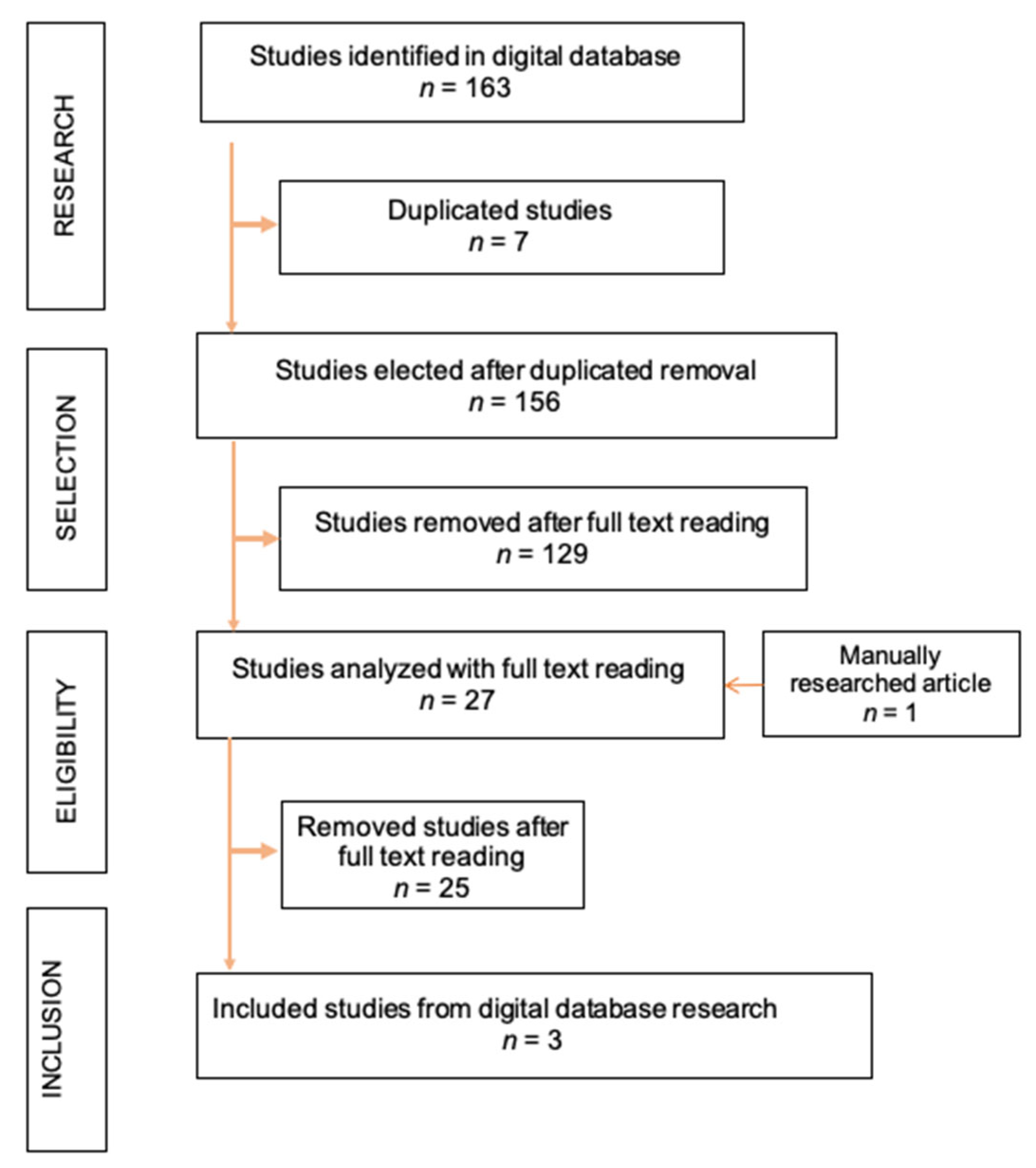
2. Material and Methods

This systematic review followed the Preferred Reporting Items for Systematic Reviews and Meta-Analysis (PRISMA) guidelines and was registered as part of a project at PROSPERO (CRD42020183268). The focus of the present review was defined according to the PICO (Patient, Intervention, Comparison, and Outcome) strategy: “Is there a relationship between the presence of anatomical abnormalities of the bone or tooth (malposition, prominences, and dehiscence or bone fenestrations) and the appearance of GRs?” (P: teeth with site-specific root/bone prominence; I: occurrence of gingival recession or difficulty in root coverage procedure; C: compared to a patient who does not present root or alveolar prominence; O: likelihood of GR and results associated).

2.1. Databases and Search Strategy

A bibliographic search was conducted using MedLine/PubMed, Web of Science, and Embase to collect articles published between January 2012 and December 2023 (12 years), recruiting only articles in the English language. A combination of specific terms was used: ((gingival recession OR retraction OR recession) AND (protuberances OR prominence OR eminence OR malposition OR exostosis OR rotation)) AND (coverage). They were adjusted according to the specific strategy in each database. Additional manual research was performed using the main journals in Periodontics.

2.2. Eligibility Criteria

This study was conducted based on any clinical study, such as a randomized clinical trial (RCT), controlled clinical trial (CCT), prospective/retrospective clinical study, case series, or case report, published in English from January 2012 to December 2023; human clinical studies; and articles reporting any involvement of the root/bone prominence approach. The exclusion criteria were clinical studies without report results/details of the case(s), studies based on questionnaires, editorial letters, any review, in vitro/in silica and animal studies, and interviews.

2.3. Study Selection and Data Extraction

After the bibliographic search, two independent researchers (G.R. and G.V.O.F.) proceeded to filter relevant articles that fitted the study by analyzing the title and abstract for study selection. Any reviewer disagreement was discussed with a third author (N.B.M.S.). Cohen’s kappa test was performed to assess the reviewers’ agreement. The same strategy of disagreement resolution was applied to the manual research.
Reviewers extracted the data independently from the selected articles for further analysis using data extraction tables which included the following parameters: author(s), year of publication, study design, main goal, the number of participants, systemic condition, exclusion criteria, and occlusal assessment method. All values and details were reported.

2.4. Quality Assessment

The risk of bias and quality assessments of the included studies were carried out independently by two reviewers (G.R. and G.V.O.F.). The Strengthening the Reporting of Observational Studies in Epidemiology (STROBE) statement was applied. Twenty-two items were evaluated, and the final percentage was obtained. If the result was between 0% and 40%, there was a high risk of bias/low-quality assessment; between 41% and 75%, moderate risk/quality; and >75%, low risk of bias/high-quality assessment. A third reviewer (N.B.M.S.) verified the ratings obtained, and group discussion resolved any discrepancy.

3. Results

After a bibliographic search on Embase (n = 31), MedLine/PubMed (n = 117), and Web of Science (n = 23), a total of 163 articles were found. Seven articles were duplicated and were removed. Then, the title and abstract were evaluated. Twenty-seven articles were successfully found in the initial assessment and were selected for full-text reading. One article was manually found and included. After full-text reading, 25 articles were excluded due to incongruency (lack of information/incomplete data related to the topic studied, no detail or report about the root/bone prominence and its relationship with the GR). Then, three articles met the criteria of interest (Figure 1). Cohen’s kappa coefficient was applied in the study of inter-examinator agreement (k = 0.98).

3.1. Study Characteristics

Three studies were enrolled (one case report and two retrospective studies). The case report was developed by Núñez et al., in 2018 (Spain) [34], and the other two articles included were published by Gil et al., in 2018 and 2019 (USA) [35,36] (Table 1). The criteria and results are presented in Table 2.

3.2. Studies Description

Study A1 [34] described a treatment strategy for anterior localized gingival recessions composed of two steps. First, odontoplasty was performed to modify the anatomy of the root surface, which was prominent to the buccal side due orthodontic treatment, creating a new emergence profile; after eight weeks, the tunnel root coverage procedure with connective tissue graft (CTG) was performed. The goal was to provide extra space for the graft, which should directly cause thickening of the gingival margin and be able to correct the root prominence. The results of this case series were favorable regarding percentual root coverage, CAL gain, and especially concerning KTW gain.
Study A2 [35] consisted of a pilot retrospective study investigating the relationship between site-specific characteristics of patients treated with the vestibular incision subperiosteal tunnel access (VISTA) technique and the outcome of the root coverage procedure. Mean root prominence was 0.8 ± 0.6 mm, and it was negatively correlated (r = −0.80) with linear root coverage. The exposed root area covered after the surgical procedure was also negatively correlated with mean root prominence (r = −0.83). This article had valuable benefits that can be derived from this study, including the proposal of an innovative measurement technique that allows the superimposition of preoperative and postoperative images for more reliable comparison results. Due to the nature of the measurement technique, new parameters, usually hard to evaluate, could be established.
Study A3 [36] utilized the same sample as A2, reporting digital volumetric measurements of preoperative and postoperative periodontal data of multiple-recession-type defects treated with the VISTA technique, seeking to evaluate which were the gingival volume changes following root coverage with the VISTA procedure. Linear and volumetric changes in gingival thickness at 1, 2, 3, 4, and 5 mm from the postoperative gingival margin were analyzed. For gingival linear gain, initial root prominence was again detrimental, with a negative correlation r = −0.18 (p = 0.02). However, initial root prominence and volumetric thickness gain presented a non-significant correlation.

3.3. Quality Assessment

The STROBE checklist was used to establish the quality of the included studies. After evaluating the 22 items, all the included studies had a high-quality assessment (greater than 75%) with values greater than 85% (Table 3).

4. Discussion

GR is one of the most debated topics in the literature. It is frequently related to root caries, hypersensitivity, cervical abrasion, and esthetic damage [37]. The most common techniques applied for GR treatment are the coronally advanced flap (CAF) [38] and its modification (MCAF), which are considered the gold-standard procedure for RC. Both techniques require periosteal incisions, eliminating the muscle tension [39]. The MCAF precludes the need for vertical incisions and sutures, thus achieving better and more stable RC. In addition, the CTG is also considered the gold standard for the biomaterials used combined with those techniques; it is strongly suggested first to remove the epithelium layer to avoid any biological complications [40,41,42].
Moreover, there are many etiological factors associated with GR, as mentioned in the introduction. It is relevant to highlight that many orthodontic treatments have movement of the tooth/teeth exceeding the anatomical limit, leading to an iatrogenic buccal protuberance of roots [21,43]. The most affected site is the buccal region of the lower incisor, especially when movements are made in an anterior direction [20]. Molars can also be affected by lateral movements [4]. Then, gingival alterations during the orthodontic treatment follow the tooth movements. When a tooth is repositioned in a buccal direction, a reduction in gingival volume is consequently observed. Furthermore, one of the etiological factors, the prominence of the root/bone, has a low number of published research studies. This fact led us to develop this systematic study. Some authors described a negative influence of root/bone prominence on flap adaptation and suture tension when covering a GR [44]. When those protuberances exceed the alveolar limits of the normal bone skeletal structure, they often create concavities between the tooth and the adjacent bone [12]. Such concavities can cause the overlying gingiva’s retraction, covering the root’s prominent face. On the other hand, it is common, especially in those patients with high hygienical standards who practice faulty toothbrushing, to find a retraction of the apical margin in those areas affected by bone prominences, which forms a wedge-shaped defect with the normal plane of the alveolar bone [20]. Even if the retraction only occurs on the prominent site of the bone, it is more common for it to progress than to stabilize [13]. Clinicians should be able to identify areas of bone or root prominences not only because it can improve the prognosis but also because it can be preventive. Counseling the patient and being aware of the risk of recession occurrence can be preventive.
Teeth characterized by an exceedingly buccal location are associated with poor periodontal characteristics (thin biotype and reduced width of attached keratinized tissue [KTW]) [10,45] and are generally more prone to mucogingival defects. The research strategy used in this systematic review resulted in the identification of studies where the effects on the periodontium of excessively prominent root were clinically studied and identification of which tools and techniques are more powerful and reliable to be able to establish the nature of these parameters.
Root prominence influences the outcome of GR treatment and has been shown to be detrimental, showing a negative correlation with mean root coverage and linear thickness gain. The unfavorable action of the root prominence was better observed in those cases where the root was >1 mm prominent [35,36]. In support of what was reported, a case series suggested reducing prominent sites in dental roots (odontoplasty), showing that this procedure was beneficial for the outcome of root coverage procedures. The treatment protocol provided extra space for graft placement and the subsequent formation of a thicker gingival profile [34].
It has been pointed out that the polishing of root surfaces did not substantially modify them, and, in cases of root curvature, even vigorous root planning did not affect the prominence but only demonstrated a slight reduction in the mesiodistal dimension, resulting in a slightly flattened root surface (6%) [11]. While normal root planning has not demonstrated differences in healing patterns in controlled experimental and clinical studies [39], it is safe to say that, in those cases where root prominence occurs, extensive root planning, even with the use of rotatory instruments to reduce the avascular bed for the flap, is suggested [46,47]. It is important to observe the volume of the root’s buccal surface, which must be removed once this procedure can achieve the root canal, leading to endodontic treatment. Moreover, in the guided tissue regeneration (GTR) technique, the same philosophy is encountered, and the creation of a little concavity on the root is suggested to create more space for tissue regeneration [48].
Evidence about tooth malposition and its significance for periodontal health has been investigated early in the past. The literature describes that the eruption pattern of the tooth appears to be correlated with the periodontal status of the tooth. For teeth erupted in a labial direction, the thickness of the attached gingiva is narrow, with an absence of keratin at the gingival margin. These conditions are considered detrimental to periodontal health [49,50,51]. Periodontal health at the level of gingival thickness could also be compromised in cases of impacted teeth in an excessively labial position before the treatment [52,53]. A study analyzing the periodontal status after orthodontic treatment in maxillary canines with a labial eruption pattern showed that, out of ninety-four canines observed, sixty-nine had an ectopic eruption pattern. Of the sixty-four ectopic canines, 15 had a gingival recession. The authors defended the fact that the value was high, considering the age of the studied population, concluding that the labial eruption pattern may affect periodontal status [54].

4.1. Existent Classifications for Gingival Recession Defects and Future Suggestions

Observing the existent classifications for gingival recession, such as those by Sullivan and Atkins (1968) [55], Mlinek (1973) [19], Liu and Solt (1980) [56], Benque (1983) [57], Miller (1985) [27], Smith (1990) [58], Pini-Prato et al. (2010) [59], and Cairo et al. (2011) [60], which, since 1968, have been strongly studied and published, it is still possible to find deficiencies. Many aspects are lacking within them, unable to provide a better prognosis for GR treatments.
Summarizing them, the first classification proposed by Sullivan and Atkins [55] divides recessions into four groups considering only the depth and the width of the defects: deep wide, shallow wide, deep narrow, and shallow narrow. Milnek et al. [19] introduced a numerical value to differentiate between shallow narrow (<3 mm) and deep wide defects (>3 mm). Liu and Solt (1980) [56] divided GR defects into two categories: visible (from the CEJ to the gingival margin) and hidden (the depth of the sulcus/pocket, measured from the gingival margin to the bottom of the sulcus/pocket). Summing these two measurements gives us the total amount of the recession. In 1983, Benque et al. [57] classified GR defects based on the shape of the defect and prognosis: (i) U-shaped, poor prognosis; (ii) V-shaped, favorable prognosis; and (iii) I-shaped, good prognosis. Therefore, it had a limitation of poor scientific support.
In 1985, one of the most famous classifications worldwide was developed. Miller [27] developed the classification essentially based on the radicular-crown extent of the marginal recession and the extent of the interproximal hard and soft tissue lost in the surrounding region [15,61]. This classification is relevant because it can help predict the prognosis of RC procedures. Miller’s classification has four classes in ascending order of severity and difficulty in treatment: Classes I and II have GR defects with no bone or soft tissue lost in interproximal areas and a good prognosis of complete root coverage expected. The only difference is the apical extension of the recession, which is limited to the attached gingiva in Class I and goes apical to the mucogingival junction (MGJ) in Class II. In Class III, GR goes to or above the MGJ and is characterized by loss of hard and soft tissue in the interproximal area that does not extend apically more than the buccal GR or by a mispositioned tooth. Total RC is not expected. Class IV is similar to Class III, but the interproximal loss goes to or beyond the buccal surface recession, which cannot make a predictive prognosis for the treatment.
In 1990, Smith [58] proposed a recession index formed by two digits; the first was used to state the horizontal and the second to state the vertical component of the GR. In horizontal extent, Score 0 is used for no evidence of root exposure; Score 1 for no clinical evidence of an exposed root but dentinal hypersensitivity in response to air blast and/or if the CEJ is exposed for up to 10% of the estimated mid-mesial to mid-distal distance; Score 2 for CEJ exposed by more than 10%, not exceeding 25% of the estimated mid-mesial to mid-distal distance; Score 3 for CEJ exposed for more than 25% of the mid-mesial to mid-distal distance but not exceeding 50%; Score 4 for CEJ exposed for more than 50% of the mid-mesial to mid-distal distance but not exceeding 75%; Score 5 for the exposure of the CEJ for more than 75% of the mid-mesial to mid-distal distance up to 100% [62]. For the vertical component: Score 0 has no exposed root clinically observed; Score 1, no clinical signs of root exposure, but the patient refers to dentinal hypersensitivity and/or the CEJ is exposed not more than 1 mm vertically to the gingival margin; Scores 2–9, root exposed from 2 mm to 9 mm to the base of the soft tissue defect. If applicable, an asterisk (*) could be used next to the second digit whenever the vertical component of the soft tissue defect encroaches the MGJ or extends beyond it into alveolar mucosa; the absence of an asterisk implies either the absence of MGJ involvement or non-involvement of the soft tissue defect [58,62].
In 2010, Pini-Prato et al. [59] proposed a new classification with the purpose of introducing more clinical parameters to analyze the GR defect. The proposal was to integrate the existent classifications with more information about the presence or absence of the CEJ and step in the tooth, which could result from the NCCL. This fact is due to Miller’s Class I or II recession type, where it can be difficult to quantify the complete root coverage without such an important landmark as the CEJ. This classification is divided into: visible CEJ = A; non-visible CEJ = B; presence of step = (+); and absence of step = (−).
Finally, Cairo et al., (2011) [60] published a classification based on the assessment of clinical attachment loss (CAL) in interproximal and buccal surfaces. The recession types (RT) were divided into RT1: GR not associated with interproximal attachment loss and CEJ is not detectable interproximal; RT2: GR with interproximal CAL (measured from interproximal CEJ to the bottom of the sulcus/pocket), which is lesser or equal to the buccal CAL; and RT3: GR with CAL where the amount of interproximal CAL is higher than the base of the recession.
Observing all existing classifications to treat GR defects panoramically, root/bone prominence and other important factors are not considered, showing deficiencies in the clinician’s predictability. Then, new studies and more complex analyses are suggested and stimulated to help professionals in the prognosis of the RC procedures.

4.2. Limitations and Strengths of the Studies

As a primary limitation, the number of studies published evaluating this parameter (root/bone prominence) can be considered extremely low. Also, few patients were included, mainly in the A1 study. A control group was lacking in all articles included. Specifically, A1 did not quantify odontoplasty, comparing before and after surgery. Also, it lacked control of investigator–operator bias. Two studies (A2 and A3) had the same patients. Moreover, A2 could not quantify the mucosal thickness adequately, and A3 presented only the thickness changes but not the actual gingival tissue thickness.
To the best of our knowledge, this is the first systematic review focused on root/bone prominence, which can open opportunities for more studies and publications. This systematic review, although limited by the number of included studies and their nature, encompasses relevant information about: 1. the benefits that could be gained by complementing mucogingival surgery with odontoplasty techniques that pursue reduced prominence before the surgery; 2. how to quantify root prominence and gingival changes with an efficient, reliable, and reproducible method; and 3. the contribution that different prominence degrees give to the outcome of root coverage treatment. Even though the obtained results confirm that root prominence may be detrimental for both periodontal health and root coverage procedures, more studies, with more robust evidence, should be carried out to assess the reproducibility of the measurement methodology applied in studies A2 and A3, checking the outcome of RC procedure preceded by odontoplasty, quantifying the influence that root or bone prominence has on the occurrence of gingival recession, and the outcome of RC procedures.

5. Conclusions

This systematic study focused on the influence of a site-specific characteristic (root or bone prominence) that is already considered negative for the outcome of root coverage procedures and as a risk factor for the onset of GRs. Within this review’s limitations, it was not possible to draw conclusions about the topic studied, even though the included studies had high-quality assessments. Otherwise, it is possible that the root prominence may impact gingival recession. Therefore, new and well-designed studies must be developed, expanding the inclusion criteria and searching strategy and including more databases to establish a significant conclusion about this condition and better understand and assess the root prominence impact.

Author Contributions

Conceptualization, G.V.O.F. and N.B.M.d.S.; methodology, G.R., N.B.M.d.S., L.M.N., A.C.V.M.-M. and G.V.O.F.; software, G.R., L.M.N. and G.V.O.F.; validation, N.B.M.d.S., A.C.V.M.-M., J.C.H.F. and G.V.O.F.; formal analysis, G.R., N.B.M.d.S., L.M.N., A.C.V.M.-M., J.C.H.F. and G.V.O.F.; investigation, G.R., N.B.M.d.S., L.M.N., A.C.V.M.-M., J.C.H.F. and G.V.O.F.; resources, A.C.V.M.-M. and G.V.O.F.; data curation, G.R., N.B.M.d.S., L.M.N., A.C.V.M.-M., J.C.H.F. and G.V.O.F.; writing—original draft preparation, G.R., N.B.M.d.S., L.M.N., A.C.V.M.-M., J.C.H.F. and G.V.O.F.; writing—review and editing, G.R., A.C.V.M.-M., J.C.H.F. and G.V.O.F.; visualization, G.R., N.B.M.d.S., L.M.N., A.C.V.M.-M., J.C.H.F. and G.V.O.F.; supervision, N.B.M.d.S., A.C.V.M.-M. and G.V.O.F.; project administration, A.C.V.M.-M. and G.V.O.F. All authors have read and agreed to the published version of the manuscript.

Funding

This research received no external funding.

Institutional Review Board Statement

Not applicable.

Informed Consent Statement

Not applicable.

Data Availability Statement

All data are available in the article.

Conflicts of Interest

The authors declare no conflicts of interest.

References

  1. Fernandes, G.V.D.O.; Santos, N.B.; Siqueira, R.A.; Wang, H.L.; Blanco-Carrion, J.; Fernandes, J.C. Autologous platelet concentrate of 2nd and 3rd generations efficacy in the surgical treatment of gingival recession: An overview of systematic reviews. J. Indian Soc. Periodontol. 2021, 25, 463–479. [Google Scholar] [CrossRef]
  2. Pilloni, A.; Marini, L.; Zeza, B.; Ferlosio, A.; Aghazada, R. Histologic Analysis of Clinically Healthy Human Gingiva in Patients with Altered Passive Eruption. Dent. J. 2021, 9, 29. [Google Scholar] [CrossRef] [PubMed]
  3. Serino, G.; Wennström, J.L.; Lindhe, J.; Eneroth, L. The prevalence and distribution of gingival recession in subjects with a high standard of oral hygiene. J. Clin. Periodontol. 1994, 21, 57–63. [Google Scholar] [CrossRef] [PubMed]
  4. Jati, A.S.; Furquim, L.Z.; Consolaro, A. Gingival recession: Its causes and types, and the importance of orthodontic treatment. Dent. Press J. Orthod. 2016, 21, 18–29. [Google Scholar] [CrossRef] [PubMed]
  5. Rubin, E.; Reisner, H. Essentials of Rubin’s Pathology, 6th ed.; Wolters Kluwer: Hong Kong, China, 2013. [Google Scholar]
  6. Zucchelli, G. Chirurgia Estetica Mucogengivale; Quintessenza Edizioni: Milan, Italy, 2012. [Google Scholar]
  7. Mythri, S.; Arunkumar, S.; Hegde, S.; Rajesh, S.; Munaz, M.; Ashwin, D. Etiology and occurrence of gingival recession—An epidemiological study. J. Indian Soc. Periodontol. 2015, 19, 671. [Google Scholar] [CrossRef] [PubMed]
  8. Zucchelli, G.; Testori, T.; de Sanctis, M. Clinical and Anatomical Factors Limiting Treatment Outcomes of Gingival Recession: A New Method to Predetermine the Line of Root Coverage. J. Periodontol. 2006, 77, 714–721. [Google Scholar] [CrossRef] [PubMed]
  9. Zucchelli, G.; Tavelli, L.; Barootchi, S.; Stefanini, M.; Wang, H.L.; Cortellini, P. Clinical Remarks on the Significance of Tooth Malposition and Papillae Dimension on the Prediction of Root Coverage. Int. J. Periodontics Restor. Dent. 2020, 40, 795–803. [Google Scholar] [CrossRef]
  10. Stefanini, M.; Marzadori, M.; Aroca, S.; Felice, P.; Sangiorgi, M.; Zucchelli, G. Decision making in root-coverage procedures for the esthetic outcome. Periodontol. 2000 2018, 77, 54–64. [Google Scholar] [CrossRef]
  11. Saletta, D.; Baldi, C.; Nieri, M.; Ceppatelli, P.; Franceschi, D.; Rotundo, R.; Cairo, F.; Pini Prato, G.P. Root Curvature: Differences Among Dental Morphotypes and Modifications After Mechanical Instrumentation. J. Periodontol. 2005, 76, 723–730. [Google Scholar] [CrossRef]
  12. El Chaar, E.S.; Oshman, S.; Danesh-Sani, S.; Pooria, D.; Fallah Abed, P.; Castaño, A. Increasing Contact Between Soft Tissue Graft and Blood Supply A Technique for Managing Connective Tissue Grafting Over Prominent Root—Clinical case studies. Curr. Opin. Cosmet. Dent. 2016, 32, 52–66. [Google Scholar]
  13. Nicolucci, M.; Arlin, M. Gingival Recession—Etiology and Treatment. Prev. Dent. Can. 2011, 2, 6–11. [Google Scholar]
  14. Zachrisson, B.U. Orthodontics and periodontics. In Clinical Periodontology and Implant Dentistry, 3rd ed.; Munksgaard: Copenhagen, Denmark, 1998; pp. 741–793. [Google Scholar]
  15. Goldstein, M.; Brayer, L.; Schwartz, Z. A Critical Evaluation of Methods for Root Coverage. Crit. Rev. Oral Biol. Med. 1996, 7, 87–98. [Google Scholar] [CrossRef] [PubMed]
  16. Thorstensson, H.; Falk, H.; Hugoson, A.; Olsson, J. Some salivary factors in insulin-dependent diabetics. Acta Odontol. Scand. 1989, 47, 175–183. [Google Scholar] [CrossRef] [PubMed]
  17. Chambrone, L.; Chambrone, D.; Pustiglioni, F.E.; Chambrone, L.A.; Lima, L.A. The influence of tobacco smoking on the outcomes achieved by root-coverage procedures: A systematic review. J. Am. Dent. Assoc. 2009, 140, 294–306. [Google Scholar] [CrossRef]
  18. Kasaj, A. Gingival Recession Management; Springer: Berlin/Heidelberg, Germany, 2018. [Google Scholar]
  19. Mlinek, A.; Smukler, H.; Buchner, A. The Use of Free Gingival Grafts for the Coverage of Denuded Roots. J. Periodontol. 1973, 44, 248–254. [Google Scholar] [CrossRef] [PubMed]
  20. Lang, N.P.; Lindhe, J. Parodontologia Clinica e Implantologia Orale, 6th ed.; Edi-Ermes: Milan, Italy, 2016; Volumes 1–2. [Google Scholar]
  21. Farsoun, C.F.; Castro, F.; Farsoun, J.; Fernandes, J.C.H.; Fernandes, G.V.O. Gingival recession in canines orthodontically aligned: A narrative Review. Int. J. Sci. Dent. 2023, 3, 100–121. [Google Scholar] [CrossRef]
  22. Wennstrom, J.L.; Lindhe, J.; Sinclair, F.; Thilander, B. Some periodontal tissue reactions to orthodontic tooth movement in monkeys. J. Clin. Periodontol. 1987, 14, 121–129. [Google Scholar] [CrossRef]
  23. Tezel, A.; Çanakçi, V.; Şiçek, Y.; Demir, T. Evaluation of Gingival Recession in Left-and Right-Handed Adults. Int. J. Neurosci. 2001, 110, 135–146. [Google Scholar] [CrossRef]
  24. Banihashemrad, S.A.; Fatemi, K.; Najafi, M.H. Effect of Smoking on Gingival Recession. Dent. Res. J. 2008, 5, 1–4. [Google Scholar]
  25. Tróia, P.M.B.P.S.; Spuldaro, T.R.; Fonseca, P.A.B.; Fernandes, G.V.O. Presence of Gingival Recession or Noncarious Cervical Lesions on Teeth under Occlusal Trauma: A Systematic Review. Eur. J. Gen. Dent. 2021, 10, 50–59. [Google Scholar] [CrossRef]
  26. Kassab, M.M.; Cohen, R.E. The etiology and prevalence of gingival recession. J. Am. Dent. Assoc. 2003, 134, 220–225. [Google Scholar] [CrossRef] [PubMed]
  27. Miller, P.D. A classification of marginal tissue recession. Int. J. Periodontics Restor. Dent. 1985, 5, 8–13. [Google Scholar]
  28. Chambrone, L.; Sukekava, F.; Araújo, M.G.; Pustiglioni, F.E.; Chambrone, L.A.; Lima, L.A. Root-Coverage Procedures for the Treatment of Localized Recession-Type Defects: A Cochrane Systematic Review. J. Periodontol. 2010, 81, 452–478. [Google Scholar] [CrossRef] [PubMed]
  29. Poormoradi, B.; Torkzaban, P.; Gholami, L.; Hooshyarfard, A.; Farhadian, M. Effect of Er, Cr (YSGG Laser Root Conditioning on the Success of Root Coverage with Subepithelial Connective Tissue Graft): A Randomized Clinical Trial with a 6-Month Follow-Up. J. Dent. 2018, 15, 230–239. [Google Scholar]
  30. Rasperini, G.; Acunzo, R.; Limiroli, E. Decision Making in Gingival Recession Treatment: Scientific Evidence and Clinical Experience. Clin. Adv. Periodontics 2011, 1, 41–52. [Google Scholar] [CrossRef] [PubMed]
  31. Marques, T.; Santos NBMd Sousa, M.; Fernandes, J.C.H.; Fernandes, G.V.O. Mixed-Thickness Tunnel Access (MiTT) through a Linear Vertical Mucosal Incision for a Minimally Invasive Approach for Root Coverage Procedures in Anterior and Posterior Sites: Technical Description and Case Series with 1-Year Follow-Up. Dent. J. 2023, 11, 235. [Google Scholar] [CrossRef]
  32. Marques, T.M.; Santos, N.B.M.; Montero, J.; Correia, A.; Fernandes, G.V.O. The Effect of the Interproximal Creeping Attachment in Aesthetic Site After Root Coverage Surgery with VISTA Technique—A Case Report. Indian J. Dent. Res. 2023, 34, 90–93. [Google Scholar] [CrossRef]
  33. Evginer, M.S.; Olgun, E.; Parlak, H.M.; Dolgun, A.B.; Keceli, H.G. Comparison of two techniques in gingival recession treatment: A randomized one-year clinical follow-up study. Dent. Med. Probl. 2022, 59, 121–130. [Google Scholar] [CrossRef]
  34. Núñez, J.; Sanz-Esporrin, J.; Caffesse, R.; Sanz, M. A Two-Step Surgical Approach with Flattening of the Root Surface to Treat Localized Gingival Recessions Affecting Mandibular Incisors: A Case Series Report. J. Esthet. Restor. Dent. 2018, 38, 683–690. [Google Scholar] [CrossRef]
  35. Gil, A.; Bakhshalian, N.; Min, S.; Zadeh, H.H. Treatment of multiple recession defects with vestibular incision subperiosteal tunnel access (VISTA): A retrospective pilot study utilizing digital analysis. J. Esthet. Restor. Dent. 2018, 30, 572–579. [Google Scholar] [CrossRef]
  36. Gil, A.; Bakhshalian, N.; Min, S.; Nart, J.; Zadeh, H. Three-Dimensional Volumetric Analysis of Multiple Gingival Recession Defects Treated by the Vestibular Incision Subperiosteal Tunnel Access (VISTA) Procedure. J. Esthet. Restor. Dent. 2019, 39, 687–695. [Google Scholar] [CrossRef]
  37. Buti, J.; Baccini, M.; Nieri, M.; La Marca, M.; Pini-Prato, G.P. Bayesian network meta-analysis of root coverage procedures: Ranking efficacy and identification of best treatment. J. Clin. Periodontol. 2013, 40, 372–386. [Google Scholar] [CrossRef] [PubMed]
  38. Dias, A.T.; Menezes, C.C.; Kahn, S.; Fischer, R.G.; Figueredo, C.M.S.; Fernandes, G.V.O. Gingival recession treatment with enamel matrix derivative associated with coronally advanced flap and subepithelial connective tissue graft: A split-mouth randomized controlled clinical trial with molecular evaluation. Clin. Oral Investig. 2022, 26, 1453–1463. [Google Scholar] [CrossRef] [PubMed]
  39. Wennström, J.L. Mucogingival Therapy. Ann. Periodontol. 1996, 1, 671–701. [Google Scholar] [CrossRef]
  40. Kahn, S.; Araújo, I.T.E.; Dias, A.T.; Souza, A.B.; Chambrone, L.; Fernandes, G.V.O. Histologic and histomorphometric analysis of connective tissue grafts harvested by the parallel incision method: A pilot randomized controlled trial comparing macro- and microsurgical approaches. Quintessence Int. 2021, 52, 772–778. [Google Scholar] [CrossRef]
  41. Kahn, S.; Oliveira, L.Z.; Dias, A.T.; Fernandes, G.V.O. Clinical evaluation and biological understanding of the early step-by-step healing after periodontal microsurgery: A case report with PES analysis comparing initial and 31-day result. J. Adv. Periodontol. Implant Dent. 2022, 14, 141–145. [Google Scholar] [CrossRef]
  42. Maia, V.T.G.; Kahn, S.; Souza, A.B.; Fernandes, G.V.O. Deepithelialized Connective Tissue Graft and The Reminiscent Epithelial Content After Harvested by the Harris’ Technique: A Histological and Morphometrical Case Series. Clin. Adv. Periodontics 2021, 11, 150–154. [Google Scholar] [CrossRef] [PubMed]
  43. Kahn, S.; Dias, A.T.; Nobre, V.; Oliveira, L.Z.; Fernandes, G.V.O. Endodontic and periodontal treatment of complete buccal root and apex exposition: A challenging case report with 17 months follow-up. Clin. Adv. Periodontics 2022, 12, 152–158. [Google Scholar] [CrossRef]
  44. Santamaria, M.P.; Ambrosano, G.M.B.; Casati, M.Z.; Nociti, F.H., Jr.; Sallum, A.W.; Sallum, E.A. The influence of local anatomy on the outcome of treatment of gingival recession associated with non-carious cervical lesions. J. Periodontol. 2010, 81, 1027–1034. [Google Scholar] [CrossRef]
  45. De Rouck, T.; Eghbali, R.; Collys, K.; De Bruyn, H.; Cosyn, J. The gingival biotype revisited: Transparency of the periodontal probe through the gingival margin as a method to discriminate thin from thick gingiva. J. Clin. Periodontol. 2009, 36, 428–433. [Google Scholar] [CrossRef]
  46. Miller, P.D. Root coverage using the free soft tissue autograft following citric acid application. III. A successful and predictable procedure in areas of deep-wide recession. Int. J. Periodontics Restor. Dent. 1985, 5, 14–37. [Google Scholar]
  47. Holbrook, T.; Ochsenbein, C. Complete coverage of the denuded root surface with a one-stage gingival graft. Int. J. Periodontics Restor. Dent. 1983, 3, 8–27. [Google Scholar]
  48. Tinti, C.; Vincenzi, G.; Cortellini, P.; Prato, G.P.; Clauser, C. Guided Tissue Regeneration in the Treatment of Human Facial Recession. A 12-Case Report. J. Periodontol. 1992, 63, 554–560. [Google Scholar] [CrossRef]
  49. Thurow, R.C. Atlas of Orthodontics Principles, 2nd ed.; Mosby: St. Louis, MI, USA, 1977. [Google Scholar]
  50. Maynard, J.G.; Ochsenbein, C. Mucogingival Problems, Prevalence and Therapy in Children. J. Periodontol. 1975, 46, 543–552. [Google Scholar] [CrossRef]
  51. Lang, N.P.; Löe, H. The Relationship Between the Width of Keratinized Gingiva and Gingival Health. J. Periodontol. 1972, 43, 623–627. [Google Scholar] [CrossRef] [PubMed]
  52. Odenrick, L.; Modéer, T. Periodontal status following surgical-orthodontic alignment of impacted teeth. Acta Odontol. Scand. 1978, 36, 233–236. [Google Scholar] [CrossRef] [PubMed]
  53. Heaney, T.G.; Atherton, J.D. Periodontal Problems Associated with the Surgical Exposure of Unerupted Teeth. Br. J. Orthod. 1976, 3, 79–85. [Google Scholar] [CrossRef]
  54. Modéer, T.; Odenrick, L. Post-treatment periodontal status of labially erupted maxillary canines. Acta Odontol. Scand. 1980, 38, 253–256. [Google Scholar] [CrossRef]
  55. Sullivan, H.C.; Atkins, J.H. Free autogenous gingival grafts. 3. Utilization of grafts in the treatment of gingival recession. Periodontics 1968, 6, 152–160. [Google Scholar]
  56. Liu, W.J.L.; Solt, C.W. A Surgical Procedure for the Treatment of Localized Gingival Recession in Conjunction with Root Surface Citric Acid Conditioning. J. Periodontol. 1980, 51, 505–509. [Google Scholar] [CrossRef]
  57. Benque, E.P.; Brunel, G.; Gineste, M.; Colin, L.; Duffort, J.; Fonvielle, E. Gingival Recession. Parodontol. J. 1984, 3, 207–241. [Google Scholar]
  58. Smith, R.G. Gingival recession Reappraisal of an enigmatic condition and a new index for monitoring. J. Clin. Periodontol. 1997, 24, 201–205. [Google Scholar] [CrossRef] [PubMed]
  59. Pini-Prato, G.; Franceschi, D.; Cairo, F.; Nieri, M.; Rotundo, R. Classification of Dental Surface Defects in Areas of Gingival Recession. J. Periodontol. 2010, 81, 885–890. [Google Scholar] [CrossRef] [PubMed]
  60. Cairo, F.; Nieri, M.; Cincinelli, S.; Mervelt, J.; Pagliaro, U. The interproximal clinical attachment level to classify gingival recessions and predict root coverage outcomes: An explorative and reliability study. J. Clin. Periodontol. 2011, 38, 661–666. [Google Scholar] [CrossRef]
  61. Mahajan, A. Mahajan’s Modification of the Miller’s Classification for Gingival Recession. Dent. Hypotheses 2010, 1, 45–50. [Google Scholar] [CrossRef]
  62. Armit, M.; Rosiline, J. Classification for Gingival Recession: A Mini review. Galore Int. J. Health Sci. Res. 2018, 3, 33–38. [Google Scholar]
Figure 1. Flow diagram for the selection process for the included articles.
Surgeries 05 00012 g001
Table 1. Studies’ details, design, objective(s), and parameters assessed.
ArticleAuthor/Year/CountryTitleObjectiveStudy DesignClinical Parameters Assessed
A1 [34]Núñez et al., 2018/SpainA two-step surgical approach with flattening of the root surface to treat localized gingival recessions affecting mandibular incisors: a case series reportTo analyze the clinical outcome of the two-step procedure in patients with localized GR and prominent roots secondary to orthodontic therapyCase series
GR, CAL, KTW, BD;
Esthetic outcome (1 year);
Dentin hypersensitivity.
A2
[35]
Gil et al., 2018/U.S.A.Treatment of multiple recession defects with VISTA: a retrospective pilot study utilizing digital analysisTo examine the correlation between initial site-specific characteristics of patients with multiple GR defects and the outcome of RC therapyRetrospective study
Recession class;
Tooth and graft type;
Root prominence;
Initial recession depth/width;
Initial gingival margin thickness;
Arch.
A3
[36]
Gil et al., 2019/U.S.A.Three-dimensional volumetric analysis of multiple gingival recession defects treated by the VISTA procedureTo evaluate gingival volume changes following RC with the VISTA procedureRetrospective study
Initial recession depth and gingival thickness;
Types of recession;
Tooth and graft type;
Root prominence;
Anatomical location in the arch.
GR = gingival recession; CAL = clinical attachment level; KTW = keratinized tissue width; BD = bone dehiscence; RC = root coverage; VISTA = vestibular incision subperiosteal tunnel access.
Table 2. Selective criteria and results presented by the included studies.
Sample/Mean AgeInclusion CriteriaExclusion CriteriaResults
A1
[34]
Ten patients
(3M–7F);
Mean age:
28 ± 10 years.
GR mandibular incisor;
Age > 18;
Systemically healthy;
Lack of KT;
Shallow vestibule.
Smokers (>10 cigarettes/day);
Systemically unhealthy;
GR associated with NCCL.
Root coverage: 100% in all RT1–2 defects and 80.5% in RT3 defects;
CAL gain (p < 0.001);
KTW gain (p < 0.001);
Recession defect reduction (p < 0.001).
A2
[35]
Twenty-one patients
(154 teeth),
8M–13F;
Mean age:
52.4 ± 9.5 years.
Age: 18–75 years;
RT1–2 (>1 mm in depth) on at least two adjacent teeth;
Presence of identifiable CEJ or restorative margin that was reliable to use as a reference;
Diagnostic casts available at preoperative (within 3 months prior to therapy) and post-therapy stages (≥12 months postoperatively).
Smokers (>10 cigarettes/day);
Miller’s Class IV (RT3) for gingival recession;
Patients taking medication that could affect the gingival health or anatomy;
Previous mucogingival surgeries performed in the area of analysis.
Mean root prominence and linear root coverage (r = −0.80; p < 0.001);
Mean root prominence and root surface area covered (r = −0.83; p < 0.001);
A precipitous drop in root coverage was observed in sites with root prominence greater than 1 mm.
A3
[36]
Twenty-one patients
(154 teeth),
8M–13F;
Mean age:
52.4 ± 9.5 years.
Patients between 18 and 75 years old;
Multiple RT1–2 defects on at least two adjacent teeth;
Presence of identifiable CEJ or restorative margin that was reliable to use as a reference;
Diagnostic casts are available at preoperative (within 3 months prior to therapy) and post-therapy stages (≥12 months postoperatively).
Heavy smoking (>10 cigarettes/day);
RT3 defects;
History of mucogingival surgeries in the area.
Initial root prominence and linear thickness gain R = −0.18 (p = 0.02) when the gains achieved at 2, 3, 4, and 5 mm levels were combined;
Initial root prominence and volume gain did not show a significant correlation (p = 0.71).
GR = gingival recession; M = male; F = female; CEJ = cementoenamel junction; RT = recession type; CAL = clinical attachment level; KTW = keratinized tissue width.
Table 3. Quality assessment for the included studies.
Items 12345678910111213141516171819202122Total (%)
Articles
A1 [34] 19 (86%)
A2 [35] 20 (91%)
A3 [36] 21 (95%)
Red = not present/found; green = present/found.
Disclaimer/Publisher’s Note: The statements, opinions and data contained in all publications are solely those of the individual author(s) and contributor(s) and not of MDPI and/or the editor(s). MDPI and/or the editor(s) disclaim responsibility for any injury to people or property resulting from any ideas, methods, instructions or products referred to in the content.

