Review

Primary Occlusion of the Fourth Ventricle: Case Report and Review of the Literature

1
Department of Neurosurgery, University Hospital Münster, 48149 Münster, Germany
2
Department of Neurosurgery, St. Barbara-Hospital, Academic Hospital of University Münster, 59073 Hamm, Germany
3
Department of Medicine, Faculty of Health, Witten/Herdecke University, 58455 Witten, Germany
*
Author to whom correspondence should be addressed.
These authors contributed equally to this work.
Surgeries 2023, 4(4), 665-677; https://doi.org/10.3390/surgeries4040063
Submission received: 12 October 2023 / Revised: 1 December 2023 / Accepted: 4 December 2023 / Published: 6 December 2023

Abstract

:
Idiopathic obstruction of the outlets of the fourth ventricle (FVOO) is a rare cause of hydrocephalus, which can be misdiagnosed as communicating hydrocephalus due to the enlargement of all four ventricles. Different surgical approaches are discussed in the literature. We present a case report of a 25-year-old male admitted with headache, vertigo, and nystagmus. The MRI scan showed a tetraventricular hydrocephalus with a patent aqueduct. After endoscopic third ventriculostomy (ETV), symptoms resolved. We performed a systematic review of the literature, covering 26 years, with the aim to investigate the symptoms, therapy, and outcome of primary FVOO, according to the PRISMA (Preferred Reporting Items for Systematic Reviews and Meta-Analysis) guidelines. We found 9 case reports and 2 case series and could extract a total of 34 cases. After ETV all symptoms resolved in 10 of 23 cases (43.5%), and in 13 of 23 cases (56.5%) symptoms improved partially. Seven cases (30.4%) required additional surgery. A decrease in ventricular volume occurred in most cases. In the 10 patients who were operated via fenestration, all symptoms resolved in 6 cases. ETV seems to be an effective treatment option for patients with idiopathic FVOO in a majority of cases. In special cases, fenestration of the foramen of Magendie may be suitable.

1. Introduction

Hydrocephalus can be divided into obstructive and communicating forms. Obstructive hydrocephalus often occurs due to obstructions of the foramen of Monro or the Sylvian aqueduct; obstructions of the outlet of the fourth ventricle, the foramina of Magendie and Luschka (FVOO) are rare and regularly secondary to infections, hemorrhages, or congenital malformations. Typical congenital causes are conditions such as tonsillar herniation Chiari malformations type I [1], hypoplasia of the cerebellar vermis in Dandy–Walker malformations [2], achondroplasia [3], or tuberous sclerosis [4]. Other reasons are acquired pathologies such as scarring after hemorrhages or infections like meningitis, arachnoiditis, or neurocysticercosis [5,6]. Other rare causes are tumors, arachnoid cysts of the posterior fossa [7], Blake’s pouch cysts [8], or Noonan syndrome [9].
In contrast, idiopathic obstructions of the outlets of the fourth ventricle are rare. Possible reasons are congenital atresia or membranous occlusion of foramina of Magendie or Luschka. While autopsies revealed membranes at the level of Magendie’s foramen in up to 2–3% and partial or total obstruction in up to 6% [10], most of the obstructions seem to be compensated by outflow through the foramen of Luschka. After endoscopic examination, Longatti distinguished thick, opaque, and thin, spiderweb-like membranes. In some cases, a valve mechanism was observed. Carpentier describes three different anatomical abnormalities of the foramen of Luschka: valve-like membranes due to interconnecting discontinuous flaps leading to an intermittent CFS flow, punctiform aperture of the Luschka’s membrane with limited adaption of the flow rate, and net-like membranes due to local inflammation leading to a poor communication of the intraventricular and the subarachnoid space. These abnormalities are referred to as Luschka dysgenesis, Luschka agenesis, and Luschka arachnoiditis [11]. Carpentier also describes a self-reinforcing process in the genesis of hydrocephalus due to obstruction of the foramina of the fourth ventricle. A reduced outflow through the foramina due to the before-mentioned anatomical variations leads to downward displacement of the posterior fossa structures which causes an adherence of the semipermeable membranes to the dura leading to a complete CSF blockage. In rare cases, the posterior inferior cerebellar artery (PICA) seems to be involved [12]. Histological examinations revealed glial or neural tissue with no sign of inflammation [13]. Still, subclinical infection is discussed as a possible cause in some cases [12]. Laszlo describes the examination of 122 foramina of Luschka in 61 formalin-fixed human brains. Primary obstruction was present in 11 of 122 cases. An overspreading rhomboid lip, which is a remnant of the roof of the fourth ventricle, was found to contribute to the obstruction. Histologically, three layers of the membrane and of the rhomboid lip were described, an inner ependymal, a middle glial, and an outer leptomeningeal layer. In some cases, a pouch in the cerebellopontine angle was described, which carries the potential of growing into a symptomatic diverticulum. However, in all examined specimens the foramen of Magendie was patent [14].
The reasons for late onset, partly in adults, are not completely understood. Some authors suggest the presence of a conatal, partial obstruction with a semipermeable membrane or a punctiform aperture. Acute decompensation could be explained by a temporal adherence of the membrane to the dura or the development of a valve mechanism [11].
In the literature, multiple symptoms are described ranging from typical symptoms of acute hydrocephalus, like headache, nausea, vomiting, papilledema, or reduced vigilance, to chronic hydrocephalus with NPH-like symptoms, like gait disturbance, urinary incontinence, and dementia, or cerebellar symptoms like ataxia or nystagmus. Some reports describe miscellaneous symptoms like the syndrome of inadequate antidiuretic hormone secretion (SIADH), seizure, and frontal syndromes. In our review, we investigate, classify, and quantify the described symptoms in the literature.
To treat clinically relevant FVOO, several surgical approaches can be considered. Besides ventriculoperitoneal (VP) shunting, which bears the risks of infection and malfunction, endoscopic third ventriculostomy (ETV) and fenestration of the foramen of Magendie or Luschka, via suboccipital, telovelar, or endoscopic transaqueductal approaches, have been described in the literature [15,16,17]. In our review, we compare the surgical approaches and the outcomes for the respective approach and discuss their advantages and disadvantages.
Two objectives of this review were defined. The first aim was to investigate, quantify, and describe the symptoms and their percentage distribution in the literature of the past 26 years. The second objective was to identify, quantify, and describe the respective treatment options and their outcomes over this period.
In this article, we described the case of a young patient with FVOO treated successfully via ETV. In addition, we performed a systematic review of the literature of the past 26 years considering the symptoms and treatment options for primary FVOO.

Case Report

A 25-year-old male patient presented to our department with vertigo for four days including nausea and vomiting for one day. The patient was otherwise healthy; in particular, there was no history of trauma, intracranial hemorrhage, neoplasm, or any infection of the central nervous system (CNS). Neurological examination revealed gait disturbance and horizontal, spontaneous nystagmus with rotatory components in both directions. Magnetic resonance imaging (MRI) of the head showed a tetraventricular hydrocephalus. The lateral recesses of the fourth ventricle were enlarged. There was no flow void signal over the foramina of Magendie or Luschka, but a clear flow void signal over the Sylvian aqueduct. The enlarged fourth ventricle pushed the medulla oblongata rostrally toward the clivus. A direct visualization of a membrane occluding the outflows of the fourth ventricle was not demonstrated. The MRI revealed no evidence of tumors, cysts, infections, or anatomical variations as the cause of hydrocephalus. Medical evaluation excluded other pathologies, such as vestibulitis or Menière’s syndrome. ETV was performed without any intraoperative adverse events (Figure 1). After surgery, headache and vertigo resolved immediately and after one week the preoperative neurological deficits were no longer detectable. MRI about two months later showed a normal size in all ventricles and a flow void signal over the stoma in the floor of the third ventricle. There was still no flow void signal over the outlets of the fourth ventricle; however, the size of the lateral recesses was clearly decreased (Figure 2a,b). Further, follow-up examination after 14 months showed a stable condition without subjective complaints or objective neurological deficits. MRI showed stable results with unaltered, thin ventricles and a patent flow over the stoma.

2. Materials and Methods

This study was conducted and reported according to the Preferred Reporting Items for Systematic Reviews and Meta-Analysis (PRISMA) [18]. The study was registered at the International Prospective Register of Systematic Reviews (PROSPERO), and the ID is CRD42023460843.

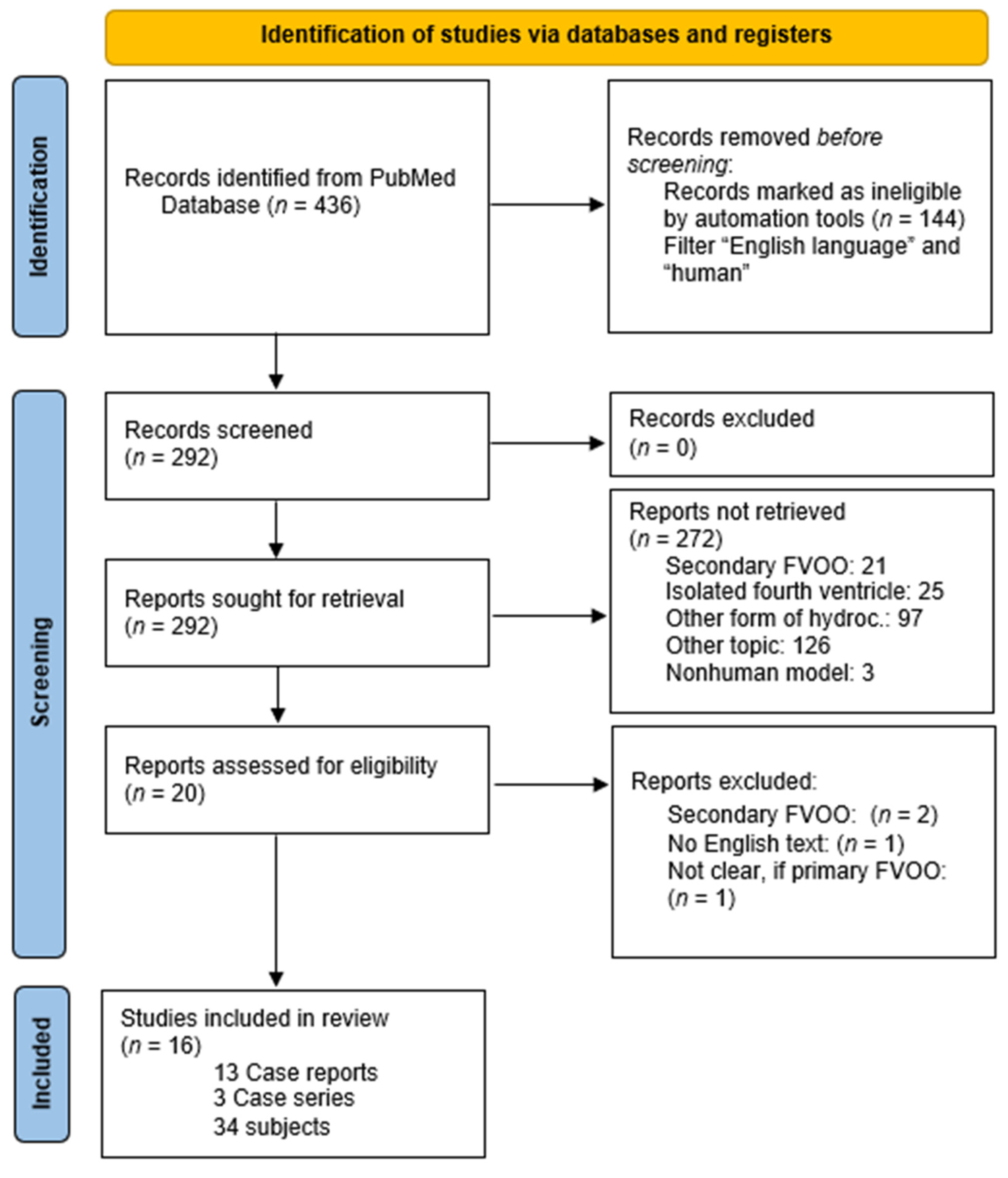
Search Strategy and Article Selection

A computerized search strategy using the MEDLINE/Pub Med database was performed to identify relevant case series and case reports describing the treatment of primary FVOO. Only articles in English published between 1 January 1997 and 31 October 2022 were included. We used combined keywords and medical subject headings as search terms for abstract and full text. The following terms were used: “obstruction fourth ventricle”, “occlusion fourth ventricle”, “occlusion Magendie”, “obstruction Magendie”, “obstruction Luschka”, “occlusion Luschka”, “fourth ventricle outlet obstruction”, “FVOO”. All articles were screened regarding their titles and abstracts. Only articles describing the treatment of primary FVOO in humans with sufficient description of the symptoms preoperatively and the clinical outcome were selected. Articles published in languages other than English were also excluded. A cross-referenced check of the citations of each included relevant literature review was done in order to ensure that no relevant studies were missed by the computerized database research.
The following characteristics from the included studies were extracted: clinical findings before and after surgery, radiological findings before and after surgery, and type of surgery. Interventional studies involving animals or humans, and other studies that require ethical approval, must list the authority that provided approval and the corresponding ethical approval code.

3. Results

The computerized search resulted in 436 articles. After the application of the inclusion and exclusion criteria mentioned above, 13 case reports and 3 case series were included, counting 34 cases (13 from case reports, 21 from case series) (Figure 3) (Table 1 and Table 2) [3,12,16,19,20,21,22,23,24]. The 34 patients were aged from 3 months to 73 years, and 4 were pediatric patients. Of the patients, 18 were male, 15 were female, and in 1 case the sex was not mentioned. In addition, the case series described 33 patients with FVOO, 7 of them with primary FVOO [5,21]; however, the outcome of the patients could not be clearly associated with primary or secondary FVOO. Therefore, these studies were only included in the analysis describing the distribution of the symptoms, and not considered for evaluating the outcome after the different types of surgery.

3.1. Clinical Features

Most symptoms were associated with elevated intracranial pressure (ICP). For instance, nausea was described in 8 (19.5%) cases, vomiting in 17 (41.5%), urinary incontinence in 7 (17.1%), Hakim triad in 2 (4.9%), papilledema in 4 (9.8%), and dizziness in 6 (14.6%) cases. Out of 41 patients, 24 (58.5%) reported headache. Besides symptoms of elevated intracranial pressure, cerebellar symptoms like cerebellar ataxia, gait disturbance, or impaired balance occurred in 23 patients (56.1%): cerebellar ataxia occurred in 6 (14.6%), gait disturbance in 21 (51.2%), vertigo in 3 (7.3%), diplopia in 3 (7.3%), or impaired balance in 3 patients (7.3%). Apart from cerebellar and ICP-related symptoms, ideomotor dysfunctions (three cases, 7.3%) and memory disturbances (four cases, 9.8%) were described. In two cases (4.9%), a syringomyelia with an associated myelopathy occurred. Other rare symptoms were the syndrome of inappropriate antidiuretic hormone secretion (SIADH), seizures, depression, frontal syndromes or disorder of higher cognitive functions in one case (2.4%) and visual impairment in three (7.3%), or cervical spine pain in two cases (4.9%), respectively. The three patients who were 12 months and younger presented with bulging fontanel and lethargy. One patient (2.4%) died before surgery.

3.2. Treatments

ETV was performed in 23 cases (69.7%). After surgery, all symptoms resolved completely in 10 of 23 cases (43.5%), and in 13 of 23 cases (56.5%) symptoms improved partially. Seven patients (30.4%) required additional surgery, five in the form of re-ETV, and three in the form of ventriculoperitoneal shunting. Fenestration of the fourth ventricular outlets was performed in 10 cases, 8 of them via an open suboccipital craniotomy and 2 with an endoscopic, transaqueductal approach. In two of two cases (100%) treated via an endoscopic transaqueductal approach, all symptoms resolved completely. Of eight patients operated via open suboccipital approach, in four patients (50.0%) symptoms resolved completely, and in three patients (37.5%) partially. Two patients (25.0%) required additional surgery, one because of epidural hematomas [27]. A radiological decrease in ventricular volume was described in most cases.

4. Discussion

We analyzed symptoms, diagnostic characteristics, and therapeutic options of primary FVOO and their outcomes in the literature.

4.1. Symptoms

Most patients suffered from hydrocephalus-associated symptoms such as headache, nausea, and vomitus. Other symptoms like cognitive impairment, urinary incontinence, or gait disturbance may be attributed to NPH-like syndromes in chronic hydrocephalus. More than half of the described patients had additional cerebellar symptoms like ataxia or gait disturbance. In solitary cases, miscellaneous symptoms like syringomyelia, seizures, vertigo, frontal symptoms, or higher cognitive function disorders occurred. In one case, SIADH in a one year and eight-month-old child was described. The increase in antidiuretic hormone (ADH) secretion in the serum was explained by acute intraventricular hypertension. The ADH levels normalized after ETV [3].
The patient described in our case report had both hydrocephalus and cerebellar symptoms including nausea, vomiting, nystagmus, and a gait disturbance.

4.2. Diagnostic

Most publications describe the enlargement of all four ventricles and the lateral recesses as pathognomonic for FVOO [3,11,12,16,17,19,20,22,23,24]. In some cases, direct visualization of the membrane in the T1 MRI was possible [19,23]. In some of these cases, a thick membrane was visualized intraoperatively. A direct visualization is probably not possible in the case of thinner membranes [11]. Many authors describe flow analysis or Cine-MRI to visualize CSF flow and outflow from the fourth ventricle [3,20,22,24]. However, flow analysis over the foramina of Magendie or Luschka seems to be less practicable than in the aqueduct. Possible reasons are turbulent flow, the bigger volume of the fourth ventricle, and the outflow in three different directions [21]. Due to the enlargement of all four ventricles, FVOO can often be misdiagnosed as communicating hydrocephalus.
Computed tomography (CT) or MRI ventriculography are the most reliable tools to verify an interruption of the CSF-flow [21]. However, due to the invasiveness of these diagnostic procedures, many authors restrict themselves to conventional MRIs with or without flow analysis [5,7,11,16,19,22,23,24].

4.3. Therapy

4.3.1. ETV

ETV is an established procedure for the treatment of occlusive hydrocephalus in the case of aqueduct stenosis. The utility of ETV for the treatment of primary FVOO was first described by Suehiro et al. in 2000 [17]. In most cases, like in our case, ETV was performed under general anesthesia via a burr hole at Kocher’s point, slightly anterior to the coronal suture. The endoscope was inserted via the foramen of Monro and the perforation was performed between the mammillary bodies and the infundibular recess. In this review of the literature, we found that in 23 cases of primary FVOO treated with ETV, in 43.5% all symptoms improved and in 56.5% a part of the symptoms improved. However, 30.4% percent of the patients required additional surgery. According to the literature, adhesions in basal cisterns correlated with the failure of ETV. Results were worse in patients with thick and opaque floors of the third ventricle [1]. Carpentier describes a spontaneous functional reopening of the foramina of the fourth ventricle after ETV in the postoperative flow rate studies with a diminished but physiological flow through the foramina of Magendie and Luschka. This is explained by a downward displacement of the posterior fossa structures during the genesis of the hydrocephalus, which causes an adherence of the semipermeable membranes to the dura leading to a complete CSF blockage. By restoring the pressure equilibrium between the intra- and extraventricular spaces, the downward displacement of the posterior fossa structures is reduced and the flow over the foramina is restored [11].

4.3.2. Open, Suboccipital Approach

The open, suboccipital approach was first described as a treatment for FVOO by Holland and Graham in 1957 [15]. It is performed directly via a suboccipital craniectomy. In most cases, a laminectomy of C1 was performed in addition. Some authors describe a splitting of the vermis to reach the foramen of Magendie; however, a mobilization of the tonsils may be sufficient in many cases [19,24]. The membrane is visualized and resected. The main advantage of this approach is the restoration of natural CSF-flow. Postoperative MRIs can demonstrate flow phenomenon through the foramen of Magendie.
However, this approach seems to be more invasive with a more severe muscular and osseous involvement, a longer duration of surgery and a potential higher blood loss, which should be considered especially in pediatric patients. In the eight cases in our review all preoperative described symptoms resolved in 50% of cases [19,24], residual symptoms were described in 37.5% [25,26,27,28,29]. Additional surgery was necessary in 25% [26,27]. Some studies describe a high failure rate for infants younger than six months and suggest a ventriculoperitoneal shunting procedure as a favorable option in this age group [1]. Few cases of postoperative epidural hematoma after posterior fossa surgery for fourth ventricular outlet obstruction are described in the literature [27].

4.3.3. Endoscopic Transaqueductal Approach

Endoscopic, transaqueductal Magendie foraminoplasty, as described by Longatti in 2006 [16], or Luschka foraminoplasty, described by Torres–Corzo in 2014 [5], are alternative options to restore the flow of CSF.
An endoscopic approach to the fourth ventricle with a flexible endoscope is possible due to the dilatation of the aqueduct in tetraventricular hydrocephalus. The endoscopic fenestration allows the restoration of the physiological flow. Endoscopic foraminoplasty is described for the foramen of Magendie, Luschka, or both. Another advantage is the avoidance of damage to structures at the floor of the third ventricle. Hence, this method seems to be a useful option, if an EVT is not feasible, like in patients with a thick floor of the third ventricle or an attachment of the basilar artery. Possible risks are the compression of the calamus scriptorius or damage to the posterior inferior cerebellar artery (PICA) at the foramen of Magendie. In a series of 52 patients who underwent transaqueductal Magendie or Luschka foraminoplasty (not only due to primary FVOO but also due to secondary FVOO), ependymal contusion occurred in 2 cases [16]. In the two cases, in which this method is described for primary FVOO, no adverse events were reported [16]. Endoscopic, transaqueductal Magendie or Luschka foraminoplasty may be good options for selected cases, when the membrane obstructing the foramen is thin, the PICA (in the case of Magendie foraminoplasty) is visible, and an ETV is not feasible. Endoscopic foraminoplasty was described for both adult and pediatric patients. The outcome of foraminoplasty seemed to be better in patients with primary FVOO than for secondary FVOO. It was often performed secondarily, after other procedures had failed or when an ETV was not feasible intraoperatively [5].

4.3.4. Shunting Procedures

In some cases, before the diagnosis of FVOO, ventriculoperitoneal shunts were implanted. After diagnosis of FVOO, other treatments like EVT or opening of the outflows of the fourth ventricle were performed [5,24]. Shunting procedures bear the risk of infection, misplacement, or malfunction. As mentioned above, some studies describe a high failure rate for ETV for infants younger than six months. A ventriculoperitoneal shunting procedure as a favorable option in this age group is suggested [1].
There are several treatment options for patients with occlusive hydrocephalus due to primary FVOO. Besides ventricular–peritoneal shunting procedures, three surgical treatment options are common and described in the literature, all of which have advantages, disadvantages, and limitations. The main advantage is avoiding the implantation of a shunt and the associated risk of infection and malfunction. ETV is a well-established procedure for the treatment of occlusive hydrocephalus, which alleviates symptoms in most cases of primary FVOO, but is limited in patients with small basal cisterns, thick, opaque third ventricle floors, and adherent basilar arteries. On the contrary endoscopic, transaqueductal fenestrations of the foramina of Magendie or Luschka seem to be an alternative treatment option. However, this approach requires a sufficient enlargement of the aqueduct. In addition, there is an option for open resection of the membrane via a suboccipital approach, entailing the necessity of a larger, more invasive approach.
The approach has to be selected depending on the individual anatomic and clinical characteristics of the patient.

4.4. Limitation

The results of the study rely retrospectively on case reports and series and could be biased due to patient selection.

5. Conclusions

In the case of a newly diagnosed hydrocephalus, the foramina of Luschka and Magendie must be closely examined on imaging to detect occlusion. In the case of FVOO, based on the results of this review, we would recommend ETV; in the case of treatment failure, direct endoscopic fenestration or implantation of a VP shunt could be performed.

Author Contributions

Conceptualization, E.W. and M.S.; methodology, E.W. and M.S.; validation, E.W. and M.S.; formal analysis, E.W. and M.S.; investigation, E.W., T.F. and M.S.; resources, E.W., T.F. and M.S.; data curation, E.W., C.E., S.Z.A., T.F. and M.L.; writing—original draft preparation, E.W.; writing—review and editing, E.W., S.S. and M.S.; visualization, E.W.; supervision, M.S.; project administration, E.W., M.S. and S.Z.A. All authors have read and agreed to the published version of the manuscript.

Funding

This research received no external funding.

Institutional Review Board Statement

The study was conducted in accordance with the Declaration of Helsinki, and approved by the Institutional Ethics Committee of Witten/Herdecke University (protocol code: 95/2020, date of approval: 7 July 2020).

Informed Consent Statement

Informed consent was obtained from all subjects involved in the study.

Data Availability Statement

Data are contained within the article.

Conflicts of Interest

The authors declare no conflict of interest.

References

  1. Mohanty, A.; Biswas, A.; Satish, S.; Praharaj, S.S.; Sastry, K.V.R. Treatment options for Dandy-Walker malformation. J. Neurosurg. 2006, 105, 348–356. [Google Scholar] [CrossRef] [PubMed]
  2. Calabro, F.; Arcuri, T.; Jinkins, J.R. Blake’s pouch cyst: An entity within the Dandy-Walker continuum. Neuroradiology 2000, 42, 290–295. [Google Scholar] [CrossRef] [PubMed]
  3. Hashimoto, H.; Maeda, A.; Kumano, K.; Kimoto, T.; Fujisawa, Y.; Akai, T. Rapid deterioration of primary fourth ventricular outlet obstruction resulting in syndrome of inappropriate antidiuretic hormone secretion. Pediatr. Int. 2014, 56, e30–e32. [Google Scholar] [CrossRef]
  4. ELLIOTT, G.B.; WOLLIN, D.W. Defect of the corpus callosum and congenital occlusion of fourth ventricle with tuberous sclerosis. Am. J. Roentgenol. Radium Ther. Nucl. Med. 1961, 85, 701–705. [Google Scholar] [PubMed]
  5. Torres-Corzo, J.; Sanchez-Rodriguez, J.; Cervantes, D.; Rodriguez-Della Vecchia, R.; Muruato-Araiza, F.; Hwang, S.W.; Rangel-Castilla, L. Endoscopic transventricular transaqueductal Magendie and Luschka foraminoplasty for hydrocephalus. Neurosurgery 2014, 74, 426–435; discussion 436. [Google Scholar] [CrossRef] [PubMed]
  6. Roth, J.; Ben-Sira, L.; Udayakumaran, S.; Constantini, S. Contrast ventriculo-cisternography: An auxiliary test for suspected fourth ventricular outlet obstruction. Childs. Nerv. Syst. 2012, 28, 453–459. [Google Scholar] [CrossRef] [PubMed]
  7. Marin-Sanabria, E.A.; Yamamoto, H.; Nagashima, T.; Kohmura, E. Evaluation of the management of arachnoid cyst of the posterior fossa in pediatric population: Experience over 27 years. Childs. Nerv. Syst. 2007, 23, 535–542. [Google Scholar] [CrossRef]
  8. Cornips, E.M.J.; Overvliet, G.M.; Weber, J.W.; Postma, A.A.; Hoeberigs, C.M.; Baldewijns, M.M.L.L.; Vles, J.S.H. The clinical spectrum of Blake’s pouch cyst: Report of six illustrative cases. Childs. Nerv. Syst. 2010, 26, 1057–1064. [Google Scholar] [CrossRef]
  9. Spennato, P.; Sacco, M.; Cinalli, G. Diverticular Enlargement of Foramen of Luschka and Hydrocephalus in Child with Noonan Syndrome. World Neurosurg. 2019, 125, 371–373. [Google Scholar] [CrossRef]
  10. BARR, M.L. Observations on the foramen of Magendie in a series of human brains. Brain 1948, 71, 281–289. [Google Scholar] [CrossRef]
  11. Carpentier, A.; Brunelle, F.; Philippon, J.; Clemenceau, S. Obstruction of Magendie’s and Luschka’s foramina. Cine-MRI, aetiology and pathogenesis. Acta Neurochir. 2001, 143, 517–521; discussion 521–522. [Google Scholar] [CrossRef]
  12. Longatti, P.; Fiorindi, A.; Martinuzzi, A.; Feletti, A. Primary obstruction of the fourth ventricle outlets: Neuroendoscopic approach and anatomic description. Neurosurgery 2009, 65, 1078–1085; discussion 1085–1086. [Google Scholar] [CrossRef]
  13. Rifkinson-Mann, S.; Sachdev, V.P.; Huang, Y.P. Congenital fourth ventricular midline outlet obstruction. J. Neurosurg. 1987, 67, 595–599. [Google Scholar] [CrossRef]
  14. Barany, L.; Baksa, G.; Patonay, L.; Racz, G.; Ganslandt, O.; Buchfelder, M.; Kurucz, P. Primary Obstruction of the Foramen of Luschka: Anatomy, Histology, and Clinical Significance. World Neurosurg. 2018, 112, e288–e297. [Google Scholar] [CrossRef]
  15. Holland, H.C.; Graham, W.L.; Holland, C.H.C.; Graham, C.W.L.; Barany, L.; Baksa, G.; Patonay, L.; Racz, G.; Ganslandt, O.; Buchfelder, M.; et al. Congenital atresia of the foramina of Luschka and Magendie with hydrocephalus; report of a case in an adult. J. Neurosurg. 1958, 15, 688–694. [Google Scholar] [CrossRef] [PubMed]
  16. Longatti, P.; Fiorindi, A.; Feletti, A.; Baratto, V. Endoscopic opening of the foramen of magendie using transaqueductal navigation for membrane obstruction of the fourth ventricle outlets. J. Neurosurg. 2006, 105, 924–927. [Google Scholar] [CrossRef] [PubMed]
  17. Suehiro, T.; Inamura, T.; Natori, Y.; Sasaki, M.; Fukui, M. Successful neuroendoscopic third ventriculostomy for hydrocephalus and syringomyelia associated with fourth ventricle outlet obstruction. J. Neurosurg. 2000, 93, 326–329. [Google Scholar] [CrossRef] [PubMed]
  18. Moher, D.; Liberati, A.; Tetzlaff, J.; Altman, D.G. Preferred reporting items for systematic reviews and meta-analyses: The PRISMA statement. Int. J. Surg. 2010, 8, 336–341. [Google Scholar] [CrossRef]
  19. Huang, Y.C.; Chang, C.N.; Chuang, H.L.; Scott, R.M. Membranous obstruction of the fourth ventricle outlet. Pediatr. Neurosurg. 2001, 35, 43–47. [Google Scholar] [CrossRef]
  20. Ishi, Y.; Asaoka, K.; Kobayashi, H.; Motegi, H.; Sugiyama, T.; Yokoyama, Y.; Echizenya, S.; Itamoto, K. Idiopathic fourth ventricle outlet obstruction successfully treated by endoscopic third ventriculostomy: A case report. Springerplus 2015, 4, 565. [Google Scholar] [CrossRef]
  21. Karachi, C.; Le Guerinel, C.; Brugieres, P.; Melon, E.; Decq, P. Hydrocephalus due to idiopathic stenosis of the foramina of Magendie and Luschka. J. Neurosurg. 2003, 98, 897–902. [Google Scholar] [CrossRef]
  22. Kawaguchi, T.; Fujimura, M.; Tominaga, T. Syringomyelia with obstructive hydrocephalus at the foramens of Luschka and Magendie successfully treated by endoscopic third ventriculostomy. Surg. Neurol. 2009, 71, 349–352; discussion 352. [Google Scholar] [CrossRef] [PubMed]
  23. Rougier, A.; Menegon, P. MRI evidence of membranous occlusion of the foramen of Magendie. Acta Neurochir. 2009, 151, 693–694. [Google Scholar] [CrossRef]
  24. Shimoda, Y.; Murakami, K.; Narita, N.; Tominaga, T. Fourth Ventricle Outlet Obstruction with Expanding Space on the Surface of Cerebellum. World Neurosurg. 2017, 100, 711.e1–711.e5. [Google Scholar] [CrossRef]
  25. Giannetti, A.V.; Malheiros, J.A.; Da Silva, M.C. Fourth ventriculostomy: An alternative treatment for hydrocephalus due to atresia of the Magendie and Luschka foramina. J. Neurosurg. Pediatr. 2011, 7, 152–156. [Google Scholar] [CrossRef] [PubMed]
  26. Duran, D.; Hadzipasic, M.; Kahle, K.T. Mystery Case: Acute hydrocephalus caused by radiographically occult fourth ventricular outlet obstruction. Neurology 2017, 88, e36–e37. [Google Scholar] [CrossRef]
  27. Tyagi, G.; Bhat, D.I.; Devi, B.I.; Shukla, D. Multiple Remote Sequential Supratentorial Epidural Hematomas-An Unusual and Rare Complication After Posterior Fossa Surgery. World Neurosurg. 2019, 128, 83–90. [Google Scholar] [CrossRef] [PubMed]
  28. Bai, J.; Yu, Q.; Sun, X.; Xiao, H.; Wang, K.; Sun, F.; Sui, Q. Hydrocephalus Due to Idiopathic Fourth Ventricle Outflow Obstruction. J. Craniofac. Surg. 2019, 30, e397–e400. [Google Scholar] [CrossRef]
  29. Krejčí, O.; Krejčí, T.; Mrůzek, M.; Večeřa, Z.; Šalounová, D.; Lipina, R. Hydrocephalus Caused by Primary Fourth Ventricle Outlet Obstruction: Our Experience and Literature Review. World Neurosurg. 2021, 148, e425–e435. [Google Scholar] [CrossRef]
Figure 1. Endoscopic view to the floor of the third ventricle after endoscopic third ventriculostomy (ETV) in fourth ventricle outflow obstruction (FVOO).
Surgeries 04 00063 g001
Figure 2. T2 image before and after endoscopic third ventriculostomy. (a) Sagittal T2 image before and after ETV. Before ETV the fourth ventricle is enlarged. Flow void over the aqueduct indicates a preserved flow over the Sylvian aqueduct. (b) Transversal T2 image before and after ETV. Before ETV the fourth ventricle and the lateral recesses are enlarged.
Surgeries 04 00063 g002
Figure 3. The search resulted in 436 articles. Thirteen case reports and three case series were included after the application of inclusion and exclusion criteria.
Surgeries 04 00063 g003
Table 1. Thirteen case reports were included in this review.
Case No.AuthorSex/AgePresentationTreatmentFollow-UpOutcome
1Suehiro et al., 2000 [17]f/27 yearsDizziness, headache, nausea, syringomyeliaETVNot reportedAll symptoms resolved completely
2Huang et al., 2001 [19]f/15 yearsAmenhorrhea, headache, nausea, vomitingSuboccipital craniotomy, C1 laminectomy, splitting of vermis, removal of membrane14 monthsAll symptoms resolved completely
3Carpentier et al., 2001 [11]f/58 yearsHeadache, nausea, vomiting, instability (frequent falls), cerebellar ataxia, pseudo-vertebrobasilar insufficiency, blindness with head rotation (maybe due to compression of VA in foramen magnum)ETV36 monthsAll symptoms resolved completely
4Longatti et al., 2006 [16]f/64 yearsCervical spine pain, nighttime headache, disturbed gait, dizziness, cerebellar ataxiaTransaqueductal Magendieplasty (ETV scheduled, not performed, since basilar tip left no space for stoma)At least 4 monthsAll symptoms resolved completely
5Rougier et al., 2009 [23]m/60 yearsHeadache, unsteady gaitSuboccipital approach, fenestration of membraneAt least 6 monthsAll symptoms resolved completely
6Kawaguchi et al., 2009 [22]m/55 yearsSyrinx, sensory disturbance, discrete movement disturbance of bilateral upper limb, dysuria, spine deformity, hyperreflection of upper limbs, hyorreflection of lower limbs, rectovesical dysfunctionETVNot reportedSome symptoms resolved completely
7Giannetti et al., 2011 [25] f/8 yearsHeadache, vomiting, decreased alertness, gait ataxiaSuboccipital endoscopic opening of the membranes36 monthsSome symptoms resolved completely
8Hashimoto et al., 2014 [3]m/20 monthsSIADH, vomitingEVD, later ETVNot reportedAll symptoms resolved completely
9Ishi et al., 2015 [20]m/3 yearsHeadache, vomitingEVD, 2 days later ETV Transaqueductal endoscpic exploration of fourth ventricle20 months after second ETVAll symptoms resolved completely
10Shimoda et al., 2017 [24]f/66 yearsFirst admission: gait disturbance, wide based gate, cerebellar ataxia, dizziness, nausea, incontinence
Second admission: disturbed balance, drowsiness
First admission: VPS (fourth ventricle enlargement 9 months after shunting),
Second admission: ETV
Not reportedImprovement of symptoms
11Duran et al., 2017 [26]f/19Diplopia, headache, papilledema, abducens nerve palsySuboccipital craniotomy, later ventriculo-peritoneal shuntingNot reportedSome symptoms (diplopia) resoled after suboccipital craniotomy, all symptoms resolves after shunting,
12Tyagi et al., 2019 [27]m/22Headache, vomiting, truncal ataxia, papilledema, gaze-evoked nystagmus, dysarthriaPosterior fossa decompression, magendieplasty Epidural hematoma, serial surgical evacuation
13Bai et al., 2019 [28]15Headache, vomitingSuboccipital craniotomy, resection of membrane12 monthsImprovement of symptoms
Table 2. Three case series with twenty-one subjects were included in this review.
Case No.AuthorSex/AgePresentationTreatmentFollow-UpOutcome (Clinic)
14–16Karachi et al., 2003 [21]14: 21/f
15: 53/f
16: 68/m
14: headache, projectile vomiting, bilateral papilledema,
15: vertigo, positional nausea, slight postural instability
16: gait disorders, sphincter disorders, disorders of higher functions, stepping gait, postural instability, urinary incontinence, frontal syndrome
14: ETV
15: ETV
16: ETV
14: 26 months
15: 24 months
16: 58 months
14–16: all symptoms resolved completely
17–26Longatti et al., 2009 [12]17: m/53 18: f/49
19: m/70 20: m/73 21: m/64
22: m/36 23: m/65 24: m/69 25: m/69 26: m/58
17: ideomotor slowdown, gait difficulties, depression
18: dizziness, gait difficulties, memory impairment
19: gait difficulties, ideomotor slowdown, incontinence
20: gait difficulties, ideomotor slowdown, memory impairment
21: gait difficulties, incontinence
22: dizziness, visusal impairment, headache, vomiting
23: cervical pain, headache, gait difficulties, dizziness, nausea, vomiting
24: gait difficulties, memory impairment
25: gait difficulties, incontinence, memory impairment
26: gait difficulties
17–22, 24–26: ETV, all transaqueductal exploration
20: additional aqueductoplasty
23: magendieplasty
17: 12 years
18: 7 years
19: 6 years
20: 6 years
21: lost to follow up
22: 2 months, lost to follow up
23: 34 months
24: 15 months
25: 5 months
26: 3 months
23: all symptoms resolved completely
17–22, 24–26: improvement of symptoms
27–34Krejci et al., 2021 [29]27: 22/f
28: 74/f
29: 54/f
30: 22/f
31: 33/m
32: 44/m
33: 30/m
34: 28/f
27: headache, vertigo, gait disturbance, diplopia,
28: headache, Hakim triad, vomiting,
29: Hakim triad,
30: headache, vomiting, gait disturbance,
31: headache, intracranial hypertension, diplopia, vomiting, vertigo, papilledema, cognitive impairment
32: headache, vertigo
33: headache, vomiting
34: headache, vomiting, vertigo, papilledema
27: ETV
28: ETV
29: ETV
30: ETV
31: craniotomy
32: craniotomy
33: ventricular drainage
34: ETV
27: 132 months
28: 84 months
29: 24 months
30: 48 months
31: 125 months
32: 108 months
33: -
34: 7 months
27: all symptoms resolved completely
28: all symptoms resolved completely
29: improvement of symptoms (later VP shunt)
30: all symptoms resolved completely
31: all symptoms resolved completely
32: all symptoms resolved completely
33: death before treatment
34: improvement of symptoms (after re-ETV)
Disclaimer/Publisher’s Note: The statements, opinions and data contained in all publications are solely those of the individual author(s) and contributor(s) and not of MDPI and/or the editor(s). MDPI and/or the editor(s) disclaim responsibility for any injury to people or property resulting from any ideas, methods, instructions or products referred to in the content.

