Article

Generation of Spherical Microparticles of Moringa Leaves through a Supercritical Antisolvent Extraction Process

Department of Chemical Engineering and Food Technology, Faculty of Sciences, University of Cádiz, International Excellence Agrifood Campus (CeiA3), 11510 Puerto Real, Cadiz, Spain
*
Author to whom correspondence should be addressed.
Sustain. Chem. 2023, 4(2), 143-153; https://doi.org/10.3390/suschem4020011
Submission received: 6 March 2023 / Revised: 20 March 2023 / Accepted: 28 March 2023 / Published: 31 March 2023

Abstract

:
The objective of this work was evaluation of the supercritical antisolvent extraction (SAE) process to generate microparticles with antioxidant activity from Moringa leaves. A biodegradable polymer was used as an inductor of particle precipitation. An ethanolic extract of 25 mg/mL was used in the SAE process, during which the influences of pressure (100–200 bar), temperature (35–55 °C) and extract–polymer ratio (0.11–0.33) on particle size and antioxidant activity were evaluated. An extract flow rate of 3 mL/min, a supercritical CO2 (scCO2) flow rate of 30 g CO2/min and a nozzle diameter of 100 µm were kept constant. The identification of several compounds of Moringa leaves, namely, coumaric acid and quercetin 3D glucoside, were determined with ultra-performance liquid chromatography coupled with mass spectrometry. The antioxidant activity of the extract and the precipitates was measured with 2,2-Diphenyl-1-picrylhydrazyl. Spherical microparticles with diameters in the range of 2–5 µm were obtained, with moderate antioxidant activity.

1. Introduction

Such is the interest in functional foods, the vegetarian and vegan diet, the increase in intolerances and allergies and the rise of green cosmetic manufacturing that new insight on the investigations of plants and natural products has had exponential growth in the scientific community [1].
The “tree of life”, as Moringa oleifera is known, is the focus of such different strategic sectors as food, nutraceutics and pharmaceutics due to its nutritional features and properties. Its composition satisfies the daily amounts recommended for many nutrients, and thus, it is a great hope to eradicate malnutrition all over the world [2]. Moringa oleifera contains carotenoids, tocopherol, flavonoids, phenolic acids and minerals. Its main compounds are quercetin, vanillin, gallic acid, cumaric acid, ferulic acid, sinapic acid, kaempferol and glucosinolates, among others [3,4,5]. This tree belongs to the Moringaceae family and has been traditionally cultivated in tropical and subtropical areas all over the world. It is fast-growing and drought-resistant and thus is an appropriate plant for cattle and even fishes of arid areas [6]. In this sense, it is considered a superfood, with more than ninety chemical nutrients. Its seeds have high content of protein and lipids as oleic, palmitic and stearic acid. Thus, the oil of the seed is already used to generate biodiesel [2]. Moreover, the plant has high content of carbohydrates and fiber. Because of this, it is already employed in the poultry industry as a supplement to produce antioxidant-enriched meats. However, the leaves have high content of potassium, calcium, retinoic and ascorbic acids, iron, protein and polyphenols, and its flowers have also high content of tannins and polyphenols as flavonoids [7,8] to be used in biomedical application.
With regard to applications in biomedicine, polyphenols present in Moringa oleifera have achieved blocking of carcinogenic cells through DNA reparation mechanisms and improving protective-enzyme production [9]. It has also been demonstrated that consumption of Moringa leaves reduces hepatic illness produced by drugs, can serve as a substitute for antibiotics and can even serve as an antidepressant in ethanolic extract, making sure its valorization to become a potential product [10]. Moringa leaves were chosen in this study due to being a main source of such flavonoids as quercetin and kaempferol, whose proportion is variable according to recollection time and culture area [11].
One of the few green processes to generate Moringa-leaf nanoparticles was carried out by Virk et al. [12] using a process of two steps: dried Moringa-leaf powder was added to methanol, and then this solution was sprayed at a rate of 0.2 mL/min with boiling water under ultrasonic conditions for 5 min, then freeze-dried. However, conventional methods of obtaining natural products from waste carry great associated consumption of energy and solvents, which produces great environmental contamination, risks for health and higher time and economic expenses. Moreover, part of the product could be lost during the long evaporation steps. In this way, supercritical fluid technology removes the main drawbacks of conventional methods, obtaining a better product, conserving its properties and minimizing risks for environmental media. Use of supercritical CO2 methods produces high selectivity of natural products and complete separation of solvent and matrix [13]. Particularly, the supercritical antisolvent extraction (SAE) process is a fractioned precipitation based on polar compounds as polyphenols, which are related to numerous biological properties.
Application of the SAE process to a natural liquid extract to obtain particles requires two conditions: the liquid solvent has to be miscible or partially miscible in supercritical CO2 (scCO2), and the compounds present in the plant material must not be soluble in CO2. In this process, the liquid extract is pumped through a nozzle to a vessel that is already filled with scCO2. Then, a rapid mass transfer of organic solvent between the generated microdroplets and bulk CO2 is produced, leading to supersaturation of the solution and powder precipitation through the antisolvent effect. Moreover, this precipitated powder often has better properties of antioxidant activity than the original extract. In this way, potent antioxidant microparticles from olive [14,15], eucalyptus leaf [16] and grape residues [17] have been produced. Spherical nanoparticles were successfully precipitated, composed of polyphenols with high antioxidant activity, from mango [18,19] and orange leaves [20]. SAE has also been used to generate particles from such plants as Achillea millefolium [21] and Curcuma longa [22].
Some authors have even encapsulated natural extracts with polymers, such as Machado et al. [23], who generated submicron particle composites from grape residue extract and polyvinylpyrrolidone (PVP). In this way, Guaman-Balcazar et al. also formed an encapsulation of mango-leaf particles with PVP [24]. Visentin et al. achieved encapsulation of rosemary antioxidants [25] and Santana and Meireles achieved coprecipitation of turmeric extracts [26].
Microcapsules of flavonoids from Moringa were prepared with xylose-modified soybean protein isolate and gelatin as wall materials but with a previous deep eutectic solvent (choline chloride–urea) extraction from Moringa leaves [27]. Other authors have even achieved encapsulation of Moringa leaf extract in biopolymers through ionic gelation. This is why automatic solvent extraction using an 80% wt. ethanol solution was carried out in order to extract active material from the plant in a previous step. Then, the active material was covered in chitosan/alginate in the presence of calcium chloride via ionic gelation [28]. However, the use of chloride compounds moved both of these processes away from green-process methods.
Formation of particles depends mainly on the pressure and temperature inside the vessel, the concentration of the extract and liquid/CO2 solution flow rate ratios, but the success of the process depends enormously on the concentration of the extract. It is necessary for the liquid extract to be concentrated enough to achieve power in the collection vessel. In this sense, PVP has already been used as promotor to force supercritical precipitation of the extract [29].
The aim of the study described here was to obtain natural microparticles with notable antioxidant activity through the SAE process applied to Moringa-leaf extracts obtained through maceration. The identification and quantification of several compounds present in Moringa-leaf precipitated particles were addressed. Moreover, the influences of the SAE process’ main parameters on particle size and antioxidant activity were investigated.

2. Materials and Methods

2.1. Solvents

For this experiment, 2,2-Diphenyl-1-picrylhydrazyl (DPPH), gallic acid (≥99%), mangiferin (≥98%), quercetin 3-β-D-glucoside (≥90%), epigallocatechin gallate (≥95%), vitexin (≥95%), kaempferol (≥99%), kaempferol 3-glucoside (≥90%), cumaric acid (≥98%), ferulic acid (≥99%), sinapic acid (≥99%) and polyvinylpyrrolidone (average mol.wt 40,000) were purchased from Sigma-Aldrich (Steinheim, Germany). Ethanol, acetonitrile and formic acid (HPLC grade) were supplied by Panreac (Barcelona, Spain). CO2 with a maximum purity of 99.8% was obtained from Linde (Jerez de la Fra., Spain).

2.2. Plant Material

Moringa oleifera leaves were collected in February 2021 by Connatur in Conil de la Fra. (Cádiz, Spain). Then, the leaves were dried until loss of weight became constant. The leaves were crushed in an electric grinder until they became a powder with an average particle diameter of 500–750 µm. Finally, the leaves were kept in a dry place, at room temperature and without light to preserve their unaltered properties.

2.3. Preparation of Moringa-Leaf Extract

The extract preparation was carried out through maceration of leaves in ethanol with magnetic stirring for 3 h at 40 °C. Then, the ethanolic solution was vacuum-filtered. The final concentration was 26.7 mg/mL.

2.4. Design of Experiment

A design of experiment (DOE) was applied to clarify the influences of the pressure and temperature inside the vessel and the polymer ratio on the particle size and antioxidant activity of the particles obtained through the SAE process. The Statgraphics 19 Centurion application (The Plains, VA, USA) was used to analyze the design. A mixture-level factorial design (3 × 22) with 12 factor points and two central points was implemented at random.
Two levels of pressure (P) (100–200 bar) and temperature (T) (35–55 °C) inside the vessel and two levels of extract–polymer ratio (r) (0.11–0.33) of liquid solution injected into the vessel were the main parameters evaluated regarding the success of Moringa-leaf precipitation and optimization of particle size distribution. The nozzle diameter (100 µm), the CO2 flow rate (30 g/min), the liquid solution flow rate (3 mL/min) and the contact time (60 min) were kept at constant values. The operating conditions used for the SAE process are shown in Table 1.

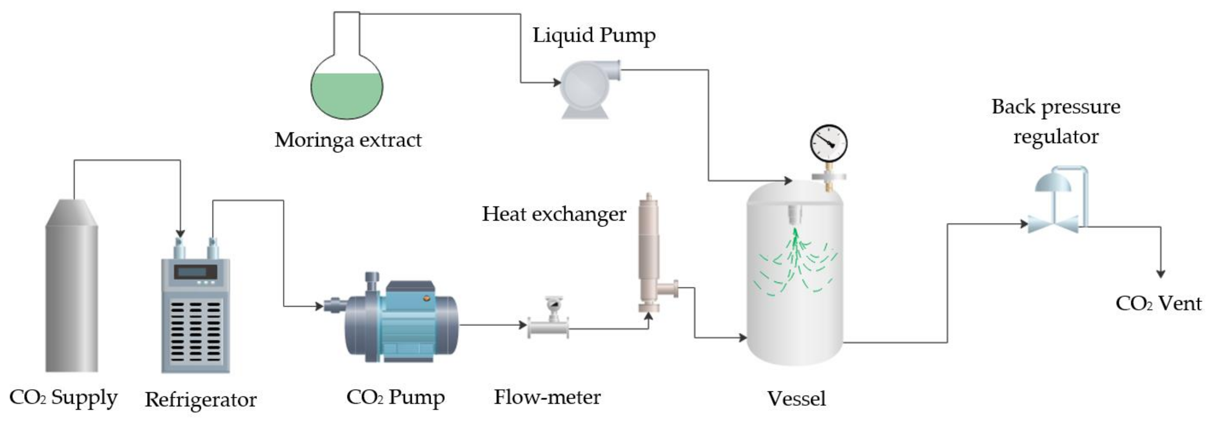
2.5. Supercritical Antisolvent Extraction (SAE)

The SAE technique was used to precipitate particles from Moringa leaves. All of the experiments were carried out in a pilot plant developed by Thar Technologies® (Pittsburgh, PA, USA) (SAS200), the scheme of which is shown in Figure 1. The apparatus was equipped with two high-pressure pumps: one for CO2 pumping and another one for pumping extract into the vessel. An electric heater and a heater exchanger were used to manipulate the CO2 temperature. The stainless-steel-made precipitator vessel (0.5 L) was provided with a pressure gauge. The vessel consisted of a main body and a frit. The pressure was controlled with an automated high-precision back-pressure regulator via software. A separator (0.5 L) with a pressure gauge and a manual back-pressure regulator were located at the end of the process to carry out separation of solvent and CO2 once pressure was released. Moreover, two electrical heating jackets surrounded the precipitator vessel and the separator, respectively, to control the temperature in the precipitation process and in the solvent separation process.
The process operated in a semicontinuous way, and all of the experiments were carried out as follows: CO2 was pumped into the vessel and the electric heater was switched on. Once the operating temperature and pressure were achieved in the vessel, the extract solution with PVP was pumped into the vessel and sprayed through a nozzle with a diameter orifice of 100 µm. Then, a rapid mass transfer happened between the microdroplets of extract solution and the bulk CO2, causing supersaturation of the solution and precipitation on the wall and frit of the vessel via the antisolvent effect. Then, fresh CO2 continued flowing for a period of time, namely, washing time, to remove the solvent from the CO2 and avoid redissolution of the precipitates in the depressurization step.

2.6. Particle Size Distribution

A Nova NanoSEMTM 450 scanning electron microscope (SEM) was used to determine the sizes and morphologies of the Moringa-leaf precipitates. Prior to the analyses, the particles were coated with a 20 nm film of gold using a sputter coater. Scion image 4.0-analysis software (Scion Corporation) was used to process the SEM images in order to obtain the mean particle size diameters.

2.7. Antioxidant Activity Assay with DPPH

Antioxidant activity was determined using the method described by Scherer and Godoy [30]. In this method, the ability of Moringa leaves to scavenge DPPH free radicals was evaluated. In total, 90 µL of six samples at different particle concentrations were mixed with 3510 µL of 6 × 10⁻⁵ mol DPPH/L ethanol (mother solution of DPPH). These solutions were mixed and allowed to react in the absence of light for 3 h. The final absorbance was measured at 517 nm. Then, a standard curve for the DPPH was prepared at different concentrations between 7 × 10⁻⁵ and 6 × 10⁻⁶ mol/L in order to quantify the antioxidant capacity. All procedures were performed in triplicate.
Then, the remaining DPPH percentage was determined with Equation (1). In this equation, CDPPHo is the initial DPPH concentration (before the reaction) and CDPPHt is the final DPPH concentration (at the end of the 3 h reaction):
%   DPPH   remaining = C DPPHt C DPPHo × 100
The sample concentrations were plotted against the % DPPH remaining, and the IC50 value was calculated graphically using a polynomial fitting curve. Thus, antioxidant activity was expressed as the antioxidant activity index (AAI) using Equation (2).
AAI = C DPPHt IC 50  

2.8. Extract Load

The composition of the particles precipitated was measured with UV-VIS spectrophotometry. Due to the extract being composed of a lot of compounds with different maximum absorptions in the visible region, measurements were focused in the UV region, which contains PVP’s maximum absorption (206 nm). A calibration curve for the PVP was carried out in acidulated water (pH 1.2). Then, 15 mg of each sample was completely dissolved over 2 h in 20 mL of acidulated water. The composition of each sample was calculated as an extract percentage comparing the total amount of sample to the amount of obtained PVP.

2.9. Moringa-Leaf Particle Composition

The analysis of the chemical composition of the Moringa-leaf particles was carried out with an Acquity™ ultra-performance liquid chromatography system (Waters, Milford, MA, USA) coupled with Xevo™ G2 QTof MS quadrupole–time-of-flight mass spectrometry (Waters, Milford, MA, USA). Analyses were processed with MassLynx version 4.1 software: the Target Lynx version. For quantification of compounds, a reverse phase column (Acquity UPLC BEH C18, 2.1 mm × 100 mm, 1.7 µm particle size) from Waters was used. The temperature of the column was set at 45 °C. The injection volume was set as 2 µL, and the solvent flow rate through the column was set at 0.4 mL/min. The phase gradient of the binary system phases, based on solvent A (water + 0.1% formic acid) and solvent B (methanol), was as follows: initial time, 95% A at 5 min, 30% A from 5 to 6 min, 5% A from 6 to 7 min and 95% A from 7 to 10 min. Electrospray was operated in negative ionization mode with a mass range of 100–1200 Da, using 2.5 kV of capillary voltage, 40 V of cone voltage and 120 °C as the source temperature. Before analysis, the samples were filtered using a 0.2 μm nylon-made syringe filter. Sample injections were performed in duplicate. In Table 2, the calibration curves for the different commercial standards and retention times, which were used to detect and quantify different compounds, are shown.

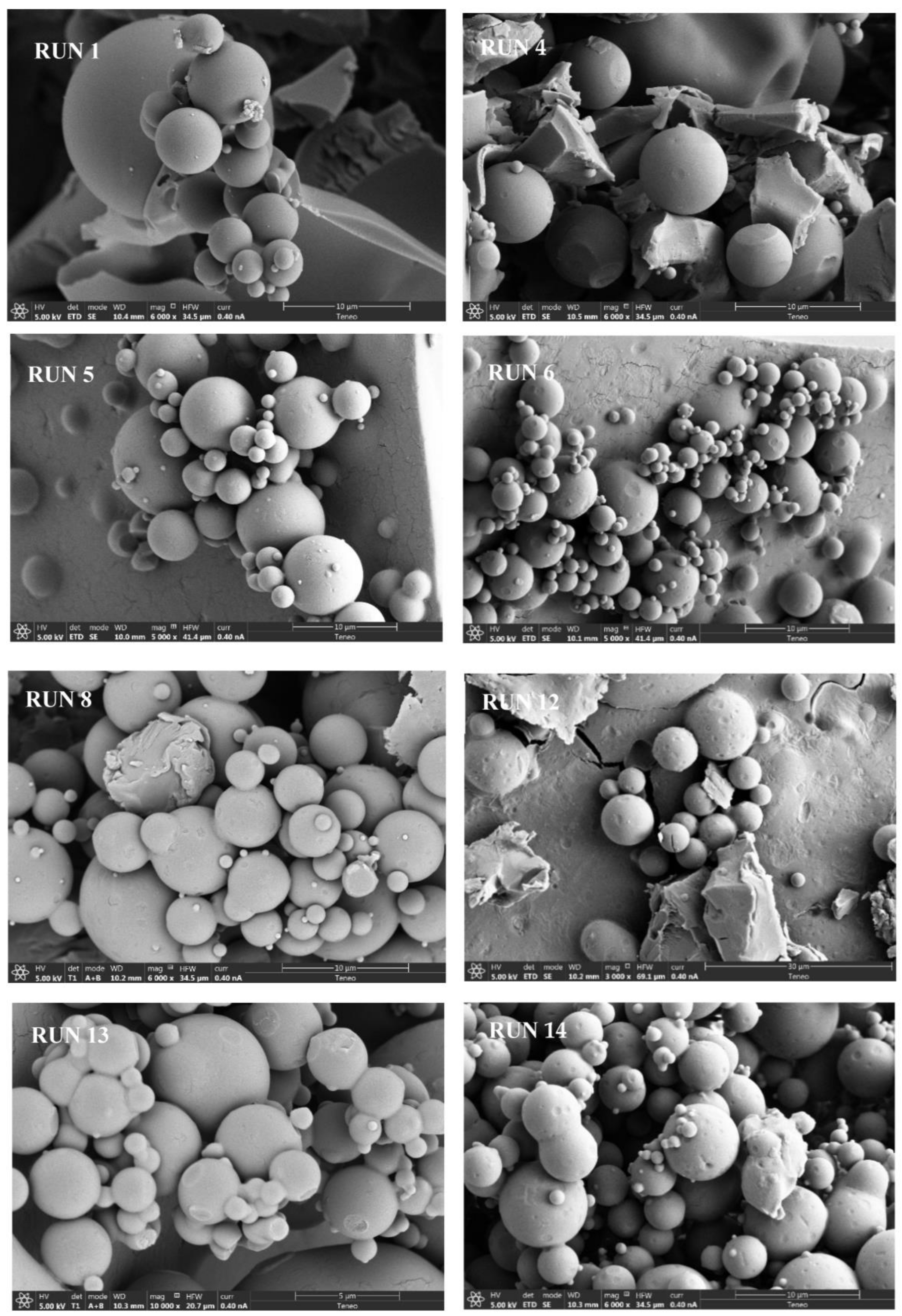
3. Results and Discussion

Ethanol was used instead of water or the other mixtures in previous extraction processes due to the fact that the yield could be higher. This can be explained due to the ethanol polarity index being lower than the water index, thus making it an effective solvent to extract polyphenols and flavonoids [31]. Moringa-leaf extract was never precipitated alone through the SAE process. Several experiments and conditions were tried, but liquid and no powder were found in the vessel. In this case, PVP was utilized as an inductor of SAE precipitation, and precipitated powder was achieved in most of the experiments, as can be observed in Table 1. Runs 1, 4, 5, 6, 8, 12, 13 and 14 were precipitated as spherical microparticles, as can be observed in Figure 2. The extract load of precipitates showed that the particles were based mainly on PVP and the extracts were about 8–10 wt%.
In this sense, the highest Moringa extract percentage had the lowest amount of polymer in the initial solution. Due to several experiments failing, no model from the described design could adjust the results. Several trends could be observed according to the size of the precipitated particles, as can be observed in Table 1. The effect of the temperature inside the vessel on particle size could be elucidated from observing runs 1 (3.02 ± 1.78 µm), and 8 (3.06 ± 2.16 µm) and runs 5 (2.76 ± 2.10 µm) and 14 (2.63 ± 1.56 µm). In both pairs of experiments, it can be seen that there is no significant difference between the runs, and this fact is independent of the polymer ratio.
With regard to pressure’s effect on particle size, it seems that at higher amounts of polymer, as in runs 1 (3.02 ± 1.78 µm) and 13 (2.77 ± 1.80 µm), particle size of precipitates increases as pressure increases. However, at lower amounts of polymer, as in runs 12 (5.32 ± 2.25 µm) and 14 (2.63 ± 1.56 µm), when pressure increases, particle size of precipitates decreases. This fact is corroborated if runs 4 (2.91 ± 2.67 µm) and 14 are compared. The significant difference test showed that these pairs of runs were not significant at a confidence level of 10%. In order to evaluate the polymer ratio, runs 1 (3.02 ± 1.78 µm) and 14 (2.63 ± 1.56 µm) can be compared, reflecting that no significant difference was found here either.
On the other hand, the Moringa-leaf extract and most of the spherical precipitates were analyzed for their radical scavenging activity with the DPPH assay, as summarized in Table 1. The antioxidant activity index (AAI) of the extract (0.62) was considered moderated, based on a study by Scherer and Godoy [30].
Zullaikah et al. [31] used subcritical extraction with ethanol and different water–ethanol mixtures to extract compounds from Moringa leaves. In that case, the highest IC50 that was obtained was 87 µg/mL. This happened when 96% ethanol was used (the higher the IC50, the lower the AAI, as can be observed in Equation (2)). In our case, the IC50 for the extract was 15.38 µg/mL; thus, our conventional extraction using ethanol had a higher antioxidant capacity. The main explanation of this difference in antioxidant activity, even in using the same solvent for extraction, is due to the temperature difference in the processes. In our process, the extraction temperature was set to 40 °C, and Zullaikah et al. used 200 °C, which could have damaged polyphenols and flavonoids responsible for antioxidant properties. Moreover, using these levels of temperature (five times higher) enormously increases the costs of the extraction process.
For that reason, Ping et al. [27] used a deep eutectic solvent (choline chloride–urea) to improve extraction of flavonoids from Moringa leaves efficiently. Nine flavonoids, hyperoside, vitexin, quercetin, cynaroside, quercetin, 3-β-d glucoside, kaempferol, taxifolin, luteolin and fisetin, were identified, achieving an IC50 of 64.1 µg/mL. This IC50 value improved upon that from Zullaikah et al. (87 µg/mL) [27] but did not improve the IC50 of the present work (15.38 µg/mL). This process included the handicap of using chloride solvent for the extraction.
On the other hand, the precipitates showed a low AAI, as can be observed in Table 1. In previous work, precipitates from ethanolic extract leaves had higher AAIs than the original extract due to selective precipitation and concentration in compounds in which a higher AAI was produced. In this sense, very-strong-antioxidant mango-leaf nanoparticles (AAI = 4.71) [13,19], very-strong-antioxidant eucalyptus-leaf submicron particles (AAI = 2.40) [16] and strong-antioxidant olive-leaf microparticles (AAI = 1.56) were achieved [14].
In this work, the opposite trend was observed, and the Moringa-leaf extract had a higher AAI than its precipitates, which could have been due to the lower amount of extract in the particles; thus, the amount of extract could have been underestimated in the DPPH method. In general, the experiments with higher percentages of Moringa particles had higher AAIs (runs 5, 12 and 14) than the rest. In Table 3, six compounds that were present in the Moringa-leaf precipitates can be observed. It can also be observed that these compounds were not quantified in the same precipitate but alternatively in the composites. For instance, in run 5, which had a higher AAI, quercetin 3-β-D glucoside, epigallocatechin gallate, gallic acid and vitexin were quantified. The mass spectra of the corresponding chromatogram of this run are shown in Figure 3.
However, run 5 had a percentage of extract lower than that of run 12, but the AAI was higher in the case of run 5 (0.41). For that reason, not only the amount of extract but also the amount of compounds with antioxidant activity governs the antioxidant properties of composites. Considering this, four compounds that could have been present in these leaves were not precipitated in any case; sinapic acid [32], ferulic acid [33], kaempferol [34] and kaempferol 3-glucoside [35] with notable antioxidant activity were not identified.
This fact could be the reason why the antioxidant activity was not very high. Particularly, ferulic acid, which is formed via partial etherification of caffeic acid, has a relative higher solubility in scCO2 than do other hydroxycinnamic acids. This difference in solubility is due to removal of a hydroxyl group, and this compound was probably removed with CO2 in the washing step [36]. In this way, kaempferol and its derivative could be extracted through supercritical CO2 in this washing [37]. However, sinapic acid has low solubility values in supercritical CO2 of the molar order of 10−8. Thus, it would be a candidate to be precipitated in the SAE process. These phenolic compounds increase solubility in scCO2 for the presence of cosolvents [38] and could be dissolved together with ethanol in the antisolvent process.

4. Conclusions

Spherical Moringa-leaf microparticles were precipitated from Moringa ethanolic extract with the aid of PVP as a precipitation inductor. The particles were precipitated in the range of 2–5 µm. It seems that pressure, temperature and polymer ratio do not significantly influence particle size at the assayed levels at a confidence level of 10%. Antioxidant activity of particles seems to be related to the proportion of PVP in the final composite. Thus, Moringa-leaf extract has higher antioxidant activity and the precipitates with higher polymer content have lower activity. Six compounds were identified in Moringa-leaf precipitates, and in general, the majority was gallic acid, followed by epigallocatechin gallate. Coumaric acid and quercetin 3D glucoside were identified in higher proportions in some precipitates.

Author Contributions

Conceptualization, A.M. and D.V.; methodology, A.M.; software, D.V.; validation, C.P. and E.M.d.l.O.; formal analysis, D.V.; investigation, A.M. and D.V.; resources, C.P.; data curation, D.V.; writing—original draft preparation, A.M.; writing—review and editing, C.P.; visualization, E.M.d.l.O.; supervision, E.M.d.l.O.; project administration, C.P. All authors have read and agreed to the published version of the manuscript.

Funding

This research received no external funding.

Institutional Review Board Statement

Not applicable.

Informed Consent Statement

Not applicable.

Data Availability Statement

Not applicable.

Acknowledgments

We gratefully acknowledge Connatur® for the donation of Moringa leaves that were used for these experiments, as well as the central services of the University of Cádiz.

Conflicts of Interest

The authors declare no conflict of interest. The funders had no role in the design of this study; in the collection, analysis, or interpretation of this data; in the writing of this manuscript; or in the decision to publish these results.

References

  1. Rubio-Sanz, L. Comparativa nutricional del cultivo de Moringa Oleifera en España. Cienc. Tecnol. UTEQ 2020, 13, 17–22. [Google Scholar] [CrossRef]
  2. Saini, R.K.; Sivanesan, I.; Keum, Y.S. Phytochemicals of Moringa oleifera: A review of their nutritional, therapeutic and industrial significance. 3 Biotech 2016, 6, 203. [Google Scholar] [CrossRef] [PubMed] [Green Version]
  3. Bhattacharya, A.; Tiwari, P.; Sahu, P.K.; Kumar, S. A Review of the Phytochemical and Pharmacological Characteristics of Moringa oleifera. J. Pharm. Bioallied Sci. 2018, 10, 181–191. [Google Scholar] [PubMed]
  4. Ammara, S.; Mohammad, S.; Muhammad, F.A.; Mirza, M.F.A.B.; Azhar, R. HPLC analysis, cytotoxicity, and safety study of Moringa oleifera Lam. (wild type) leaf extract. J. Food Biochem. 2020, 44, 3400. [Google Scholar]
  5. Pari, L.; Karama, M.; Kosiska, A.; Rybarczyk, A.; Amarowicz, R. Antioxidant activity of the crude extracts of drumstick tree (moringa oleifera lam.) and sweet broomweed (Scoparia dulcis L.) leaves. Pol. J. Food Nutr. Sci. 2007, 57, 203–208. [Google Scholar]
  6. Antonio-Alegría, L.; Sánchez-Mundo, M.L.; Sánchez-Zacarías, M.A.; Hernández-Nava, R.G. Perfil proteico, compuestos fenólicos y capacidad antioxidante de hoja de moringa (Moringa Oleífera). Rinderesu 2020, 5, 965–976. [Google Scholar]
  7. Brilhante, R.S.N.; Sales, J.A.; Pereira, V.S.; Castelo-Branco, D.S.C.M.; Cordeiro, R.A.; de Souza, C.M.; de Araújo Neto Paiva, M.; Feitosa dos Santos, J.B.; Costa, J.J.; Gadelha Rocha, M.F. Research advances on the multiple uses of Moringa Oleifera: A sustainable alternative for socially neglected population. Asian Pac. J. Trop. Med. 2017, 10, 621–630. [Google Scholar] [CrossRef]
  8. Guzmán-Maldonado, S.H.; López-Manzano, M.J.; Madera-Santana, T.J.; Núñez-Colín, C.A.; Grijalva-Verdugo, C.P.; Villa-Lerma, A.G.; Rodríguez-Núñez, J.R. Nutritional characterization of Moringa Oleifera leaves, seeds, husks and flowers from two regions of Mexico. Agron. Colomb. 2020, 38, 287–297. [Google Scholar] [CrossRef]
  9. Mumtaz, M.Z.; Kausar, F.; Hassan, M.; Javaid, S.; Malik, A. Anticancer activities of phenolic compounds from Moringa oleifera leaves: In vitro and in silico mechanistic study. Beni-Suef Univ. J. Basic Appl. Sci. 2021, 10, 12. [Google Scholar] [CrossRef]
  10. Karthivashan, G.; Arulselvan, P.; Razak, A.A.; Safinar, I.I.; Fakurazi, S. Competing Role of Bioactive Constituyents in Moringa oleifera Extract and Conventional Nutrition Feed on the Performance of Cobb 500 Broilers. Biomed Res. Int. 2015, 2015, 970398. [Google Scholar] [CrossRef] [Green Version]
  11. Leone, A.; Spada, A.; Battezzati, A.; Schiraldi, A.; Aristil, J.; Bertoli, S. Cultivation, Genetic, Ethnopharmacology, Phytochemistry and Pharmacology of Moringa oleifera Leaves: An Overview. Int. J. Mol. Sci. 2015, 16, 12791–12835. [Google Scholar] [CrossRef]
  12. Promy, V.; Manal, A.A.; Sarah, S.A.A.; Awatif, A.H.; Mai, E.; Khalid, O.; Rabia, Q.; Manal, F.E.-K.; Hany, M.Y.; Mohamed, F.S.E.-D.; et al. Green synthesis of Moringa oleifera leaf nanoparticles and an assessment of their therapeutic potential. J. King Saud Univ.-Sci. 2023, 35, 102576. [Google Scholar]
  13. Guamán-Balcázar, M.C.; Montes, A.; Fernández-Ponce, M.T.; Casas, L.; Mantell, C.; Pereyra, C.; Martínez de la Ossa, E. Generation of potent antioxidant nanoparticles from mango leaves by supercritical antisolvent extraction. J. Supercrit. Fluids 2018, 138, 92–101. [Google Scholar] [CrossRef]
  14. Chinnarasu, C.; Montes, A.; Fernández-Ponce, M.T.; Casas, L.; Mantell, C.; Pereyra, C.; Martínez de la Ossa, E. Precipitation of antioxidant fine particles from Olea europaea leaves using supercritical antisolvent process. J. Supercrit. Fluids 2015, 97, 125–132. [Google Scholar] [CrossRef]
  15. Baldino, L.; Della Porta, G.; Osseo, L.S.; Reverchon, E.; Adami, R. Concentrated oleuropein powder from olive leaves using alcoholic extraction and supercritical CO2 assisted extraction. J. Supercrit. Fluids 2018, 133, 65–69. [Google Scholar] [CrossRef]
  16. Chinnarasu, C.; Montes, A.; Fernández-Ponce, M.T.; Casas, L.; Mantell, C.; Pereyra, C.; Martínez de la Ossa, E.; Pattabhi, S. Natural antioxidant fine particles recovery from Eucalyptus globulus leaves using supercritical carbon dioxide assisted processes. J. Supercrit. Fluids 2015, 101, 161–169. [Google Scholar] [CrossRef]
  17. Natolino, A.; Da Porto, C.; Rodriguez-Rojo, S.; Moreno, T.; Cocero, M.J. Supercritical antisolvent precipitation of polyphenols from grape marc extract. J. Supercrit. Fluids 2016, 118, 54–63. [Google Scholar] [CrossRef]
  18. Meneses, M.A.; Caputo, G.; Scognamiglio, M.; Reverchon, E.; Adami, R. Antioxidant phenolic compounds recovery from Mangifera indica L. by-products by supercritical antisolvent extraction. J. Food Eng. 2015, 163, 45–53. [Google Scholar] [CrossRef]
  19. Guamán-Balcázar, M.C.; Montes, A.; Pereyra, C.; Martínez de la Ossa, E. Precipitation of mango leaves antioxidants by supercritical antisolvent process. J. Supercrit. Fluids 2017, 128, 218–226. [Google Scholar] [CrossRef]
  20. Montes, A.; Williamson, D.; Hanke, F.; Guaman-Balcazar, M.C.; Valor, D.; Pereyra, C.; Martínez de la Ossa, E.; Teipel, U. Precipitation of powerful antioxidant nanoparticles from orange leaves by means of supercritical CO2. J. CO2 Util. 2019, 31, 235–243. [Google Scholar] [CrossRef]
  21. Villanueva-Bermejo, D.; Zahran, F.; Troconis, D.; Villalva, M.; Reglero, G.; Fornari, T. Selective precipitation of phenolic compounds from Achillea millefolium L. extracts by supercritical anti-solvent technique. J. Supercrit. Fluids 2017, 120, 52–58. [Google Scholar] [CrossRef]
  22. Osorio-Tobón, J.F.; Carvalho, P.I.N.; Rostagno, M.A.; Petenate, A.J.; Meireles, M.A.A. Precipitation of curcuminoids from an ethanolic turmeric extract using a supercritical antisolvent process. J. Supercrit. Fluids 2016, 108, 26–34. [Google Scholar] [CrossRef]
  23. Machado, A.P.d.F.; Montes, A.; Valor, D.; Fernández- Ponce, M.T.; Fernández Barbero, G.; Maróstica Júnior, M.R.; Pereyra, C.; Martínez de la Ossa, E. Co-precipitation of grape residue extract using sub- and supercritical CO2 technology. J. CO2 Util. 2022, 61, 102010. [Google Scholar] [CrossRef]
  24. Guamán-Balcázar, M.C.; Montes, A.; Pereyra, C.; Martínez de la Ossa, E. Production of submicron particles of the antioxidants of mango leaves/PVP by supercritical antisolvent extraction process. J. Supercrit. Fluids 2019, 143, 294–304. [Google Scholar] [CrossRef]
  25. Visentin, A.; Rodríguez-Rojo, S.; Navarrete, A.; Maestri, D.; Cocero, M.J. Precipitation and encapsulation of rosemary antioxidants by supercritical antisolvent process. J. Food Eng. 2012, 109, 9–15. [Google Scholar] [CrossRef]
  26. Santana, Á.L.; Meireles, M.A.A. Coprecipitation of turmeric extracts and polyethylene glycol with compressed carbon dioxide. J. Supercrit. Fluids 2017, 125, 31–41. [Google Scholar] [CrossRef]
  27. Wei, P.; Zhang, Y.; Wang, Y.Y.; Dong, J.F.; Liao, B.N.; Su, Z.C.; Li, W.; Xu, J.C.; Lou, W.Y.; Su, H.H.; et al. Efficient extraction, excellent activity, and microencapsulation of flavonoids from Moringa oleifera leaves extracted by deep eutectic solvent. Biomass Conv. Bioref. 2023. [Google Scholar] [CrossRef]
  28. Ebru, K.; Raneen, A.; Mehmet, T.; Selin, Ş. Encapsulation of Moringa oleifera leaf extract in chitosan-coated alginate microbeads produced by ionic gelation. Food Biosci. 2022, 50, 102158. [Google Scholar]
  29. Prosapio, V.; Reverchon, E.; De Marco, I. Coprecipitation of Polyvinylpyrrolidone/β-Carotene by Supercritical Antisolvent Processing. Ind. Eng. Chem. Res. 2015, 54, 11568–11575. [Google Scholar] [CrossRef]
  30. Scherer, R.; Godoy, H.T. Antioxidant activity index (AAI) by the 2,2-diphenyl-1-picrylhydrazyl method. Food Chem. 2009, 112, 654–658. [Google Scholar] [CrossRef]
  31. Zullaikah, S.; Naulina, R.Y.; Meinawati, P.; Fauziyah, K.; Rachimoellah, M.; Rachmaniah, O.; Nurkhamidah, S.; Suari, N.M.I.P.; Prasetyo, E.N. Enhanced Extraction of Phenolic Compounds from Moringa Oleifera Leaves Using Subcritical Water Ethanol Mixture. IOP Conf. Ser. Mater. Sci. Eng. 2019, 543, 012021. [Google Scholar] [CrossRef] [Green Version]
  32. Chen, C. Sinapic Acid and Its Derivatives as Medicine in Oxidative Stress-Induced Diseases and Aging. Oxidative Med. Cell. Longev. 2016, 2016, 3571614. [Google Scholar] [CrossRef] [Green Version]
  33. Zduska, K.; Dana, A.; Kolodziejczak, A.; Rotsztejn, H. Antioxidant Properties of Ferulic Acid and Its Possible Application. Skin Pharmacol. Physiol. 2018, 31, 332–336. [Google Scholar] [CrossRef]
  34. Simunkova, M.; Barbierikova, Z.; Jomova, K.; Hudecova, L.; Lauro, P.; Alwasel, S.H.; Alhazza, I.; Rhodes, C.J.; Valko, M. Antioxidant vs. Prooxidant Properties of the Flavonoid, Kaempferol, in the Presence of Cu(II) Ions: A ROS-Scavenging Activity, Fenton Reaction and DNA Damage Study. Int. J. Mol. Sci. 2021, 22, 1619. [Google Scholar] [CrossRef]
  35. Kluska, M.; Juszczak, M.; Zuchowski, J.; Stochmal, A.; Wozniak, K. Effect of Kaempferol and Its Glycoside Derivatives on Antioxidant Status of HL-60 Cells Treated with Etoposide. Molecules 2022, 27, 333. [Google Scholar] [CrossRef]
  36. Murga, R.; Sanz, M.T.; Beltran, S.; Cabeza, J.L. Solubility of three hydroxycinnamic acids in supercritical carbon dioxide. J. Supercrit. Fluids 2003, 27, 239–245. [Google Scholar] [CrossRef]
  37. Cid-Ortega, S.; Monroy-Rivera, J.A. Extraction of Kaempferol and Its Glycosides Using Supercritical Fluids from Plant Sources: A Review. Food Technol. Biotechnol. 2018, 56, 480–493. [Google Scholar] [CrossRef]
  38. Paula, J.T.; Sousa, I.M.O.; Foglio, M.A.; Cabral, F.A. Solubility of protocatechuic acid, sinapic acid and chrysin in supercritical carbon dioxide. J. Supercrit. Fluids 2016, 112, 89–94. [Google Scholar] [CrossRef]
Figure 1. Schematic diagram of SAS 200 pilot plant.
Suschem 04 00011 g001
Figure 2. SEM images of Moringa-leaf particles precipitated using the SAE process.
Suschem 04 00011 g002
Figure 3. Mass spectra for the identification of compounds in run 5.
Suschem 04 00011 g003
Table 1. Design of experiment.
RunP (bar)T (°C)Moringa:PVP Ratio (wt:wt)MPS (µm)Extract Load (%)AAI
1200550.113.02 ± 1.78 9.380.12
2150350.33Agglomerated
3100350.11Agglomerated
4100550.332.91 ±2.67 11.500.25
5200350.332.76 ± 2.10 12.090.41
6150450.162.00 ± 1.25 12.380.34
7100550.11---
8200350.113.06 ± 2.1610.240.22
9150350.11Agglomerated
10100350.33---
11150450.16Agglomerated
12150550.335.32 ± 2.25 12.970.29
13150550.112.77± 1.808.780.18
14200550.332.63 ± 1.569.160.17
Table 2. The analytical characteristics for determination of Moringa-leaf compounds.
CompoundLinear EquationR2
Gallic Acid y = 11.2933 x 11.8771 0.994
Epigallocatechin Gallate y = 19.5814 x 24.3043 0.990
Mangiferin y = 100.163 x   + 6.60468 0.999
Cumaric Acid y = 74.2688 x   + 214.558 0.988
Vitexin y = 97.5725 x 20.1444 0.999
Sinapic Acid y = 63.0448 x   + 87.5507 0.995
Quercetin 3-β-D-Glucoside y = 51.0679 x   522.173 0.996
Kaempferol y = 132.434 x   + 3461.71 0.934
Ferulic Acid y = 30.0034 x   + 117.217 0.983
Kaempferol 3-Glucoside y = 114.896 x   + 345.462 0.984
Table 3. Identified compounds in precipitates via UPLC-MS.
SampleMangiferinQuercetin 3-β-D GlucosideVitexinEpigallocatechin GallateCoumaric AcidGallic Acid
µg/L
Run 10.2---0.43.1---17.5
Run 4---1.60.32.5---17.7
Run 5---7.30.52.3---16.7
Run 6------0.42.63.915.9
Run 80.41.80.42.2------
Run 12------0.7------17.1
Run 13------0.42.4------
Run 140.9------2.2---16.7
Disclaimer/Publisher’s Note: The statements, opinions and data contained in all publications are solely those of the individual author(s) and contributor(s) and not of MDPI and/or the editor(s). MDPI and/or the editor(s) disclaim responsibility for any injury to people or property resulting from any ideas, methods, instructions or products referred to in the content.

