Article

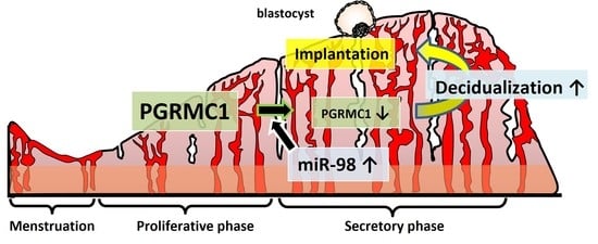
Possible Involvement of miR-98 in the Regulation of PGRMC1 During Decidualization

1
Department of Endocrine Pharmacology, Tokyo University of Pharmacy and Life Sciences, Tokyo 192-0392, Japan
2
Department of Obstetrics and Gynecology, Tokyo Medical University, Tokyo 160-0023, Japan
*
Author to whom correspondence should be addressed.
These authors contributed equally to this work.
Reprod. Med. 2022, 3(2), 189-200; https://doi.org/10.3390/reprodmed3020015
Submission received: 16 February 2022 / Revised: 8 June 2022 / Accepted: 14 June 2022 / Published: 15 June 2022
(This article belongs to the Special Issue Endometrial Physiology and Pregnancy Success)

Abstract

:
Human endometrial stromal cells (ESCs) differentiate into decidual cells for embryo implantation during the mid-secretory phase of the menstrual cycle. Decidualization is characterized by enhanced production of insulin-like growth factor-binding protein 1 (IGFBP1) and prolactin (PRL) by ESCs and their morphological transformation into polygonal cells. Progesterone (P4) receptor membrane component 1 (PGRMC1) is a member of a P4-binding complex implicated in function in female reproduction. In this study, we explored the mechanisms that regulate PGRMC1 during decidualization of human ESCs. Immunohistochemical analysis of endometrial samples showed that PGRMC1 was expressed in endometrial glandular and luminal epithelial cells and stromal cells throughout the menstrual cycle; however, the protein level in stroma was reduced in the secretory phase. Incubation of ESCs with dibutyryl (db)-cAMP and P4 in vitro, which induces decidualization, decreased the PGRMC1 protein abundance. Further, treatment with a PGRMC1-targeting siRNA or PGRMC1 inhibitor (AG-205) promoted mRNA expression of the db-cAMP/P4- and db-cAMP-induced decidual markers IGFBP1 and PRL. Moreover, the microRNA miR-98, a potential repressor of PGRMC1, was upregulated during decidualization, and transfection of ESCs with a miR-98 mimic decreased the PGRMC1 protein level. These findings suggest that miR-98-mediated downregulation of endometrial PGRMC1 may promote decidualization for the establishment of pregnancy.

Graphical Abstract

1. Introduction

In humans, increased estrogen levels in the proliferative phase of the menstrual cycle stimulate endometrial cell proliferation and endometrial gland formation. After ovulation, human endometrial stromal cells (ESCs) differentiate into decidual cells under the influence of progesterone (P4) secreted from the corpus luteum during the mid-to-late secretory phase. Differentiation of ESCs into decidual cells, referred to as decidualization, is essential for appropriate embryo implantation and subsequent placenta formation [1,2,3]. Decidualization is accompanied by morphological transition of the cells from a fibroblastic phenotype to an enlarged and rounded shape, and induction of the specific markers insulin-like growth factor-binding protein-1 (IGFBP1) and prolactin (PRL) [1,3,4,5].
P4 is an essential regulator of the events that occur during the menstrual cycle, pregnancy establishment and maintenance (such as ovulation, fertilization, decidualization, and implantation), and parturition [6,7,8,9]. These effects of P4 are predominantly exerted through interactions with classical P4 receptors (PRs), PRA and PRB. When P4 binds to its intracellular PRs, the complex translocates into the nucleus and interacts directly with specific DNA elements in genes to regulate their transcription [10]. PR-mediated P4 activity and diseases are evidenced by reports that PR-deficient mice are infertile and that dysregulation of P4/PR signaling is associated with endometriosis, breast cancer, miscarriage, and preterm birth [10,11,12]. However, ovarian granulosa cells are reported to respond to P4 even in the absence of classical PRs, suggesting the involvement of non-classical P4 receptors distinct from PRs [13,14]. One such non-classical P4 receptor is progesterone receptor membrane component 1 (PGRMC1). It was originally cloned during a search for membrane receptors with a high affinity for P4 and is considered to mediate the action of P4 via a non-canonical P4 signaling pathway [15]. PGRMC1 may have multiple biological functions, including in cell proliferation, apoptosis, steroid metabolism, lipid metabolism, membrane trafficking, angiogenesis, and the P4 response. Moreover, PGRMC1 may play a role in carcinogenesis [16]. Further, PGRMC1-mediated P4 signaling can suppress apoptosis of human ovarian granulosa cells [17], whereas conditional ablation of Pgrmc1 in the reproductive tract results in subfertility and the development of endometrial cysts in female mice [18]. Salsano et al. [19] reported that endometrial PGRMC1 expression decreases in the late secretory phase of the menstrual cycle and that overexpression of PGRMC1 in ESCs abrogates decidualization. However, the mechanisms by which PGRMC1 is downregulated during decidualization have not been elucidated.
MicroRNAs (miRNAs) are small, endogenous, noncoding RNAs that play important roles in post-transcriptional gene regulation by binding to the 3′ untranslated region of their target mRNA [20]. Upon binding, miRNAs can inhibit expression of the target gene and thereby affect various biological processes such as cell differentiation, development, apoptosis, and proliferation. Recent studies showed that miRNAs are differentially expressed in the human endometrium during decidualization and the window period of embryo implantation [21,22]. For example, loss of miR-29a attenuates proliferation and in vitro decidualization of human ESCs [23].
In this study, we explored the involvement of miRNAs in the regulation of decidual PGRMC1. Our research focused on (1) the expression of PGRMC1 in the endometrium and (2) the effect of differential stimuli on PGRMC1 expression in an in vitro decidualization model and, conversely, the effect of functional inhibition of PGRMC1 on decidual markers. Finally, we explored miRNAs as candidate PGRMC1 regulators and the effects of db-cAMP/P4 on miRNA expression in ESCs.

2. Materials and Methods

2.1. Endometrial Tissues

Endometrial samples were obtained from Japanese patients with a normal menstrual cycle who were younger than 45 years and underwent surgery for endometriosis or leiomyoma and were not on hormonal therapy at the time of surgery. In accordance with the requirements of the Clinical Research Ethics Committee of the Tokyo Medical University Hospital (No. 3018), informed consent was obtained from all patients. The use of these tissues in the experiments described herein was approved by the Clinical Research Ethics Committee of the Tokyo University of Pharmacy and Life Sciences (No. 1511).
Tissues in proliferative (n = 4) and secretory (n = 4) phases were fixed in 4% paraformaldehyde for immunohistochemistry, and primary ESCs were isolated from patients with a normal menstrual cycle who underwent surgery for leiomyoma (n = 3) as described below.

2.2. Immunohistochemistry Reagents and Antibodies

Paraffin-embedded endometrial tissue sections were deparaffinized and rehydrated. After boiling for 20 min with 10 mM citrate buffer (pH 6.0) for antigen retrieval, sections were soaked in 3% H2O2 for 30 min and blocked with 10% normal goat serum diluted in phosphate-buffered saline. Sections were then incubated with a polyclonal anti-PGRMC1 antibody (1:200; D5M5M; Cell Signaling Technology, Danvers, MA, USA) or normal rabbit IgG as a negative control overnight at 4 °C. Next, sections were incubated with anti-rabbit IgG Fab labeled with horseradish peroxidase (Histofine Simple Stain MAX-PO MULTI; Nichirei, Tokyo, Japan), developed with Histofine Simple Stain DAB solution (Nichirei), and counterstained with methyl green.

2.3. Cell Culture

Primary ESCs were obtained from patients with leiomyoma in the proliferative phase (days 16–18 of the menstrual cycle, n = 3) according to a previously described method [24]. Briefly, endometrial tissue was digested with 0.25% collagenase (Type IA; Sigma-Aldrich, St. Louis, MO, USA) and then strained through 250 and 38 µm stainless-steel sieves to remove undigested tissue and glandular cells, respectively. ESCs were maintained in phenol red-free Dulbecco’s Modified Eagle’s/F12 medium (Life Technologies, Tokyo, Japan) supplemented with 10% (v/v) charcoal-treated fetal bovine serum (HyClone, South Logan, UT, USA), 50 U/mL penicillin, 50 µg/mL streptomycin, and 0.25 µg/mL fungizone (Life Technologies). Isolated ESCs were positive for vimentin, but negative for cytokeratin staining. For the experiment, ESCs at early passages (between passage 2 and 6) were used without freezing. Cells can be cryopreserved in CELLBANKER 1 (Nippon Zenyaku Kogyo, Fukushima, Japan). To induce decidualization, ESCs were treated with 0.5 mM db-cAMP (#D0260, Sigma-Aldrich) and 1 µM P4 (#P0130, Sigma-Aldrich) (db-cAMP/P4) or db-cAMP alone for 2 or 6 days.

2.4. Identification of MiRNAs That Regulate PGRMC1 Expression

To gain insight into the mechanism underlying PGRMC1 downregulation in decidualizing cells, miRNAs that can modulate the abundance of PGRMC1 were explored. The miRNA target prediction database miRDB (http://www.mirdb.org, accessed on 11 February 2018) was screened to identify miRNAs that potentially interact with the 3′ untranslated region of PGRMC1 with high target scores.

2.5. AG-205 Treatment and siRNA/miRNA Transfection

ESCs were pretreated for 1 h with 10 μM AG-205 (#A1487, Sigma-Aldrich) and then stimulated with db-cAMP/P4 or db-cAMP for 2 days.
ESCs were transfected with PGRMC1-specific siRNA (30 pmol/well, EHU003641, Sigma-Aldrich), non-targeting control siRNA (Qiagen, Mississauga, ON, Canada), an miR-98 mimic (5′-CUA UAC AAC UUA CUA CUU UCC C-3′, No. SMM-001; Bioneer Inc., Alameda, CA, USA), or control miRNA (miRNA mimic negative control #1, No. SMC-2001; Bioneer) overnight using Lipofectamine RNAiMAX transfection reagent (Invitrogen, Carlsbad, CA, USA).

2.6. Total RNA Extraction and Real-Time RT-PCR Analysis

Total RNA was extracted using ISOGEN II reagent (Nippon Gene, Tokyo, Japan) according to the manufacturer’s instructions. RNA was amplified by real-time RT-PCR using a SYBR Green Luna Universal One-Step RT-qPCR Kit (New England Biolabs, Beverly, MA, USA). Fold changes in the expression level of each gene were calculated using the ΔΔCt method, with GAPDH as an internal control [25]. The sequences of the sense (S) and antisense (AS) primers used are listed in Table 1.
To detect miR-98, reverse transcription was performed using a Mir-XTM miRNA First Strand Synthesis Kit (Takara Bio USA, Inc., Mountain View, CA, USA). The relative expression level of miR-98 was normalized to that of U6. The sequence of the miR-98 primer used in PCR is listed in Table 1. The primers for U6 were included in the kit described above.

2.7. Immunoblot Analysis

Cell lysates were prepared in RIPA buffer (Cell Signaling Technology). Equal amounts of protein (20 µg) were separated by SDS-PAGE (5–20% gradient gel) and transferred electrophoretically to polyvinylidene difluoride membranes (8 cm × 8.5 cm; Millipore, Billerica, MA, USA) for 60 min at a constant current of 128 mA using a semi-dry transfer system (ATTO, Tokyo, Japan). Membranes were then probed with primary antibodies specific to PGRMC1 (1:5000, D6M5M, Cell Signaling Technology) and GAPDH (1:10,000, 5A12; FUJIFILM Wako Pure Chemical Corporation, Osaka, Japan). After incubation with the appropriate horseradish peroxidase-labeled secondary antibody (1:5000, PI-1000 and PI-2000; Vector Laboratories, Burlingame, CA, USA), immunoreactive bands were visualized using an enhanced chemiluminescence system (Western Lightning; PerkinElmer, Inc., Waltham, MA, USA) and analyzed using an ImageQuant LAS 500 device (semi-auto mode; GE Healthcare Japan, Tokyo, Japan). Relative band intensities were assessed by densitometric analysis of digitalized autographic images using ImageJ software (Gel Plot plug-in; NIH, Bethesda, MD, USA) and normalized to that of GAPDH.

2.8. Statistical Analysis

Data obtained from cell cultures are expressed as the mean ± standard deviation (SD) from three independent experiments. All reported results were observed in at least two independent experiments. Statistical significance was assessed using the Student’s t-test or a two-way analysis of variance (ANOVA) followed by the Tukey–Kramer multiple comparisons test. p < 0.05 was considered statistically significant.

3. Results

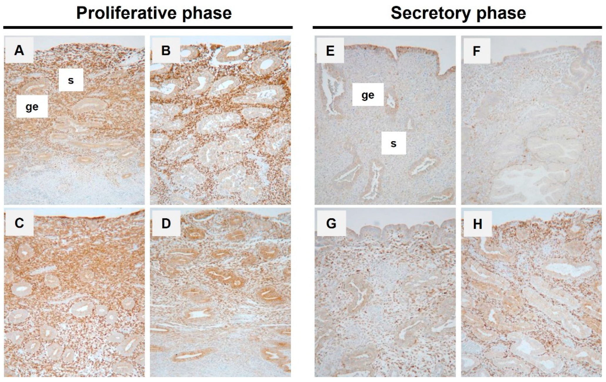
3.1. Expression of PGRMC1 in Human Endometrium Throughout the Menstrual Cycle

The expression and localization of PGRMC1 in human endometrial tissues during the proliferative and secretory phases were assessed by immunohistochemistry (Figure 1). PGRMC1 was expressed by stromal cells in the functional layer and in some glandular epithelia in the proliferative endometrium (Figure 1A–D). Consistent with the results from another group [19], the PGRMC1 level in stroma was clearly lower in the secretory phase than in the proliferative phase.

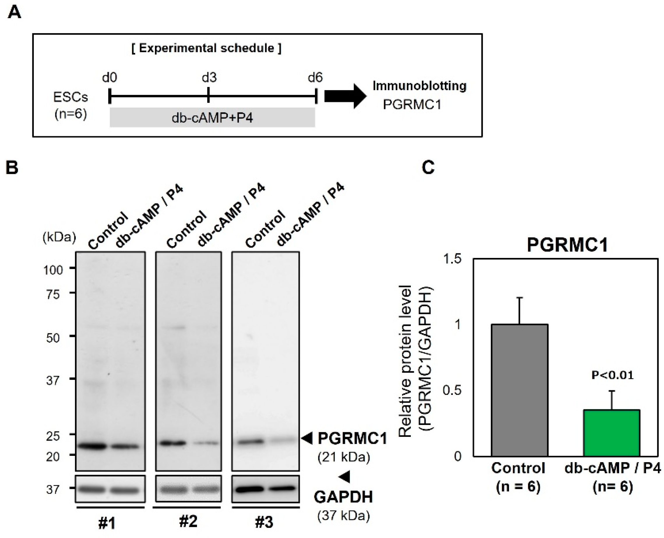
3.2. Downregulation of PGRMC1 During Decidualization of ESCs In Vitro

To determine whether downregulation of PGRMC1 expression during the secretory phase is associated with decidualization, the PGRMC1 level was examined during the decidualization of primary cultured ESCs in vitro (Figure 2). ESCs were treated with db-cAMP/P4 to induce decidualization. Immunoblot analysis showed that the level of PGRMC1 was substantially decreased in db-cAMP/P4-treated decidualizing ESCs (Figure 2B,C). These results suggest that downregulation of PGRMC1 is closely associated with the decidualization of ESCs.

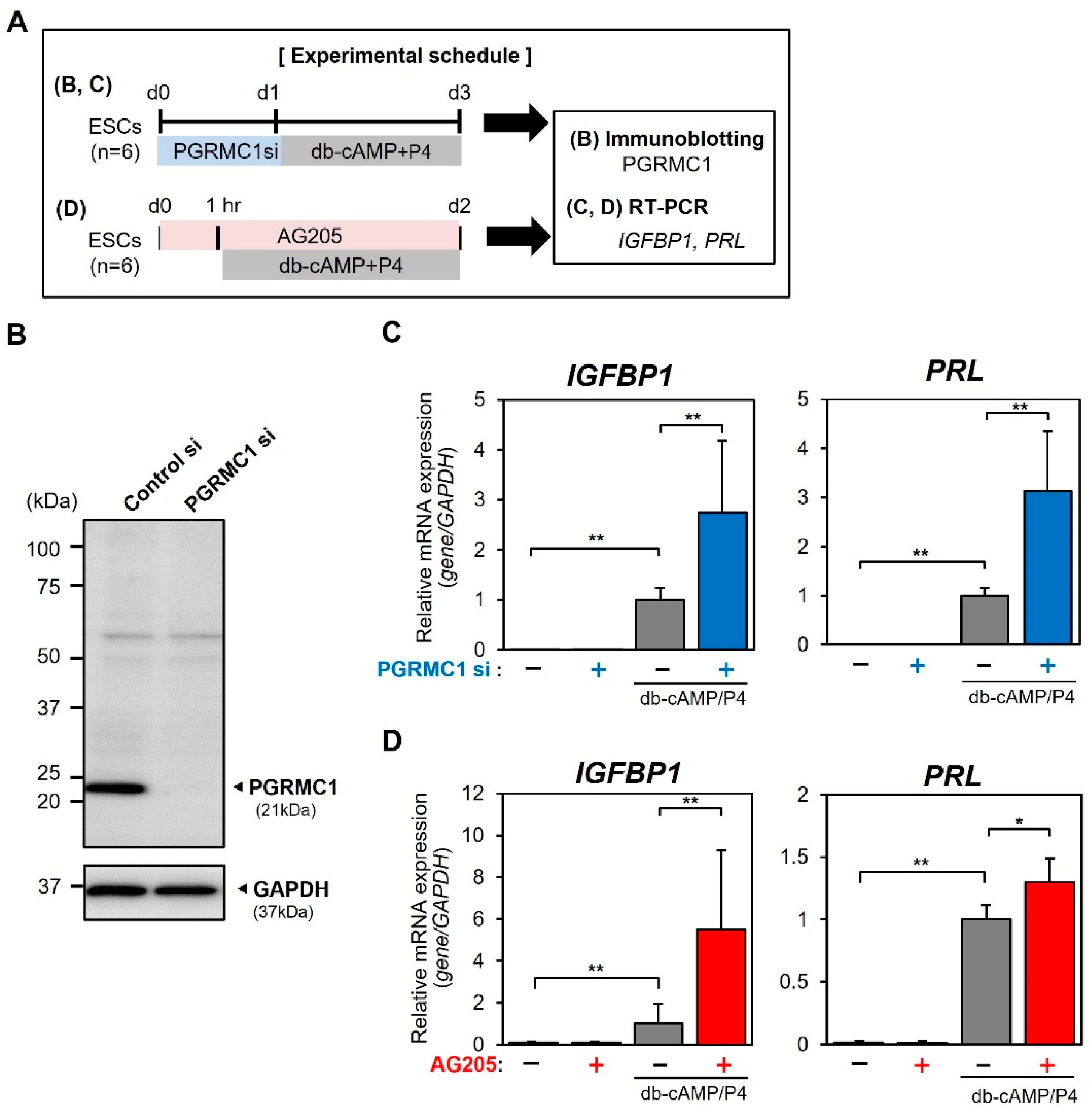
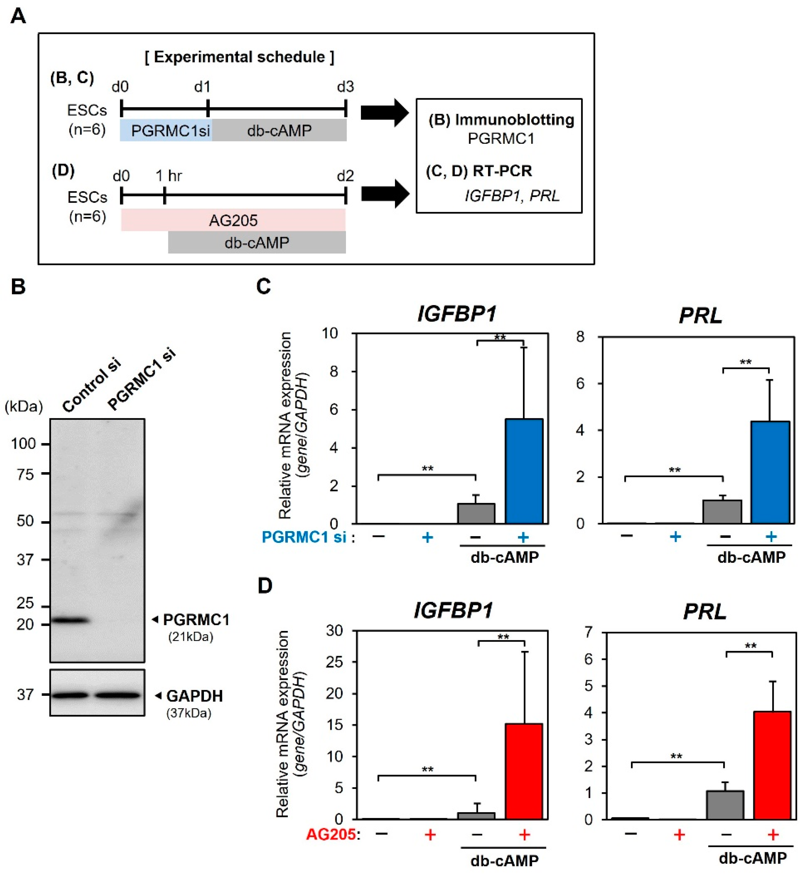
3.3. Effects of PGRMC1 Knockdown and Inhibition on Decidualization of ESCs In Vitro

To evaluate the function of PGRMC1 during the secretory phase, the effects of siRNA-mediated PGRMC1 knockdown and treatment with a PGRMC1 inhibitor (AG-205) on in vitro decidualization were examined (Figure 3). ESCs were transfected for 1 day with non-targeting control or PGRMC1-specific siRNA, or treated for 1 h with the PGRMC1 inhibitor AG-205 and then stimulated with db-cAMP/P4 (Figure 3A). Immunoblotting confirmed that transfection of PGRMC1-specific siRNA reduced PGRMC1 protein levels in ESCs (Figure 3B). In control siRNA-transfected ESCs, treatment with db-cAMP/P4 stimulated expression of the decidual markers IGFBP1 and PRL (Figure 3C). Notably, PGRMC1 knockdown significantly promoted db-cAMP/P4-induced IGFBP1 and PRL expression (Figure 3C).
In addition to the effects of PGRMC1 knockdown on decidualization, we investigated the effects of the PGRMC1 inhibitor AG-205 on db-cAMP/P4-induced decidualization (Figure 3D). Whereas treatment with AG-205 alone had no effect on IGFBP1 or PRL expression, db-cAMP/P4-induced decidual marker expression was significantly enhanced in the presence of AG-205 (Figure 3D). These results suggest that functional inhibition of PGRMC1 is involved in the process of ESC decidualization.
PGRMC1 has an affinity for P4 and is involved in the actions of P4 (15); therefore, we investigated whether P4 is involved in PGRMC1 activity during decidualization. The effects of PGRMC1 knockdown and inhibition on decidualization induced by db-cAMP, but not a combination of db-cAMP and P4, were examined (Figure 4). Treatment with db-cAMP alone significantly elevated IGFBP1 and PRL expression (Figure 4C,D). Even in the absence of P4, db-cAMP-stimulated IGFBP1 and PRL expression levels were significantly upregulated by PGRMC1 knockdown (Figure 4C) and inhibition (Figure 4D), indicating that PGRMC1 activity during decidualization is independent of P4.

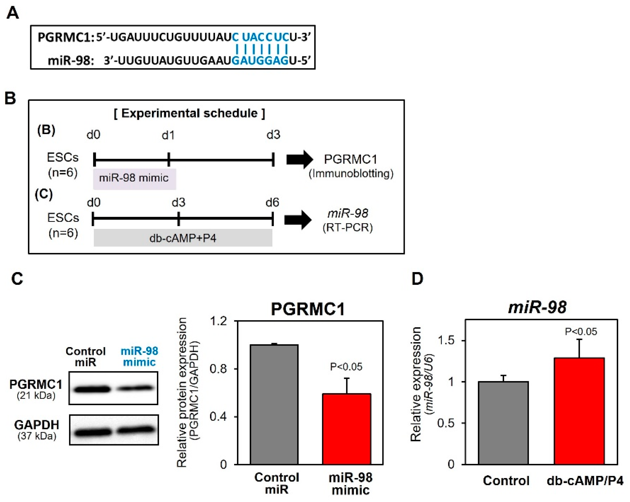
3.4. MiR-98-Mediated Downregulation of PGRMC1 During Decidualization

Based upon analysis performed using the miRDB database, miR-98 was selected as an miRNA that may regulate the PGRMC1 level (Figure 5A). Interestingly, miR-98 reportedly targets PGRMC1 in endometrial adenocarcinoma cells [26] and ovarian cancer cells [27]. To confirm the interaction of miR-98 with PGRMC1, ESCs were transfected with an miR-98 mimic (Figure 5B,C). As expected, transfection of the miR-98 mimic decreased PGRMC1 protein levels (Figure 5C). Furthermore, miR-98 levels were much higher in db-cAMP/P4-induced decidualizing cells than in undifferentiated control ESCs (Figure 5D). These results indicate that increased miR-98 expression in response to decidual stimuli attenuates PGRMC1 levels during the process of decidualization.

4. Discussion

In the present study, treatment of cultured ESCs with the decidual stimuli db-cAMP/P4 reduced the PGRMC1 protein abundance, siRNA-mediated knockdown of PGRMC1 enhanced db-cAMP/P4-stimulated IGFBP1 and PRL expression, and treatment with the PGRMC1 inhibitor AG-205 also promoted expression of these decidualization markers. AG-205 has been widely used as an inhibitor of PGRMC1, although it has some PGRMC1-independent effects. This compound inhibits galactosylceramide synthesis and induces the formation of large vesicular structures in Chinese hamster ovary cells and cells that lack PGRMC1 [28,29]. AG-205 upregulates cholesterol biosynthesis- and steroidogenesis-associated enzymes in human endometrial cells independently of PGRMC1 [30]. We cannot exclude the possibility that AG-205 has a non-specific effect on ESCs, but both AG-205 treatment and PGRMC1 knockdown promoted in vitro decidualization, suggesting the physiological significance of endometrial PGRMC1.
PGRMC1 was substantially expressed in endometrial stromal and epithelial cells. Furthermore, the PGRMC1 levels in ESCs were much lower during the secretory phase than during the proliferative phase, indicative of stage-specific PGRMC1 regulation during the menstrual cycle. PGRMC1 expression was also reported to be decreased in endometrial stroma in the late secretory phase [19]. Kao et al. [31] conducted global gene profiling of the human endometrium during the window of implantation and identified PGRMC1 as a gene that is downregulated in the secretory phase. Our immunohistochemical data are consistent with these observations. Notably, Salsano et al. [19] recently reported that PGRMC1 overexpression in ESCs represses cAMP analog- and progestin-induced PRL secretion, indicative of compromised decidualization. These findings suggest that PGRMC1 downregulation may promote ESC decidualization during the secretory phase.
Although the endometrial PGRMC1 level is downregulated in the secretory phase, the underlying mechanisms have not been demonstrated. We found that transfection of an miR-98 mimic decreased PGRMC1 levels in ESCs. Similarly, miR-98 was reported to target PGRMC1 in endometrial adenocarcinoma cells [26] and ovarian cancer cells [27]. Furthermore, miR-98 upregulation in parallel with PGRMC1 downregulation in db-cAMP/P4-induced decidual cells indicates that miR-98 is involved in the regulation of PGRMC1 during decidualization. It has been reported that downregulation of miR-98 promotes the proliferation of ESCs and represses apoptosis of ESCs by targeting the anti-apoptotic protein Bcl-xl in the rat uterus [32]. Intriguingly, Wang et al. [33] reported that miR-98 expression is low in decidual tissues of patients who experience spontaneous abortion at 6–8 weeks of gestation relative to that in tissues of women with normal pregnancies. Thus, it is also conceivable that abnormal regulation of miR-98 in decidua may affect PGRMC1 expression and lead to spontaneous abortion. miRNA-mediated regulation of the PGRMC1 abundance could fluctuate during the process of decidualization. Further study is required to determine the relationship between miR-98-mediated PGRMC1 regulation and pregnancy. Comparison of miR-98/PGRMC1 expression between the proliferative and secretory phases using endometrial biopsies may help to predict the uterine receptivity for embryo implantation, and controlling this expression may improve the outcome of in vitro fertilization.
In the secretory phase of the menstrual cycle, P4 is abundantly secreted from the corpus luteum, but endometrial PGRMC1 is downregulated. To examine whether P4 is involved in the stimulatory effects of PGRMC1 knockdown and inhibition on decidualization, ESCs pretreated with PGRMC1-specific siRNA or AG-205 were stimulated with db-cAMP only, without P4. Both PGRMC1 knockdown and inhibition promoted db-cAMP-induced decidualization, even in the absence of P4. Thus, PGRMC1 downregulation may predominantly stimulate decidualization of ESCs via cAMP signaling, rather than via P4/PR-mediated activity. This idea is supported by the finding that inhibition of PGRMC1 by AG-205 does not influence E2- or P4-induced PRL secretion [34]. However, as described above, AG-205 is not a specific inhibitor of PGRMC1. A further study is needed to investigate the effects of selective inhibition of PGRMC1 on decidualization.
Post-translational modifications of PGRMC1 such as sumoylation, acetylation, and ubiquitylation at multiple lysine residues have been reported. Peruso et al. reported that P4 treatment stimulates sumoylation of PGRMC1, which results in the generation of PGRMC1 with a high molecular mass and its nuclear translocation in immortalized ovarian granulosa cells [35]. Furthermore, the P4-induced Warburg effect is associated with post-translational modification and proteasomal degradation of PGRMC1 in HEK293 cells [36]. These reports suggest that PGRMC1 protein levels can be modulated by altered rates of transcription, translation, or degradation. Thus, further studies are needed to investigate the post-translational modification and degradation of PGRMC1, which may regulate its protein stability in ESCs during decidualization.
During the process of decidualization, some ESCs display a senescence-like phenotype characterized by increased senescence-associated β-galactosidase activity and secretion of proapoptotic factors [37,38,39,40,41]. In chorion cells, oxidative stress-induced cellular senescence is thought to be a mechanism underlying preterm premature rupture of membranes [42,43]. PGRMC1 knockdown enhances hydrogen peroxide-induced cellular senescence in chorion cells, suggesting that PGRMC1 plays a protective role in cellular senescence by maintaining fetal membrane integrity via inhibition of oxidative stress-induced chorion cell aging [44]. By contrast, it was reported that PGRMC1 (Hpr6.6) increases the rate of cell death via a non-apoptotic mechanism in response to hydrogen peroxide in MCF-7 breast cancer cells [45]. Interestingly, reproductive tract-specific PGRMC1 knockdown leads to development of endometrial cysts in mice (18). Endometrial cysts develop with aging; therefore, uterine PGRMC1 may be involved in ESC senescence. These reports suggest that secretory phase-specific downregulation of PGRMC1 promotes cellular senescence, which is accompanied by ESC decidualization.

5. Strengths and Limitations of This Study

In this study, we propose that (1) functional inhibition of PGRMC1 is involved in the process of ESCs decidualization, (2) PGRMC1 activity during decidualization is independent of P4 and downregulation of PGRMC1 may promote ESC decidualization, and (3) increased expression of miR-98 in response to decidual stimuli attenuates PGRMC1 levels during the process of decidualization. Our results suggested that miR-98-mediated PGRMC1 downregulation may be involved in decidualization. However, miR-98 may not be the only miRNA that modulates PGRMC1 expression. Therefore, comprehensive analysis of transcriptional, post-transcriptional, and post-translational regulation of endometrial PGRMC1 during decidualization is required. In addition, PGRMC1 can bind to various proteins, including receptors for epidermal growth factor [46] and insulin [47], cytochrome P450 [48], and serpine mRNA-binding protein 1 (SERBP1) [19]. Further, PGRMC1 may interact with proteins associated with endomembrane trafficking/cytoskeleton and mitochondrial functions in decidualizing ESCs [49]. Thus, we plan to investigate modulation of endometrial PGRMC1 expression and the interaction between PGRMC1 and intracellular proteins during decidualization in the future.

6. Conclusions

In this study, we demonstrated that endometrial PGRMC1 level is downregulated in the secretory phase of menstrual cycle and that the inhibition of PGRMC1 may promote the decidualization of ESCs in a P4-independent manner. Furthermore, miR-98 may be involved in PGRMC1 downregulation during decidualization.

Author Contributions

Conceptualization, M.Y. and K.T.; methodology, M.Y. and K.T.; validation, M.Y., A.T. and R.Y.; formal analysis, M.Y., A.T. and R.Y.; investigation, M.Y., A.T. and R.Y.; resources, J.K. and H.N.; data curation, M.Y., A.T., R.Y. and K.T.; writing—original draft preparation, M.Y. and A.T.; writing—review and editing, M.Y., A.T., R.Y., M.A., K.K., J.K., H.N. and K.T.; visualization, M.Y., A.T. and R.Y.; supervision, M.Y. and K.T.; project administration, M.Y. and K.T.; funding acquisition, M.Y. All authors have read and agreed to the published version of the manuscript.

Funding

This research was partially funded by KAKENHI Grants-in-Aid for Scientific Research (numbers 16K11113 and 19K09765 to M.Y.) from the Japan Society for the Promotion of Science.

Institutional Review Board Statement

This study was approved by the Institutional Review Board of Tokyo Medical University (No. 3018, 29 May 2017) and the Clinical Research Ethics Committee of the Tokyo University of Pharmacy and Life Sciences (No. 1511, 9 January 2015). Samples were collected in accordance with the Declaration of Helsinki and the requirements of the Clinical Research Ethics Committee of the Tokyo Medical University Hospital.

Informed Consent Statement

Signed informed consent to participate in the study was obtained from all patients prior to surgery.

Data Availability Statement

The data that support the findings of this study are available from the corresponding author upon reasonable requests.

Conflicts of Interest

The authors declare that there is no conflict of interest associated with this manuscript.

References

  1. Tang, B.; Guller, S.; Gurpide, E. Cyclic adenosine 3′,5′-monophosphate induces prolactin expression in stromal cells isolated from human proliferative endometrium. Endocrinology 1993, 133, 2197–2203. [Google Scholar] [CrossRef] [PubMed]
  2. Gellersen, B.; Brosens, J. Cyclic AMP and progesterone receptor cross-talk in human endometrium: A decidualizing affair. J. Endocrinol. 2003, 178, 357–372. [Google Scholar] [CrossRef] [PubMed] [Green Version]
  3. Tang, B.; Guller, S.; Gurpide, E. Mechanisms involved in the decidualization of human endometrial stromal cells. Acta Eur. Fertil. 1993, 24, 221–223. [Google Scholar] [PubMed]
  4. Giudice, L.C.; Dsupin, B.A.; Irwin, J.C. Steroid and peptide regulation of insulin-like growth factor-binding proteins secreted by human endometrial stromal cells is dependent on stromal differentiation. J. Clin. Endocrinol. Metab. 1992, 75, 1235–1241. [Google Scholar] [CrossRef] [PubMed]
  5. Maslar, I.A.; Riddick, D.H. Prolactin production by human endometrium during the normal menstrual cycle. Am. J. Obstet. Gynecol. 1979, 135, 751–754. [Google Scholar] [CrossRef]
  6. Salazar, E.L.; Calzada, L. The role of progesterone in endometrial estradiol- and progesterone-receptor synthesis in women with menstrual disorders and habitual abortion. Gynecol. Endocrinol. 2007, 23, 222–225. [Google Scholar] [CrossRef]
  7. Graham, J.D.; Clarke, C.L. Physiological action of progesterone in target tissues. Endocr. Rev. 1997, 18, 502–519. [Google Scholar] [CrossRef] [Green Version]
  8. Yoshinaga, K. Progesterone and its downstream molecules as blastocyst implantation essential factors. Am. J. Reprod. Immunol. 2014, 72, 117–128. [Google Scholar] [CrossRef]
  9. Kaya, H.S.; Hantak, A.M.; Stubbs, L.J.; Taylor, R.N.; Bagchi, I.C.; Bagchi, M.K. Roles of progesterone receptor A and B isoforms during human endometrial decidualization. Mol. Endocrinol. 2015, 29, 882–895. [Google Scholar] [CrossRef] [Green Version]
  10. Patel, B.; Elguero, S.; Thakore, S.; Dahoud, W.; Bedaiwy, M.; Mesiano, S. Role of nuclear progesterone receptor isoforms in uterine pathophysiology. Hum. Reprod. Update 2015, 21, 155–173. [Google Scholar] [CrossRef] [Green Version]
  11. Bulun, S.E.; Cheng, Y.H.; Yin, P.; Imir, G.; Utsunomiya, H.; Attar, E.; Innes, J.; Julie Kim, J. Progesterone resistance in endometriosis: Link to failure to metabolize estradiol. Mol. Cell Endocrinol. 2006, 248, 94–103. [Google Scholar] [CrossRef] [PubMed]
  12. Hirota, Y.; Cha, J.; Dey, S.K. Revisiting reproduction: Prematurity and the puzzle of progesterone resistance. Nat. Med. 2010, 16, 529–531. [Google Scholar] [CrossRef] [PubMed] [Green Version]
  13. Thomas, P. Characteristics of membrane progestin receptor alpha (mPRalpha) and progesterone membrane receptor component 1 (PGMRC1) and their roles in mediating rapid progestin actions. Front. Neuroendocrinol. 2008, 29, 292–312. [Google Scholar] [CrossRef] [PubMed] [Green Version]
  14. Peluso, J.J.; Pappalardo, A.; Losel, R.; Wehling, M. Progesterone membrane receptor component 1 expression in the immature rat ovary and its role in mediating progesterone’s antiapoptotic action. Endocrinology 2006, 147, 3133–3140. [Google Scholar] [CrossRef] [PubMed] [Green Version]
  15. Meyer, C.; Schmid, R.; Scriba, P.C.; Wehling, M. Purification and partial sequencing of high-affinity progesterone-binding site(s) from porcine liver membranes. Eur. J. Biochem. 1996, 239, 726–731. [Google Scholar] [CrossRef] [PubMed]
  16. Cahill, M.A.; Jazayeri, J.A.; Catalano, S.M.; Toyokuni, S.; Kovacevic, Z.; Richardson, D.R. The emerging role of progesterone receptor membrane component 1 (PGRMC1) in cancer biology. Biochim. Biophys. Acta 2016, 1866, 339–349. [Google Scholar] [CrossRef] [PubMed]
  17. Engmann, L.; Losel, R.; Wehling, M.; Peluso, J.J. Progesterone regulation of human granulosa/luteal cell viability by an RU486-independent mechanism. J. Clin. Endocrinol. Metab. 2006, 91, 4962–4968. [Google Scholar] [CrossRef] [Green Version]
  18. McCallum, M.L.; Pru, C.A.; Niikura, Y.; Yee, S.P.; Lydon, J.P.; Peluso, J.J.; Pru, J.K. Conditional Ablation of Progesterone Receptor Membrane Component 1 Results in Subfertility in the Female and Development of Endometrial Cysts. Endocrinology 2016, 157, 3309–3319. [Google Scholar] [CrossRef] [Green Version]
  19. Salsano, S.; Quiñonero, A.; Pérez, S.; Garrido Gómez, T.; Simón, C.; Dominguez, F. Dynamic expression of PGRMC1 and SERBP1 in human endometrium: An implication in the human decidualization process. Fertil. Steril. 2017, 108, 832–842.e831. [Google Scholar] [CrossRef] [Green Version]
  20. Bushati, N.; Cohen, S.M. microRNA functions. Annu. Rev. Cell Dev. Biol. 2007, 23, 175–205. [Google Scholar] [CrossRef]
  21. Liang, J.; Wang, S.; Wang, Z. Role of microRNAs in embryo implantation. Reprod. Biol. Endocrinol. 2017, 15, 90. [Google Scholar] [CrossRef] [PubMed]
  22. Ferlita, A.; Battaglia, R.; Andronico, F.; Caruso, S.; Cianci, A.; Purrello, M.; Pietro, C.D. Non-Coding RNAs in Endometrial Physiopathology. Int. J. Mol. Sci. 2018, 19, 2120. [Google Scholar] [CrossRef] [PubMed] [Green Version]
  23. Liu, A.; Jin, M.; Xie, L.; Jing, M.; Zhou, Y.; Tang, M.; Lin, T.; Wang, D. Loss of miR-29a impairs decidualization of endometrial stromal cells by TET3 mediated demethylation of Col1A1 promoter. iScience 2021, 24, 103065. [Google Scholar] [CrossRef] [PubMed]
  24. Yoshie, M.; Miyajima, E.; Kyo, S.; Tamura, K. Stathmin, a microtubule regulatory protein, is associated with hypoxia-inducible factor-1alpha levels in human endometrial and endothelial cells. Endocrinology 2009, 150, 2413–2418. [Google Scholar] [CrossRef] [PubMed] [Green Version]
  25. Yoshie, M.; Kaneyama, K.; Kusama, K.; Higuma, C.; Nishi, H.; Isaka, K.; Tamura, K. Possible role of the exchange protein directly activated by cyclic AMP (Epac) in the cyclic AMP-dependent functional differentiation and syncytialization of human placental BeWo cells. Hum. Reprod. 2010, 25, 2229–2238. [Google Scholar] [CrossRef] [PubMed] [Green Version]
  26. Panda, H.; Chuang, T.D.; Luo, X.; Chegini, N. Endometrial miR-181a and miR-98 expression is altered during transition from normal into cancerous state and target PGR, PGRMC1, CYP19A1, DDX3X, and TIMP3. J. Clin. Endocrinol. Metab. 2012, 97, E1316–E1326. [Google Scholar] [CrossRef] [Green Version]
  27. Wendler, A.; Keller, D.; Albrecht, C.; Peluso, J.J.; Wehling, M. Involvement of let-7/miR-98 microRNAs in the regulation of progesterone receptor membrane component 1 expression in ovarian cancer cells. Oncol. Rep. 2011, 25, 273–279. [Google Scholar] [CrossRef] [Green Version]
  28. Wang-Eckhardt, L.; Becker, I.; Eckhardt, M. The PGRMC1 Antagonist AG-205 Inhibits Synthesis of Galactosylceramide and Sulfatide. Cells 2021, 10, 3520. [Google Scholar] [CrossRef]
  29. Wang-Eckhardt, L.; Eckhardt, M. A progesterone receptor membrane component 1 antagonist induces large vesicles independent of progesterone receptor membrane component 1 expression. Biol. Chem. 2020, 401, 1093–1099. [Google Scholar] [CrossRef]
  30. Thieffry, C.; Van Wynendaele, M.; Aynaci, A.; Maja, M.; Dupuis, C.; Loriot, A.; Marbaix, E.; Henriet, P. AG-205 Upregulates Enzymes Involved in Cholesterol Biosynthesis and Steroidogenesis in Human Endometrial Cells Independently of PGRMC1 and Related MAPR Proteins. Biomolecules 2021, 11, 1472. [Google Scholar] [CrossRef]
  31. Kao, L.C.; Tulac, S.; Lobo, S.; Imani, B.; Yang, J.P.; Germeyer, A.; Osteen, K.; Taylor, R.N.; Lessey, B.A.; Giudice, L.C. Global gene profiling in human endometrium during the window of implantation. Endocrinology 2002, 143, 2119–2138. [Google Scholar] [CrossRef] [PubMed]
  32. Xia, H.F.; Jin, X.H.; Cao, Z.F.; Shi, T.; Ma, X. MiR-98 is involved in rat embryo implantation by targeting Bcl-xl. FEBS Lett. 2014, 588, 574–583. [Google Scholar] [CrossRef] [Green Version]
  33. Wang, Y.; Lv, Y.; Wang, L.; Gong, C.; Sun, J.; Chen, X.; Chen, Y.; Yang, L.; Zhang, Y.; Yang, X.; et al. MicroRNAome in decidua: A new approach to assess the maintenance of pregnancy. Fertil. Steril. 2015, 103, 980–989.e986. [Google Scholar] [CrossRef] [PubMed]
  34. Salsano, S.; González-Martín, R.; Quiñonero, A.; Pérez-Debén, S.; Domínguez, F. Deciphering the Role of PGRMC1 During Human Decidualization Using an In Vitro Approach. J. Clin. Endocrinol. Metab. 2021, 106, 2313–2327. [Google Scholar] [CrossRef] [PubMed]
  35. Peluso, J.J.; Lodde, V.; Liu, X. Progesterone regulation of progesterone receptor membrane component 1 (PGRMC1) sumoylation and transcriptional activity in spontaneously immortalized granulosa cells. Endocrinology 2012, 153, 3929–3939. [Google Scholar] [CrossRef]
  36. Sabbir, M.G. Progesterone induced Warburg effect in HEK293 cells is associated with post-translational modifications and proteasomal degradation of progesterone receptor membrane component 1. J. Steroid Biochem. Mol. Biol. 2019, 191, 105376. [Google Scholar] [CrossRef]
  37. Brighton, P.J.; Maruyama, Y.; Fishwick, K.; Vrljicak, P.; Tewary, S.; Fujihara, R.; Muter, J.; Lucas, E.S.; Yamada, T.; Woods, L.; et al. Clearance of senescent decidual cells by uterine natural killer cells in cycling human endometrium. eLife 2017, 6, e31274. [Google Scholar] [CrossRef]
  38. Leno-Durán, E.; Ruiz-Magaña, M.J.; Muñoz-Fernández, R.; Requena, F.; Olivares, E.G.; Ruiz-Ruiz, C. Human decidual stromal cells secrete soluble pro-apoptotic factors during decidualization in a cAMP-dependent manner. Hum. Reprod. 2014, 29, 2269–2277. [Google Scholar] [CrossRef] [Green Version]
  39. Rawlings, T.M.; Makwana, K.; Taylor, D.M.; Molè, M.A.; Fishwick, K.J.; Tryfonos, M.; Odendaal, J.; Hawkes, A.; Zernicka-Goetz, M.; Hartshorne, G.M.; et al. Modelling the impact of decidual senescence on embryo implantation in human endometrial assembloids. eLife 2021, 10, e69603. [Google Scholar] [CrossRef]
  40. Kusama, K.; Yoshie, M.; Tamura, K.; Nakayama, T.; Nishi, H.; Isaka, K.; Tachikawa, E. The role of exchange protein directly activated by cyclic AMP 2-mediated calreticulin expression in the decidualization of human endometrial stromal cells. Endocrinology 2014, 155, 240–248. [Google Scholar] [CrossRef] [Green Version]
  41. Kusama, K.; Yamauchi, N.; Yoshida, K.; Azumi, M.; Yoshie, M.; Tamura, K. Senolytic treatment modulates decidualization in human endometrial stromal cells. Biochem. Biophys. Res. Commun. 2021, 571, 174–180. [Google Scholar] [CrossRef] [PubMed]
  42. Menon, R.; Boldogh, I.; Hawkins, H.K.; Woodson, M.; Polettini, J.; Syed, T.A.; Fortunato, S.J.; Saade, G.R.; Papaconstantinou, J.; Taylor, R.N. Histological evidence of oxidative stress and premature senescence in preterm premature rupture of the human fetal membranes recapitulated in vitro. Am. J. Pathol. 2014, 184, 1740–1751. [Google Scholar] [CrossRef] [PubMed]
  43. Woods, J.R., Jr. Reactive oxygen species and preterm premature rupture of membranes-a review. Placenta 2001, 22 (Suppl. A), S38–S44. [Google Scholar] [CrossRef] [PubMed]
  44. Feng, L.; Allen, T.K.; Marinello, W.P.; Murtha, A.P. Roles of Progesterone Receptor Membrane Component 1 in Oxidative Stress-Induced Aging in Chorion Cells. Reprod. Sci. 2019, 26, 394–403. [Google Scholar] [CrossRef]
  45. Hand, R.A.; Craven, R.J. Hpr6.6 protein mediates cell death from oxidative damage in MCF-7 human breast cancer cells. J. Cell. Biochem. 2003, 90, 534–547. [Google Scholar] [CrossRef]
  46. Ahmed, I.S.; Rohe, H.J.; Twist, K.E.; Craven, R.J. Pgrmc1 (progesterone receptor membrane component 1) associates with epidermal growth factor receptor and regulates erlotinib sensitivity. J. Biol. Chem. 2010, 285, 24775–24782. [Google Scholar] [CrossRef] [Green Version]
  47. Hampton, K.K.; Anderson, K.; Frazier, H.; Thibault, O.; Craven, R.J. Insulin Receptor Plasma Membrane Levels Increased by the Progesterone Receptor Membrane Component 1. Mol. Pharmacol. 2018, 94, 665–673. [Google Scholar] [CrossRef]
  48. Hughes, A.L.; Powell, D.W.; Bard, M.; Eckstein, J.; Barbuch, R.; Link, A.J.; Espenshade, P.J. Dap1/PGRMC1 binds and regulates cytochrome P450 enzymes. Cell Metab. 2007, 5, 143–149. [Google Scholar] [CrossRef] [Green Version]
  49. Salsano, S.; González-Martín, R.; Quiñonero, A.; López-Martín, S.; Gómez-Escribano, A.P.; Pérez-Debén, S.; Yañez-Mo, M.; Domínguez, F. Novel nonclassic progesterone receptor PGRMC1 pulldown-precipitated proteins reveal a key role during human decidualization. Fertil. Steril. 2020, 113, 1050–1066.e1057. [Google Scholar] [CrossRef]
Figure 1. Expression of PGRMC1 in the endometrium. Sections of human endometrium in the proliferative (AD) (n = 4) and secretory (EH) (n = 4) phases were subjected to immunostaining for PGRMC1 (brown staining) and counterstained with hematoxylin. ge: glandular epithelial cells, s: stromal cells.
Reprodmed 03 00015 g001
Figure 2. Expression of PGRMC1 in db-cAMP/P4-induced decidualized ESCs. (A) Experimental schedule. ESCs isolated from three women (#1–3) were treated without (control) or with db-cAMP (0.5 mM) and P4 (1 μM) for 6 days. (B) Immunoblot analysis of PGRMC1 expression. GAPDH levels served as a loading control. (C) Graph showing the relative levels of PGRMC1 normalized to those of GAPDH. Data are from three independent experiments.
Reprodmed 03 00015 g002
Figure 3. Effects of PGRMC1 knockdown and inhibition on db-cAMP/P4-induced decidualization. (A) Experimental schedules. (B) ESCs were transfected for 1 day with non-targeting control or PGRMC1-specific siRNA and cultured for an additional 2 days. Immunoblot analysis of PGRMC1. (C) The siRNA-transfected cells were stimulated with db-cAMP/P4 for 2 days. (D) Cells were treated for 1 h with the PGRMC1 inhibitor AG-205 and then stimulated with db-cAMP/P4 for 2 days. (C,D) Total RNA was subjected to real-time RT-PCR analysis to determine the expression levels of IGFBP1 and PRL. GAPDH was used as an internal control. Data represent the mean ± SEM of three independent experiments. * p < 0.05, ** p < 0.01.
Reprodmed 03 00015 g003
Figure 4. Effects of PGRMC1 knockdown and inhibition on db-cAMP-induced decidualization. (A) Experimental schedules. (B,C) ESCs transfected with control or PGRMC1-specific siRNA were stimulated with db-cAMP for 2 days. (B) Immunoblot analysis of PGRMC1 expression. (D) Cells were treated with AG-205 and then stimulated with db-cAMP for 2 days. (C,D) Total RNA was subjected to real-time RT-PCR analysis to determine the expression levels of IGFBP1 and PRL. GAPDH served as an internal control. Data represent the mean ± SEM of three independent experiments. ** p < 0.01.
Reprodmed 03 00015 g004
Figure 5. Effects of miR-98 mimic transfection on PGRMC1 expression and of db-cAMP/P4 treatment on miR-98 expression in ESCs. (A) Sequence of miR-98 and its possible interaction with PGRMC1. (B) Experimental schedules. (C) ESCs were transfected with the control miR or miR-98 mimic for 24 h and then cultured for 2 days. PGRMC1 levels were assessed by immunoblot analysis. The graph shows the relative levels of PGRMC1 normalized to those of GAPDH in three independent experiments. (D) ESCs were treated without (control) or with db-cAMP/P4 for 6 days. Total RNA was subjected to real-time RT-PCR analysis to determine the expression levels of miR-98. U6 served as an internal control. Data represent the mean ± SD of three independent experiments.
Reprodmed 03 00015 g005
Table 1. Primers used for RT-PCR.
NameSequence
(Accession No.)(5′–3′)
IGFBP1SAATGGATTTTATCACAGCAGACAG
(NM_000596.4)ASGGTAGACGCACCAGCAGAGT
PRLSAAAGGATCGCCATGGAAAG
(NM_000948.6)ASGGTCTCGAAGGGTCACCTG
GAPDHSAGCCACATCGCTCAGACA
(NM_002046.7)ASGCCCAATACGACCAAATCC
miR-98 TGAGGTAGGAGGTTGTATAGTT
Publisher’s Note: MDPI stays neutral with regard to jurisdictional claims in published maps and institutional affiliations.

