Review

The Evolution and Complications of Long-Term Mechanical Circulatory Support Devices

by
Christian R. Sargent
1,*,
Sabrina Ali
1 and
Venkateswarlu Kanamarlapudi
2
1
Calon Cardio Technology Ltd., ILS II, Swansea University, Swansea SA2 8PP, UK
2
Singleton Park Campus, Swansea University Medical School, ILS I, Swansea SA2 8PP, UK
*
Author to whom correspondence should be addressed.
Hearts 2024, 5(1), 105-121; https://doi.org/10.3390/hearts5010008
Submission received: 11 December 2023 / Revised: 1 February 2024 / Accepted: 26 February 2024 / Published: 28 February 2024
(This article belongs to the Topic Mechanical Circulatory Support in Heart Failure)

Abstract

:
Heart failure, a common clinical syndrome caused by functional and structural abnormalities of the heart, affects 64 million people worldwide. Long-term mechanical circulatory support can offer lifesaving treatment for end-stage systolic heart failure patients. However, this treatment is not without complications. This review covers the major complications associated with implantable mechanical circulatory support devices, including strokes, pump thrombosis and gastrointestinal bleeding. These complications were assessed in patients implanted with the following devices: Novacor, HeartMate XVE, CardioWest, Jarvik 2000, HeartMate II, EVAHEART, Incor, VentrAssist, HVAD and HeartMate 3. Complication rates vary among devices and remain despite the introduction of more advanced technology, highlighting the importance of device design and flow patterns. Beyond clinical implications, the cost of complications was explored, highlighting the difference in costs and the need for equitable healthcare, especially with the expected rise in the use of mechanical circulatory support. Future directions include continued improvement through advancements in design and technology to reduce blood stagnation and mitigate high levels of shear stress. Ultimately, these alterations can reduce complications and enhance cost-effectiveness, enhancing both the survival and quality of life for patients receiving mechanical circulatory support.

1. Introduction

Cardiovascular disease (CVD) includes a range of disorders that affect the myocardium and/or the vascular system. It stands as the number one cause of death worldwide, accounting for one-third of all deaths. The prevalence of CVD contributes to the rise in healthcare costs, which is attributed to increased rates of hospitalisation, primary care usage and prescription medication [1,2]. Heart failure (HF) is a common clinical syndrome caused by functional and structural abnormalities of the heart, imposing a burden of disability, reduced quality of life and increased mortality rate [3]. Currently, there are over 64 million cases of HF worldwide, generating substantial socioeconomic implications and incurring management costs of USD 346.17 billion [4]. Whilst conventional therapeutic options for the treatment of HF include medication and lifestyle modifications, heart transplantation is considered the gold standard treatment for end-stage systolic HF. The Registry of the International Society for Heart and Lung Transplantation indicates that one-year survival rates following transplantation are approximately 80% in Europe and 90% in North America, with a median survival of 12 years [5]. Unfortunately, a significant challenge is the high mortality rate of 10–15% of patients on the waiting list, and an additional 10–15% are deemed ineligible due to medical complications [6]. In the United States of America (U.S.A) the highest number of heart transplants are performed annually, with 3817 in 2021 alone [7]. Nevertheless, organ and donor shortages often limit this therapeutic option, and patients may experience complications from immunosuppression medications following transplantation [8]. Considering these limitations and the rise in the ageing population, there is a growing demand for alternative end-stage HF treatments. Durable mechanical circulatory support (MCS) devices have become progressively more common as a long-term therapeutic option. Many patients are initially deemed eligible for MCS device implantation as a bridge to transplant but face the harsh reality of limited organ availability. Consequently, patients are transitioned to destination therapy, especially when complications arise during the long wait for transplantation. However, the number of deaths on the heart transplantation waiting list is falling [8]. This shift highlights the evolving landscape of MCS and emphasizes the need for therapeutic options in the face of organ shortages and patient complexities.

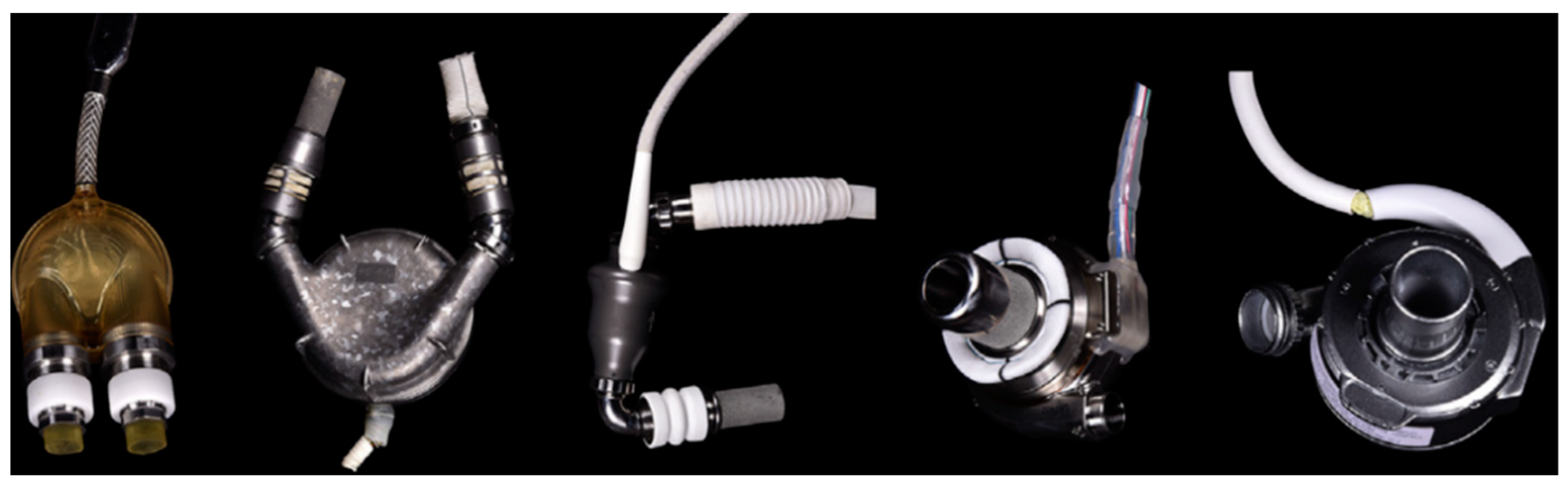
The Evolution of Device Design

Durable MCS devices function using either a pulsatile or continuous flow (CF) mechanism. An overview of long-term implantable MCS devices is summarised in Table 1 and can be observed in Figure 1. The first generation of devices were pulsatile devices, including the Novacor (World Heart Corporation, Oakland, CA, USA), Thoratec PVAD (Paracorporeal Ventricular Assist Device; Thoratec Corporation, Pleasanton, CA, USA) and HeartMate XVE (Thoratec Corp., USA). In addition to ventricular assist devices (VADs), the CardioWest total artificial heart (TAH; SynCardia Systems, Inc., Tucson, AZ, USA) was also introduced, which is an implantable device designed to replace the native valves and ventricles [9]. These devices aim to mimic the physiological pumping of the heart and maintain arterial pulsatility. Pulsatile devices function using volume displacement, which undergoes cyclic filling into an internal reservoir chamber through an electrical or pneumatic drive arrangement. Although the devices mimic physiological heart pumping and maintain arterial pulsatility, they are larger, require more invasive surgery and are often prone to malfunction [10]. In response to these limitations, the second generation of devices emerged, characterised by a reduced size and increased durability. These devices transitioned to the use of CF, as seen in the axial HeartMate II (HMII; Thoratec Corporation, Pleasanton, CA, USA) and EVAHEART (EVAHEART Inc., Sun Medical Technology Research Corporation, Nagano, Japan) pumps (Figure 1). CF-VADs possess a rotating component that unloads the ventricle, allowing for increased blood flow using either axial or centrifugal forces. Axial flow pumps contain a propeller that spins to increase blood flow into the pump through a pipe on a parallel axis. Conversely, centrifugal flow pumps contain blades that revolve within a cavity, which captures the blood and guides it through the device on a perpendicular axis [11]. CF-VAD patients show significantly less anaemia and inflammation compared to patients with first-generation TAHs [12]. However, second-generation CF-VADs contain mechanical bearings and generate flow with little to no arterial pulsatility, resulting in low pulse pressure. These design alterations make them more prone to complications such as pump thrombosis [6]. As such, the progression and introduction of third-generation devices was characterised by the development of centrifugal pumps. Such devices include the VentrAssist (Ventracor Ltd., Chatswood, NSW, Australia), HeartWare HVAD (Medtronic, Minneapolis, MN, USA), Incor® (Berlin Heart, Berlin, Germany) and HeartMate 3 (HM3; Abbott, Chicago, IL, USA). These pumps utilized novel technologies, such as contactless magnetic bearings, to reduce blood damage, as well as speed modulation settings to create a washout effect to prevent blood stagnation. These novel changes represent a step forward in the evolution of pump technology, aiming to reduce complications and improve patient care (Figure 1) [9].
The evolution in device design over time is linked with the adjustment of device speed, which can improve both blood flow and pressure dynamics. Alterations in device speed result in a change in left ventricular (LV) unloading, whereby a higher flow reduces peak LV pressure whilst raising systemic arterial pressure. To understand these relationships, HQ curves are used to characterise the relationship between device flow (Q) and differential pressure (H) between the inflow and outflow cannulas. Due to differences in device design, these factors vary between axial and centrifugal devices. Axial devices have similar pressure differentials, which means fewer fluctuations in flow and a consequential reduction in flow pulsatility. Alternately, centrifugal pumps offer a large range of flows for small changes in differential pressure, which also makes them less likely to experience suction. These devices rely on the amount of blood returning to the heart and are responsive to the resistance encountered during pumping. As such, these devices are preload-dependent and afterload-sensitive. These differences, coupled with ventricular and aortic loading conditions, contribute to the complex dynamics between the physiological response of the patient and the device itself [13].
Figure 1. Implantable Mechanical Circulatory Support Devices. An example of clinically used ventricular assist devices from first, second and third generations. From left to right: PVAD, HeartMate XVE, HeartMate II, HVAD and the HeartMate 3. Image obtained with permission and modified from Elsevier and Kim et al., 2018 [14].
Hearts 05 00008 g001
Table 1. Summary of Clinically Implanted Durable MCS Devices.
Gen.DeviceManufacturerTypeFlowMechanism
FirstNovacorWorld Heart
Inc. (Oakland, CA, USA)
VADPulsatileThe Novacor is a VAD with a polyurethane pumping chamber, pusher plates and fitted valves. It has the shortest blood flow path to reduce the risk of thromboembolism, with a blood flow of up to 10 L/min [15,16].
FirstHeartMate (I) HVEThoratec (Pleasanton, CA, USA)VADPulsatileThe HeartMate XVE is a pulsatile VAD with an electric motor. It operates a pusher plate that expands and decompresses a central chamber to control pumping and blood flow. It contains bioprosthetic unidirectional valves to prevent backflow [17].
FirstCardioWestSyncardia
Systems (Tucson, AZ, USA)
TAHPulsatileThe CardioWest is a total artificial heart (TAH) with an external driver. It is a positive displacement pump that delivers pneumatic pulses into the ventricle chambers using unidirectional valves. It has a cardiac output of up to 9 L/min [18].
SecondJarvik 2000Jarvik Heart
Inc. (New York, NY, USA)
VADContinuous (Axial)The Jarvik 2000 is a VAD that contains a single rotating vaned impeller, which accelerates blood through a central rotor with ceramic bearings. It is powered by a small motor [19].
SecondHeartMate IIThoratec (Pleasanton, CA, USA)VADContinuous (Axial)The HMII is a VAD with a mechanical blood-contacting bearing. It contains a continuously spinning impeller along a central shaft, which draws the blood from the blades of the impeller to produce a flow of up to 10 L/min [17].
SecondEVAHEARTEvaheart Inc.( Sun Medical Technology Research Corporation, Nagano, Japan)VADContinuous (Centrifugal)The EVAHEART is a hydrodynamically levitated device with one journal bearing and an open-vane impeller. The blood-contacting surfaces in the device are covered in an antithrombogenic coating. The EVAHEART has a blood flow of up to 12 L/min [19].
ThirdINCORBerlin Heart (Berlin, Germany)VADContinuous (Axial)The Incor is a VAD which passes blood flow through an inducer. The inducer contains blades that direct the blood to the impeller. This process is wear-free due to contactless magnetic bearing. The Incor also contains a stationary diffuser wheel to increase the pressure required for a blood flow of up to 7 L/min [20].
ThirdVentrAssistVentracor Ltd. (Chatswood, NSW, Australia)VADContinuous (Centrifugal)The VentrAssist is a centrifugal device that uses non-contact impellers with hydrodynamic suspension and is coated in diamond-like carbon. It weighs 298 g and has four rotor blades. The VentrAssist is electromagnetically driven and can run up to 3000 RPM, with a blood flow of up to 10 L/min [21].
ThirdHVADMedtronic (Minneapolis, MN, USA)VADContinuous (Centrifugal)The HVAD is a centrifugal device with a combination of passive magnetic levitation and hydrodynamic suspension. It does not contain any mechanical bearings. The HVAD has a blood flow of up to 10 L/min [22].
ThirdHeartMate 3Abbott (Chicago, IL, USA)VADContinuous (Centrifugal)The HM3 is a centrifugal device that has intermittent speed modulation. It has magnetic levitation and wide gap spaces to reduce blood damage. It does not contain any mechanical or hydrodynamic bearings [23,24].
Gen (Generation), VAD (ventricular assist device), TAH (total artificial heart), HM II (HeartMate II), HM3 (HeartMate 3), min (minute).

2. Complications in MCS

2.1. Patient Selection and Challenges in MCS Therapy

Patient selection is a crucial factor in determining MCS suitability, risk of complications and patient outcome.
Three main patient categories are considered, including basal characteristics, comorbidities and cardiac conditions. Basal characteristics encompass an assessment of the patient’s age (<75) and overall physical health, with younger healthier patients generally having lower mortality rates. Comorbidities include factors which also negatively impact patient outcomes, such as a high BMI (>40), diabetes (with poor glycemic control), a cancer diagnosis with <1-year survival, irreversible liver dysfunction, kidney disease and renal dysfunction (with a glomerular filtration rate [GFR] of <30). Cardiac conditions are also examined for device suitability, including an assessment of the patient’s left and right ventricular size and function. The presence of valve disease is also considered, with severe mitral stenosis and mild aortic regurgitation classified as contraindications. A neurological and cognitive assessment will also be conducted, since moderate–severe cognitive impairment and dementia are contraindications for MCS [25]. Together, these considerations enable healthcare providers to make informed decisions regarding patient suitability for device implantation, aiming to improve patient care. Although these categories are considered, the use of MCS therapy is not without some challenges.
While durable MCS devices offer numerous advantages, they can be associated with high levels of shear stress. Some shear stress is necessary to prevent coagulation, however, excessive levels of shear stress can lead to downstream blood damage [26]. Despite careful patient selection, there are various complications linked to MCS devices, including stroke, pump thrombosis and GI bleeding. Typically, the risk for complications tends to be the highest in the early post-operative period and gradually reduces thereafter [27]. Nevertheless, rates vary from device to device, suggesting that device design and flow patterns may be a factor in the development of these complications and could affect patient outcomes (Table 2). In Table 2 we clarify distinctions between devices, especially those sharing the same flow types, highlighting that flow dynamics alone cannot fully explain the observed differences. Therefore, it is important to determine other differentiating factors, including device designs, to enable comparisons among all individual devices through generations. This approach allows for a comprehensive evaluation rather than grouping devices solely based on flow characteristics.

2.2. Strokes

Strokes remain one of the leading causes of death in VAD patients [26]. The types of strokes that affect MCS patients include ischaemic and haemorrhagic. An ischaemic stroke is caused by a blocked blood vessel that results in reduced oxygen to the brain tissue. Whereas a haemorrhagic stroke is caused either by a ruptured blood vessel in the brain or a haemorrhagic transformation following an ischaemic stroke [50]. Numerous patient-specific and device-specific risk factors can increase the incidence of stroke. Patient-specific factors include patient lifestyle, demographics, co-morbidities and pre-existing conditions. Device-related risk factors include antithrombotic/anticoagulation regimes, thrombosis and incidence of non-surgical bleeding events [51]. Strategies to reduce the risk of stroke include lifestyle modifications and administration of anticoagulants, such as warfarin. While anticoagulants reduce the risk of thromboembolic events, it is worth noting that increased anticoagulation can also increase the risk of GI bleeding. The incidence of stroke also increases following GI bleeds and is related to the reduced anticoagulation regime in response to the bleeding episode. This complex interplay highlights the challenges in managing the fine line between strokes and bleeding events [52]. There are some limitations associated with traditional anticoagulants like warfarin, including frequent assessments of time spent in therapeutic range (TTR), as well as international normalized ratio (INR) measurements to maintain patients within a target range of 2.0–3.0. Deviations from this range could increase the risk of bleeding. As such, there is research into novel anticoagulants known as direct oral anticoagulants (DOACs), that inhibit thrombin (dabigatran) or Factor Xa (apixaban and rivaroxaban) [53,54]. One multi-centre, phase II, randomised clinical trial (RE-ALIGN) was conducted to investigate the impact of dabigatran compared to warfarin in patients with mechanical heart valves. However, the trial was prematurely terminated due to the presence of increased thrombosis and bleeding events in patients prescribed with dabigatran. This study suggests that not all anticoagulants are suitable for MCS and highlights the possible dangers associated with the use of DOACs in device patients. As such, caution is urged with the use of DOACs in the future until more reliable data are available [55]. Gender differences have been also observed in stroke rates after VAD implantation. For instance, female HMII patients showed increased stroke rates compared to male patients, despite similarities in blood pressure, INR and platelet counts [50,56]. These discrepancies may be due to age, life span, genetics, hormonal (mainly oestrogen) and additional unknown factors that require research to provide a more complete understanding [57]. Furthermore, there are differences in stroke rates between different devices. Jarvik 2000 patients experience stroke rates as high as 20.5%, compared with the lower stroke rates of 6.3% in HM3 patients after two years. This difference could be due to the axial flow used by the Jarvik 2000 [26,34,56]. However, not all patients with centrifugal flow devices have a low incidence of strokes, as observed with the HVAD (14.9–17.6%) (Table 2) [58]. Consequently, in 2021, the distribution of the HeartWare HVAD was terminated due to device issues and high levels of neurological adverse events that increased the risk of mortality among patients [59]. In contrast to device design, an investigation was conducted into the influence of outflow cannula alignment on the risk of stroke among VAD patients. This study revealed that patients with a VAD outflow cannula to aortic angle of <37.5°, or a graft diameter of anastomosis of <1.5 cm, had a significantly higher incidence of stroke [60]. As such, additional studies are required to investigate the optimal cannula alignment and the mechanisms involved in the development of strokes in VAD patients, to ultimately reduce both morbidity and mortality.

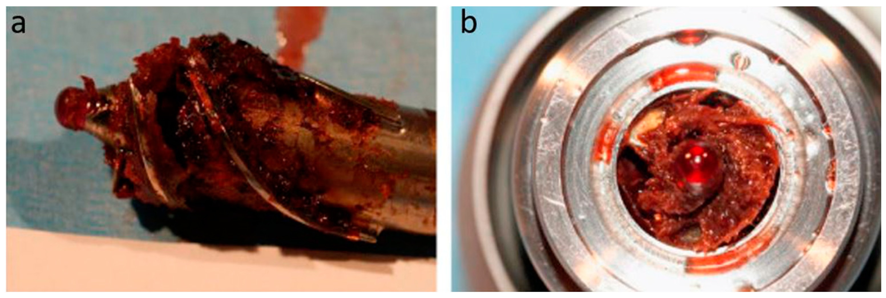
2.3. Pump Thrombosis

Pump thrombosis is characterised by the formation of a thrombus within the flow path of a device, which also includes the inflow cannula and outflow graft. When pump thrombosis occurs, it requires immediate intervention to prevent consequences such as device failure, further thromboembolic events and death (Figure 2). If the initial thrombolytic treatment is unsuccessful, device replacement may be necessary to save a patient’s life. However, this occurrence can substantially decrease the overall cost-effectiveness of MCS therapy [49]. As such, pump thrombosis is an adverse event that requires careful attention and the implementation of effective strategies to overcome. The exact cause of pump thrombosis remains unknown, although there are common factors to consider: the device, the patient and clinical management. Device factors include the type of flow, bearing and biomaterials used. Patient factors include sex, age and body mass index (BMI), whereas clinical factors include the management of anticoagulation/antithrombotic therapies, pump and cannula positioning and device operating speed. Pump thrombosis manifests clinically in MCS patients as an increase in pump power, with a reduction in pulsatility and blood pressure. Stagnant blood zones also pose a substantial risk that could increase the occurrence of pump thrombosis. Currently, there is no single biomarker that can be used to predict thrombosis with certainty. Platelets play a key role in this multifactorial process, however, changes in platelet receptor expression are insufficient for the identification of thrombosis. As such, ongoing research is underway to identify additional biomarkers for predicting thrombus formation in MCS patients [61]. A multi-centre study, known as PREVENT (PREVENTtion of HeartMate II Pump Thrombosis Through Clinical Management), was conducted with 300 patients and aimed to reduce the occurrence of pump thrombosis. The signs and symptoms of pump thrombosis were classified as the presence of clinical hemolysis, worsening of heart failure and abnormal pump parameters. The study suggested that pump thrombosis can be reduced by following a protocol of structured device implantation, which includes the presence of an optimal pump pocket and adequate positioning of the pump, inflow and outflow cannulas. Afterwards, they recommend adequate anticoagulation/antithrombotic medications and optimal device speed. The results showed that these interventions are feasible and can successfully reduce the risk of pump thrombosis in many patients [62]. Despite these findings being for HMII patients, they may be useful with additional devices. Furthermore, the MOMENTUM 3 trial also yielded valuable insights, demonstrating that pump thrombosis rates were significantly lower in the centrifugal HM3 device compared to its predecessor, the axial HMII (1.4% (n = 7/516) versus 13.9% (n = 70/512), respectively). However, these findings do not imply that axial flow is directly related to pump thrombosis. For instance, axial flow devices such as the Jarvik 2000 and Incor have similar rates of pump thrombosis to the HM3 (0–1.2%) [32,43]. Furthermore, pump thrombosis rates range from 6.4–14% with the HVAD, despite its centrifugal flow (Table 2) [36,45,46]. The intricacies of device design and flow type are principal factors in the development of thrombosis. Consequently, tailoring strategies to manage pump thrombosis might also require device-specific approaches. Device evaluation is therefore required to ensure the safety and haemocompatibility of future devices, whilst acknowledging the interplay between the design and the associated risks. This suggests that one verified technique may not translate to patients with another device, highlighting the need for rigorous testing.

2.4. Strategies to Overcome Pump Thrombosis

To optimise the efficiency and cost-effectiveness of durable MCS device therapy, it is crucial to minimise the risk of thrombosis. While aspirin has demonstrated its effectiveness in reducing pump thrombosis, device design is a crucial factor to consider. In HVAD patients, 325 mg of aspirin was necessary to successfully reduce the occurrence of pump thrombosis and device malfunction [46]. Several studies have also shown that a reduction or discontinuation of aspirin in HMII patients with bleeding complications increased the risk of thromboembolic events within this patient population [64,65]. Conversely, HM3 patients have benefited from advancements in device design, leading to a near removal of pump thrombosis occurrence [66]. Consequently, aspirin would have little effect on pump thrombosis in HM3 patients and may only increase the risk of bleeding complications. A small multicentre study involving HM3 patients has revealed promising results, whereby individuals who continued anticoagulation treatment, but discontinued aspirin treatment, experienced no pump thrombosis or thromboembolic events. However, out of the 23 patients who remained on aspirin, 9 encountered episodes of bleeding [67]. Thus, aspirin may not be necessary for all VAD patients, emphasizing the need for personalised treatment strategies to balance the risk of thrombosis and bleeding-related complications. Achieving this balance requires the meticulous evaluation and vigilant monitoring of aspirin use, including considerations of dosage and duration. Moreover, the close monitoring of patients’ clinical status and observations for potential signs of GI bleeding are essential to optimise patient care. Ongoing research is crucial for an enhanced understanding of the impact of aspirin on patient outcomes. The recently completed Antiplatelet Removal and Hemocompatibility Events with the HeartMate 3 Pump (ARIES HM3) clinical trial marks an important milestone for understanding the role of aspirin, particularly in HM3 patients on a vitamin K antagonist. This double-blind, randomised and placebo-controlled study provides valuable insight into the clinical benefits of personalised treatment approaches for pump thrombosis and improved patient outcomes [68,69]. The multi-centre trial revealed that patients who did not receive aspirin as part of their antithrombotic regimen had a significant reduction (34%) in bleeding, but no raised incidence of stroke or thromboembolic events [70]. Therefore, omitting aspirin in HM3 patients with a vitamin K antagonist could be a safe and effective strategy, potentially reducing hospitalization rates and the cost of care due to reduced complications. However, it is essential to acknowledge that these findings could be device-specific and further studies are necessary with additional devices.
Ongoing research is actively addressing the challenges of pump thrombosis. One avenue involves the development of adaptive algorithms that are designed to monitor device power in VAD patients and could play a crucial role in patient care. A recent study showed that one such algorithm demonstrated the ability to provide early warnings three and one-half days before thrombosis occurred, showing a high sensitivity (83.3%) and specificity (98.9%), accompanied by a low false alarm rate. This novel approach paves the way for a future where algorithms become crucial in early detection and intervention for patients at risk of pump thrombosis. Ultimately, this patient-specific management aims to improve patient outcomes and reduce associated complications [71]. Additionally, cannula positioning has been highlighted as an important consideration in reducing the risk of pump thrombosis. The PREVENT study revealed that the mispositioning of the outflow graft could reduce flow through the device, and lead to an increased risk of pump thrombosis. This encompasses both inflow cannula obstruction as well as outflow graft kinking [71]. To reduce the risk of thrombosis, a study was conducted that compared differing outflow graft configurations. This research demonstrated that 45° and 60° angles caused lower levels of shear stress and platelet activation, consequently reducing the risk of thrombosis. However, at a 90° angle, there were higher levels of shear stress and an increased risk of thrombosis. As such, this study demonstrates that shallow graft angles result in more favourable conditions, which reduces the risk of thrombosis [72]. Collectively, these findings provide valuable guidance, highlighting the multifaceted conditions that influence thrombosis and how they can contribute to its mitigation in future applications.

2.5. Gastrointestinal (GI) Bleeding

GI bleeding encompasses a range of clinical manifestations, including haematemesis (vomiting blood), haematochezia (blood in stools), bleeding identified during colonoscopy and/or a reduction in haemoglobin and HCT levels, often necessitating blood transfusion [58]. Research indicates that up to 24.5% of HM3 patients still experience GI bleeding events [37,73,74,75,76]. There appears to be a relationship between the loss of pulsatility and an increase in bleeding in MCS device patients. A retrospective analysis revealed that VAD patients with pulsatile devices (HeartMate XVE, n = 109) had lower rates of GI bleeding after one year. While GI bleeding events were 6.5% and 21.8% for pulsatile and non-pulsatile, respectively [30]. Moreover, further studies demonstrate that survival is higher in HM3 patients (74.7%) compared with patients with a pulsatile device (24%) after two years [31,37]. However, variations in bleeding rates exist even between devices that utilise the same flow type. For instance, devices with centrifugal flow, such as the HVAD (35.1%), HM3 (24.5%) and VentrAssist (12%) exhibit varying rates of GI bleeding. Similarly, axial devices like the HMII (19–34.2%) and Jarvik 2000 (10.8–14%) also show differences in GI bleeding rates [61,73]. Therefore, these discrepancies could suggest that device design plays a role in the development of GI bleeding. In contrast, there were no occurrences of GI bleeding with the EVAHEART (centrifugal) or INCOR (axial), although this could be due to small patient numbers [20,21,36,37,41,42,43,44].

2.6. Risk Factors for Gastrointestinal Bleeding

The risk factors associated with increased GI bleeding are multifaceted and encompass various aspects. These factors include vWF degradation, platelet dysfunction, the presence of angiodysplasia and arteriovenous malformations (AVMs). Clinical factors that can increase the risk of GI bleeding include antithrombotic and anticoagulant medication, increased INR, a history of previous bleeding and device flow type [77]. Patients who had experienced GI bleeding were also more likely to develop stroke compared to those who did not have any bleeding [78]. Thus, further research can gain insights into the underlying mechanisms of GI bleeding and lead to the identification of strategies to mitigate this risk in MCS patients. Perioperative bleeding can also occur in MCS patients and is associated with higher mortality rates and an increased risk of re-bleeding episodes. High right atrial pressure was identified as a risk factor for perioperative bleeding, therefore, careful management before MCS device implantation could potentially reduce the risk of bleeding complications in these patients. This approach can improve patient outcomes and the overall success of device implantation [79]. The integration of smart, real-time monitoring for relevant parameters further enhances the ability to detect and promptly respond to bleeding episodes. Rapid responses to GI bleeding events are vital for effective and timely management. Furthermore, several recent studies have used bio-banked samples from the PREVENT study to analyse pre- and post-operative serum from HMII patients. Patients with pre-operative elevations in angiopoietin-2 and TNF-α (tumour necrosis factor-alpha) were more likely to experience bleeding events following device implantation [80]. In addition, increased thrombin formation post-implantation was found to be associated with increased bleeding episodes [81]. As such, clinicians can use these findings to implement additional parameters to haematology testing to facilitate the identification of high-risk bleeders, paving the way for tailored patient management. This integration could enhance patient care and optimise outcomes for MCS device patients. Extensive research is essential to gain a comprehensive understanding of these factors and their impact on patient outcomes, especially before implantation, to prevent perioperative bleeding episodes. Implementing strategies for the prediction, prevention, early detection and management of GI bleeding at any stage is crucial. This includes routine blood tests, imaging techniques, endoscopic interventions or adjustments in device settings to mitigate the risk of GI bleeding.
Additionally, elevated mechanical shear stress levels have been shown to activate platelets, prompting cytoskeletal changes to increase the surface area for binding [82,83]. Continued exposure to mechanical shear stress causes platelet damage and impairs platelet function [84]. Clinical studies further support this by showing that platelet function is compromised in MCS patients, resulting in prolonged platelet plug formation and impaired platelet aggregation [85,86]. In fact, patients with MCS devices display a 2.5-fold reduction in platelet function compared to healthy controls, increasing the risk of GI bleeding [86]. However, platelet dysfunction alone is not a reliable predictor of GI bleeding. Moreover, MCS-induced shear stress can also lead to the degradation of vWF, a large glycoprotein involved in clotting. High levels of shear stress results in two mechanisms of vWF degradation: firstly, vWF unravelling and an increased enzymatic-induced cleavage at the vWF A2 domain by ADAMTS13, and secondly, the mechanical degradation of vWF multimers [47]. If required, device flow adjustments can also be made to minimise the risk of shear stress-induced complications. Further mechanisms could be involved in the development of bleeding events, including the absence of pulsatility [77]. Pulsatile flow has been linked to reduced vWF degradation compared to continuous flow, revealing that pulsatility modulates the level of vWF degradation and can induce physiological vWF release from endothelial cells [87]. Continued vWF degradation in MCS patients may lead to the development of acquired von Willebrand syndrome (aVWS) and an increased risk of GI bleeding [88]. Despite significant vWF degradation in MCS patients, approximately 30.9% of these patients experienced GI bleeding, indicating that vWF degradation alone does not adequately predict or explain the exact cause of GI bleeding [37,77,89]. The most common cause of GI bleeding in CF-VAD patients is due to AVMs, accounting for 61% of cases. A low pulsatility index, indicative of reduced pulse pressure, significantly increases GI bleeding at AVM sites [75]. This low pulse pressure may also lead to hypoperfusion of the GI lining and mucosal ischemia, leading to the formation of immature vessels that are prone to bleeding. Increased intraluminal pressure coupled with reduced pulse pressure can also lead to angiodysplasia, mucosal hypoxia and, ultimately, GI bleeding [30]. The pulsatile nature of physiological blood flow governs nitric oxide (NO) release from endothelial cells. NO is responsible for increasing vessel dilation for the regulation of blood pressure and flow. Thus, a CF device could reduce NO release, leading to lower blood pressure and flow within the GI tract, which may increase susceptibility to GI bleeding due to the development of angiodysplasia/AVMs [30,74,90]. The interplay between pulse pressure, endothelial cell function and NO release highlights the complex mechanisms underlying GI bleeding in CF-VAD patients. Therefore, the mechanisms that govern GI bleeding require regular management and monitoring, encompassing aspects such as INR, platelet dysfunction, vWF breakdown, NO levels and AVM formation. This approach could help to increase safety and improve patient outcomes in MCS therapy. The multifactorial impact of MCS on the risk of patient GI bleeding should be forefront of the aims of the next generation of novel devices, aiming to overcome the impacts of low pulsatility and high levels of shear stress. Altogether, this information can be utilised to develop comprehensive patient risk profiling systems. These systems can then integrate patient- and device-related factors, allowing for the personalisation of management plans and accounting for individual variations in response to MCS therapy, such as vital signs and haematology data. Establishing techniques to identify high-risk individuals and predict bleeding remains a challenge, but clinicians must ensure regular long-term training and open communication to enhance patient safety.

3. The Financial Impact of MCS Device Complications

The increasing adoption of MCS therapy brings with it a consequential rise in the number of patients experiencing complications, which can have significant economic implications. These complications can increase the incremental cost-effectiveness ratio (ICER) of MCS devices, raising concerns about their impact on healthcare costs and resource allocation. It is essential to recognise that the cost-effectiveness and hospitalisation rates associated with MCS devices can vary across different countries and healthcare settings. This variation highlights the importance of assessing the economic implications of these devices in each specific context to ensure equitable healthcare delivery. By understanding the economic impacts, healthcare services can make informed decisions regarding the allocation of resources and consider the potential financial implications associated with increased MCS therapy. Currently, GI bleeding events and driveline infections cost GBP 6899 and GBP 7662 in England but cost USD 9990 and USD 13,681 in the USA [91]. As such, the use of MCS devices still poses a challenge in providing equitable healthcare. It is of the utmost ethical and moral importance for all researchers to play a significant role in eradicating complications and improving patient quality of life, particularly considering the growing prevalence of these devices. Earlier studies demonstrate that MCS implantation is not yet as cost-effective compared to heart transplantation, but a reduction in complication rates and improvements in device design can improve overall costs [92]. A recent analysis has shown that NHS England (National Health Service) has a willingness-to-pay threshold of GBP 50,000 per quality age life-year (QALY) for MCS device implantation. The ICER of MCS devices as destination therapy was placed at GBP 43,207 per QALY, which is less than the NHS threshold. With improvements in device design, MCS as destination therapy appears to be a cost-effective therapy for the NHS in the U.K [93]. The rapid evolution of the next generation of devices aims to reduce the costs and complications associated with MCS therapy and to improve patient survival and quality of life. However, potential challenges could impact their widespread implementation, such as regulatory considerations, reimbursement issues and patient acceptance. Nevertheless, given the current financial implications, novel devices should enhance the cost-effectiveness and efficacy of future therapies. This can be achieved through ongoing research into improved product design, advanced technologies and patient assessments.

4. Devices in Development

The use of long-term MCS therapy has risen in recent times, offering a lifeline to patients living with HF. This rise coincides with ongoing research and development in the field, with a focus on the production of novel devices. An illustrating case comes from the initial experience with the EVAHEART device and the complications observed during its clinical use (summarised in Table 2). Consequently, this prompted the development of a new iteration, the EVAHEART 2. This device is currently undergoing evaluation in the COMPETENCE clinical trial, which is investigating both its short- and long-term use. In the transition from the EVAHEART to the EVAHEART 2, improvements have been made to the device design. Although the pump has identical blood pathways, it is smaller in weight and volume and has novel features including reduced pump speeds, larger blood gaps and an altered inflow cannula. These enhancements aim to avoid any associated adverse events, such as pump thrombosis and suction [94,95]. One avenue that takes innovation further involves the reimagining of traditional impeller blades, such as blunting, to reduce the high-velocity regions at the tip of the blades. Alternately, impeller blades could be removed entirely, as exemplified by CorWave’s bladeless LVAD. This device employs a wave membrane to induce blood flow with low fluid shear stress and reduced blood damage [96]. The CorWave LVAD operates using an average flow rate of 6 L/min, a gentle speed of 1.5 m/sec and an oscillation cycle of 25 ms. It achieves this through the use of an undulating polymer membrane that can be regulated by altering the oscillation frequency and magnitude, to produce a physiologic pulsatile blood flow [97]. Furthermore, Realheart® is currently developing a novel four-chamber TAH for clinical use. The device is comprised of two independent pumps, left and right. Each pump contains an artificial atrium, ventricle and two valves with a continuous inflow and a pulsatile outflow. Each side of the device is controlled to account for different volumes of blood, with an atrioventricular plane similar to the physiological functioning of the heart [98]. These novel devices have demonstrated low levels of shear stress and increased pulsatility, which highlights their potential to shape the future landscape of MCS device therapy. The future of MCS devices holds great promise, with possibilities that extend further than mechanical improvements. Smart technologies and sensors with real-time monitoring and assessment of patient activity could dynamically adjust their operating speed in line with the physiological status and needs of the patient, particularly during exercise. Monitoring could be facilitated using a smartwatch, which continuously collects real-time data that can be sent to clinicians, allowing for the earlier identification of potential issues and timely intervention. Such innovations hold the potential to enhance the next generation of smart and patient-specific devices and enhance the overall efficacy of MCS therapy. The medical device industry will continue to prioritise the optimisation of product design and technology to produce pumps with enhanced haemocompatibility and reduced costs, ultimately benefitting patients and healthcare providers alike. Ongoing research that investigates patient-centred outcomes and quality-of-life measures is essential to assess the impact of MCS devices on patients’ daily lives, physical functioning, psychological well-being, social interactions and overall satisfaction with the therapy. With advancements in machine learning, artificial intelligence, remote monitoring and smart devices, these techniques can revolutionise device design, patient management and treatment outcomes, leading to better clinical outcomes. Their integration into MCS therapy can introduce an era of healthcare characterised by innovation, enhanced precision and superior clinical outcomes. Advancements in technology could successfully analyse multiple variables and generate electronic health records. These records could provide predictive models for RV failure, risks, prognosis, complications and mortality rates. Moreover, they could provide real-time alert signals to patients and clinicians. These advancements could result in reduced complications, improved patient outcomes and a better quality of life for patients.

Future Directions

As the need for alternative therapeutic options for HF increases, the use of MCS is expected to expand worldwide. However, major adverse events are most common in the first 60 days following MCS device implantation. Most occur in the first 30 days, with bleeding emerging as one of the most common adverse events. These occurrences highlight patient vulnerabilities within the perioperative period and the increased risks going forward. As such, more preventative strategies should be employed to target this critical period, including improved techniques to facilitate the early detection and management of complications, to lower the risk of morbidity and mortality [27,99]. Emphasising the importance of early detection is crucial. Moreover, to enhance MCS therapy, the search for improved device performance necessitates changes in device design. As such, there are ongoing efforts by engineers, clinicians and scientists that are dedicated to enhancing haemocompatibility, reducing complications and exploring innovative technologies such as biomaterials or miniaturised devices.
Future designs could also prioritise a reduction in the amount of blood stagnation and mitigate regions with elevated levels of shear stress. Innovative designs could eliminate or reduce areas with blood pooling to ensure unimpeded flow and improved washing. To mitigate excessive shear stress, device geometry should be optimised efficiently to gently direct and evenly distribute the blood flow. A potential avenue for improvement is the reintroduction of pulsatile flow as a configuration option in novel devices, to mimic physiological conditions and provide the optimal pulsatility for vascular function. Moreover, blood is a highly complex fluid that is sensitive to changes in flow dynamics and shear stress alterations. As such, a small increase in force could have large downstream effects on blood components. Careful engineering and advanced modelling should be employed to guide the design process with this in mind. To address ongoing research gaps, additional studies can focus on understanding how variations in patient populations, comorbidities and underlying causes of systolic heart failure influence the outcomes and complications associated with MCS devices, allowing for more personalised approaches to device selection and management. Interestingly, the MOMENTUM clinical trial revealed that after two years of support, female HM3 patients had a significantly increased risk of stroke, GI bleeding and infection compared to male patients. However, these differences were not observed in patients aged 65 and older, and both male and female patients showed similar survival rates. Further investigations are required to identify and reveal insights into the sex-related differences in adverse events among HM3 patients [100]. Furthermore, it is crucial to explore strategies that enhance early diagnosis and infection prevention, as these measures can significantly impact patient outcomes. Novel device designs that incorporate wireless power systems with transcutaneous energy transfer systems are of particular interest as potential solutions for driveline infections. Implementing such designs has the potential to improve morbidity and mortality rates in MCS patients by reducing the risk of infection associated with traditional drivelines. However, there are safety concerns associated with wireless technology, such as heat production and tissue damage, although ongoing research aims to enhance this technology for MCS device application, thus significantly improving patient quality of life [101]. In addition to improving device design, efforts have been made to refine surgical techniques to enhance MCS patient recovery. A single-centre study investigated the impact of conventional sternotomy versus a less-invasive surgery for device implantation. Briefly, the less-invasive surgery involved identification of the LV apex via echocardiography and a partial J-shaped sternotomy was performed in the third intercostal space. An anterolateral thoracotomy was then conducted to insert the device. This procedure resulted in significantly reduced hospital mortality rates and less time in intensive care compared with conventional methods. Although this less-invasive method did not reduce the occurrence of adverse events, it effectively improved post-operative stability. As such, further research into the optimisation of surgical techniques could be conducted to improve patient outcomes following MCS device implantation [102]. Overall, there is a need for research on predicting complications, identifying additional biomarkers and exploring the multifactorial impacts of flow type and device design. By focusing on these areas, the importance of ongoing research efforts as well as advancements in device design should improve MCS therapy and patient quality of life.

5. Conclusions

HF is a major global health issue, carrying a considerable risk of both morbidity and mortality. With an ever-increasing ageing population, HF cases are expected to rise substantially. If eligible, HF patients can be put forward for heart transplantation. However, the number of heart transplants does not meet the rising demand, and organ shortages often limit this therapeutic option. Considering the limitations of heart transplantation, the rise in ageing populations and consequential health costs, the need for alternative HF treatments is higher now than ever before. As such, long-term MCS devices, such as VADs and TAHs, have become progressively more common as a therapeutic option, improving quality of life and reducing mortality rates among end-stage HF patients. However, MCS therapy comes with challenges, including an increased risk of stroke (both ischaemic and haemorrhagic), pump and outflow graft thrombosis and GI bleeding. These complications can significantly impact patient outcomes and increase healthcare costs. Understanding the mechanisms underlying these complications is essential to develop strategies for prevention, early detection and patient management. Furthermore, complication rates vary amongst devices, indicating that device-specific characteristics could play a role in their development and should be evaluated carefully. Improvements have been made, including efforts to enhance haemocompatibility, reduce costs and mimic physiological conditions. These improvements have aimed to minimise complications and enhance patient outcomes. However, some complications remain, highlighting the need for patient-specific approaches to prevent and predict complications. As such, ongoing research should be focused on addressing the multifaceted impact of these complications on patients, healthcare and society. Consequently, the next generation of devices will continue to advance, thereby offering improvements in many factors, such as complication rates, cost-effectiveness and patient outcomes, following MCS device implantation.

Author Contributions

Conceptualization, C.R.S., S.A. and V.K.; methodology, C.R.S., S.A. and V.K.; validation, C.R.S., S.A. and V.K.; formal analysis, C.R.S.; investigation, C.R.S.; resources, C.R.S.; data curation, C.R.S.; writing—original draft preparation, C.R.S.; writing—review and editing, C.R.S., S.A. and V.K.; visualization, C.R.S., S.A. and V.K.; supervision, C.R.S., S.A. and V.K.; project administration, C.R.S., S.A. and V.K. All authors have read and agreed to the published version of the manuscript.

Funding

This research received no external funding.

Acknowledgments

This work was supported by Calon Cardio Technology Ltd. and Swansea University Medical School.

Conflicts of Interest

The authors C.R.S. and S.A. are employed by Calon Cardio-Technology Ltd. The remaining author declares no conflicts of interest.

References

  1. Bhatnagar, P.; Wickramasinghe, K.; Williams, J.; Rayner, M.; Townsend, N. The epidemiology of cardiovascular disease in the UK 2014. Heart 2015, 101, 1182–1189. [Google Scholar] [CrossRef]
  2. Virani, S.S.; Alonso, A.; Benjamin, E.J.; Bittencourt, M.S.; Callaway, C.W.; Carson, A.P.; Chamberlain, A.M.; Chang, A.R.; Cheng, S.; Delling, F.N.; et al. Heart Disease and Stroke Statistics-2020 Update: A Report from the American Heart Association. Circulation 2020, 141, e139–e596. [Google Scholar] [CrossRef]
  3. Lippi, G.; Sanchis-Gomar, F. Global epidemiology and future trends of heart failure. AME Med. J. 2020, 5, 15. [Google Scholar] [CrossRef]
  4. Savarese, G.; Becher, P.M.; Lund, L.H.; Seferovic, P.; Rosano, G.M.; Coats, A.J. Global burden of heart failure: A comprehensive and updated review of epidemiology. Cardiovasc. Res. 2023, 118, 3272–3287, Erratum in Cardiovasc. Res. 2023, 119, 1453. [Google Scholar] [CrossRef]
  5. Hsich, E.; Singh, T.P.; Cherikh, W.S.; Harhay, M.O.; Hayes, D.; Perch, M.; Potena, L.; Sadavarte, A.; Lindblad, K.; Zuckermann, A.; et al. The International thoracic organ transplant registry of the international society for heart and lung transplantation: Thirty-ninth adult heart transplantation report-2022; focus on transplant for restrictive heart disease. J. Heart Lung Transplant. 2022, 41, 1366–1375. [Google Scholar] [CrossRef]
  6. Williams, M.L.; Trivedi, J.R.; McCants, K.C.; Prabhu, S.D.; Birks, E.J.; Oliver, L.; Slaughter, M.S. Heart transplant vs left ventricular assist device in heart transplant-eligible patients. Ann. Thorac. Surg. 2011, 91, 1330–1333. [Google Scholar] [CrossRef]
  7. Statista [Internet]: Heart Transplants in the U.S. 1975–2021. Available online: https://www.statista.com/statistics/671451/heart-transplants-number-us/ (accessed on 10 October 2023).
  8. MacGowan, G.A.; Crossland, D.S.; Hasan, A.; Schueler, S. Considerations for patients awaiting heart transplantation-Insights from the UK experience. J. Thorac. Dis. 2015, 7, 527–531. [Google Scholar]
  9. Rigatelli, G.; Santini, F.; Faggian, G. Past and present of cardiocirculatory assist devices: A comprehensive critical review. J. Geriatr. Cardiol. JGC 2012, 9, 389–400. [Google Scholar] [PubMed]
  10. Pagani, F.D.; Miller, L.W.; Russell, S.D. Extended mechanical circulatory support with a continuous-flow rotary left ventricular assist device. J. Am. Coll. Cardiol. 2009, 54, 312–321. [Google Scholar] [CrossRef] [PubMed]
  11. Moazami, N.; Fukamachi, K.; Kobayashi, M.; Smedira, N.G.; Hoercher, K.J.; Massiello, A.; Lee, S.; Horvath, D.J.; Starling, R.C. Axial and centrifugal continuous-flow rotary pumps: A translation from pump mechanics to clinical practice. J. Heart Lung Transplant. 2013, 32, 1–11. [Google Scholar] [CrossRef] [PubMed]
  12. Mankad, A.K.; Tang, D.G.; Clark, W.B.; Flattery, M.; Harton, S.; Katlaps, G.J.; Stribling, W.K.; Cooke, R.H.; Hess, M.L.; Kasirajan, V.; et al. Persistent anemia after implantation of the total artificial heart. J. Card. Fail. 2012, 18, 433–438. [Google Scholar] [CrossRef]
  13. Narang, N.; Raikhelkar, J.; Sayer, G.; Uriel, N. Hemodynamic Pump-Patient Interactions and Left Ventricular Assist Device Imaging. Cardiol. Clin. 2018, 36, 561–569. [Google Scholar] [CrossRef]
  14. Kim, J.H.; Cowger, J.A.; Shah, P. The Evolution of Mechanical Circulatory Support. Cardiol. Clin. 2018, 36, 443–449. [Google Scholar] [CrossRef]
  15. Vetter, H.O.; Kaulbach, H.G.; Schmitz, C.; Forst, A.; Überfuhr, P.; Kreuzer, E.; Pfeiffer, M.; Brenner, P.; Dewald, O.; Reichart, B. Experience With the Novacor Left Ventricular Assist System as a Bridge to Cardiac Transplantation, Including the New Wearable System. J. Thorac. Cardiovasc. Surg. 1995, 109, 74–80. [Google Scholar] [CrossRef]
  16. Loebe, M.; Koster, A.; Sänger, S.; Potapov, E.V.; Kuppe, H.; Noon, G.P.; Hetzer, R. Inflammatory response after implantation of a left ventricular assist device: Comparison between the axial flow MicroMed DeBakey VAD and the pulsatile Novacor device. ASAIO J. 2001, 47, 272–274. [Google Scholar] [CrossRef]
  17. Wilson, S.R.; Givertz, M.M.; Stewart, G.C.; Mudge, G.H. Ventricular assist devices the challenges of outpatient management. J. Am. Coll. Cardiol. 2009, 54, 1647–1659. [Google Scholar] [CrossRef]
  18. Imamura, T.; Narang, N.; Kim, G.; Nitta, D.; Fujino, T.; Nguyen, A.; Grinstein, J.; Rodgers, D.; Ota, T.; Jeevanandam, V.; et al. Aortic Insufficiency during HeartMate 3 Left Ventricular Assist Device Support. J. Card. Fail. 2020, 26, 863–869. [Google Scholar] [CrossRef] [PubMed]
  19. Yamazaki, K.; Kihara, S.; Akimoto, T.; Tagusari, O.; Kawai, A.; Umezu, M.; Tomioka, J.; Kormos, R.L.; Griffith, B.P.; Kurosawa, H. EVAHEART: An implantable centrifugal blood pump for long-term circulatory support. Jpn. J. Thorac. Cardiovasc. Surg. 2002, 50, 461–465. [Google Scholar] [CrossRef] [PubMed]
  20. Schmid, C.; Tjan, T.D.; Etz, C.; Schmidt, C.; Wenzelburger, F.; Wilhelm, M.; Rothenburger, M.; Drees, G.; Scheld, H.H. First clinical experience with the Incor left ventricular assist device. J. Heart Lung Transplant. 2005, 24, 1188–1194. [Google Scholar] [CrossRef] [PubMed]
  21. Esmore, D.; Spratt, P.; Larbalestier, R.; Tsui, S.; Fiane, A.; Ruygrok, P.; Meyers, D.; Woodard, J. VentrAssist left ventricular assist device: Clinical trial results and Clinical Development Plan update. Eur. J. Cardiothorac. Surg. 2007, 32, 735–744. [Google Scholar] [CrossRef] [PubMed]
  22. Wiedemann, D.; Haberl, T.; Riebandt, J.; Simon, P.; Laufer, G.; Zimpfer, D. Ventricular Assist Devices—Evolution of Surgical Heart Failure Treatment. Eur. Cardiol. 2014, 9, 54–58. [Google Scholar] [CrossRef]
  23. Hanke, J.S.; Rojas, S.V.; Dogan, G.; Feldmann, C.; Beckmann, E.; Deniz, E.; Wiegmann, B.; Michaelis, J.-E.; Napp, L.C.; Berliner, D.; et al. First series of left ventricular assist device exchanges to HeartMate 3. Eur. J. Cardiothorac. Surg. 2017, 51, 887–892. [Google Scholar] [CrossRef]
  24. Alvarez, J.; Rao, V. HeartMate 3-a “Step” in the right direction. J. Thorac. Dis. 2017, 9, E457–E460. [Google Scholar] [CrossRef]
  25. Melendo-Viu, M.; Dobarro, D.; Roubin, S.R.; Pernas, C.L.; Cordón, C.M.; Lamas, M.V.; Esteban, M.P.; Martínez, M.V.; Abu Assi, E.; Romero, R.P.; et al. Left Ventricular Assist Device as a Destination Therapy: Current Situation and the Importance of Patient Selection. Life 2023, 13, 1065. [Google Scholar] [CrossRef] [PubMed]
  26. Mehra, M.R.; Goldstein, D.J.; Uriel, N.; Cleveland, J.C.; Yuzefpolskaya, M.; Salerno, C.; Walsh, M.N.; Milano, C.A.; Patel, C.B.; Ewald, G.A.; et al. Two-Year Outcomes with a Magnetically Levitated Cardiac Pump in Heart Failure. N. Engl. J. Med. 2018, 378, 1386–1395. [Google Scholar] [CrossRef] [PubMed]
  27. Molina, E.J.; Shah, P.; Kiernan, M.S.; Cornwell, W.K.; Copeland, H.; Takeda, K.; Fernandez, F.G.; Badhwar, V.; Habib, R.H.; Jacobs, J.P.; et al. The Society of Thoracic Surgeons Intermacs 2020 Annual Report. Ann. Thorac. Surg. 2021, 111, 778–792. [Google Scholar] [CrossRef] [PubMed]
  28. Strauch, J.T.; Spielvogel, D.; Haldenwang, P.L.; Correa, R.K.; Richard, A.D.; Seissler, P.E.; Baran, D.A.; Gass, A.L.; Lansman, S.L. Recent improvements in outcome with the Novacor left ventricular assist device. J. Heart Lung Transplant. 2003, 22, 674–680. [Google Scholar] [CrossRef]
  29. El-Banayosy, A.; Arusoglu, L.; Kizner, L.; Tenderich, G.; Minami, K.; Inoue, K.; Körfer, R. Novacor left ventricular assist system versus Heartmate vented electric left ventricular assist system as a long-term mechanical circulatory support device in bridging patients: A prospective study. J. Thorac. Cardiovasc. Surg. 2000, 119, 581–587. [Google Scholar] [CrossRef]
  30. Crow, S.; John, R.; Boyle, A.; Shumway, S.; Liao, K.; Colvin-Adams, M.; Toninato, C.; Missov, E.; Pritzker, M.; Martin, C.; et al. Gastrointestinal bleeding rates in recipients of nonpulsatile and pulsatile left ventricular assist devices. J. Thorac. Cardiovasc. Surg. 2009, 137, 208–215. [Google Scholar] [CrossRef]
  31. Slaughter, M.S.; Rogers, J.G.; Milano, C.A.; Russell, S.D.; Conte, J.V.; Feldman, D.; Sun, B.; Tatooles, A.J.; Delgado, R.M.; Long, J.W.; et al. Advanced Heart Failure Treated with Continuous-Flow Left Ventricular Assist Device. N. Eng. J. Med. 2009, 361, 2241–2251. [Google Scholar] [CrossRef]
  32. Copeland, J. Results with an anticoagulation protocol in 99 SynCardia total artificial heart recipients. ASAIO J. 2013, 59, 216–220. [Google Scholar] [CrossRef]
  33. El-Banayosy, A.; Arusoglu, L.; Morshuis, M.; Kizner, L.; Tenderich, G.; Sarnowski, P.; Milting, H.; Koerfer, R. CardioWest total artificial heart: Bad Oeynhausen experience. Ann. Thorac. Surg. 2005, 80, 548–552. [Google Scholar] [CrossRef]
  34. Kohno, H.; Matsumiya, G.; Sawa, Y.; Ono, M.; Saiki, Y.; Shiose, A.; Yamazaki, K.; Matsui, Y.; Niinami, H.; Matsuda, H.; et al. The Jarvik 2000 left ventricular assist device as a bridge to transplantation: Japanese Registry for Mechanically Assisted Circulatory Support. J. Heart Lung Transplant. 2017, 37, 71–78. [Google Scholar] [CrossRef] [PubMed]
  35. Letsou, G.V.; Shah, N.; Gregoric, I.D.; Myers, T.J.; Delgado, R.; Frazier, O. Gastrointestinal bleeding from arteriovenous malformations in patients supported by the Jarvik 2000 axial-flow left ventricular assist device. J. Heart. Lung. Transplant. 2005, 24, 105–109. [Google Scholar] [CrossRef]
  36. Rogers, J.G.; Pagani, F.D.; Tatooles, A.J.; Bhat, G.; Slaughter, M.S.; Birks, E.J.; Boyce, S.W.; Najjar, S.S.; Jeevanandam, V.; Anderson, A.S.; et al. Intrapericardial Left Ventricular Assist Device for Advanced Heart Failure. N. Engl. J. Med. 2017, 376, 451–460. [Google Scholar] [CrossRef] [PubMed]
  37. Mehra, M.R.; Uriel, N.; Naka, Y.; Cleveland, J.C.; Yuzefpolskaya, M.; Salerno, C.T.; Walsh, M.N.; Milano, C.A.; Patel, C.B.; Hutchins, S.W.; et al. A Fully Magnetically Levitated Left Ventricular Assist Device—Final Report. N. Engl. J. Med. 2019, 380, 1618–1627. [Google Scholar] [CrossRef] [PubMed]
  38. Malick, A.; Ning, Y.; Kurlansky, P.A.; Melehy, A.; Yuzefpolskaya, M.; Colombo, P.C.; Sayer, G.; Uriel, N.; Naka, Y.; Takeda, K. Development of De Novo Aortic Insufficiency in Patients with HeartMate 3. Ann. Thorac. Surg. 2021, 114, 450–456. [Google Scholar] [CrossRef]
  39. Morgan, J.A.; Paone, G.; Nemeh, H.W.; Henry, S.E.; Patel, R.; Vavra, J.; Williams, C.T.; Lanfear, D.E.; Tita, C.; Brewer, R.J. Gastrointestinal bleeding with the HeartMate II left ventricular assist device. J. Heart Lung Transplant. 2012, 31, 715–718. [Google Scholar] [CrossRef] [PubMed]
  40. Demirozu, Z.T.; Radovancevic, R.; Hochman, L.F.; Gregoric, I.D.; Letsou, G.V.; Kar, B.; Bogaev, R.C.; Frazier, O. Arteriovenous malformation and gastrointestinal bleeding in patients with the HeartMate II left ventricular assist device. J. Heart Lung Transplant. 2011, 30, 849–853. [Google Scholar] [CrossRef] [PubMed]
  41. Saito, S.; Yamazaki, K.; Nishinaka, T.; Ichihara, Y.; Ono, M.; Kyo, S.; Nishimura, T.; Nakatani, T.; Toda, K.; Sawa, Y.; et al. Post-approval study of a highly pulsed, low-shear-rate, continuous-flow, left ventricular assist device, EVAHEART: A Japanese multicenter study using J-MACS. J. Heart Lung Transplant. 2014, 33, 599–608. [Google Scholar] [CrossRef]
  42. Chen, H.B.; Wang, X.Q.; Du, J.; Shi, J.; Ji, B.Y.; Shi, L.; Shi, Y.S.; Zhou, X.T.; Yang, X.H.; Hu, S.S. Long-term outcome of EVAHEART I implantable ventricular assist device for the treatment of end stage heart failure: Clinical 3-year follow-up results of 15 cases. Zhonghua Xin Xue Guan Bing Za Zhi 2023, 51, 393–399. [Google Scholar] [PubMed]
  43. Iacovoni, A.; Centofanti, P.; Attisani, M.; Verde, A.; Terzi, A.; Senni, M.; Maiani, M.; Baronetto, A.; Livi, U.; Frigerio, M.; et al. Low incidence of gastrointestinal bleeding and pump thrombosis in patients receiving the INCOR LVAD system in the long-term follow-up. Int. J. Artif. Organs. 2015, 38, 542–547. [Google Scholar] [CrossRef] [PubMed]
  44. Esmore, D.; Kaye, D.; Spratt, P.; Larbalestier, R.; Ruygrok, P.; Tsui, S.; Meyers, D.; Fiane, A.E.; Woodard, J. A prospective, multicenter trial of the VentrAssist left ventricular assist device for bridge to transplant: Safety and efficacy. J. Heart Lung Transplant. 2008, 27, 579–588. [Google Scholar] [CrossRef] [PubMed]
  45. Strueber, M.; O’driscoll, G.; Jansz, P.; Khaghani, A.; Levy, W.C.; Wieselthaler, G.M. Multicenter evaluation of an intrapericardial left ventricular assist system. J. Am. Coll. Cardiol. 2011, 57, 1375–1382. [Google Scholar] [CrossRef] [PubMed]
  46. Najjar, S.S.; Slaughter, M.S.; Pagani, F.D.; Starling, R.C.; McGee, E.C.; Eckman, P.; Tatooles, A.J.; Moazami, N.; Kormos, R.L.; Hathaway, D.R.; et al. An analysis of pump thrombus events in patients in the HeartWare ADVANCE bridge to transplant and continued access protocol trial. J. Heart Lung Transplant. 2014, 33, 23–34. [Google Scholar] [CrossRef] [PubMed]
  47. Bartoli, C.R.; Restle, D.J.; Zhang, D.M.; Acker, M.A.; Atluri, P. Pathologic von Willebrand factor degradation with a left ventricular assist device occurs via two distinct mechanisms: Mechanical demolition and enzymatic cleavage. J. Thorac. Cardiovasc. Surg. 2015, 149, 281–289. [Google Scholar] [CrossRef]
  48. Gustafsson, F.; Shaw, S.; Lavee, J.; Saeed, D.; Pya, Y.; Krabatsch, T.; Schmitto, J.; Morshuis, M.; Chuang, J.; Damme, L.; et al. Six-month outcomes after treatment of advanced heart failure with a full magnetically levitated continuous flow left ventricular assist device: Report from the ELEVATE registry. Eur. Heart J. 2018, 39, 3454–3460. [Google Scholar] [CrossRef]
  49. Colombo, P.C.; Mehra, M.R.; Goldstein, D.J.; Estep, J.D.; Salerno, C.; Jorde, U.P.; Cowger, J.A.; Cleveland Jr, J.C.; Uriel, N.; Sayer, G.; et al. Comprehensive Analysis of Stroke in the Long-Term Cohort of the MOMENTUM 3 Study. Circulation 2019, 139, 155–168. [Google Scholar] [CrossRef]
  50. Ojaghihaghighi, S.; Vahdati, S.S.; Mikaeilpour, A.; Ramouz, A. Comparison of neurological clinical manifestation in patients with hemorrhagic and ischemic stroke. World J. Emerg. Med. 2017, 8, 34–38. [Google Scholar] [CrossRef]
  51. Bartoli, C.R.; Kang, J.; Restle, D.J.; Zhang, D.M.; Shabahang, C.; Acker, M.A.; Atluri, P. Inhibition of ADAMTS-13 by Doxycycline Reduces von Willebrand Factor Degradation During Supraphysiological Shear Stress: Therapeutic Implications for Left Ventricular Assist Device-Associated Bleeding. JACC Heart Fail. 2015, 3, 860–869. [Google Scholar] [CrossRef]
  52. Stulak, J.M.; Lee, D.; Haft, J.W.; Romano, M.A.; Cowger, J.A.; Park, S.J.; Aaronson, K.D.; Pagani, F.D. Gastrointestinal bleeding and subsequent risk of thromboembolic events during support with a left ventricular assist device. J. Heart Lung Transplant. 2014, 33, 60–64. [Google Scholar] [CrossRef]
  53. Van der Hulle, T.; Kooiman, J.; Den Exter, P.L.; Dekkers, O.M.; Klok, F.A.; Huisman, M.V. Effectiveness and safety of novel oral anticoagulants as compared with vitamin K antagonists in the treatment of acute symptomatic venous thromboembolism: A systematic review and meta-analysis. J. Thromb. Haemost. 2014, 12, 320–328. [Google Scholar] [CrossRef] [PubMed]
  54. Li, S.; Mahr, C. Anticoagulation in the HeartMate 3 Left Ventricular Assist Device: Are We Finally Moving the Needle? ASAIO J. 2022, 68, 323–324. [Google Scholar] [CrossRef] [PubMed]
  55. Van de Werf, F.; Brueckmann, M.; Connolly, S.J.; Friedman, J.; Granger, C.B.; Härtter, S.; Harper, R.; Kappetein, A.P.; Lehr, T.; Mack, M.J.; et al. A comparison of dabigatran etexilate with warfarin in patients with mechanical heart valves: THE Randomized, phase II study to evaluate the safety and pharmacokinetics of oral dabigatran etexilate in patients after heart valve replacement (RE-ALIGN). Am. Heart J. 2012, 163, 931–937.e1. [Google Scholar] [CrossRef] [PubMed]
  56. Bogaev, R.C.; Pamboukian, S.V.; Moore, S.A.; Chen, L.; John, R.; Boyle, A.J.; Sundareswaran, K.S.; Farrar, D.J.; Frazier, O.H.; HeartMate II Clinical Investigators. Comparison of Outcomes in Women Versus Men Using a Continuous-Flow Left Ventricular Assist Device as a Bridge to Transplantation. J. Heart Lung Transplant. 2011, 30, 515–522. [Google Scholar] [CrossRef]
  57. Wilson, M.E. Stroke: Understanding the differences between males and females. Pflügers Arch. Eur. J. Physiol. 2013, 465, 595–600. [Google Scholar] [CrossRef]
  58. Tchantchaleishvili, V.; Sagebin, F.; Ross, R.E.; Hallinan, W.; Schwarz, K.Q.; Massey, H.T. Evaluation and treatment of pump thrombosis and hemolysis. Ann. Cardiothorac. Surg. 2014, 3, 490–495. [Google Scholar]
  59. FDA [Internet]. Available online: https://www.fda.gov/medical-devices/medical-device-recalls/medtronic-stops-distribution-and-sale-heartware-hvad-system-due-risk-neurological-adverse-events (accessed on 10 October 2023).
  60. Kassi, M.; Agrawal, T.; Xu, J.; Marcos-Abdala, H.G.; Araujo-Gutierrez, R.; Macgillivray, T.; Suarez, E.E.; Yousefzai, R.; Fida, N.; Kim, J.H.; et al. Outflow cannula alignment in continuous flow left ventricular devices is associated with stroke. Int. J. Artif. Organs 2023, 46, 226–234. [Google Scholar] [CrossRef]
  61. Koliopoulou, A.; McKellar, S.H.; Rondina, M.; Selzman, C.H. Bleeding and thrombosis in chronic ventricular assist device therapy: Focus on platelets. Curr. Opin. Cardiol. 2016, 31, 299–307. [Google Scholar] [CrossRef]
  62. Maltais, S.; Kilic, A.; Nathan, S.; Keebler, M.; Emani, S.; Ransom, J.; Katz, J.N.; Sheridan, B.; Brieke, A.; Egnaczyk, G.; et al. PREVENtion of HeartMate II Pump Thrombosis Through Clinical Management: The PREVENT multi-center study. J. Heart Lung Transplant. 2017, 36, 1–12. [Google Scholar] [CrossRef]
  63. Uriel, N.; Han, J.; Morrison, K.A.; Nahumi, N.; Yuzefpolskaya, M.; Garan, A.R.; Duong, J.; Colombo, P.C.; Takayama, H.; Thomas, S.; et al. Device thrombosis in HeartMate II continuous-flow left ventricular assist devices: A multifactorial phenomenon. J. Heart Lung Transplant. 2014, 33, 51–59. [Google Scholar] [CrossRef]
  64. Katz, J.N.; Adamson, R.M.; John, R.; Tatooles, A.; Sundareswaran, K.; Kallel, F.; Farrar, D.J.; Jorde, U.P. Safety of reduced anti-thrombotic strategies in HeartMate II patients: A one-year analysis of the US-TRACE Study. J. Heart Lung Transplant. 2015, 34, 1542–1548. [Google Scholar] [CrossRef]
  65. Netuka, I.; Litzler, P.-Y.; Berchtold-Herz, M.; Flecher, E.; Zimpfer, D.; Damme, L.; Sundareswaran, K.S.; Farrar, D.J.; Schmitto, J.D. Outcomes in HeartMate II Patients With No Antiplatelet Therapy: 2-Year Results From the European TRACE Study. Ann. Thorac. Surg. 2017, 103, 1262–1268. [Google Scholar] [CrossRef] [PubMed]
  66. Lim, H.S.; Ranasinghe, A.; Chue, C.; Mascaro, J. Two-year outcome of warfarin monotherapy in HeartMate 3 left ventricular assist device: A single-center experience. J. Heart Lung Transplant. 2020, 39, 1149–1151. [Google Scholar] [CrossRef]
  67. Consolo, F.; Raimondi Lucchetti, M.; Tramontin, C.; Lapenna, E.; Pappalardo, F. Do we need aspirin in HeartMate 3 patients? Eur. J. Heart Fail. 2019, 21, 815–817. [Google Scholar] [CrossRef]
  68. Whitlock, E.P.; Burda, B.U.; Williams, S.B.; Guirguis-Blake, J.M.; Evans, C.V. Bleeding Risks With Aspirin Use for Primary Prevention in Adults: A Systematic Review for the U.S. Preventive Services Task Force. Ann. Intern. Med. 2016, 164, 826–835. [Google Scholar] [CrossRef]
  69. Mehra, M.R.; Crandall, D.L.; Gustafsson, F.; Jorde, U.P.; Katz, J.N.; Netuka, I.; Uriel, N.; Connors, J.M.; Sood, P.; Heatley, G.; et al. Aspirin and left ventricular assist devices: Rationale and design for the international randomized, placebo-controlled, non-inferiority ARIES HM3 trial. Eur. J. Heart Fail. 2021, 23, 1226–1237. [Google Scholar] [CrossRef]
  70. Mehra, M.R.; Netuka, I.; Uriel, N.; Katz, J.N.; Pagani, F.D.; Jorde, U.P.; Gustafsson, F.; Connors, J.M.; Ivak, P.; Cowger, J.; et al. Aspirin and Hemocompatibility Events With a Left Ventricular Assist Device in Advanced Heart Failure: The ARIES-HM3 Randomized Clinical Trial. JAMA 2023, 330, 2171–2181. [Google Scholar] [CrossRef] [PubMed]
  71. Gross, C.; Dimitrov, K.; Riebandt, J.; Wiedemann, D.; Laufer, G.; Schima, H.; Moscato, F.; Brown, M.C.; Kadrolkar, A.; Stadler, R.W.; et al. Validation of Intrinsic Left Ventricular Assist Device Data Tracking Algorithm for Early Recognition of Centrifugal Flow Pump Thrombosis. Life 2022, 12, 563. [Google Scholar] [CrossRef] [PubMed]
  72. Aliseda, A.; Chivukula, V.K.; Mcgah, P.; Prisco, A.R.; Beckman, J.A.; Garcia, G.J.M.; Mokadam, N.A.; Mahr, C. LVAD Outflow Graft Angle and Thrombosis Risk. ASAIO J. 2017, 63, 14–23. [Google Scholar] [CrossRef] [PubMed]
  73. Aggarwal, A.; Pant, R.; Kumar, S.; Sharma, P.; Gallagher, C.; Tatooles, A.J.; Pappas, P.S.; Bhat, G. Incidence and management of gastrointestinal bleeding with continuous flow assist devices. Ann. Thorac. Surg. 2012, 93, 1534–1540. [Google Scholar] [CrossRef]
  74. Stern, D.R.; Kazam, J.; Edwards, P.; Maybaum, S.; Bello, R.A.; D’alessandro, D.A.; Goldstein, D.J. Increased incidence of gastrointestinal bleeding following implantation of the HeartMate II LVAD. J. Cardiac. Surg. 2010, 25, 352–356. [Google Scholar] [CrossRef]
  75. Wever-Pinzon, O.; Selzman, C.H.; Drakos, S.G.; Saidi, A.; Stoddard, G.J.; Gilbert, E.M.; Labedi, M.; Reid, B.B.; Davis, E.S.; Kfoury, A.G.; et al. Pulsatility and the risk of nonsurgical bleeding in patients supported with the continuous-flow left ventricular assist device HeartMate II. Circ. Heart Fail. 2013, 6, 517–526. [Google Scholar] [CrossRef]
  76. Akhter, S.A.; Badami, A.; Murray, M.; Kohmoto, T.; Lozonschi, L.; Osaki, S.; Lushaj, E.B. Hospital Readmissions After Continuous-Flow Left Ventricular Assist Device Implantation: Incidence, Causes, and Cost Analysis. Ann. Thorac. Surg. 2015, 100, 884–889. [Google Scholar] [CrossRef] [PubMed]
  77. Crow, S.; Chen, D.; Milano, C.; Thomas, W.; Joyce, L.; Piacentino, V., III; Sharma, R.; Wu, J.; Arepally, G.; Bowles, D.; et al. Acquired von Willebrand syndrome in continuous-flow ventricular assist device recipients. Ann. Thorac. Surg. 2010, 90, 1263–1269. [Google Scholar] [CrossRef] [PubMed]
  78. Szymanski, T.W.; Weeks, P.A.; Patel, C.J.; Jezovnik, M.K.; Gulbis, B.; Nathan, S.S.; Jumean, M.F.; Radovancevic, R.; Kar, B.; Gregoric, I.D. Risk of pump thrombosis and stroke in patients with continuous-flow left ventricular assist devices and gastrointestinal bleeding. Artif. Organs 2020, 44, 1171–1175. [Google Scholar] [CrossRef] [PubMed]
  79. Elzeneini, M.; Mahmoud, A.; Elsayed, A.H.; Taha, Y.; Meece, L.E.; Al-Ani, M.; Jeng, E.I.; Arnaoutakis, G.J.; Vilaro, J.R.; Parker, A.M.; et al. Predictors of perioperative bleeding in left ventricular assist device implantation. AHJ Plus Cardiol. Res. Pract. 2021, 2, 100006. [Google Scholar] [CrossRef]
  80. Kim, G.; Sayer, G.; Ransom, J.; Keebler, M.; Katz, J.; Kilic, A.; Lindenfeld, J.; Egnaczyk, G.; Shah, P.; Brieke, A.; et al. Association of Angiopoetin-2 and TNF-α With Bleeding During Left Ventricular Assist Device Support: Analysis From the PREVENT Biorepository. ASAIO J. 2023, 69, 742–748. [Google Scholar] [CrossRef] [PubMed]
  81. Jeske, W.; Ransom, J.; Katz, J.N.; Kilic, A.; Lindenfeld, J.; Egnaczyk, G.; Shah, P.; Brieke, A.; Uriel, N.; Crandall, D.; et al. Enhanced Thrombin Formation in Patients with Ventricular Assist Devices Experiencing Bleeding: Insights from the Multicenter PREVENT Study. ASAIO J. 2023, 69, 278–283. [Google Scholar] [CrossRef] [PubMed]
  82. Eckman, P.; John, R. Bleeding and Thrombosis in Patients with Continuous-Flow Ventricular Assist Devices. Circulation 2012, 125, 3038–3047. [Google Scholar] [CrossRef]
  83. Aslan, J.E.; Itakura, A.; Gertz, J.M.; McCarty, O.J. Platelet shape change and spreading. Methods Mol. Biol. 2012, 788, 91–100. [Google Scholar]
  84. Sheriff, J.; Bluestein, D.; Girdhar, G.; Jesty, J. High-shear stress sensitizes platelets to subsequent low-shear conditions. Ann. Biomed. Eng. 2010, 38, 1442–1450. [Google Scholar] [CrossRef]
  85. Klovaite, J.; Gustafsson, F.; Mortensen, S.A.; Sander, K.; Nielsen, L.B. Severely impaired von Willebrand factor-dependent platelet aggregation in patients with a continuous-flow left ventricular assist device (HeartMate II). J. Am. Coll. Cardiol. 2009, 53, 2162–2167. [Google Scholar] [CrossRef]
  86. Steinlechner, B.; Dworschak, M.; Birkenberg, B.; Duris, M.; Zeidler, P.; Fischer, H.; Milosevic, L.; Wieselthaler, G.; Wolner, E.; Quehenberger, P.; et al. Platelet dysfunction in outpatients with left ventricular assist devices. Ann. Thorac. Surg. 2009, 87, 131–137. [Google Scholar] [CrossRef]
  87. Vincent, F.; Rauch, A.; Loobuyck, V.; Robin, E.; Nix, C.; Vincentelli, A.; Smadja, D.M.; Leprince, P.; Amour, J.; Lemesle, G.; et al. Arterial Pulsatility and Circulating von Willebrand Factor in Patients on Mechanical Circulatory Support. J. Am. Coll. Cardiol. 2018, 71, 2106–2118. [Google Scholar] [CrossRef]
  88. Nicholson, C.; Paz, J.C. Total artificial heart and physical therapy management. Cardiopulm. Phys. Ther. J. 2010, 21, 13–21. [Google Scholar] [CrossRef] [PubMed]
  89. Uriel, N.; Pak, S.-W.; Jorde, U.P.; Jude, B.; Susen, S.; Vincentelli, A.; Ennezat, P.-V.; Cappleman, S.; Naka, Y.; Mancini, D. Acquired von Willebrand syndrome after continuous-flow mechanical device support contributes to a high prevalence of bleeding during long-term support and at the time of transplantation. J. Am. Coll. Cardiol. 2010, 56, 1207–1213. [Google Scholar] [CrossRef]
  90. Nakano, T.; Tominaga, R.; Nagano, I.; Okabe, H.; Yasui, H.; Koning, N.J.; Vonk, A.B.A.; van Barneveld, L.J.; Beishuizen, A.; Atasever, B.; et al. Pulsatile flow enhances endothelium-derived nitric oxide release in the peripheral vasculature. Am. J. Physiol. Heart Circ. Physiol. 2000, 278, H1098–H1104. [Google Scholar] [CrossRef] [PubMed]
  91. Mahr, C.; McGee, E.J.; Cheung, A.; Mokadam, N.A.; Strueber, M.; Slaughter, M.S.; Danter, M.R.; Levy, W.C.; Cheng, R.K.; Beckman, J.A.; et al. Cost-Effectiveness of Thoracotomy Approach for the Implantation of a Centrifugal Left Ventricular Assist Device. ASAIO J. 2020, 66, 855–861. [Google Scholar] [CrossRef]
  92. Alba, A.C.; Alba, L.F.; Delgado, D.H.; Rao, V.; Ross, H.J.; Goeree, R. Cost-effectiveness of ventricular assist device therapy as a bridge to transplantation compared with nonbridged cardiac recipients. Circulation 2013, 127, 2424–2435. [Google Scholar] [CrossRef] [PubMed]
  93. Schueler, S.; Silvestry, S.C.; Cotts, W.G.; Slaughter, M.S.; Levy, W.C.; Cheng, R.K.; Beckman, J.A.; Villinger, J.; Ismyrloglou, E.; Tsintzos, S.I.; et al. Cost-effectiveness of left ventricular assist devices as destination therapy in the United Kingdom. ESC Heart Fail. 2021, 8, 3049–3057. [Google Scholar] [CrossRef] [PubMed]
  94. Allen, S.R.; Slaughter, M.S.; Ahmed, M.M.; Bartoli, C.R.; Dhingra, R.; Egnaczyk, G.F.; Gulati, S.K.; Kiernan, M.S.; Mahr, C.; Meyer, D.M.; et al. COMPETENCE Trial: The EVAHEART 2 continuous flow left ventricular assist device. J. Heart Lung Transplant. 2023, 42, 33–39. [Google Scholar] [CrossRef] [PubMed]
  95. Motomura, T.; Tuzun, E.; Yamazaki, K.; Tatsumi, E.; Benkowski, R.; Yamazaki, S. Preclinical Evaluation of the EVAHEART 2 Centrifugal Left Ventricular Assist Device in Bovines. ASAIO J. 2019, 65, 845–854. [Google Scholar] [CrossRef] [PubMed]
  96. Martinolli, M.; Cornat, F.; Vergara, C. Computational Fluid-Structure Interaction Study of a New Wave Membrane Blood Pump. Cardiovasc. Eng. Technol. 2022, 13, 373–392. [Google Scholar] [CrossRef] [PubMed]
  97. RealHeart. The Artificial Heart. Available online: https://www.corwave.com/press/ (accessed on 12 November 2023).
  98. CorWave. Press Releases. Available online: https://realheart.se/the-artificiel-heart/ (accessed on 12 November 2023).
  99. Genovese, E.A.; Dew, M.A.; Teuteberg, J.J.; Simon, M.A.; Kay, J.; Siegenthaler, M.P.; Bhama, J.K.; Bermudez, C.A.; Lockard, K.L.; Winowich, S.; et al. Incidence and patterns of adverse event onset during the first 60 days after ventricular assist device implantation. Ann. Thorac. Surg. 2009, 88, 1162–1170. [Google Scholar] [CrossRef]
  100. Ramu, B.; Cogswell, R.; Ravichandran, A.K.; Cleveland, J.; Mehra, M.R.; Goldstein, D.; Uriel, N.; Dirckx, N.; Ahmed, S.; Yuzefpolskaya, M. Clinical Outcomes with a Fully Magnetically Levitated Left Ventricular Assist Device among Women and Men. JACC Heart Fail. 2023, 11, 1692–1704. [Google Scholar] [CrossRef]
  101. Lucke, L.; Bluvshtein, V. Safety considerations for wireless delivery of continuous power to implanted medical devices. Annu. Int. Conf. IEEE Eng. Med. Biol. Soc. 2014, 2014, 286–289. [Google Scholar]
  102. Nozdrzykowski, M.; Bauer, J.M.; Schulz, U.; Jawad, K.; Bireta, C.; Eifert, S.; Sandri, M.; Jozwiak-Nozdrzykowska, J.; Borger, M.A.; Saeed, D. Stroke and pump thrombosis following left ventricular assist device implantation: The impact of the implantation technique. Front. Cardiovasc. Med. 2023, 10, 974527. [Google Scholar] [CrossRef]
Figure 2. Pump Thrombosis. HeartMate II with pump thrombosis. (a) The HeartMate II impeller with a red clot (blood only). (b) Cross-section of the HeartMate II motor with pump thrombosis. Image obtained with permission and modified from Elsevier and Uriel et al., 2014 [63].
Hearts 05 00008 g002
Table 2. Complication Rates in Patients with Implantable MCS Devices.
Device NameHaemorrhagic StrokeIschaemic StrokePump ThrombosisGI BleedingReferences
Novacor9.0%15.0%N/A20.0%[28,29]
HeartMate (I) HVE8.0%7.0%0%6.5%[30,31]
CardioWest0.0–2.3%2.0–2.3%N/A4.0%[32,33]
Jarvik 2000Overall stroke rate: 20.5%1.2%10.8–14%[34,35]
HeartMate II4.0–9.3%8.1–13.4%10.7–13.9%19–34.2%[26,36,37,38,39,40]
EVAHEART6.6–13.5%17.7–20.0%1.0%0.0%[41,42]
INCOR14.3%2.4%0.0%0.0%[43]
VentrAssist8%16%15%12%[21,44]
HVAD8–14.9%4.9–17.6%6.4–14%35.1[36,45,46]
HeartMate 31.5–4.2%3.90–6.3%0–1.4%6.1–24.5%[26,37,47,48]
% (percent), n (numbers in study), GI (gastrointestinal). Individual haemorrhagic/ischaemic stroke rates were not available for the Jarvik 2000, but it was reported that 18 haemorrhagic and 6 ischaemic strokes occurred in the 17 patients [49].
Disclaimer/Publisher’s Note: The statements, opinions and data contained in all publications are solely those of the individual author(s) and contributor(s) and not of MDPI and/or the editor(s). MDPI and/or the editor(s) disclaim responsibility for any injury to people or property resulting from any ideas, methods, instructions or products referred to in the content.

