Article

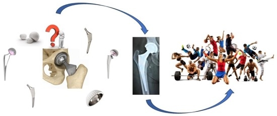
Is It Possible to Create an “Ideal Endoprosthesis” for an “Ideal Total Hip Replacement”?

by
Valentin L. Popov
1,*,
Aleksandr M. Poliakov
2,* and
Vladimir I. Pakhaliuk
2
1
Department of System Dynamics and Friction Physics, Institute of Mechanics, Technische Universität Berlin, 10623 Berlin, Germany
2
Polytechnic Institute, Sevastopol State University, 299053 Sevastopol, Russia
*
Authors to whom correspondence should be addressed.
Prosthesis 2023, 5(4), 1020-1036; https://doi.org/10.3390/prosthesis5040071
Submission received: 6 September 2023 / Revised: 5 October 2023 / Accepted: 9 October 2023 / Published: 13 October 2023
(This article belongs to the Special Issue State of Art in Hip and Knee Replacement)

Abstract

:
Since the end of the 19th and the beginning of the 20th centuries, technological equipment, implant designs (endoprosthesis) and the surgical technique of total hip replacement (THR) have been constantly improved and reached a high level of functionality and quality. Therefore, at present, THR is one of the most high-tech, reliable and popular surgical operations that allow a large number of people suffering from osteoarthritis and other serious diseases of the hip joint to return to an active lifestyle. At the same time, it is known that even operations at this level do not always guarantee the achievement of the desired result and can lead to various complications. The question arises: are there potential opportunities for creating an “ideal endoprosthesis” that allows one to perform an “ideal THR”? In this paper, based on a critical analysis of modern endoprosthesis designs for THR, technologies for their implantation and systemic postoperative complications, the most probable, according to the authors, ways of their development are formulated, which allow asymptotically approaching the “ideal”.

Graphical Abstract

1. Introduction

One of the articles published in the Lancet in 2007 argues that for many reasons THR can be considered the operation of the century [1]. This statement is difficult to dispute, since it really contributes to the return to active life of a large number of people of different ages suffering from serious diseases and injuries of the hip joint. In particular, THR often remains the only means of combating osteoarthritis of the joints, a disease that, according to the World Health Organization, most often leads to disability of a large number of people, especially the elderly [2].
The demand for THR is a powerful incentive for the development of technologies and technological tools necessary to ensure the high quality of this surgical operation. It is quite natural that much attention is paid to these processes in the scientific and industrial spheres. This is confirmed by a huge number of scientific articles published over more than a century of history of THR development, the beginning of which is considered the end of the 19th to the beginning of the 20th centuries [1]. At the same time, it is generally accepted that the first endoprosthesis for THR in the modern sense was created by P. Wiles in 1938 [3], and revolutionary changes in the design of endoprostheses, justified in the 1960s of the 20th century and which allowed for significantly increasing the quality of THR, are associated with J. Charnley [4]. These changes included three basic principles:
  • − ensuring a low friction moment between the contact pairs of endoprosthesis components (low friction arthroplasty);
  • − the use of acrylic cement to fix the components of the endoprosthesis in the bone tissue of the host;
  • − the use of ultra-high-density polyethylene as a material for the endoprosthesis modular cup.
It should be noted that the above requirements for THR endoprosthesis designs remain relevant to one degree or another at present. But, despite the obvious advantages achieved with their use, they also determine a number of disadvantages inherent in modern endoprostheses for THR. However, in most cases, THR provides significant intermediate and long-term benefits in terms of both disease-specific quality of life and general health, especially pain and functionality, leading to positive patient satisfaction [5]. However, developers of endoprostheses for THR are constantly looking for the best designs in order to achieve a minimum of failures and post-operative complications. In this regard, the question arises: are there potential opportunities for creating an “ideal endoprosthesis” that allows one to perform an “ideal THR”? Obviously, from a practical point of view, the creation of such an endoprosthesis within the framework of the established paradigm of total joint arthroplasty is impossible because it implies the obligatory use of an artificial analogue of the joint, which cannot be better than the natural one by definition. Nevertheless, it can be assumed that the desire to achieve the “ideal” may ultimately lead to the creation of an endoprosthesis that meets the functional needs of the patient and provides him with a high quality of life for a long period.
What steps should be taken to create such an endoprosthesis, given the current level of science development, engineering and technology? The answer to this question cannot be unambiguous for many reasons: objective and subjective, technical and physiological. In this paper, taking into account the current state of the issue and our own experience in designing and testing endoprostheses for THR and their main components, we have formulated the most likely, in our opinion, ways to improve them, allowing asymptotically approaching the “ideal”.

2. Materials and Methods

2.1. Designs of Modern Endoprostheses for THR

To substantiate the actual ways to improve endoprostheses for THR, it is necessary to systematize all the information about their advantages and disadvantages, service life, causes of failure and periprosthetic complications. Given the demand for THR, a huge amount of such information has been published in the literature, which greatly simplifies its systematization. However, here we are also within the framework of the existing paradigm of total joint arthroplasty and are limited to considering artificial analogues of natural joints. In this regard, the initial stage of systematization should begin with a review of modern endoprosthesis designs.
Typically, modular endoprostheses for THR include three main structural components (Figure 1): acetabular (Cup), femoral (Stem), and intermediate (Femoral Head). Obviously, each of them has its own history and currently represents a theoretically and practically substantiated set of structures adapted for various clinical cases and physiological and anthropometric characteristics of patients. For example, in [6], the history of the development for the endoprosthesis stem is presented in sufficient detail, and in [7], its design of a modular type is substantiated. At the same time, in all cases, in addition to describing changes in structures, important attention is paid to the reasons for their evolution. That is, in essence, the creation of each new design was due to the need to eliminate the shortcomings inherent in earlier versions.
Similarly, one can follow the evolution of the acetabular component [8,9], including the creation of modular cups with dual mobility [10,11,12]. Finally, despite the apparent constructive simplicity of the femoral head, it also has its own history of development and is characterized by many design features [13,14,15].
It is clear that modular endoprostheses for THR are obtained by combining various options for the main components, which allow obtaining a fairly large number of designs that can be selected by the surgeon depending on the specific clinical case. This is a rather complicated task, to simplify the solution of which a number of systems for classifying patient conditions have been developed that allow for planning of personalized treatment strategies for a particular joint, including primary and revision arthroplasty [16,17,18,19,20,21,22]. Here we do not discuss the principles of classification of the conditions and planning for joint arthroplasty, but only note the fact that in order to ensure the quality of a surgical operation, it is necessary to choose high-quality preoperative, intraoperative and postoperative treatment strategies, including the choice of an appropriate endoprosthesis design.
In addition to the design of the endoprosthesis itself, an important role is played by the method of fixing its components in bone structures. At the same time, two types of fixation of the stem and cup are distinguished: cemented and cementless [23]. Sometimes, hybrid fixation is also considered as another type. Each of these types has its advantages and disadvantages and is chosen by the surgeon depending on the general condition of the patient and his damaged joint, in particular [24,25,26,27].
The surfaces of cementless stems and cups of hip endoprostheses are made of materials having a porous structure or they are coated with hydroxyapatite. They are installed in bone structures using the “tight fit” method, which in the postoperative period contributes to the osseointegration of the implant. In contrast to this method, cementless endoprosthesis components are fixed in bone structures using a special cement based on polymethyl methacrylate.
Replacement of uncemented endoprosthesis components, if revision arthroplasty of the joint is required, is usually easier than replacement of cemented ones. In addition, the lifespan of such endoprostheses is quite high and is about 20–25 years. Therefore, they are usually recommended for relatively young patients, in whom the rate of bone tissue regeneration is sufficiently high and the immune system quickly adapts to a foreign body.
With a loose bone structure, it is impossible to ensure the strength of its connection with the implant. In such cases, the components of the endoprosthesis are recommended to be fixed in the bone with cement. Since bone density and strength decrease with age, cemented endoprosthesis components are usually recommended for the treatment of elderly patients.
Thus, the entire set of modern endoprostheses for THR is divided into two subsets: cemented and cementless, each of which is represented by a wide range of product sizes that differ, among other things, in the materials from which the main components are made, including the femoral head and modular cup liner forming a friction pair. Currently, the following friction pairs are mainly used in modular endoprostheses for THR [28,29,30]:
  • hard-on-soft bearings (metal-on-polyethylene (MOP) is a metal femoral head and a polyethylene acetabular liner, and ceramic-on-polyethylene (COP) is a ceramic femoral head and a polyethylene acetabular liner);
  • hard-on-hard bearings (metal-on-metal (MOM), ceramic-on-ceramic (COC) and ceramic-on-metal (COM) are ceramic femoral head and a metal acetabular liner).
The advantages and disadvantages of each of these pairs are described in the literature, for example [30]. Nevertheless, in the context of this article, it is necessary to note the main drawback inherent in all artificial friction pairs, which consists in the fact that, as a result of wear, a large number of the smallest particles from the materials of the pair accumulate in the periprosthetic tissues. The immune system recognizes them as foreign bodies and seeks to remove them from the body, which in most cases leads to serious postoperative complications.

2.2. Complications following THA

In 2011, the Hip Society Board of Directors created a Total Hip Arthroplasty Complications Workgroup that approved and validated 19 medical complications of THA with the aim of further standardizing their definitions and developing a stratification and validation system [31]. They included
  • Bleeding;
  • Wound complication;
  • Thromboembolic disease;
  • Neural deficit;
  • Vascular injury;
  • Dislocation/instability;
  • Periprosthetic fracture;
  • Abductor muscle disruption;
  • Deep periprosthetic joint infection;
  • Heterotopic ossification;
  • Bearing surface wear;
  • Osteolysis;
  • Implant loosening;
  • Cup-liner dissociation;
  • Implant fracture;
  • Reoperation;
  • Revision;
  • Readmission;
  • Death.
It should be noted that at the first stage of agreement, less than 80% of experts included bearing surface wear and osteolysis among the main complications following THA. However, given the fact that these processes are closely related to each other, as well as such a complication as implant loosening when interpreting the corresponding clinical scenario, they were eventually included in the list of major complications [31].
In practice, the manifestation and development of all the complications listed above depend on a combination of many dissimilar factors, but all of them can be minimized in each clinical case, provided that the endoprosthesis available on the market for medical services is chosen correctly. At the same time, a number of complications only indirectly depend on the endoprosthesis and are determined by the general condition of the patient, the qualifications of the surgeon, the quality of the surgical instrument and additional technical means, as well as the implementation of therapeutic and rehabilitation programs in the perioperative period. Depending to a certain extent directly on the design of the endoprosthesis can be considered:
  • Dislocation/instability—a complication of THR in most cases associated with patient noncomplicance with post-operative precautions, implant malposition, or soft-tissue deficiency. However, a common cause of dislocation is wear of the acetabular cup liner in the medium term (about 5 years) after surgery [32,33];
  • Periprosthetic fracture—a complication after THR which can occur due to trauma to the hip area, high-impact falls and in other cases. However, it may also be directly related to the implant, which contributes to the development of osteolysis and incorrect remodeling of the periprosthetic bone [34,35];
  • Bearing surface wear—arises from local stresses that exceed the mechanical strength of the articulating materials. At the same time, wear rates increase with factors such as increased sliding distance in hard-on-soft bearings, or suboptimal fluid film lubrication in the case of hard-on-hard implants. Therefore, this complication of THR directly depends on the design of the implant [36,37,38,39,40];
  • Osteolysis is one of the most serious complications after THR, which is the active resorption of bone matrix by osteoclasts. Osteolysis is mediated by wear particles of implant materials and may cause an immune response or alteration of the periprosthetic bone structure [41,42,43,44,45,46];
  • Implant loosening is one of the main complications following THR. Its nature is determined by patient factors (obesity, bone quality, activity level, patient genetics), surgical technique factors and implant factors (primarily the type of friction pair, method of fixation and the ability of the implant to osseointegrate) [47,48,49,50];
  • Cup–liner dissociation—a relatively rare complication of THR associated with misplacement of the component and/or impingement. To a greater extent, it is determined by the design of the implant [51,52,53];
  • Implant fracture—a complication after THK resulting from the loss of strength of the implant, i.e., to the greatest extent determined directly by the design of the implant [54,55].
Improving the designs of endoprostheses in order to minimize the causes of these complications contributes to an increase in their lifespan and patient satisfaction with the quality of THR and, as a result, brings them closer to some “ideal”.

3. Results

3.1. Improving the Components Design for Total Hip Arthroplasty

It should be noted that the improvement of total hip arthroplasty designs is ongoing. The authors of this article also contributed to this process. At the same time, we tried to use a systematic approach to the analysis and synthesis of new constructions, the methodology of which was given in [56]. Some of our inventions are presented in the Section 6 “Patents”, and the main directions for the development of THR are formulated in [23,30]. In general, they correlate with the directions formulated in [57,58].
A systematic analysis of the observed complications allows us to conclude that in order to improve the quality of THR, it is necessary to develop navigational and robotic surgical technologies, minimally invasive surgery, manufacturing methods, and materials and designs of endoprostheses (including their friction pairs) [23,57]. From our point of view, therapy, rehabilitation and monitoring of the patient’s condition in the perioperative period are also significant factors that require priority attention [30].
Below are a number of examples illustrating the process of continuous improvement of the components for a modular endoprosthesis, aimed at eliminating or mitigating the manifestations of typical complications after THR, improving the quality of life and positive patient satisfaction in the postoperative period.
For example, a new design of the polymer liner for the acetabular component was developed with an increased degree of the femoral head fixation in it, which helps to reduce the likelihood of its dislocation, leading to an obvious revision intervention. When it was created, along with the solution of the main problem, an increased area of joint mobility was achieved [59].
Reliability of pairing the endoprosthesis components in the new design is ensured due to the fact that the insert hole through which the femoral head is installed is located at the maximum possible distance from the diametrical plane. It is clear that in such cases it is necessary to ensure sufficiently large deformations of the liner during the installation of the head. A two-criterion problem with conflicting quality criteria, consisting in choosing the maximum possible distance of the liner hole from the diametric plane with the minimum possible deformation, was solved by numerical simulation. Ultimately, the forces required to install and remove the femoral head from the polymer liner were calculated, and an improved design of the liner was proposed to increase the endoprosthesis stability and reduce the likelihood of head dislocation.
Improving the quality of total hip arthroplasty can be achieved by improving the components of friction pairs. At present, it can be considered that a certain consensus has been reached as to which friction pairs are most acceptable in various clinical cases. However, there are various ways to improve their quality.
For example, it has been found that the tribological properties of polymers used in medical devices are affected by surface texturing [60]. In this case, as shown in [61,62], in a MOP friction pair with a textured polymer, the coefficient of dry friction decreases, but, at the same time, such a material resists wear less. At the same time, it is known that under conditions of liquid lubrication, friction decreases more noticeably than with dry friction. Thus, texturing without lubrication of surfaces leads to a decrease in friction by 4–7% and under lubricated conditions by 45.5–60.3% [60]. The effect of reducing friction increases with increasing texture density, which is explained by a decrease in the effective contact area due to the presence of dimples.
Previously, we proposed the use of texturing the surface of the femoral head to reduce the wear of the polymer liner in the MOP friction pair [14]. The layout of the texture elements is shown in Figure 2.
From the standpoint of the Kragelsky I.V. theory [63] and the hypothesis of film starvation, the following relations were obtained for the friction coefficient f and the ratio of the parameters S S d :
f = A p c 1 2 ν + 1 + β + B p c 1 2 ν + 1
S S d = p c p c 2 ν 2 ν + 1 ,
where s is an average distance between dimples (spacing); s d is their average extent (Figure 1); p c and p c are the contour pressures without oil pockets and with pockets, respectively; ν ,   b are the approximation parameters of the initial part of the reference surface roughness curve [64]; A ,   B are constants determined depending on the elastic characteristics of the friction pair materials (Young’s moduli E 1 ,   E 2 and Poisson’s ratios μ 1 ,   μ 2 ).
Expressing p c from Equation (2) and substituting into Equation (1), we obtain
f = A S S d 1 2 ν · 1 p c 2 ν + 1 + β + B S S d 1 2 ν · 1 p c 2 ν + 1 ,
Differentiating Equation (3) with respect to S S d and equating the result to zero, we obtain the necessary condition for the extremum (minimum) of the friction coefficient f for given parameter values:
S S d = C Δ 2 ν 2 ν + 1 · p a 0.4 2 ν + 1 ν ,
where C is a constant, determined depending on the combination of parameters of the friction pair materials, determined according to [64], and Δ is a complex roughness parameter, which, taking into account the nominal pressure p a , is determined by the expression [64]:
Δ = 1 S S d 1 ν 1 C p a 0.4 2 ν + 1 2 ν + 1 2 ν
Equations (4) and (5) were used to determine the optimal parameters of the friction pair, providing the lowest coefficient of friction and wear of the material. As a criterion confirming the optimality of the texture parameters, a decrease in the nominal contact pressures p a and equivalent stresses in the Mises form on the surface with dimples with respect to a smooth surface was chosen.
Since the function p a = f h d , d , S cannot be specified analytically, it is quite difficult to predict its behavior in the parameter space. In this regard, its study was carried out in a number of trial points. For the purpose of a systematic analysis of the parameter space, test points were selected using L Π τ sequences of points uniformly distributed in three-dimensional space h d , d , S   [64].
As a result of numerical experiments, it was found that the recommended rational parameters of the head surface texture in a MOP friction pair are h d = 0.08 0.25 ,   d = 50 190   μ m ,   S = 200 480   μ m , a n d     h = 8 30   μ m , but for a MOM pair, the space S should not exceed 250 μm. It may be justified to increase the depth of the dimple h more than 30 μm so that the wear particles accumulate in the dimples for a longer time, thereby reducing the surface wear of the pair and the release of particles into the periarticular space. The recommended ratio for both pairs is S S d = 1.1 2.5 , which corresponds to a texture density range of approximately 0.02…0.46.
Thus, the use of texturing the surface of the femoral head was theoretically substantiated in order to reduce the coefficient of friction and wear of materials in the friction pair. However, in order to introduce such pairs into surgical practice, it is necessary to perform in vitro wear tests recommended by the standards on specialized test machines, as well as in vivo clinical trials.
It should be noted here that the friction forces arising from the relative movement of the head and cup liner surfaces have a significant impact on the endoprosthesis lifespan. In this regard, when developing new implant designs, assessments of these forces and the wear of materials mediated by them, which can be performed as a result of modeling the friction pair operation, acquire an important role [65]. However, to develop adequate models of friction and wear, it is necessary to take into account the real nature of the friction pair surface lubrication, which in general is not an easy task. In addition, such models should be built taking into account the similarity of artificial and complex natural human synovial joints, in which biological, hydrodynamic and tribological phenomena occur [66], as well as depending on the type of human motor activity [67]. But even when the above conditions are met, estimates of material wear made on the basis of modeling turn out to be very inaccurate. In order to obtain more reliable results, it is advisable to determine wear in in vitro experiments performed on specialized simulator rigs [68]. Therefore, endoprostheses in which the wear of friction pair materials, obtained as a result of in vitro tests, turns out to be the least are considered to be of the highest quality.
Another way is to reduce the coefficients of friction and wear in MOP and COP friction pairs by special treatment of the ultra-high molecular weight polyethylene (UHMWPE) liner surface, which increases its hardness and, therefore, wear resistance. In [69], it was shown that the treatment of UHMWPE in helium plasma at low pressure leads to the appearance in the IR spectrum of UHMWPE of a new absorption line corresponding to wavelength λ = 965 cm−1 (Figure 3), which may correspond to double bonds.
The concentration of double bonds, proportional to the relative intensity of this absorption line to the absorption line with λ = 1470 cm−1, corresponding to the deformation vibrations of the C-H bond, reaches saturation in 12 min of treatment at a power of 50 W and a helium flow of 5 cm3/min. The formation of double bonds apparently occurs under the action of chemically active plasma components, such as vacuum ultraviolet radiation and electron and ion fluxes incident on the surface of the polymer sample. The primary process in this case is the abstraction of atomic or molecular hydrogen from the polymer molecule with the formation of alkyl radicals. Subsequently, these radicals recombine with the formation of double bonds, depending on if the radicals belong to neighboring atoms of the polymer molecule, or intermolecular crosslinking, and if the radicals are located on neighboring molecules.
Thus, the formation of double bonds is always accompanied by the formation of intermolecular crosslinks. Unfortunately, in IR spectra, in accordance with the rules of symmetry, cross-links do not appear. However, publications are known in which the formation of double bonds is used as an internal dosimeter to determine the degree of crosslinking in UHMWPE [70]. In our case, it is important that the formation of crosslinks in the surface layer leads to an increase in the surface strength characteristics of UHMWPE and, ultimately, to an increase in its wear resistance.
An important influence on the quality of a hip joint endoprosthesis is exerted by the method of fixing a stem with a femoral head currently made from metal alloys (cobalt–chromium, molybdenum alloy, titanium alloy) or ceramics (Al2O3, ZrO2, delta-ceramics) and their mixtures. These materials have a sufficiently high hardness, which ensures minimal wear of the friction pair. However, due to the toxic nature of cobalt and chromium ions in the metal head, which tend to accumulate in body tissues and contribute to the occurrence of severe allergic reactions in the patient, there is a clear tendency to increase the use of ceramic heads, since ceramics and their wear products are biologically compatible for the body [23]. In this regard, modular femoral heads, which have the properties of metal in connection with the neck of the stem and the properties of ceramics on the bearing surface of the friction pair, become relevant. Their manufacture is possible based on technologies that provide a reliable connection of ceramics and titanium alloy.
With the participation of this work’s authors, a technology for soldering these materials was developed, which provides a connection with high tensile, compressive and shear strength [71]. As a result, it became possible to develop a modular femoral head, the general view and 3D model of which are shown in Figure 4.
The modular head consists of an external ceramic component (3) with a spherical outer surface and an internal blind hole at its base, in which the inner component (2) is located—a titanium alloy sleeve with a cylindrical outer surface and an axial blind taper hole. The surface of the hole in the ceramic component (3) contains a specially made macrotexture to improve the adhesion of the solder to the soldered ceramics. Sleeve 2 in the specified hole of component 3 is installed along its surface with a gap of approximately 50–200 μm on the radius and with a similar gap along the surface perpendicular to the longitudinal axis to fill the gap with biocompatible solder, and is fixedly connected to component 3 by means of high-temperature soldering.
This design of the modular femoral head allows for a significant reduction or, in practice, the elimination of the occurrence of fretting corrosion in the taper connection of the endoprosthesis stem neck and the head sleeve, made of titanium alloy. Such a pair thereby excludes the occurrence of an electrolytic pair between them in an aggressive organism environment and accumulation of its products in periprosthetic tissues and their probable migration and accumulation in vital human organs.
The above design of the modular head combines all the positive qualities of metal and ceramic heads. In it, the presence of a metal sleeve with a traditionally taper axial hole provides a fixed connection with the stem neck, thereby significantly increasing the stability of the endoprosthesis, and prevents the head from possible splitting in the connection, as in the case of entirely ceramic head. All these factors create conditions under which there is an improvement in the quality of life for the patient by increasing the lifespan of the entire endoprosthesis.
The examples presented above indicate that the improvement of total hip replacement designs occurs in different directions. At the same time, constructive solutions that help eliminate or reduce the impact of systemic complications after THA are found not only because of theoretical and experimental studies but also by heuristic methods. However, if such solutions contribute to the improvement of a specific design and reduce the likelihood of complications after THA, they are considered significant and further efforts are made to find their theoretical and experimental justification. As a rule, such solutions are issued in the form of patents for inventions (see, for example, the Section 6 “Patents”) and in many cases are used in commercially available endoprostheses.
Thus, each constructive solution, justified theoretically, experimentally or found heuristically, contributing to one degree or another to reduce the likelihood of complications or their impact on the general health of the patient after THA is a real step towards creating an “ideal” endoprosthesis. However, in order to fulfill the “ideal” THR with the help of this “ideal”, it is also necessary to ensure the fulfillment of many other conditions, taking into account, among other things, the factors of a particular patient and surgical, therapeutic and rehabilitation technologies.

3.2. Condition Monitoring and Perioperative Measures Aimed at Increasing the Lifespan of Total Hip Replacement

Largely, the quality of THR is determined by patient factors, so monitoring of his condition throughout the perioperative period plays an important role. This allows not only high-quality planning of the operation but also ensuring its success during and after THR by implementing adequate therapeutic and rehabilitation strategies and technologies.
For quite a long period, various implantable sensor systems have been used to monitor the human condition [72]. With their help, various information about the state of tissues and systems of the body in vivo is obtained. Implantable sensory systems have made it possible to clarify and expand knowledge about the biomechanics of the human musculoskeletal system, as well as about the processes occurring in skeletal connective tissues under various conditions [73,74]. Studies have focused on the development of implantable sensor systems adapted directly for orthopedic use and, in particular, for monitoring the state of implants [75,76,77].
A number of studies in the literature report on the development of so-called intelligent implants [78,79]. The results obtained using these devices in vivo allowed us to solve a number of clinically significant problems related to adequate patient care in the postoperative period. As noted in [79], smart-implants provided answers to questions about the recommended types of rehabilitation devices and made it possible to plan rehabilitation schemes for patients after THR. They can be used to test the endoprosthesis for loosening [80], to determine realistic loads for in vitro endoprostheses studies under various modes of motor activity [81,82,83], to measure the temperature of the friction pair [84], to evaluate forces and torques [85], as well as to monitor the wear of materials of friction pairs and the state of periprosthetic tissues [86].
That is, modern implantable sensor systems can be used to obtain various information about the state of the patient and his tissues during most of the perioperative period. At the initial stage, this may contribute to a more accurate choice of a therapeutic strategy to prepare the patient for surgery, as well as to THR planning, including a reasoned choice of an appropriate endoprosthesis and, if necessary, cellular technologies. If we assume that, taking into account adequate therapeutic preparation and the correct choice of the endoprosthesis, the operation will be successful, then after THR, it is necessary to use therapeutic and rehabilitation procedures aimed at the fastest healing of periprosthetic tissues and osseointegration of the endoprosthesis components without cement fixation. The effectiveness of these procedures will be significantly higher if their planning is carried out on a scientific basis, using appropriate mathematical models, the parameters of which are determined or refined using sensor systems. Moreover, the combination of a mathematical model with an implantable sensory system allows for creating a “Digital Twin” of the periprosthetic area with real-time settings. One of its tasks will be to determine optimal or close-to-optimal strategies for the healing of periprosthetic tissues and osseointegration of endoprosthesis components.
It should be noted that the phenomenon of the osseointegration process for the components of cementless endoprostheses is of great theoretical and practical interest due to the rejuvenation of the contingent of people who need THR and, as a result, due to the observed increase in the use of cementless endoprostheses for THR. To study all the features of this process, various means are used, including studies in vitro, in vivo and in silico. The efficiency of the first two research methods increases in the case of using implantable sensor systems, and the predictions developed based on the in silico models become more realistic when they use parameters obtained in studies in vitro and in vivo. But, as is known, such models should adequately reflect the object or process under study.
With regard to THR, in silico studies can use many mathematical models of osseointegration with varying degrees of detail, as shown, for example, in [30]. The same can be said about the models of periprosthetic (primarily bone) tissue healing. The main point is that the mathematical model used in the assessment of osseointegration or healing, if possible, directly takes into account those factors that are decisive for the course of a particular process. That is, it must take into account, at a minimum, factors of a chemical, biological and mechanical nature. In addition, if possible, the model should have a minimum number of degrees of freedom if a certain key parameter is adequately estimated. For example, the model of P. Moreo et al. [87,88] makes it possible to study the processes of density evolution for platelets ( c ) , osteogenic cells ( m ) , osteoblasts ( b ) and growth factors ( s 1 ) and ( s 2 ) . However, under certain conditions, the process of platelet density evolution can be considered as a one-parameter problem, the model of which is built taking into account the linear diffusion of cells with the coefficient ( D c ) , their adhesion to the implant surface and kinetic terms. Cell adhesion in the first approximation can be modeled as a linear “taxis”, depending on the gradient of proteins adsorbed on the implant surface ( p ) with a certain coefficient ( H c ). In this case, the evolution of platelets is described by the differential equation
c t D c c + A c c = · H c c p ,
where t is time; A c is a coefficient characterizing the rate of cell removal due to inflammation.
It should be noted that the value of p is assumed to be known and depends on the implant surface micro texture, and its value can be determined in experiments in vivo using implantable sensors. The maximum value of p is on the surface and decreases with distance from it, reaching a zero value, which is preserved in the rest of the region. Graphs characterizing the change in platelet density at different initial values of p are shown in Figure 5 [30].
The results of many models in the study of periprosthetic tissue healing and implant osseointegration are available in a variety of literature and are not considered in detail here. It is only important to emphasize that the simulation of the above processes, and even more so the analysis of “Digital Twins”, based on adequate models is a powerful tool that allows for effectively managing the THR surgical operation throughout the entire perioperative period.

4. Discussion

The main goal of creating new designs of total endoprostheses for THR, as well as improving surgical, therapeutic and rehabilitation technologies, is to improve the quality of life of patients who need this complex and traumatic surgical operation. As noted above, THR is currently called the operation of the twentieth century [1] due to its high quality, provided under certain conditions, including patient factors, endoprosthesis design, surgeon qualifications, implantation technology, perioperative therapy and rehabilitation. In principle, most of the above factors can be adjusted to contribute to the success of THR, with the exception of patient factors. This indicates that this surgical operation is personalized and its outcome largely depends on the physiological state of the patient. This fact alone suggests that the creation of a universal endoprosthesis for THR is a very difficult task. At the same time, one of the potential ways to solve it can be associated, for example, with the development of cellular, therapeutic and rehabilitation technologies that help improve and maintain a stable state of bone tissues in the perioperative period. Hypothetically, this can be achieved, at least for patients of certain age groups. In this case, the standardization level of the factors that ensure the success of THR can be significantly increased, including in relation to the choice of a specific endoprosthesis. The development of just such endoprostheses will contribute to a gradual approach to some conditional “ideal”. That is, an endoprosthesis can be considered “ideal” if a number of conditions are met. For example, an endoprosthesis that does not require revision for patients of a certain age group can be considered “conditionally ideal” until the end of their life. On the other hand, given the impossibility of achieving a normal state of bone tissue in a group of patients using therapeutic and rehabilitation methods, a “conditionally ideal” endoprosthesis should provide the maximum possible lifespan without revision for patients in this group. At the same time, in addition to the above, other conditions must be met to prevent the occurrence of any complications after THR, leading to a decrease in the quality of life, satisfaction and comfort of patients.
The examples given in this paper, as well as many other examples available in the literature, allow us to state that the process of moving towards the creation of an “ideal” endoprosthesis occurs constantly based on objective reasons due to complications observed in practice after THR. Therefore, the objective goal of this product evolution is minimizing postoperative complications. Its achievement will be possible with the creation of an endoprosthesis that is not the source or cause of any complications. It is this design that can be considered “ideal”.
At the same time, it cannot be assumed that the use of an “ideal endoprosthesis” always corresponds to an “ideal THR”. The quality of this operation, as noted above, depends on many other factors. Therefore, only such THR can be considered “ideal”, in which all factors influencing the occurrence of complications are minimized.
Thus, the answer to the question posed in the title of this article can be formulated as follows: the creation of an “ideal endoprosthesis” for an “ideal THR” is possible by asymptotic approximation of real structures to a certain “conditional ideal”, subject to continuous improvement of perioperative technologies. This process is currently observed and its development will be relevant for an indefinite period, at least until a new paradigm of total joint arthroplasty is adopted, based, for example, on the regeneration of their tissues.

5. Conclusions

As is known, THR is currently one of the most high-tech and high-quality operations, but even it is not without problems. Statistics show that the number of failed THRs is very small. However, behind every fraction of a percent failure is a large number of people suffering from the need for revision prosthetics. In this regard, improving the quality of THR, as well as the factors determining its quality, is an urgent and important social task. It is clear that its solution can only be complex, and in order to understand the connectedness of all factors, a systematic analysis of the advantages and disadvantages associated with THR is needed. In this article, based on the analysis of literature and our own experience in the development and testing of hip endoprostheses components, we tried to systematize information related to the improvement of THR designs and technologies and predict the further evolution of this process.
The answers to the questions discussed in the article are not unambiguous even in the understanding of the authors. Each of them can be the subject of a separate article to provide an opportunity for discussion among a wide range of practicing surgeons and theoretical experts.

6. Patents

Author Contributions

Conceptualization, A.M.P.; investigation, V.L.P. and V.I.P.; software, A.M.P.; writing—original draft preparation, A.M.P.; writing—review and editing, V.L.P. and V.I.P. All authors have read and agreed to the published version of the manuscript.

Funding

This research received no external funding.

Institutional Review Board Statement

Not applicable.

Informed Consent Statement

Not applicable.

Data Availability Statement

Data is contained within the article.

Conflicts of Interest

The authors declare no conflict of interest.

References

  1. Learmonth, I.D.; Young, C.; Rorabeck, C. The operation of the century: Total hip replacement. Lancet 2007, 370, 1508–1519. [Google Scholar] [CrossRef]
  2. Popov, V.L.; Poliakov, A.M.; Pakhaliuk, V.I. Synovial Joints. Tribology, Regeneration, Regenerative Rehabilitation and Arthroplasty. Lubricants 2021, 9, 15. [Google Scholar] [CrossRef]
  3. Wiles, P. The surgery of the osteoarthritic hip. Br. J. Surg. 1958, 45, 488–497. [Google Scholar] [CrossRef] [PubMed]
  4. Charnley, J. Arthroplasty of the hip. A new operation. Lancet 1961, 1, 1129–1132. [Google Scholar] [CrossRef]
  5. Shan, L.; Shan, B.; Graham, D.; Saxena, A. Total hip replacement: A systematic review and meta-analysis on mid-term quality of life. Osteoarthr. Cartil. 2014, 22, 389–406. [Google Scholar] [CrossRef]
  6. Wellauer, H.; Heuberger, R.; Gautier, E.; Tannast, M.; Steinke, H.; Wahl, P. The history of the development of the regular straight stem in hip arthroplasty. EFORT Open Rev. 2023, 8, 548–560. [Google Scholar] [CrossRef] [PubMed]
  7. Srinivasan, A.; Jung, E.; Levine, B.R. Modularity of the femoral component in total hip arthroplasty. J. Am. Acad. Orthop. Surg. 2012, 20, 214–222. [Google Scholar] [CrossRef] [PubMed]
  8. Hamilton, W.G.; Calendine, C.L.; Beykirch, S.E.; Hopper, R.H., Jr.; Engh, C.A. Acetabular fixation options: First-generation modular cup curtain calls and caveats. J. Arthroplasty 2007, 22 (Suppl. 1), 75–81. [Google Scholar] [CrossRef]
  9. Powers, C.C.; Ho, H.; Beykirch, S.E.; Huynh, C.; Hopper, R.H., Jr.; Engh, C.A., Jr.; Engh, C.A. A comparison of a second- and a third-generation modular cup design: Is new improved? J. Arthroplasty 2010, 25, 514–521. [Google Scholar] [CrossRef]
  10. Vajapey, S.P.; Fideler, K.L.; Lynch, D.; Li, M. Use of dual mobility components in total hip arthroplasty: Indications and outcomes. J. Clin. Orthop. Trauma 2020, 11 (Suppl. 5), S760–S765. [Google Scholar] [CrossRef]
  11. Aguado-Maestro, I.; de Blas-Sanz, I.; Sanz-Peñas, A.E.; Campesino-Nieto, S.V.; Diez-Rodríguez, J.; Valle-López, S.; Espinel-Riol, A.; Fernández-Díez, D.; García-Alonso, M. Dual Mobility Cups as the Routine Choice in Total Hip Arthroplasty. Medicina 2022, 58, 528. [Google Scholar] [CrossRef] [PubMed]
  12. Manson, T.T.; Adrados, M.; Gililland, J.M.; Mahmood, B.M.; Samuel, L.T.; Moskal, J.T. The Role of Dual-Mobility Components in Total Hip Arthroplasty. J. Bone Jt. Surg. 2023, 105, 250–261. [Google Scholar] [CrossRef]
  13. Tankut, O.V. Substantiation of Hip Arthroplasty Using Single Crystal Sapphire in the Joint of Hip Prosthesis. Ph.D. Thesis, Sytenko Institute of Spine and Joint Pathology, Kharkiv, Ukraine, 2010. [Google Scholar]
  14. Pakhaliuk, V.I.; Polyakov, A.M.; Kalinin, M.I.; Bratan, S.M. Evaluating the impact and norming the parameters of partially regular texture on the surface of the articulating ball head in a total hip joint prosthesis. Tribol. Online 2016, 11, 527–539. [Google Scholar] [CrossRef]
  15. Patent Prosthesis Ball-Joint. Available online: https://patents.google.com/patent/EP0406040A2/en (accessed on 16 August 2023).
  16. D’Antonio, J.; McCarthy, J.C.; Bargar, W.L.; Borden, L.S.; Cappelo, W.N.; Collis, D.K.; Steinberg, M.E.; Wedge, J.H. Classification of femoral abnormalities in total hip arthroplasty. Clin. Orthop. Relat. Res. 1993, 296, 133–139. [Google Scholar] [CrossRef]
  17. Dossick, P.H.; Dorr, L.D.; Gruen, T.M.; Saberi, M.T. Techniques for preoperative planning and postoperative evaluation of noncemented hip arthroplasty. Tech. Orthop. 1991, 6, 1–6. [Google Scholar] [CrossRef]
  18. Brady, O.H.; Garbuz, D.S.; Masri, B.A.; Duncan, C.P. The reliability and validity of the Vancouver classification of femoral fractures after hip replacement. J. Arthroplasty 2000, 15, 59–62. [Google Scholar] [CrossRef]
  19. Toom, A.; Fischer, K.; Märtson, A.; Rips, L.; Haviko, T. Inter-observer reliability in the assessment of heterotopic ossification: Proposal of a combined classification. Int. Orthop. 2005, 29, 156–159. [Google Scholar] [CrossRef]
  20. Gómez, L.F.U.; Gaitán-Lee, H.; Duarte, M.A.; Halley, P.D.; Jaramillo, A.R.; García, E.L. Precision and accuracy of pre-surgical planning of non-cemented total hip replacement with calibrated digital images and acetates. J. Orthop. Surg. Res. 2021, 16, 431. [Google Scholar] [CrossRef]
  21. Di Martino, A.; Rossomando, V.; Brunello, M.; D’Agostino, C.; Pederiva, D.; Frugiuele, J.; Pilla, F.; Faldini, C. How to perform correct templating in total hip replacement. Musculoskelet. Surg. 2023, 107, 19–28. [Google Scholar] [CrossRef]
  22. Colombi, A.; Schena, D.; Castelli, C.C. Total hip arthroplasty planning. EFORT Open Rev. 2019, 4, 626–632. [Google Scholar] [CrossRef]
  23. Poliakov, A.M.; Pakhaliuk, V.I.; Popov, V.L. Current Trends in Improving of Artificial Joints Design and Technologies for Their Arthroplasty. Front. Mech. Eng. Sec. Tribol. 2020, 6, 4. [Google Scholar] [CrossRef]
  24. Pala, E.; Mavrogenis, A.F.; Angelini, A.; Henderson, E.R.; Douglas, L.G.; Ruggieri, P. Cemented versus cementless endoprostheses for lower limb salvage surgery. J. BUON 2013, 18, 496–503. [Google Scholar]
  25. Szypuła, J.; Cabak, A.; Kiljański, M.; Boguszewski, D.; Tomaszewski, W. Comparison of Biocompatibility of Cemented vs. Cementless Hip Joint Endoprostheses Based on Postoperative Evaluation of Proinflammatory Cytokine Levels. Med. Sci. Monit. 2016, 22, 4830–4835. [Google Scholar] [CrossRef]
  26. Mäkelä, K.T.; Eskelinen, A.; Pulkkinen, P.; Paavolainen, P.; Remes, V. Total hip arthroplasty for primary osteoarthritis in patients fifty-five years of age or older. An analysis of the Finnish arthroplasty registry. J. Bone Jt. Surg. Am. 2008, 90, 2160–2170. [Google Scholar] [CrossRef] [PubMed]
  27. Lewis, P.M.; Khan, F.J.; Feathers, J.R.; Lewis, M.H.; Morris, K.H.; Waddell, J.P. Uncemented total hip arthroplasty can be used safely in the elderly population. Bone Jt. Open 2021, 2, 293–300. [Google Scholar] [CrossRef]
  28. Thomsen, M.; von Strachwitz, B.; Mau, H.; Cotta, H. Survey of materials in hip endoprosthes. Z. Orthop. Ihre Grenzgeb. 1995, 133, 1–6. [Google Scholar] [CrossRef]
  29. Khalifa, A.A.; Bakr, H.M. Updates in biomaterials of bearing surfaces in total hip arthroplasty. Arthroplasty 2021, 3, 32. [Google Scholar] [CrossRef]
  30. Popov, V.L.; Poliakov, A.M.; Pakhaliuk, V.I. Improving the Endoprosthesis Design and the Postoperative Therapy as a Means of Reducing Complications Risks after Total Hip Arthroplasty. Lubricants 2022, 10, 38. [Google Scholar] [CrossRef]
  31. Healy, W.L.; Iorio, R.; Clair, A.J.; Pellegrini, V.D.; Della Valle, C.J.; Berend, K.R. Complications of Total Hip Arthroplasty: Standardized List, Definitions, and Stratification Developed by The Hip Society. Clin. Orthop. Relat. Res. 2016, 474, 357–364. [Google Scholar] [CrossRef] [PubMed]
  32. Brooks, P.J. Dislocation following total hip replacement. Bone Jt. J. 2013, 95-B, No 11_Supple_A, 67–69. [Google Scholar] [CrossRef]
  33. Werner, B.C.; Brown, T.E. Instability after total hip arthroplasty. World J. Orthop. 2012, 3, 122–130. [Google Scholar] [CrossRef]
  34. Young, S.W.; Pandit, S.; Munro, J.T.; Pitto, R.P. Periprosthetic femoral fractures after total hip arthroplasty. ANZ J. Surg. 2007, 77, 424–428. [Google Scholar] [CrossRef]
  35. Patsiogiannis, N.; Kanakaris, N.K.; Giannoudis, P.V. Periprosthetic hip fractures: An update into their management and clinical outcomes. EFORT Open Rev. 2021, 6, 75–92. [Google Scholar] [CrossRef] [PubMed]
  36. Hosseinzadeh, S.; Reza, H.; Eajazi, A.; Sina, A. The Bearing Surfaces in Total Hip Arthroplasty—Options, Material Characteristics and Selection. In Recent Advances in Arthroplasty; Intech Open: London, UK, 2012. [Google Scholar] [CrossRef]
  37. Paxton, E.W.; Inacio, M.C.; Namba, R.S.; Love, R.; Kurtz, S.M. Metal-on-conventional Polyethylene Total Hip Arthroplasty Bearing Surfaces Have a Higher Risk of Revision Than Metal-on-highly Crosslinked Polyethylene: Results from a US Registry. Clin. Orthop. Relat. Res. 2015, 473, 1011–1021. [Google Scholar] [CrossRef]
  38. Urban, J.A.; Garvin, K.L.; Boese, C.K.; Bryson, L.; Pedersen, D.R.; Callaghan, J.J.; Miller, R.K. Ceramic-on-polyethylene bearing surfaces in total hip arthroplasty. Seventeen to twenty-one-year results. J. Bone Jt. Surg. Am. 2001, 83, 1688–1694. [Google Scholar] [CrossRef] [PubMed]
  39. Bierbaum, B.E.; Nairus, J.; Kuesis, D.; Morrison, J.C.; Ward, D. Ceramic-on-ceramic bearings in total hip arthroplasty. Clin. Orthop. Relat. Res. 2002, 405, 158–163. [Google Scholar] [CrossRef]
  40. Silverman, E.J.; Ashley, B.; Sheth, N.P. Metal-on-metal total hip arthroplasty: Is there still a role in 2016? Curr. Rev. Musculoskelet. Med. 2016, 9, 93–96. [Google Scholar] [CrossRef]
  41. Vallés, G.; Vilaboa, N. Osteolysis after Total Hip Arthroplasty: Basic Science. In Acetabular Revision Surgery in Major Bone Defects, 1st ed.; García-Rey, E., García-Cimbrelo, E., Eds.; Springer: Cham, Switzerland, 2019; pp. 1–31. [Google Scholar] [CrossRef]
  42. Howie, D.W.; Neale, S.D.; Haynes, D.R.; Holubowycz, O.T.; McGee, M.A.; Solomon, L.B.; Callary, S.A.; Atkins, G.J.; Findlay, D.M. Periprosthetic osteolysis after total hip replacement: Molecular pathology and clinical management. Inflammopharmacology 2013, 21, 389–396. [Google Scholar] [CrossRef]
  43. Xing, D.; Li, R.; Li, J.J.; Tao, K.; Lin, J.; Yan, T.; Zhou, D. Catastrophic Periprosthetic Osteolysis in Total Hip Arthroplasty at 20 Years: A Case Report and Literature Review. Orthop. Surg. 2022, 14, 1918–1926. [Google Scholar] [CrossRef]
  44. Beck, R.T.; Illingworth, K.D.; Saleh, K.J. Review of periprosthetic osteolysis in total joint arthroplasty: An emphasis on host factors and future directions. J. Orthop. Res. 2012, 30, 541–546. [Google Scholar] [CrossRef]
  45. Noordin, S.; Masri, B. Periprosthetic osteolysis: Genetics, mechanisms and potential therapeutic interventions. Can. J. Surg. 2012, 55, 408–417. [Google Scholar] [CrossRef]
  46. Dattani, R. Femoral osteolysis following total hip replacement. Postgrad Med. J. 2007, 83, 312–316. [Google Scholar] [CrossRef] [PubMed]
  47. Desy, N.M.; Abdel, M.P. Aseptic Implant Loosening. In Complications after Primary Total Hip Arthroplasty; Abdel, M., Della Valle, C., Eds.; Springer: Cham, Switzerland, 2017; pp. 183–194. [Google Scholar] [CrossRef]
  48. Cherian, J.J.; Jauregui, J.J.; Banerjee, S.; Pierce, T.; Mont, M.A. What Host Factors Affect Aseptic Loosening after THA and TKA? Clin. Orthop. Relat. Res. 2015, 473, 2700–2709. [Google Scholar] [CrossRef]
  49. Toni, A.; Viceconti, M.; Sudanese, A.; Baruffaldi, F.; Giunti, A. Bone remodelling after total hip arthroplasty. J. Mater. Sci. Mater. Med. 1996, 7, 149–152. [Google Scholar] [CrossRef]
  50. Dapunt, U.; Prior, B.; Kretzer, J.P.; Hänsch, G.M.; Gaida, M.M. The effect of surgical suture material on osteoclast generation and implant-loosening. Int. J. Med. Sci. 2021, 18, 295–303. [Google Scholar] [CrossRef]
  51. Beckmann, N.A.; Schonhoff, M.; Bastian, J.D.; Renkawitz, T.; Jaeger, S. Dissociation of liner from cup in THA: Does liner damage affect the risk of dissociation? Arch. Orthop. Trauma Surg. 2023, 143, 2747–2754. [Google Scholar] [CrossRef] [PubMed]
  52. Ciolli, G.; Silva, R.; Giovannetti de Sanctis, E.; Proietti, L.; Mocini, F.; Corona, K.; Mazzoleni, M.G.; Romanini, E.; Marescalchi, M.; Brancaccio, V.; et al. Liner dissociation in total hip arthroplasty: A systematic review. Eur. Rev. Med. Pharmacol.Sci. 2022, 26 (Suppl. 1), 138–150. [Google Scholar] [CrossRef]
  53. De Martino, I.; D’Apolito, R.; Soranoglou, V.G.; Poultsides, L.A.; Sculco, P.K.; Sculco, T.P. Dislocation following total hip arthroplasty using dual mobility acetabular components: A systematic review. Bone Jt. J. 2017, 99-B (Suppl. 1), 18–24. [Google Scholar] [CrossRef] [PubMed]
  54. Sadoghi, P.; Pawelka, W.; Liebensteiner, M.C.; Williams, A.; Leithner, A.; Labek, G. The incidence of implant fractures after total hip arthroplasty. Int. Orthop. 2014, 38, 39–46. [Google Scholar] [CrossRef]
  55. Tallarico, M.; Meloni, S.M.; Park, C.-J.; Zadrożny, Ł.; Scrascia, R.; Cicciù, M. Implant Fracture: A Narrative Literature Review. Prosthesis 2021, 3, 267–279. [Google Scholar] [CrossRef]
  56. Polyakov, A.; Pakhaliuk, V.; Kalinin, M.; Kramar, V.; Kolesova, M.; Kovalenko, O. System analysis and synthesis of total hip joint endoprosthesis. Proc. Eng. 2015, 100, 530–538. [Google Scholar] [CrossRef]
  57. Eingartner, C. Current trends in total hip arthroplasty. Ortop. Traumatol. Rehabil. 2007, 9, 8–14. [Google Scholar]
  58. Shubnyakov, I.I.; Riahi, A.; Denisov, A.O.; Korytkin, A.A.; Aliyev, A.G.; Veber, E.V.; Muravyeva, Y.V.; Sereda, A.P.; Tikhilov, R.M. The Main Trends in Hip Arthroplasty Based on the Data in the Vreden’s Arthroplasty Register from 2007 to 2020. Traumatol. Orthop. Russia 2021, 27, 119–142. [Google Scholar] [CrossRef]
  59. Pakhaliuk, V.I.; Poliakov, A.M. Modeling deformation of the polymeric liner of the hip joint acetabulary component in couple with the head. Fundam. Appl. Probl. Eng. Technol. 2021, 4, 87–96. [Google Scholar] [CrossRef]
  60. Evangelista, I.; Wencel, D.; Beguin, S.; Zhang, N.; Gilchrist, M.D. Influence of Surface Texturing on the Dry Tribological Properties of Polymers in Medical Devices. Polymers 2023, 15, 2858. [Google Scholar] [CrossRef]
  61. Korpela, T.; Suvanto, M.; Pakkanen, T.T. Friction and wear of periodically micro-patterned polypropylene in dry sliding. Wear 2012, 289, 1–8. [Google Scholar] [CrossRef]
  62. Korpela, T.; Suvanto, M.; Pakkanen, T.T. Wear and friction behavior of polyacetal surfaces with micro-structure controlled surface pressure. Wear 2015, 328–329, 262–269. [Google Scholar] [CrossRef]
  63. Kragelsky, I.V.; Dobychin, M.N.; Kombalov, V.S. Fundamentals of Calculations for Friction and Wear; Mashinostroenie: Moscow, Russia, 1977; p. 526. (In Russian) [Google Scholar]
  64. Sobol, I.M.; Statnikov, R.B. Choice of Optimal Parameters in Problems with Many Criteria; Science: Moscow, Russia, 1981; p. 110. (In Russian) [Google Scholar]
  65. Ruggiero, A.; Sicilia, A. Lubrication modeling and wear calculation in artificial hip joint during the gait. Tribol. Int. 2020, 142, 105993. [Google Scholar] [CrossRef]
  66. Ruggiero, A. Milestones in Natural Lubrication of Synovial Joints. Front. Mech. Eng. 2020, 6, 52. [Google Scholar] [CrossRef]
  67. Ruggiero, A.; Sicilia, A. Implementation of a Finite Element Deformation Model within an Elasto-Hydrodynamic Lubrication Numerical Solver for a Ball in Socket Tribopair. Front. Mech. Eng. 2022, 8, 909156. [Google Scholar] [CrossRef]
  68. Ruggiero, A.; Sicilia, A.; Affatato, S. In silico total hip replacement wear testing in the framework of ISO 14242-3 accounting for mixed elasto-hydrodynamic lubrication effects. Wear 2020, 460–461, 203420. [Google Scholar] [CrossRef]
  69. Pakhaliuk, V.I.; Vasilets, V.N.; Poliakov, A.M.; Torkhov, N.A. Reducing the Wear of the UHMWPE Used in the Total Hip Replacement after Low-Pressure Plasma Treatment. J. Appl. Comp. Mech. 2022, 8, 1035–1042. [Google Scholar] [CrossRef]
  70. Lyons, B.J.; Johnson, W.C. Irradiation of Polymeric Materials: Processes, Mechanisms, and Applications, 3rd ed.; Reichmanis, E., Frank, C.W., O’Donnell, J.H., Eds.; American Chemical Society Publication: Washington, DC, USA, 1993; p. 346. [Google Scholar]
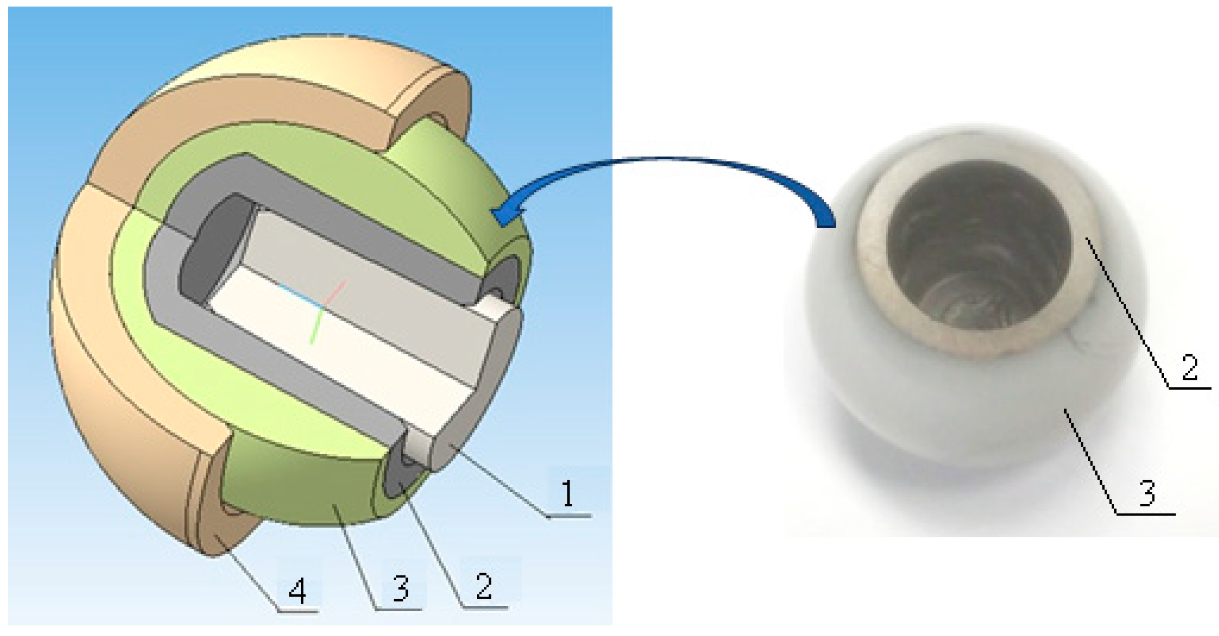
  71. Pakhaliuk, V.I.; Poliakov, A.M.; Fedotov, I.V. The ceramic modular head improvement in the design of a total hip replacement. Facta Univ. Ser. Mech. Eng. 2021, 19, 67–78. [Google Scholar] [CrossRef]
  72. Ledet, E.H.; D’Lima, D.; Westerhoff, P.; Szivek, J.A.; Wachs, R.A.; Bergmann, G. Implantable sensor technology: From research to clinical practice. J. Am. Acad. Orthop. Surg. 2012, 20, 383–392. [Google Scholar] [CrossRef]
  73. D’Lima, D.D.; Fregly, B.J.; Colwell, C.W. Implantable sensor technology: Measuring bone and joint biomechanics of daily life in vivo. Arthritis Res. Ther. 2013, 15, 203. [Google Scholar] [CrossRef]
  74. Jeyaraman, M.; Jayakumar, T.; Jeyaraman, N.; Nallakumarasamy, A. Sensor Technology in Fracture Healing. Indian J. Orthop. 2023, 57, 1196–1202. [Google Scholar] [CrossRef] [PubMed]
  75. Anderson, W.D.; Wilson, S.L.M.; Holdsworth, D.W. Development of a Wireless Telemetry Sensor Device to Measure Load and Deformation in Orthopaedic Applications. Sensors 2020, 20, 6772. [Google Scholar] [CrossRef]
  76. Shiying, H.; Taylor, S. A closed-loop inductive power control system for an instrumented strain sensing tibial implant. In Proceedings of the IEEE Engineering in Medicine and Biology Society, Chicago, IL, USA, 26–30 August 2014; pp. 6553–6556. [Google Scholar] [CrossRef]
  77. Hao, S.; Gorjon, J.; Taylor, S. SCIMITAR: Subject-carried implant monitoring inductive telemetric ambulatory reader for remote data acquisition from implanted orthopaedic prostheses. Med. Eng. Phys. 2014, 36, 405–411. [Google Scholar] [CrossRef] [PubMed]
  78. Iyengar, K.P.; Gowers, B.T.V.; Jain, V.K.; Ahluwalia, R.S.; Botchu, R.; Vaishya, R. Smart sensor implant technology in total knee arthroplasty. J. Clin. Orthop. Trauma 2021, 22, 01605. [Google Scholar] [CrossRef]
  79. Ledet, E.H.; Liddle, B.; Kradinova, K.; Harper, S. Smart implants in orthopedic surgery, improving patient outcomes: A review. Innov. Entrepreneurship Health 2018, 5, 41–51. [Google Scholar] [CrossRef] [PubMed]
  80. Sauer, S.; Marschner, U.; Jettkant, B.; Fischer, W.-J.; Clasbrummel, B. A wireless integrated hip prosthesis loosening detection system—Influence of mechanical cross-sensitivities on resonance frequencies. Biomed. Eng. 2012, 57 (SI-1-Track-S), 869. [Google Scholar] [CrossRef]
  81. Damm, P.; Dymke, J.; Bender, A.; Duda, G.; Bergmann, G. In vivo hip joint loads and pedal forces during ergometer cycling. J. Biomech. 2017, 60, 197–202. [Google Scholar] [CrossRef]
  82. Damm, P.; Schwachmeyer, V.; Dymke, J.; Bender, A.; Bergmann, G. In vivo hip joint loads during three methods of walking with forearm crutches. Clin. Biomech. 2013, 28, 530–535. [Google Scholar] [CrossRef]
  83. Bergmann, G.; Graichen, F.; Rohlmann, A.; Bender, A.; Heinlein, B.; Duda, G.N.; Heller, M.O.; Morlock, M.M. Realistic loads for testing hip implants. Biomed. Mater. Eng. 2010, 20, 65–75. [Google Scholar] [CrossRef] [PubMed]
  84. Graichen, F.; Bergmann, G.; Rohlmann, A. Hip endoprosthesis for in vivo measurement of joint force and temperature. J. Biomech. 1999, 32, 111–1117. [Google Scholar] [CrossRef] [PubMed]
  85. Damm, P.; Graichen, F.; Rohlmann, A.; Bender, A.; Bergmann, G. Total hip joint prosthesis for in vivo measurement of forces and moments. Med. Eng. Phys. 2010, 32, 95–100. [Google Scholar] [CrossRef] [PubMed]
  86. Kelmers, E.; Szuba, A.; King, S.W.; Palan, J.; Freear, S.; Pandit, H.G.; van Duren, B.H. Smart Knee Implants: An Overview of Current Technologies and Future Possibilities. Indian J. Orthop. 2022, 57, 635–642. [Google Scholar] [CrossRef] [PubMed]
  87. Moreo, P.; García-Aznar, J.M.; Doblaré, M. Bone ingrowth on the surface of endosseous implants. Part 1: Mathematical model. J. Theor. Biol. 2009, 260, 1–12. [Google Scholar] [CrossRef]
  88. Moreo, P.; García-Aznar, J.M.; Doblaré, M. Bone ingrowth on the surface of endosseous implants. Part 2: Theoretical and numerical analysis. J. Theor. Biol. 2009, 260, 13–26. [Google Scholar] [CrossRef]
Figure 1. Main components of a modular THR endoprosthesis are indicated. The acetabular component, as a rule, includes an additional element—a liner, which forms a friction pair in contact with the head.
Prosthesis 05 00071 g001
Figure 2. The scheme of texturing the surface of the femoral head in order to reduce the wear of the polymer liner: 1. Femoral head; 2. Polymer liner; 3. Cup; 4. Stem; 5. Lubrication layer.
Prosthesis 05 00071 g002
Figure 3. IR spectra of UHMWPE before (black—1) and after (red—2) treatment in helium plasma at low pressure (discharge power 50 W, helium flow5 cm3/min).
Prosthesis 05 00071 g003
Figure 4. General view and 3D-model of a cup with a modular head obtained using the technology of soldering alumina ceramics with a titanium alloy: 1. Tapered neck of a total hip joint endoprosthesis stem; 2. Titanium alloy blind hole sleeve; 3. Alumina ceramic femoral head; 4. Cup.
Prosthesis 05 00071 g004
Figure 5. Change in platelet population density ( p 1 = 0.1   m g m m 2 ; p 2 = 0.5   m g m m 2 ; x —distance from the implant surface): (a) p x = p 1 e 2 x ; (b) p x = p 2 e 2 x .
Prosthesis 05 00071 g005
Disclaimer/Publisher’s Note: The statements, opinions and data contained in all publications are solely those of the individual author(s) and contributor(s) and not of MDPI and/or the editor(s). MDPI and/or the editor(s) disclaim responsibility for any injury to people or property resulting from any ideas, methods, instructions or products referred to in the content.

