Review

Prosthodontic Treatment in Patients with Temporomandibular Disorders and Orofacial Pain and/or Bruxism: A Review of the Literature

1
Multidisciplinary Department of Medical-Surgical and Odontostomatological Specialties, University of Campania “Luigi Vanvitelli”, 80121 Naples, Italy
2
School of Dentistry Department of Biomedical and Dental Sciences and Morphofunctional Imaging, University of Messina, via Consolare Valeria, 1, 98125 Messina, Italy
3
Department of Dentistry, University of Aldent, 1000 Tirana, Albania
*
Author to whom correspondence should be addressed.
Prosthesis 2022, 4(2), 253-262; https://doi.org/10.3390/prosthesis4020025
Submission received: 22 April 2022 / Revised: 31 May 2022 / Accepted: 6 June 2022 / Published: 7 June 2022
(This article belongs to the Special Issue Feature Review Papers for Prosthesis)

Abstract

:
Temporomandibular disorders are a group of conditions affecting the temporomandibular joints, the jaw muscles, and related structures. Patients with temporomandibular signs and/or symptoms frequently present with indications for prosthetic treatment. The management of these patients aims to achieve patient comfort, occlusal stability, and the complex restoration of the teeth. The goal of this review is to provide an overview of the relationship between prosthodontics and temporomandibular disorders and/or bruxism with a focus on the cause-and-effect implications and the strategies for planning prosthetic treatments in patients with temporomandibular disorders and/or bruxism.

1. Introduction

Temporomandibular disorders (TMDs) are a heterogeneous group of conditions affecting the temporomandibular joints (TMJs), the jaw muscles, and related structures [1,2,3,4,5,6]. They have a multifactorial cause, with an interaction of systemic, psychosocial [7], genetic [8,9], trauma-related [10], hormonal [11], neurological [12,13,14], and anatomic or facial morphology factors [15,16,17,18]. The Diagnostic Criteria for Temporomandibular Disorders (DC/TMD) are employed to diagnose patients. The DC/TMD protocol comprises two domains: a physical domain in Axis I (clinical condition) and a psychosocial domain in Axis II (psychosocial distress) [19]. The clinical examination for Axis I diagnostics requires pain history, assessed by a questionnaire, and a well-defined and structured clinical examination. The criteria for DC/TMD Axis I comprise TMJ arthralgia, masticatory muscle myalgia, headache attributed to TMD, degenerative joint disease, and TMJ disc displacements. DC/TMD Axis II assesses the patient’s psychosocial function and distress as well as pain-related disability. Axis II is based on validated instruments (questionnaires) and interpretation guidelines. It includes instruments for assessing pain behavior, jaw function, and psychosocial functioning and distress. Several papers suggested that the relationship between TMDs and dental occlusion is weak [20,21]. Nevertheless, patients with TMD symptoms often need a prosthetic treatment, including partial edentulism, esthetic deficiencies, or functional problems [21,22]. Those patients should be managed carefully after a detailed evaluation [21,22,23]. Different studies introducing iatrogenic changes to dental occlusion reported some interesting considerations [20,21]. Furthermore, as far as bruxism is concerned, several systematic reviews analyzing implant-supported restorations suggest that bruxism may be associated more with mechanical than biological causes [24,25,26,27]. The aim of this review is to provide an overview of the relationship between prosthodontics and TMD with a focus on the cause-and-effect implications and the strategies for planning prosthetic treatments in patients with TMD and/or bruxism.

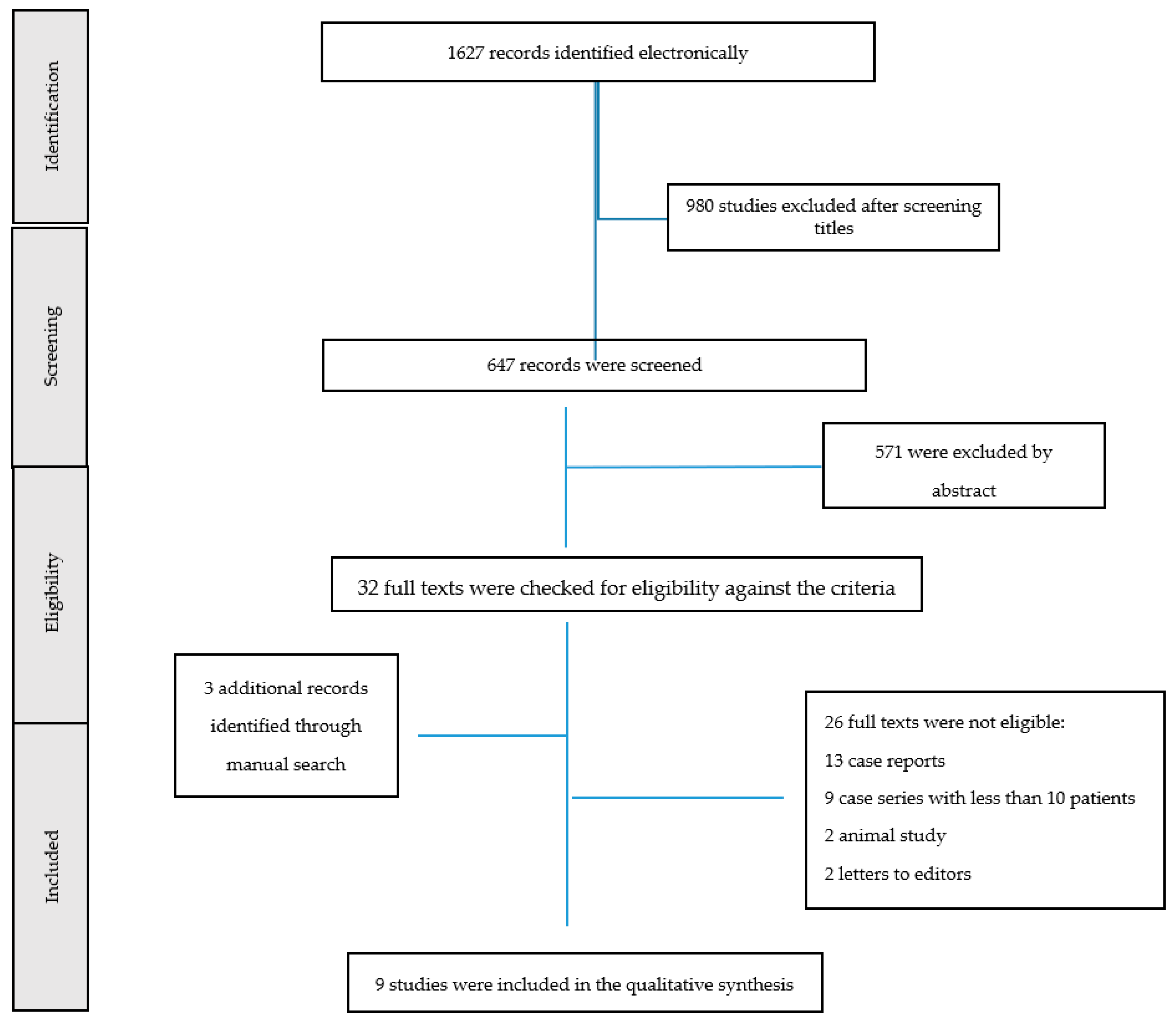
2. Materials and Methods

The literature search was conducted in the Scopus, Web of Science, and PubMed electronic databases. Document type was limited to articles written in English, without time restrictions.
The review protocol was registered with PROSPERO (registration number CRD42022326411).
The search terms included “temporomandibular disorder”, OR “TMD” OR “orofacial pain” OR “bruxism” combined with “prosthesis” OR “denture” OR “fixed prosthesis”, OR “removable prosthesis” OR “implant-supported restorations” OR “laminate veneers”.
The following inclusion criteria were used: articles in English, human studies, clinical trials, systematic and narrative review article, case series with more than 10 patients treated, and case reports. The following exclusion criteria were used: articles that did not answer the key questions, duplicate articles, books, letters to editors, and experimental studies.
The database search was further supplemented with a hand search of relevant articles in the reference lists.
A total of 1627 published articles were found from the electronic searches. Two independent reviewers (F.D. and G.M.) carried out the screening and selection process for the studies.
First, duplicate citations were removed. Then, the two authors independently reviewed the retrieved articles by the title and abstract of each citation to determine its suitability for inclusion. In the initial scan, articles were eliminated if they were clearly outside the aim of the review.
All titles were checked, and 647 articles were selected for abstract reading. Then, following the analysis of the abstracts, 571 articles that did not satisfy the eligibility criteria were excluded. Thus, 32 full-text articles were identified. In addition, checking the reference lists of the most recent systematic reviews produced three full-text studies, resulting in a total of 35 articles. Finally, 9 full-text articles satisfied the inclusion criteria. The features of the included studies are described in Table 1.
The authors’ decisions to include/exclude each article were compared. Discrepancies were discussed and an agreement was finally reached. The flowchart of data selection is shown in Figure 1.
The quality of the included studies was evaluated independently by two reviewers (F.D. and A.L.). The quality of non-randomized clinical studies was assessed using the Newcastle–Ottawa Scale (NOS), as shown in Table 2 [28]. This scale uses a star system by which a study is judged on three broad aspects: the selection of the study groups (up to 4 points), the comparability of the groups (up to 2 points), and the exposure or outcome of interest for case–control or cohort studies, respectively (up to 3 points). Studies that met five or more of the NOS score criteria were considered as good quality and were included in the study. For other types of studies, the quality assessment was evaluated using a tool focusing on eight items developed by den Hartog et al. [29], as shown in Table 3. The studies scoring five or more plus signs were considered acceptable.

3. Results

3.1. Prosthodontic Treatment in Patients Affected by TMDs

Prosthetic treatments often produce a change in the interarch relationship, thus potentially requiring more adjustment. Changes in the interarch relationship may be induced by increases in the occlusal vertical dimension of occlusion (VDO) and mandible repositioning treatments [30]. The occlusal vertical dimension represents the distance between two anatomical or marked points in maximal intercuspal position. An increase in VDO might cause clinical drawbacks, such as elevation of bite forces, muscle hypersensitivity, symptoms of temporomandibular disorders, phonetic limitations, and teeth tenderness [31]. Some prosthetic therapies often need to increase the interarch distance and then modify the VDO in order to obtain optimal outcomes [32]. Several authors have showed that the VDO is a fixed and specific parameter that cannot be modified. Moreover, the increasing or decreasing of the VDO could cause serious problems such as muscle pain, temporomandibular joint (TMJ) disorders, headaches, tooth grinding, and clenching [33,34,35]. A recent literature review analyzing the association between TMJ disorders and modification of the VDO concluded that many commonly held concepts related to this topic were not supported by scientific evidence and that more studies are necessary to understand this relationship more accurately [32]. To date, there is a lack of evidence concerning the incidence of prosthetic and functional complications in patients treated with a VDO increase by means of teeth-supported, mixed, and implant-supported restorations [33,34]. However, the masticatory system has an excellent ability of adjustment, both to natural dental-skeletal irregularities and to iatrogenic variations [24]. Therefore, the safest prosthodontic strategy against the possible beginning of TMD symptoms is not to plan occlusal modifications that can negatively influence the capacity for accommodation. Rehabilitations based on organized occlusal schemes or interarch relations are not recommended, since they do not consider the muscle engrams and the functional adaptation that the neuromuscular system of an asymptomatic patient has created naturally [1]. The role of occlusion as a risk factor for temporomandibular disorders (TMDs) is controversial [21]. Nowadays, studies suggest that the cause of TMDs is less linked to occlusal morphology [23,36]. However, irreversible occlusal changes of prosthodontic or orthodontic rehabilitations cannot be recommended for the management or even the prevention of such conditions [21]. In healthy individuals, the placement of a restoration in supraocclusion can cause only a local lesion such as transient dental and/or muscle pain and can be solved through the removal of the interference [23,37]. Furthermore, there is a reduction in the EMG activity of the masseter muscles and no effect on the pressure pain threshold [10,17]. This indicates the establishment of an avoidance adaptation pattern, as confirmed by clinical observations that patients are not able to chew on restorations in supraocclusion and try to avoid contacts with that tooth. Thus, TMD cannot be triggered by iatrogenic changes to dental occlusion [21]. Interestingly, patients with a history of TMD may suffer an increased risk for palpation-elicited muscle pain in response to artificially introduced occlusal interferences. This concept should be highlighted when carrying out rehabilitation treatments involving periods of occlusal instability due, for example, to interim restorations, increases in VDO, or shifting of teeth [11,21]. Several epidemiological studies reported TMD in complete denture wearers, highlighting that occlusal instability was one of the potential factors contributing to the development of TMD among complete denture wearers; in particular, the TMD signs and symptoms were correlated with the quality of the dentures and the denture wearing habits [38,39,40,41]. It has also been suggested that incorrect vertical dimension and centric relation were the most frequent causes of TMD [38]. Despite the multifactorial character of TMD and the controversial role of occlusal factors, some authors consider that it has been suggested to remodel poor dentures in denture-wearing patients affected by TMD [9,38,42]. Then, fitting new complete dentures had positive effects on the signs and symptoms of TMD. Reviewing the dental literature revealed that the influence of condylar disc position and defective occlusion on TMD remains a controversial issue, as does the influence of replacement by a new removable prosthesis [38]. Abdelnabi et al. showed that new dentures with corrected occlusion significantly improved clinical signs and symptoms of TMD in complete denture wearers and disc position [38]. Moreover, new complete dentures can also significantly reduce MRI signal intensification that corresponds to joint effusion [18].
In routine clinical practice, the existence of clicking disc displacement sounds does not represent a contraindication to occlusal rehabilitations. On the contrary, in patients with open-ended TMDs, their symptoms should be treated before beginning any prosthetic treatment. Patients affected by TMDs are very sensitive to stressors and may thus adapt less easily than healthy patients to the occlusal and psychological stress of a modification to their occlusal scheme because of their delicate psychophysiological equilibrium [18]. As far as the restorative materials are concerned, the literature does not support any clinical evidence [23,37]. The choice of material for an extensive rehabilitation in patients with TMDs is often based on the clinician’s predilections and patient expectations. No evidence-based recommendations are available on how to perform the increasing of the original VDO in patients affected by bruxism or TMD [18,43]. The presence of severe tooth wear preventing retentive crown preparations, insufficient interarch space to restore or replace missing teeth, and esthetic reasons can represent prosthetic reasons which cause positional changes of the mandible and/or an increase in the vertical dimension of occlusion (VDO) [44,45]. The occlusal pattern of extensive rehabilitations in patients with bruxism or TMDs should be as simple as possible. Basic requisites such as a symmetrical distribution of interarch contacts, occlusal stability, and subjective comfort are generally enough to optimize function [18]. The reproducibility of any centric relation is not mandatory, and there is evidence that high neuroplasticity-based adaptability of the system is better supported rather than any real functional advantages of centric relation [46].

3.2. Prosthodontic Treatment in Patients Affected by Bruxism

Among the temporomandibular disorders and prosthodontics, bruxism needs to be evaluated in more detail. According to the American Academy of Orofacial Pain, bruxism is a diurnal or nocturnal parafunctional activity (recognized as non-functional jaw movements) which includes bracing, clenching, gnashing, and grinding of the teeth [11,47]. The biological hypothesis that a centrally mediated phenomenon such as bruxism may be caused by a prosthetic treatment is nonexistent [48]. Currently, no specific treatment exists that can stop bruxism, although a lot of treatments, including prosthetic treatment, have been tried over the years. Conversely, various treatments have been suggested based on behavior modification, such as habit awareness, habit reversal therapy, and relaxation techniques, which may eliminate awake bruxism [11]. Patients affected by bruxism can be treated with occlusal splints. In general, the occlusal splint is used to treat muscle hyperactivity [8,49,50]. Several studies showed that these splints can decrease bruxism activity generated during periods of stress [12,46,47]. Therefore, the use of these devices is recommended in patients with suspected bruxism following prosthodontic treatment including full coverage crowns or with laminate veneers [47,48,49,50,51,52,53]. A functional design should have restorations installed in patients affected by some type of bruxism activity, especially in conditions where the patient has already lost some tooth structure. Furthermore, these restorations should be performed according to a correct anterior and canine guidance [47,54,55]. Some authors suggested that bruxism can represent a contraindication to these bonded restorations [47,56]. Granell-Ruíz et al. showed that the success rate for veneers is reduced to 60% in patients with bruxism activity [47]. Similarly, some studies showed this percentage for metal–ceramic restorations in the same clinical conditions [47,56]. If bruxism is controlled, the success rates may increase. Therefore, a preventive measure such as a nocturnal and/or diurnal splint is recommended in order to reduce the risk of failure, especially in patients affected by bruxism [57,58,59]. Studies analyzing the materials employed in patients treated with fixed dental prosthesis are scarce, and the choice often needs to be made on the basis of clinical experience rather than scientific data. With an opposing natural tooth, most clinicians agree that a metal occlusal surface, preferably one of high noble matter, should be selected in order to minimize wear of the natural dentition. The use of ceramics could be especially dangerous to opposing natural teeth. However, new ceramics such as zirconia have demonstrated improved mechanical properties in laboratory studies and may be promising in the treatment of bruxism-related tooth wear [60,61]. However, a systematic review of zirconia fixed dental prostheses (FDPs) has shown that complications can occur when clinicians meet realistic technical and clinical situations [62]. As for the relationship between implant-supported restorations and bruxism, several studies analyzing complications with implant-supported restorations suggested that bruxism may be associated more with mechanical causes (screw loosening, ceramic fracture or chipping, and abutment or fixture fracture) than biological (loss of marginal bone attachment) ones [55,59,63]. Clinical strategies can be engaged in order to decrease mechanical trauma, such as reducing cusp steepness, enlarging contact areas, and providing slight occlusal under-contact. From a practical viewpoint, the clinician should adopt a safe and cautionary approach to a patient with bruxism who needs a prosthetic treatment by employing strategies in order to reduce the potentially negative effects of bruxism [20]. These include both prosthetic (occlusal design) and surgical (number, size, and location of implants) aspects [22,26]. Connection between implants may provide a better load distribution and reduce peri-implant bone stress, and cantilevers and immediate-loading protocols should be avoided [26]. In addition, flatter cuspal planes are recommended in order to obtain a positive liberty of movements around the occlusal contact areas in maximum intercuspation and to protect the prosthesis during eccentric movements.

4. Discussion

The relationship between temporomandibular disorders and bruxism and prosthodontics is a topic requiring further discussion. The question of whether occlusion can represent a risk factor for temporomandibular disorders is controversial. Patients with temporomandibular disorders may adapt less easily than healthy patients to the occlusal and psychological stress of a modification to their occlusal scheme because of their delicate psychophysiological equilibrium. Thus, patients affected by TMDs or bruxism need a simple occlusal design. The biological hypothesis that a centrally mediated phenomenon such as bruxism may be caused by a prosthetic treatment is nonexistent. Restorations placed in patients affected by some type of bruxism activity should have a functional design, especially in circumstances where the patient has already lost some tooth structure and where these restorations support the patient. Patients with implant-supported restorations affected by bruxism may run into mechanical complications; thus, the choice of a metal occlusal surface as the material, connection between implants, and flatter cuspal planes are recommended, while cantilevers and immediate-loading protocols should be avoided [64].

5. Conclusions

The relationship between temporomandibular disorders and prosthesis requires further discussion. The role of occlusion as a risk factor for TMD is controversial. Studies analyzing the incidence of prosthetic and functional complications in patients treated with a VDO increase in teeth-supported, mixed, and implant-supported restorations should be conducted. On the other hand, patients affected by bruxism can be treated with occlusal splints, and a functional design of the restorations is recommended. Furthermore, prosthetic (occlusal design) and surgical (number, size, and location of implants) aspects have to be considered for implant rehabilitation in patients affected by bruxism.

Author Contributions

Conceptualization, A.L. and G.M.; methodology, F.D.F.; software, D.R.; validation, C.D. and A.M.; writing—original draft preparation, G.M.; writing—review and editing, F.D.F.; visualization, D.R.; supervision, L.F.; project administration, G.C. All authors have read and agreed to the published version of the manuscript.

Funding

This research received no external funding.

Institutional Review Board Statement

Not applicable.

Informed Consent Statement

Not applicable.

Data Availability Statement

Data are available on request to the authors.

Conflicts of Interest

The authors declare no conflict of interest.

Abbreviations

TMDTemporomandibular disorder
TMJTemporomandibular joint
EMGElectromyography
MRIMagnetic resonance imaging
VDOVertical dimension of occlusion
DC/TMDDiagnostic Criteria for Temporomandibular Disorders
FDPsFixed dental prostheses

References

  1. Manfredini, D.; Lombardo, L.; Siciliani, G. Temporomandibular disorders and dental occlusion. A systematic review of association studies: End of an era? J. Oral Rehabil. 2017, 44, 908–923. [Google Scholar] [CrossRef]
  2. Racich, M.J. Occlusion, temporomandibular disorders, and orofacial pain: An evidence-based overview and update with recommendations. J. Prosthet. Dent. 2018, 120, 678–685. [Google Scholar] [CrossRef]
  3. Moccia, S.; Nucci, L.; Spagnuolo, C.; d’Apuzzo, F.; Grazia, M.; Minervini, G. Polyphenols as potential agents in the management of temporomandibular disorders. Appl. Sci. 2020, 10, 5305. [Google Scholar] [CrossRef]
  4. Ingawalé, S.; Goswami, T. Temporomandibular joint: Disorders, treatments, and biomechanics. Ann. Biomed. Eng. 2009, 37, 976–996. [Google Scholar] [CrossRef] [PubMed]
  5. Zhang, Y.; Zhang, J.; Wang, L.; Wang, K.; Svensson, P. Effect of transcutaneous electrical nerve stimulation on jaw movement-evoked pain in patients with TMJ disc displacement without reduction and healthy controls. Acta Odontol. Scand. 2020, 78, 309–320. [Google Scholar] [CrossRef] [PubMed]
  6. Tiirp, J.C.; Dent, M.; Strub, J.R.; Dentb, M. Prosthetic rehabilitation in patients with temporomandibular disorders. J. Prosthet. Dent. 1996, 76, 418–423. [Google Scholar] [CrossRef]
  7. Minervini, G.; Nucci, L.; Lanza, A.; Femiano, F.; Contaldo, M.; Grassia, V. Temporomandibular disc displacement with reduction treated with anterior repositioning splint: A 2-year clinical and magnetic resonance imaging (MRI) follow-up. J. Biol. Regul. Homeost. Agents 2020, 34 (Suppl. 1), 151–160. [Google Scholar] [PubMed]
  8. Manfredini, D.; Ahlberg, J.; Aarab, G.; Bracci, A.; Durham, J.; Ettlin, D.; Gallo, L.M.; Koutris, M.; Wetselaar, P.; Svensson, P.; et al. Towards a Standardized Tool for the Assessment of Bruxism (STAB)—Overview and general remarks of a multidimensional bruxism evaluation system. J. Oral Rehabil. 2020, 47, 549–556. [Google Scholar] [CrossRef]
  9. Al-Jabrah, O.A.; Al-Shumailan, Y.R. Prevalence of temporomandibular disorder signs in patients with complete versus partial dentures. Clin. Oral Investig. 2006, 10, 167–173. [Google Scholar] [CrossRef] [PubMed]
  10. Minervini, A.L.G.; Romano, A.; Petruzzi, M.; Maio, C.; Serpico, R.; di Stasio, D. Oral-Facial-Digital Syndrome (OFD): 31-Year follow-up management and monitoring. J. Biol. Regul. Homeost. Agents 2018, 32, 127–130. [Google Scholar]
  11. Johansson, A.; Omar, R.; Carlsson, G.E. Bruxism and prosthetic treatment: A critical review. J. Prosthodont. Res. 2011, 55, 127–136. [Google Scholar] [CrossRef] [Green Version]
  12. Minervini, G.; Lucchese, A.; Perillo, L.; Serpico, R.; Minervini, G. Unilateral superior condylar neck fracture with dislocation in a child treated with an acrylic splint in the upper arch for functional repositioning of the mandible. Cranio-J. Craniomandib. Pract. 2017, 35, 337–341. [Google Scholar] [CrossRef] [PubMed]
  13. Carlsson, G.E. Some dogmas related to prosthodontics, temporomandibular disorders and occlusion. Acta Odontol. Scand. 2010, 68, 313–322. [Google Scholar] [CrossRef]
  14. Bousema, E.J.; Koops, E.A.; van Dijk, P.; Dijkstra, P.U. Association between Subjective Tinnitus and Cervical Spine or Temporomandibular Disorders: A Systematic Review. Trends Hear. 2018, 22, 2331216518800640. [Google Scholar] [CrossRef] [PubMed]
  15. Deregibus, A.; Ferrillo, M.; Piancin, M.G.; Domini, M.C.; de Sire, A.; Castroflorio, T. Are occlusal splints effective in reducing myofascial pain in patients with muscle-related temporomandibular disorders? A randomized-controlled trial. Turk. J. Phys. Med. Rehabil. 2021, 67, 32–40. [Google Scholar] [CrossRef] [PubMed]
  16. de Sire, A.; Marotta, N.; Ferrillo, M.; Agostini, F.; Sconza, C.; Lippi, L.; Respizzi, S.; Giudice, A.; Invernizzi, M.; Ammendolia, A. Oxygen-Ozone Therapy for Reducing Pro-Inflammatory Cytokines Serum Levels in Musculoskeletal and Temporomandibular Disorders: A Comprehensive Review. Int. J. Mol. Sci. 2022, 23, 2528. [Google Scholar] [CrossRef]
  17. Minervini, G.; Russo, D.; Herford, A.S.; Gorassini, F.; Meto, A.; D’Amico, C.; Cervino, G.; Cicciù, M.; Fiorillo, L. Teledentistry in the Management of Patients with Dental and Temporomandibular Disorders. BioMed Res. Int. 2022, 2022, 7091153. [Google Scholar] [CrossRef] [PubMed]
  18. Manfredini, D.; Castroflorio, T.; Perinetti, G.; Guarda-Nardini, L. Dental occlusion, body posture and temporomandibular disorders: Where we are now and where we are heading for. J. Oral Rehabil. 2012, 39, 463–471. [Google Scholar] [CrossRef]
  19. Schiffman, E.; Ohrbach, R.; Truelove, E.; Look, J.; Anderson, G.; Goulet, J.-P.; List, T.; Svensson, P.; Gonzalez, Y.; Lobbezoo, F.; et al. Diagnostic Criteria for Temporomandibular Disorders (DC/TMD) for Clinical and Research Applications: Recommendations of the International RDC/TMD Consortium Network* and Orofacial Pain Special Interest Group†. J. Oral Facial Pain Headache 2014, 28, 6–27. [Google Scholar] [CrossRef]
  20. di Francesco, F.; de Marco, G.; Capcha, E.B.; Lanza, A.; Cristache, C.M.; Vernal, R.; Cafferata, E.A. Patient satisfaction and survival of maxillary overdentures supported by four or six splinted implants: A systematic review with meta-analysis. BMC Oral Health 2021, 21, 247. [Google Scholar] [CrossRef] [PubMed]
  21. Manfredini, D.; Poggio, C.E. Prosthodontic planning in patients with temporomandibular disorders and/or bruxism: A systematic review. J. Prosthet. Dent. 2017, 117, 606–613. [Google Scholar] [CrossRef]
  22. Sessle, B.J. Mechanisms of oral somatosensory and motor functions and their clinical correlates. J. Oral Rehabil. 2006, 33, 243–261. [Google Scholar] [CrossRef]
  23. Carlsson, G.E. Dental occlusion: Modern concepts and their application in implant prosthodontics. Odontology 2009, 97, 8–17. [Google Scholar] [CrossRef]
  24. Levartovsky, S.; Pilo, R.; Shadur, A.; Matalon, S.; Winocur, E. Complete rehabilitation of patients with bruxism by veneered and non-veneered zirconia restorations with an increased vertical dimension of occlusion: An observational case-series study. J. Prosthodont. Res. 2019, 63, 440–446. [Google Scholar] [CrossRef] [PubMed]
  25. Chatzopoulos, G.S.; Wolff, L.F. Symptoms of temporomandibular disorder, self-reported bruxism, and the risk of implant failure: A retrospective analysis. Cranio-J. Craniomandib. Pract. 2020, 38, 50–57. [Google Scholar] [CrossRef]
  26. di Francesco, F.; de Marco, G.; Sommella, A.; Lanza, A. Splinting vs Not Splinting Four Implants Supporting a Maxillary Overdenture: A Systematic Review. Int. J. Prosthodont. 2019, 32, 509–518. [Google Scholar] [CrossRef]
  27. di Francesco, F.; de Marco, G.; Carnevale, U.A.G.; Lanza, M.; Lanza, A. The number of implants required to support a maxillary overdenture: A systematic review and meta-analysis. J. Prosthodont. Res. 2019, 63, 15–24. [Google Scholar] [CrossRef] [PubMed]
  28. Wells, G.A.; Shea, B.; O’Connell, D.; Peterson, J.; Welch, V.; Losos, M.; Tugwell, P. The Newcastle-Ottawa Scale (NOS) for Assessing the Quality of Nonrandomised Studies in Meta-Analyses. 2019. Available online: http://www.ohri.ca/programs/clinical_epidemiology/oxford.asp (accessed on 12 February 2022).
  29. den Hartog, L.; Slater, J.J.R.H.; Vissink, A.; Meijer, H.J.A.; Raghoebar, G.M. Treatment outcome of immediate, early and conventional single-tooth implants in the aesthetic zone: A systematic review to survival, bone level, soft-tissue, aesthetics and patient satisfaction. J. Clin. Periodontol. 2008, 35, 1073–1086. [Google Scholar] [CrossRef] [PubMed]
  30. Calamita, M.; Coachman, C.; Sesma, N.; Kois, J. Occlusal vertical dimension: Treatment planning decisions and management considerations. Int. J. Esthet. Dent. 2019, 14, 166–181. [Google Scholar]
  31. Fabbri, G.; Sorrentino, R.; Cannistraro, G.; Mintrone, F.; Bacherini, L.; Turrini, R.; Bombardelli, T.; Nieri, M.; Fradeani, M. Increasing the Vertical Dimension of Occlusion: A Multicenter Retrospective Clinical Comparative Study on 100 Patients with Fixed Tooth-Supported, Mixed, and Implant-Supported Full-Arch Rehabilitations. Int. J. Periodontics Restor. Dent. 2018, 38, 323–335. [Google Scholar] [CrossRef] [PubMed]
  32. Alhajj, M.N.; Khalifa, N.; Abduo, J.; Amran, A.G.; Ismail, I.A. Determination of occlusal vertical dimension for complete dentures patients: An updated review. J. Oral Rehabil. 2017, 44, 896–907. [Google Scholar] [CrossRef]
  33. Moreno-Hay, I.; Okeson, J.P. Does altering the occlusal vertical dimension produce temporomandibular disorders? A literature review. J. Oral Rehabil. 2015, 42, 875–882. [Google Scholar] [CrossRef]
  34. Abduo, J. Safety of increasing vertical dimension of occlusion: A systematic review. Quintessence Int. 2012, 43, 369–380. [Google Scholar] [PubMed]
  35. Vailati, F.; Carciofo, S. Treatment planning of adhesive additive rehabilitations: The progressive wax-up of the three-step technique. Int. J. Esthet. Dent. 2016, 11, 356–377. [Google Scholar]
  36. Bourdiol, P.; Hennequin, M.; Peyron, M.-A.; Woda, A. Masticatory Adaptation to Occlusal Changes. Front. Physiol. 2020, 11, 263. [Google Scholar] [CrossRef] [Green Version]
  37. Johansson, A.; Johansson, A.-K.; Omar, R.; Carlsson, G.E. Rehabilitation of the worn dentition. J. Oral Rehabil. 2008, 35, 548–566. [Google Scholar] [CrossRef]
  38. Abdelnabi, M.H.; Swelem, A.A. Influence of defective complete dentures renewal on TMD; An MRI and clinical controlled prospective study. Gerodontology 2015, 32, 211–221. [Google Scholar] [CrossRef] [PubMed]
  39. Poštić, S.D. Specific occlusal scheme for partially edentulous patients with TMD signs-preliminary report. J. Stomatol. Oral Maxillofac. Surg. 2018, 119, 337–347. [Google Scholar] [CrossRef]
  40. Fiorillo, L.; D’Amico, C.; Turkina, A.Y.; Nicita, F.; Amoroso, G.; Risitano, G. Endo and Exoskeleton: New Technologies on Composite Materials. Prosthesis 2020, 2, 1–9. [Google Scholar] [CrossRef] [Green Version]
  41. Sreekumar, S.; Janakiram, C.; Mathew, A. Effects of Prosthetic Rehabilitation on Temporomandibular Disorders: Protocol for a Randomized Controlled Trial. JMIR Res. Protoc. 2021, 10, e33104. [Google Scholar] [CrossRef] [PubMed]
  42. D’Amico, C.; Bocchieri, S.; Sambataro, S.; Surace, G.; Stumpo, C.; Fiorillo, L. Occlusal Load Considerations in Implant-Supported Fixed Restorations. Prosthesis 2020, 2, 252–265. [Google Scholar] [CrossRef]
  43. Ribeiro, J.A.M.; de Resende, C.M.B.M.; Lopes, A.L.C.; Farias-Neto, A.; Carreiro, A.D.F.P. Association between prosthetic factors and temporomandibular disorders in complete denture wearers. Gerodontology 2014, 31, 308–313. [Google Scholar] [CrossRef]
  44. Muts, E.J.; van Pelt, H.; Edelhoff, D.; Krejci, I.; Cune, M. Tooth wear: A systematic review of treatment options. J. Prosthet. Dent. 2014, 112, 752–759. [Google Scholar] [CrossRef]
  45. Yılmaz, D.; Kamburoğlu, K. Comparison of the effectiveness of high resolution ultrasound with MRi in patients with temporomandibular joint dısorders. Dentomaxillofac. Radiol. 2019, 48, 20180349. [Google Scholar] [CrossRef]
  46. de Boever, J.A.; Carlsson, G.E.; Klineberg, I.J. Need for occlusal therapy and prosthodontic treatment in the management of temporomandibular disorders. Part II. Tooth loss and prosthodontic treatment. J. Oral Rehabil. 2000, 27, 647–659. [Google Scholar] [CrossRef]
  47. Granell-Ruíz, M.; Agustín-Panadero, R.; Fons-Font, A.; Román-Rodríguez, J.L.; Solá-Ruíz, M.F. Influence of bruxism on survival of porcelain laminate veneers. Med. Oral Patol. Oral Cir. Bucal 2014, 19, e426–e432. [Google Scholar] [CrossRef] [PubMed]
  48. Lobbezoo, F.; Ahlberg, J.; Manfredini, D.; Winocur, E. Are bruxism and the bite causally related? J. Oral Rehabil. 2012, 39, 489–501. [Google Scholar] [CrossRef] [PubMed]
  49. Botelho, J.; Machado, V.; Proença, L.; Rua, J.; Martins, L.; Alves, R.; Cavacas, M.A.; Manfredini, D.; Mendes, J.J. Relationship between self-reported bruxism and periodontal status: Findings from a cross-sectional study. J. Periodontol. 2020, 91, 1049–1056. [Google Scholar] [CrossRef] [PubMed]
  50. Huhtela, O.; Näpänkangas, R.; Joensuu, T.; Raustia, A.; Kunttu, K.; Sipilä, K. Self-Reported Bruxism and Symptoms of Temporomandibular Disorders in Finnish University Students. J. Oral Facial Pain Headache 2016, 30, 311–317. [Google Scholar] [CrossRef] [PubMed]
  51. Koenig, V.; Wulfman, C.; Bekaert, S.; Dupont, N.; le Goff, S.; Eldafrawy, M.; Vanheusden, A.; Mainjot, A. Clinical behavior of second-generation zirconia monolithic posterior restorations: Two-year results of a prospective study with Ex vivo analyses including patients with clinical signs of bruxism. J. Dent. 2019, 91, 103229. [Google Scholar] [CrossRef] [PubMed]
  52. Brignardello-Petersen, R. There are probably similar outcomes between a glass-hybrid restorative and a nanoceramic composite resin after 2 years of restoring noncarious cervical lesions in patients with bruxism. J. Am. Dent. Assoc. 2020, 151, e6. [Google Scholar] [CrossRef]
  53. Contaldo, M.; Serpico, R.; Lucchese, A. In vivo imaging of enamel by reflectance confocal microscopy (RCM): Non-invasive analysis of dental surface. Odontology 2014, 102, 325–329. [Google Scholar] [CrossRef]
  54. Gentile, E.; di Stasio, D.; Santoro, R.; Contaldo, M.; Salerno, C.; Serpico, R.; Lucchese, A. In Vivo Microstructural Analysis of Enamel in Permanent and Deciduous Teeth. Ultrastruct. Pathol. 2015, 39, 131–134. [Google Scholar] [CrossRef] [Green Version]
  55. Chrcanovic, B.R.; Albrektsson, T.; Wennerberg, A. Bruxism and dental implants: A meta-analysis. Implant. Dent. 2015, 24, 505–516. [Google Scholar] [CrossRef]
  56. Faus-Matoses, V.; Ruiz-Bell, E.; Faus-Matoses, I.; Özcan, M.; Salvatore, S.; Faus-Llácer, V.J. An 8-year prospective clinical investigation on the survival rate of feldspathic veneers: Influence of occlusal splint in patients with bruxism. J. Dent. 2020, 99, 103352. [Google Scholar] [CrossRef]
  57. Cicciù, M.; Cervino, G.; Terranova, A.; Risitano, G.; Raffaele, M.; Cucinotta, F.; Santonocito, D.; Fiorillo, L. Prosthetic and Mechanical Parameters of the Facial Bone under the Load of Different Dental Implant Shapes: A Parametric Study. Prosthesis 2019, 1, 41–53. [Google Scholar] [CrossRef] [Green Version]
  58. Manfredini, D.; Poggio, C.E.; Lobbezoo, F. Is Bruxism a Risk Factor for Dental Implants? A Systematic Review of the Literature. Clin. Implant. Dent. Relat. Res. 2014, 16, 460–469. [Google Scholar] [CrossRef]
  59. Manfredini, D.; Ahlberg, J.; Mura, R.; Lobbezoo, F. Bruxism is unlikely to cause damage to the periodontium: Findings from a systematic literature assessment. J. Periodontol. 2015, 86, 546–555. [Google Scholar] [CrossRef]
  60. Ortorp, A.; Kihl, M.L.; Carlsson, G.E. A 3-year retrospective and clinical follow-up study of zirconia single crowns performed in a private practice. J. Dent. 2009, 37, 731–736. [Google Scholar] [CrossRef]
  61. Koutayas, S.O.; Vagkopoulou, T.; Pelekanos, S.; Koidis, P.; Strub, J.R. Zirconia in dentistry: Part 2. Evidence-based clinical breakthrough. Eur. J. Esthet. Dent. 2009, 4, 348–380. [Google Scholar]
  62. Schley, J.-S.; Heussen, N.; Reich, S.; Fischer, J.; Haselhuhn, K.; Wolfart, S. Survival probability of zirconia-based fixed dental prostheses up to 5 yr: A systematic review of the literature. Eur. J. Oral Sci. 2010, 118, 443–450. [Google Scholar] [CrossRef] [PubMed]
  63. Contaldo, M.; della Vella, F.; Raimondo, E.; Minervini, G.; Buljubasic, M.; Ogodescu, A.; Sinescu, C.; Serpico, R. Early Childhood Oral Health Impact Scale (ECOHIS): Literature review and Italian validation. Int J. Dent. Hyg. 2020, 18, 396–402. [Google Scholar] [CrossRef] [PubMed]
  64. Chrcanovic, B.R.; Kisch, J.; Albrektsson, T.; Wennerberg, A. Bruxism and dental implant failures: A multilevel mixed effects parametric survival analysis approach. J. Oral Rehabil. 2016, 43, 813–823. [Google Scholar] [CrossRef] [PubMed]
Figure 1. Flow diagram of the selection of the studies.
Prosthesis 04 00025 g001
Table 1. Features of included studies.
AuthorYearStudy DesignTopic
Levartovsky et al.2019RetrospectiveTMD/Bruxism
Ribeiro et al.2014RetrospectiveTMD
Yilmaz et al.2019ProspectiveTMD
Al-Jabrah et al.2006RetrospectiveTMD
Granell-Ruíz et al.2014ProspectiveBruxism
Koenig et al.2019ProspectiveBruxism
Brignardello et al.2020RetrospectiveBruxism
Faus-Matoses et al.2020RetrospectiveTMD
Ortorp et al.2009RetrospectiveBruxism
Table 2. Quality of included studies using Newcastle–Ottawa scale (NOS). Studies that met five or more of the NOS score criteria were considered as good quality (the number of * indicate the maximum score of each column accordingly to NOS questionnaire).
StudySelection ****Comparability **Outcome ***Score
Levartovsky et al.********8
Yilmaz et al.********8
Koenig et al.********8
Brignardello et al.*******7
Faus-Matoses et al.********8
Table 3. Quality of included studies using a tool developed by den Hartog et al., focusing on eight items. Studies scoring five or more plus signs were considered acceptable.
Study1. Are the Characteristics of the Study Group Clearly Described? 2. Is there a High Risk of Selection Bias? Are the Inclusion and Exclusion Criteria Clearly Described? 3. Is the Intervention Clearly Described? Are all Patients Treated According to the Same Intervention? 4. Are the Outcomes Clearly Described? Are Adequate Methods Used to Assess the Outcome?5. Is Blinding Used to Assess the Outcome?6. Is there a Sufficient Follow-Up?7. Can Selective Loss-to Follow-Up Sufficiently Be Excluded?8. Are the Most Important Confounders or Prognostic Factors Identified and Are these Taken into Consideration with Respect to the Study Design and Analysis?
Ribeiro et al.+++++++
Granell-Ruíz et al.+++++++
Ortorp et al.++++?++
Al-Jabrah et al.+++++++
Publisher’s Note: MDPI stays neutral with regard to jurisdictional claims in published maps and institutional affiliations.

