
Biofabrication of Silver Nanoparticles Using Teucrium Apollinis Extract: Characterization, Stability, and Their Antibacterial Activities
Abstract
:1. Introduction
2. Materials and Methods
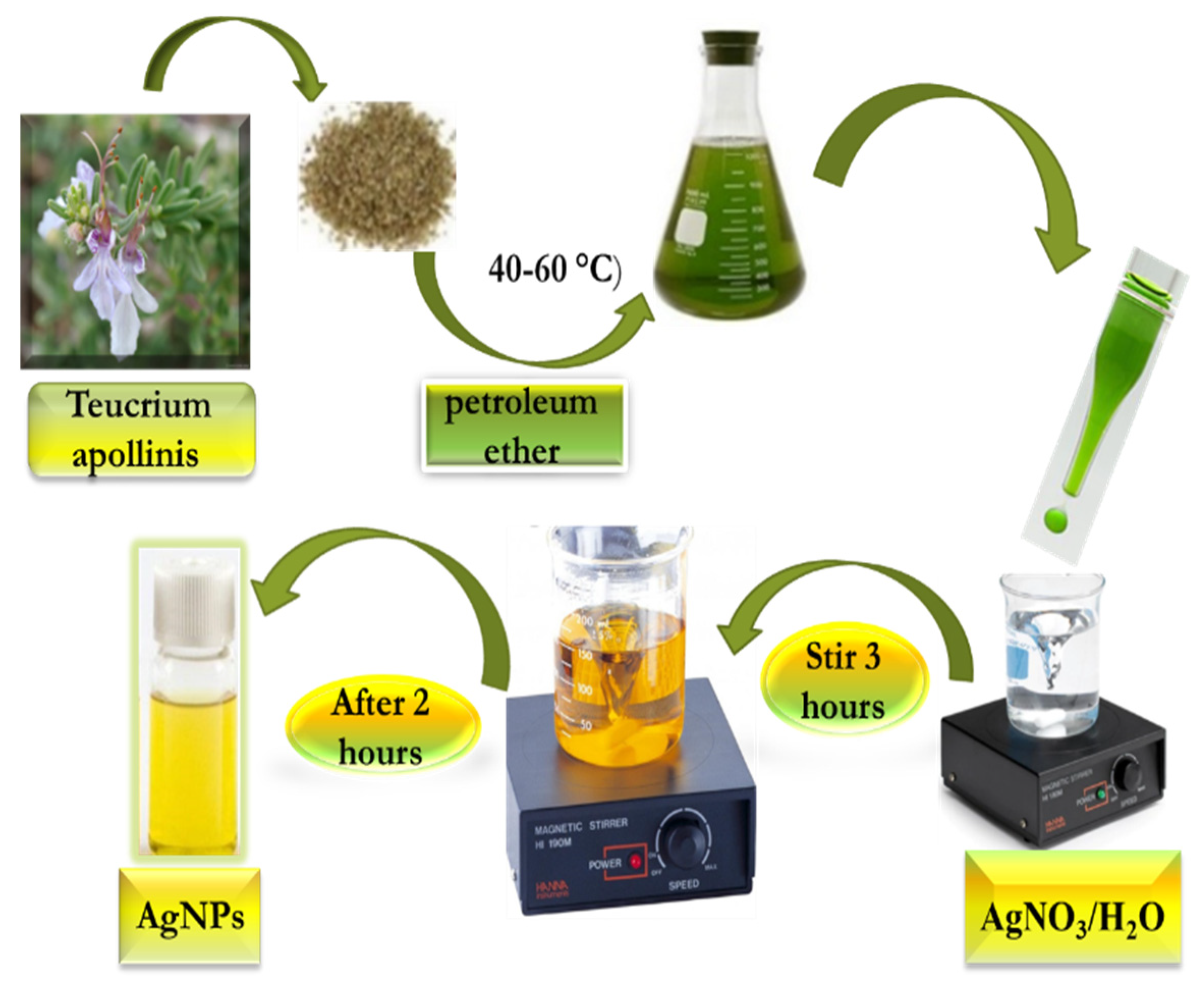
2.1. Green Synthesis of AgNP by Using Teucrium Apollinis Extract
2.1.1. Teucrium apollinis Extraction Procedure
2.1.2. Synthesis of Silver Nanoparticles
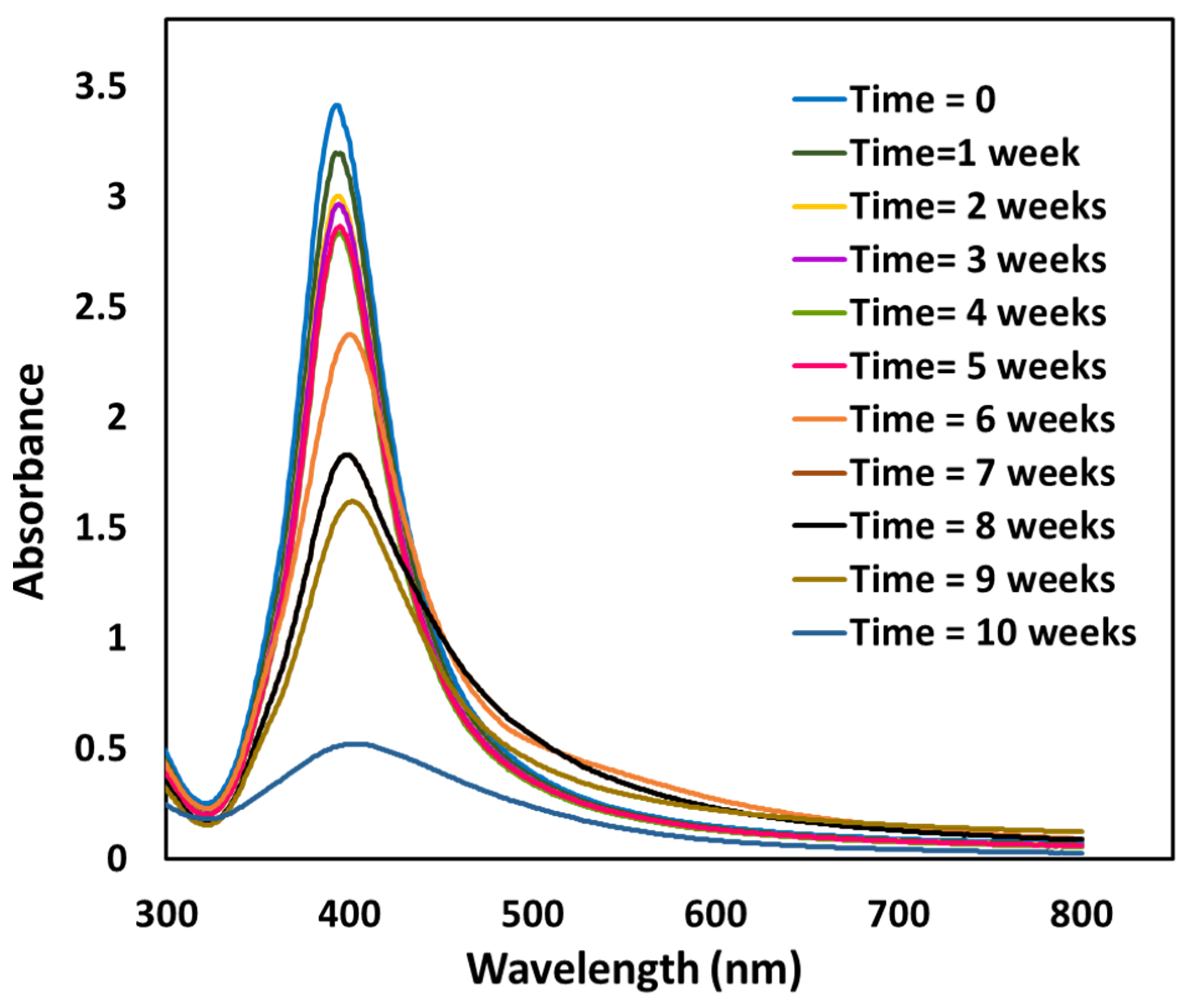
2.2. UV-Visible Spectroscopic Investigation
Study of the Silver Nanoparticle’s Stability
2.3. pH Stability Study of AgNPs
pH Study of the Stability of Teucrium Apollinis-AgNPs- in Acid and Basic Conditions of 0.1 M HCl and 0.1 M NaOH
3. Results
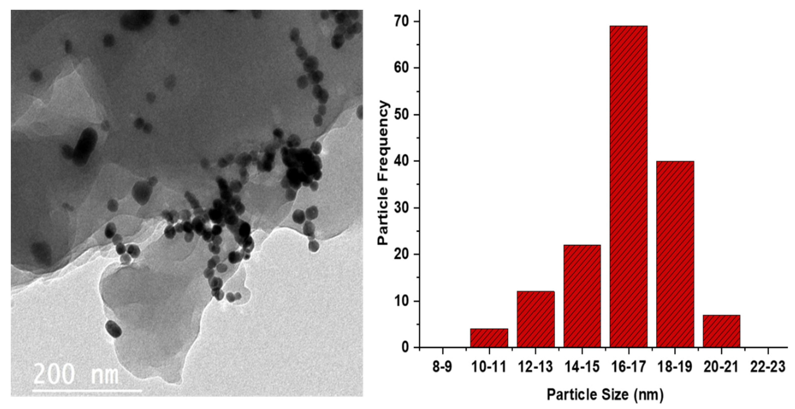
3.1. TEM Results
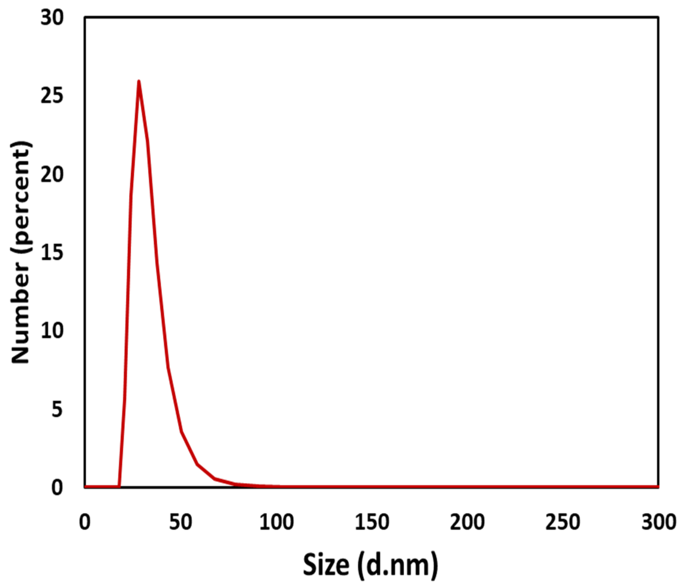
3.2. Dynamic Light Scattering (DLS) Analysis
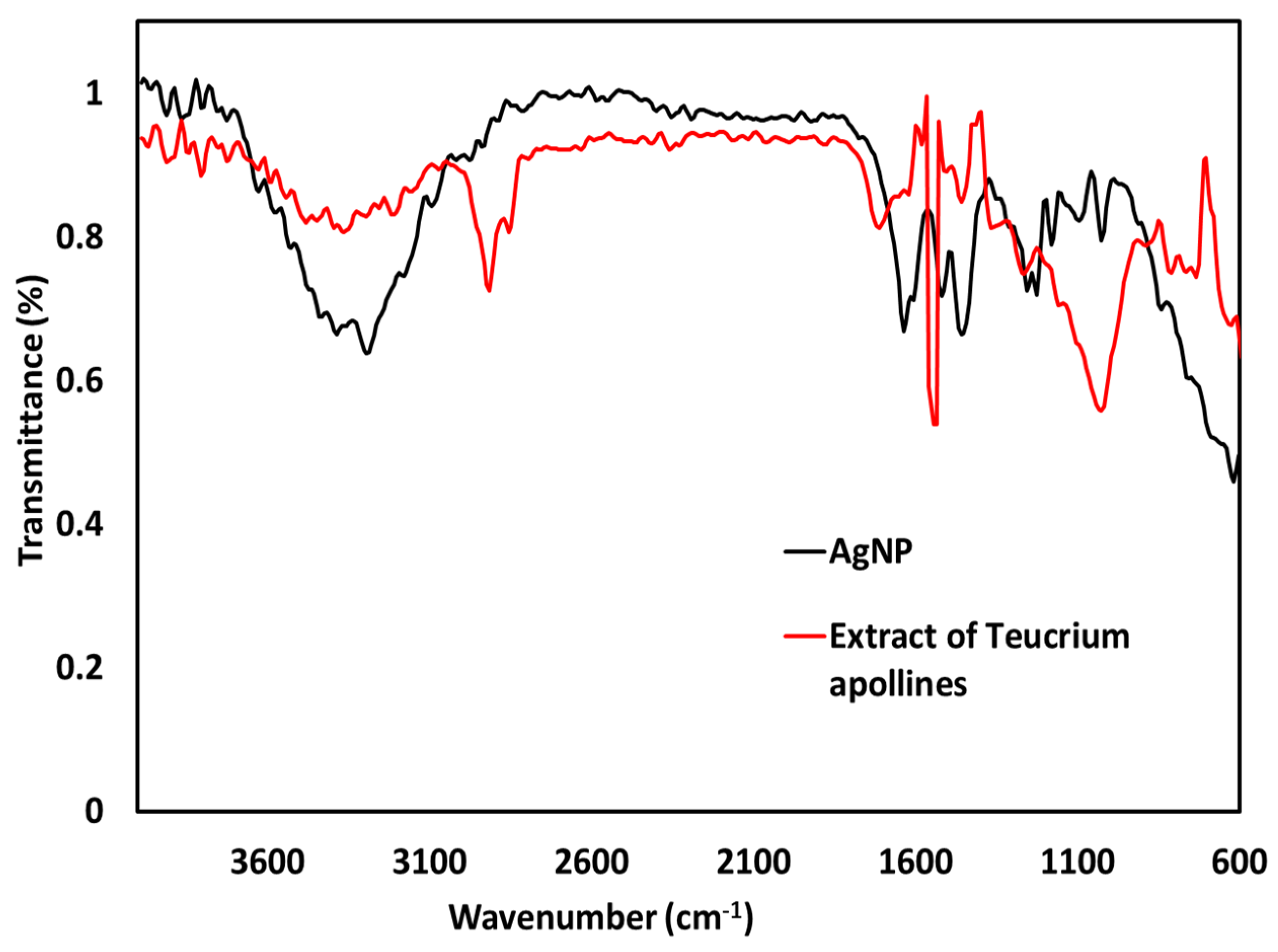
3.3. Attenuated Total Reflection Fourier Transform Infrared Spectroscopy (ATR-FTIR)
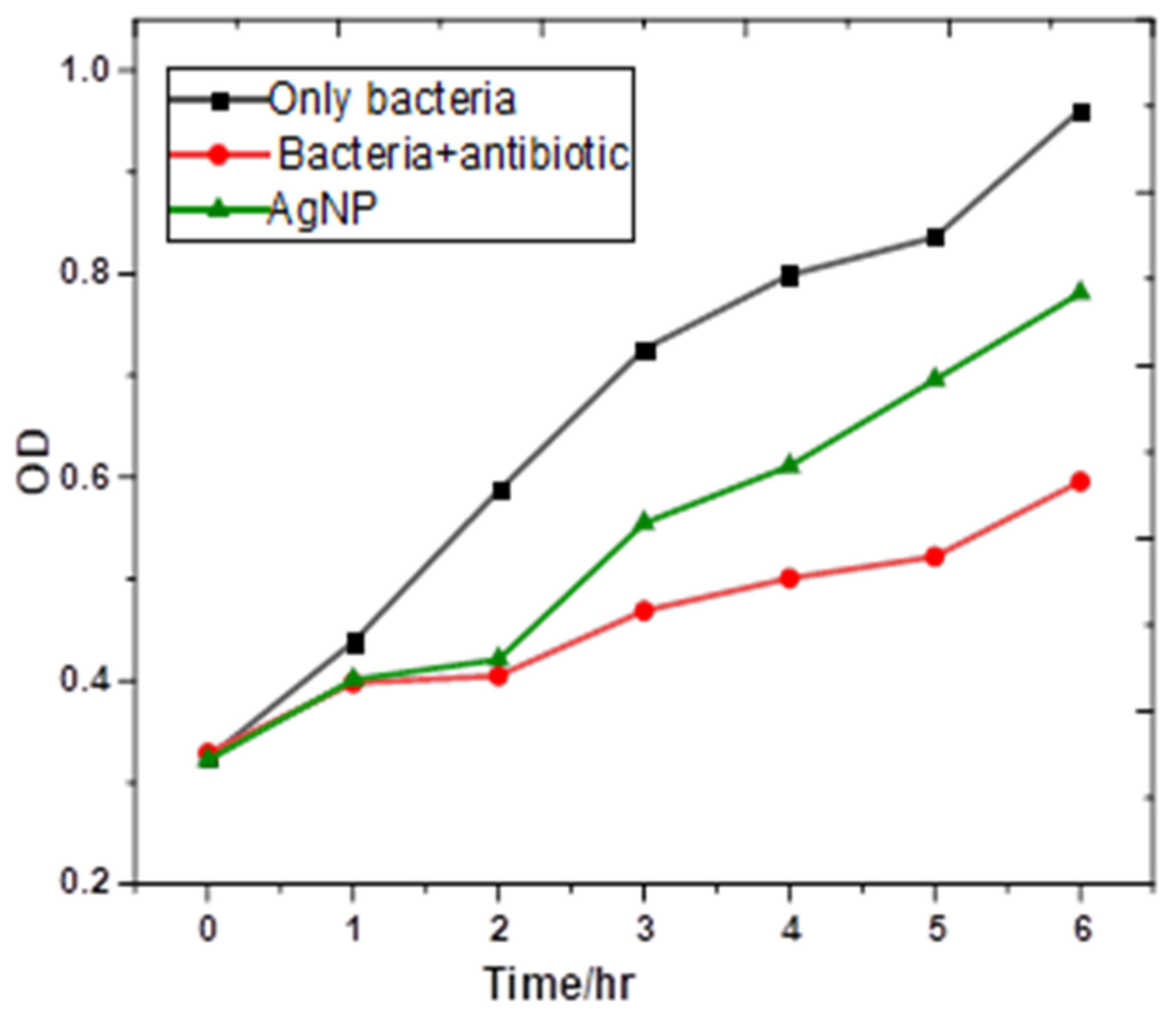
3.4. Antibacterial Activities of Silver Nanoparticles
4. Conclusions
Author Contributions
Funding
Institutional Review Board Statement
Informed Consent Statement
Data Availability Statement
Acknowledgments
Conflicts of Interest
References
- Zhang, X.F.; Liu, Z.G.; Shen, W.; Gurunathan, S. Silver nanoparticles: Synthesis, characterization, properties, applications, and therapeutic approaches. Int. J. Mol. Sci. 2016, 17, 1534. [Google Scholar] [CrossRef] [PubMed]
- Bruna, T.; Maldonado-Bravo, F.; Jara, P.; Caro, N. Silver nanoparticles and their antibacterial applications. Int. J. Mol. Sci. 2021, 22, 7202. [Google Scholar] [CrossRef] [PubMed]
- Yin, I.X.; Zhang, J.; Zhao, I.S.; Mei, M.L.; Li, Q.; Chu, C.H. The antibacterial mechanism of silver nanoparticles and its application in dentistry. Int. J. Nanomed. 2020, 15, 2555. [Google Scholar] [CrossRef] [PubMed] [Green Version]
- Salomoni, R.; Léo, P.; Montemor, A.F.; Rinaldi, B.G.; Rodrigues, M.F.A. Antibacterial effect of silver nanoparticles in Pseudomonas aeruginosa. Nanotechnol. Sci. Appl. 2017, 10, 115. [Google Scholar] [CrossRef] [Green Version]
- Balkrishna, A.; Sharma, N.; Sharma, V.K.; Mishra, N.D.; Joshi, C.S. Green synthesis, characterisation and biological studies of AgNPs prepared using Shivlingi (Bryonia laciniosa) seed extract. IET Nanobiotechnol. 2018, 12, 371–375. [Google Scholar] [CrossRef]
- Yamamoto, M.; Kashiwagi, Y.; Nakamoto, M. Size-controlled synthesis of monodispersed silver nanoparticles capped by long-chain alkyl carboxylates from silver carboxylate d tertiary amine. Langmuir 2006, 22, 8581–8586. [Google Scholar] [CrossRef]
- Malhotra, S.P.K.; Alghuthaymi, M.A. Biomolecule-assisted biogenic synthesis of metallic nanoparticles. In Agri-Waste Microbes Production of Sustainable Nanomaterials; Elsevier: Amsterdam, The Netherlands, 2022; pp. 139–163. [Google Scholar]
- Salem, S.S.; Fouda, A. Green synthesis of metallic nanoparticles and their prospective biotechnological applications: An overview. Biol. Trace Elem. Res. 2021, 199, 344–370. [Google Scholar] [CrossRef]
- Vishwanath, R.; Negi, B. Conventional and green methods of synthesis of silver nanoparticles and their antimicrobial properties. Curr. Res. Green Sustain. Chem. 2021, 4, 100205. [Google Scholar] [CrossRef]
- Candela, R.G.; Rosselli, S.; Bruno, M.; Fontana, G. A review of the phytochemistry, traditional uses and biological activities of the essential oils of genus Teucrium. Planta Med. 2021, 87, 432–479. [Google Scholar] [CrossRef]
- Al-Marhaby, F.A.; Seoudi, R. Preparation and characterization of silver nanoparticles and their use in catalytic reduction of 4-Nitrophenol. World J. Nano Sci. Eng. 2016, 6, 29–37. [Google Scholar] [CrossRef]
- Czechowska-Biskup, R.; Rokita, B.; Ulański, P.; Rosiak, J.M. Preparation of gold nanoparticles stabilized by chitosan using irradiation and sonication methods. Prog. Chem. Appl. Chitin Its Deriv. 2015, 20, 18–33. [Google Scholar] [CrossRef] [Green Version]
- Mock, J.J.; Smith, D.R.; Schultz, S. Local refractive index dependence of plasmon resonance spectra from individual nanoparticles. Nano Lett. 2003, 3, 485–491. [Google Scholar] [CrossRef]
- Mukaratirwa-Muchanyereyi, N.; Gusha, C.; Mujuru, M.; Guyo, U.; Nyoni, S. Synthesis of silver nanoparticles using plant extracts from Erythrina abyssinica aerial parts and assessment of their anti-bacterial and anti-oxidant activities. Results Chem. 2022, 4, 100402. [Google Scholar] [CrossRef]
- Singh, M.; Sinha, I.; Mandal, R.K. Role of pH in the green synthesis of silver nanoparticles. Mater. Lett. 2009, 63, 425–427. [Google Scholar] [CrossRef]
- Stankovic, M.S.; Curcic, M.G.; Zizic, J.B.; Topuzovic, M.D.; Solujic, S.R.; Markovic, S.D. Teucrium plant species as natural sources of novel anticancer compounds: Antiproliferative, proapoptotic and antioxidant properties. Int. J. Mol. Sci. 2011, 12, 4190–4205. [Google Scholar] [CrossRef]
- Heuer-Jungemann, A.; Feliu, N.; Bakaimi, I.; Hamaly, M.; Alkilany, A.; Chakraborty, I.; Masood, A.; Casula, M.F.; Kostopoulou, A.; Oh, E.; et al. The role of ligands in the chemical synthesis and applications of inorganic nanoparticles. Chem. Rev. 2019, 119, 4819–4880. [Google Scholar] [CrossRef] [Green Version]
- Vanlalveni, C.; Lallianrawna, S.; Biswas, A.; Selvaraj, M.; Changmai, B.; Rokhum, S.L. Green synthesis of silver nanoparticles using plant extracts and their antimicrobial activities: A review of recent literature. RSC Adv. 2021, 11, 2804–2837. [Google Scholar] [CrossRef]
- Alqadi, M.K.; Noqtah, O.A.A.; Alzoubi, F.Y.; Alzouby, J.; Aljarrah, K. pH effect on the aggregation of silver nanoparticles synthesized by chemical reduction. Mater. Sci. Pol. 2014, 32, 107–111. [Google Scholar] [CrossRef]
- Molleman, B.; Hiemstra, T. Time, pH, and size dependency of silver nanoparticle dissolution: The road to equilibrium. Environ. Sci. Nano 2017, 4, 1314–1327. [Google Scholar] [CrossRef]
- Miranda, A.; Akpobolokemi, T.; Chung, E.; Ren, G.; Raimi-Abraham, B.T. pH Alteration in Plant-Mediated Green Synthesis and Its Resultant Impact on Antimicrobial Properties of Silver Nanoparticles (AgNPs). Antibiotics 2022, 11, 1592. [Google Scholar] [CrossRef]
- Saliani, M.; Jalal, R.; Goharshadi, E.K. Effects of pH and temperature on antibacterial activity of zinc oxide nanofluid against E. coli O157: H7 and Staphylococcus aureus. Jundishapur J. Microbiol. 2015, 8, e17115. [Google Scholar] [CrossRef] [Green Version]
- Smith, D.J. Characterization of nanomaterials using transmission electron microscopy. Nanocharacterisation 2015, 37, 1–29. [Google Scholar]
- Kouhbanani, M.A.J.; Beheshtkhoo, N.; Fotoohiardakani, G.; Hosseini-Nave, H.; Taghizadeh, S.; Amani, A.M. Green synthesis and characterization of spherical structure silver nanoparticles using wheatgrass extract. J. Environ. Treat. Tech. 2019, 7, 142–149. [Google Scholar]
- Wang, L.; Housel, L.M.; Bock, D.C.; Abraham, A.; Dunkin, M.R.; McCarthy, A.H.; Wu, Q.; Kiss, A.; Thieme, J.; Takeuchi, E.S. Deliberate Modification of Fe3O4 Anode Surface Chemistry: Impact on Electrochemistry. ACS Appl. Mater. Interfaces 2019, 11, 19920–19932. [Google Scholar] [CrossRef] [PubMed]
- Singh, P.; Mijakovic, I. Strong antimicrobial activity of silver nanoparticles obtained by the green synthesis in Viridibacillus sp. extracts. Front. Microbiol. 2022, 13, 820048. [Google Scholar] [CrossRef]
- Ahmadi, M.; Adibhesami, M. The effect of silver nanoparticles on wounds contaminated with Pseudomonas aeruginosa in mice: An experimental study. Iran. J. Pharm. Res. IJPR 2017, 16, 661. [Google Scholar]
- Fadwa, A.O.; Alkoblan, D.K.; Mateen, A.; Albarag, A.M. Synergistic effects of zinc oxide nanoparticles and various antibiotics combination against Pseudomonas aeruginosa clinically isolated bacterial strains. Saudi J. Biol. Sci. 2021, 28, 928–935. [Google Scholar] [CrossRef]
- Borcherding, J.; Baltrusaitis, J.; Chen, H.; Stebounova, L.; Wu, C.-M.; Rubasinghege, G.; Mudunkotuwa, I.A.; Caraballo, J.C.; Zabner, J.; Grassian, V.H. Iron oxide nanoparticles induce Pseudomonas aeruginosa growth, induce biofilm formation, and inhibit antimicrobial peptide function. Environ. Sci. Nano 2014, 1, 123–132. [Google Scholar] [CrossRef] [Green Version]
- Abdussalam-Mohammed, W.; Najem, M.Y.; Errayes, A.O.; Shamsi, S.S.; Darwish, M.O.; Mezoughi, A.B. Synthesis of Highly Stabilized AuNPs Using 3, 5-Dinitrobenzoic Acid and Sodium Acetate as Capping Agents in an Aqueous Solution and their Bioactivity. J. Nano Res. 2021, 70, 67–79. [Google Scholar] [CrossRef]
- Nayem, S.A.; Sultana, N.; Haque, M.A.; Miah, B.; Hasan, M.M.; Islam, T.; Hasan, M.M.; Awal, A.; Uddin, J.; Aziz, M.A.; et al. Green synthesis of gold and silver nanoparticles by using amorphophallus paeoniifolius tuber extract and evaluation of their antibacterial activity. Molecules 2020, 25, 4773. [Google Scholar] [CrossRef]
- Usman, M.S.; Zowalaty, M.E.E.; Shameli, K.; Zainuddin, N.; Salama, M.; Ibrahim, N.A. Synthesis, characterization, and antimicrobial properties of copper nanoparticles. Int. J. Nanomed. 2013, 8, 4467–4479. [Google Scholar]


Disclaimer/Publisher’s Note: The statements, opinions and data contained in all publications are solely those of the individual author(s) and contributor(s) and not of MDPI and/or the editor(s). MDPI and/or the editor(s) disclaim responsibility for any injury to people or property resulting from any ideas, methods, instructions or products referred to in the content. |
© 2023 by the authors. Licensee MDPI, Basel, Switzerland. This article is an open access article distributed under the terms and conditions of the Creative Commons Attribution (CC BY) license (https://creativecommons.org/licenses/by/4.0/).
Share and Cite
Abdussalam-Mohammed, W.; Mohamed, L.; Abraheem, M.S.; Mansour, M.M.A.; Sherif, A.M. Biofabrication of Silver Nanoparticles Using Teucrium Apollinis Extract: Characterization, Stability, and Their Antibacterial Activities. Chemistry 2023, 5, 54-64. https://doi.org/10.3390/chemistry5010005
Abdussalam-Mohammed W, Mohamed L, Abraheem MS, Mansour MMA, Sherif AM. Biofabrication of Silver Nanoparticles Using Teucrium Apollinis Extract: Characterization, Stability, and Their Antibacterial Activities. Chemistry. 2023; 5(1):54-64. https://doi.org/10.3390/chemistry5010005
Chicago/Turabian StyleAbdussalam-Mohammed, Wanisa, Laila Mohamed, Mohammed S. Abraheem, Mohmeed M.A Mansour, and Akram Mansour Sherif. 2023. "Biofabrication of Silver Nanoparticles Using Teucrium Apollinis Extract: Characterization, Stability, and Their Antibacterial Activities" Chemistry 5, no. 1: 54-64. https://doi.org/10.3390/chemistry5010005